EP0372119B2 - Lithotripteur - Google Patents
Lithotripteur Download PDFInfo
- Publication number
- EP0372119B2 EP0372119B2 EP88120646A EP88120646A EP0372119B2 EP 0372119 B2 EP0372119 B2 EP 0372119B2 EP 88120646 A EP88120646 A EP 88120646A EP 88120646 A EP88120646 A EP 88120646A EP 0372119 B2 EP0372119 B2 EP 0372119B2
- Authority
- EP
- European Patent Office
- Prior art keywords
- shock wave
- wave head
- ray system
- ray
- lithotriptor
- Prior art date
- Legal status (The legal status is an assumption and is not a legal conclusion. Google has not performed a legal analysis and makes no representation as to the accuracy of the status listed.)
- Expired - Lifetime
Links
- 230000035939 shock Effects 0.000 claims description 51
- 230000005855 radiation Effects 0.000 claims description 10
- 238000002686 lithotriptor Methods 0.000 claims 8
- 238000002604 ultrasonography Methods 0.000 description 8
- 230000008878 coupling Effects 0.000 description 6
- 238000010168 coupling process Methods 0.000 description 6
- 238000005859 coupling reaction Methods 0.000 description 6
- 239000012530 fluid Substances 0.000 description 3
- 239000007788 liquid Substances 0.000 description 2
- 238000001514 detection method Methods 0.000 description 1
- 230000005520 electrodynamics Effects 0.000 description 1
- 230000003760 hair shine Effects 0.000 description 1
- 238000003384 imaging method Methods 0.000 description 1
- 238000002560 therapeutic procedure Methods 0.000 description 1
- XLYOFNOQVPJJNP-UHFFFAOYSA-N water Substances O XLYOFNOQVPJJNP-UHFFFAOYSA-N 0.000 description 1
Images
Classifications
-
- A—HUMAN NECESSITIES
- A61—MEDICAL OR VETERINARY SCIENCE; HYGIENE
- A61B—DIAGNOSIS; SURGERY; IDENTIFICATION
- A61B17/00—Surgical instruments, devices or methods
- A61B17/22—Implements for squeezing-off ulcers or the like on inner organs of the body; Implements for scraping-out cavities of body organs, e.g. bones; for invasive removal or destruction of calculus using mechanical vibrations; for removing obstructions in blood vessels, not otherwise provided for
- A61B17/225—Implements for squeezing-off ulcers or the like on inner organs of the body; Implements for scraping-out cavities of body organs, e.g. bones; for invasive removal or destruction of calculus using mechanical vibrations; for removing obstructions in blood vessels, not otherwise provided for for extracorporeal shock wave lithotripsy [ESWL], e.g. by using ultrasonic waves
- A61B17/2256—Implements for squeezing-off ulcers or the like on inner organs of the body; Implements for scraping-out cavities of body organs, e.g. bones; for invasive removal or destruction of calculus using mechanical vibrations; for removing obstructions in blood vessels, not otherwise provided for for extracorporeal shock wave lithotripsy [ESWL], e.g. by using ultrasonic waves with means for locating or checking the concrement, e.g. X-ray apparatus, imaging means
- A61B17/2258—Implements for squeezing-off ulcers or the like on inner organs of the body; Implements for scraping-out cavities of body organs, e.g. bones; for invasive removal or destruction of calculus using mechanical vibrations; for removing obstructions in blood vessels, not otherwise provided for for extracorporeal shock wave lithotripsy [ESWL], e.g. by using ultrasonic waves with means for locating or checking the concrement, e.g. X-ray apparatus, imaging means integrated in a central portion of the shock wave apparatus
-
- A—HUMAN NECESSITIES
- A61—MEDICAL OR VETERINARY SCIENCE; HYGIENE
- A61B—DIAGNOSIS; SURGERY; IDENTIFICATION
- A61B6/00—Apparatus or devices for radiation diagnosis; Apparatus or devices for radiation diagnosis combined with radiation therapy equipment
- A61B6/12—Arrangements for detecting or locating foreign bodies
-
- A—HUMAN NECESSITIES
- A61—MEDICAL OR VETERINARY SCIENCE; HYGIENE
- A61B—DIAGNOSIS; SURGERY; IDENTIFICATION
- A61B17/00—Surgical instruments, devices or methods
- A61B2017/00831—Material properties
- A61B2017/00902—Material properties transparent or translucent
Definitions
- the invention relates to a lithotripter with an x-ray system for locating concrements and a shock wave head for generating shock waves for crushing the concrements.
- a lithotripter of this type is known in which there are two x-ray systems, each with an x-ray emitter and an x-ray image intensifier with a television chain connected downstream (EP-A-0 273 256).
- the patient is irradiated from two directions so that the concrements can be located.
- Two shock wave heads are adjustable so that one of them can be moved laterally to the patient for treatment.
- the central beam of the emitted shock waves and the central beams of the two X-ray systems intersect in an isocenter in which the concrement to be treated is positioned. Since the plane which is delimited by the two central beams of the sound beams of the shock wave heads is perpendicular to the plane delimited by the two central beams of the X-ray systems, the lateral space requirement is relatively large.
- FR-A-2 591 467 discloses a lithotripter with an ultrasound location system, in which the shock wave generator encloses the ultrasound location system. This principle cannot be used for X-ray detection.
- EP 0 369 177 A2 which is older but not prepublished, contains a lithotripter with an X-ray system for locating concrements and a shock wave head described for the generation of shock waves, the shock wave head of which has a central, hollow cylindrical space penetrated by the X-rays without radiation attenuation.
- this publication describes a lithotripter which, instead of the x-ray system, has an ultrasound locating system which is arranged at the same point where, in the case of the aforementioned embodiment, the central, hollow-cylindrical space penetrated by the x-rays is located without radiation attenuation.
- EP 0 372 174 A1 which is also older, but not prepublished, describes a lithotripter with an X-ray system and an ultrasound location system for locating concrements, and a shock wave head for generating shock waves for crushing the concrements, which centric is penetrated by the X-rays , hollow cylindrical space without radiation attenuation.
- the ultrasound location system is firmly connected to the shock wave head and received in a bore of the shock wave head. The arrangement is such that the axis of the ultrasound locating system and the central beam of the X-ray system running through said space intersect.
- the invention has for its object to design a lithotripter of the type mentioned so that there is a compact structure.
- the shock wave head can be arranged after the X-ray tube when viewed in the beam direction. Lateral space under the patient table is not necessary.
- the shock wave head has a central, hollow-cylindrical region penetrated by the X-rays without radiation attenuation, e.g. an air-filled room. This enables particularly good imaging and simple observation during therapy in the sonicated area.
- the shock wave head can be on the primary aperture of the x-ray system or in front of the radiation receiver, e.g. the image intensifier, the X-ray system.
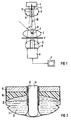
- FIG. 1 shows a bed 1 on which a patient 2 lies, which contains a concretion 3.
- the concretion 3 is to be smashed by shock waves.
- An X-ray system with an X-ray emitter 4 with a primary beam diaphragm 5 and an X-ray image intensifier 6 with a television chain 7 connected downstream is provided for locating the calculus 3. So that spatial information about the position of the calculus 3 is obtained, the X-ray system 4, 5, 6 can be pivoted about an axis 8 by an angle ⁇ , so that the patient 2 can be irradiated in different directions.
- the axis 8 should coincide with the concretion 3.
- the bed 1 is adjustable accordingly.
- the concrement 3 is sonicated with shock waves.
- a shock wave head 9 is attached to the primary beam diaphragm 5, which emits shock waves focused on a focus area and is set such that the focus area also coincides with the concretion 3.
- the shock wave head 9 can be adjustably mounted in the direction of the central beam 10 of the X-ray system 4, 5, 6. The adjustment can be made individually, but also together with the X-ray emitter 4.
- the shock wave head 9 is covered on its application side by a flexible film 11 which is applied to the surface of the patient 2 for acoustic coupling.
- the shock wave head 9 is arranged in relation to the x-ray system 4, 5, 6 such that the central beam 10 of the x-ray system 4, 5, 6 runs approximately centrally through the shock wave head 9.
- the shock wave head 9 has a central, hollow cylindrical space 12 penetrated by the X-rays without radiation attenuation.
- the room 12 is filled with air. Its hollow cylindrical wall delimits the space of the shock wave head 9 filled with coupling fluid. This is explained in more detail below with reference to FIG. 2.
- the central beam 10 of the x-ray system 4, 5, 6 coincides with the central beam of the sound lobe emitted by the shock wave head 9.
- the location is carried out by pivoting the X-ray system 4, 5, 6.
- the X-ray beam that can be used for image generation is also determined by the dimensions of the space 12.
- Fig. 2 shows that the shock wave head 9 is a closed, from a coupling liquid, e.g. Water, filled space 13.
- a coupling liquid e.g. Water
- the application side of the shock wave head 9 is covered by the flexible film 11, which is pulled over the space 12.
- the hollow cylindrical wall of the space 12 delimits the space 13 filled with coupling liquid.
- An ultrasound location system 14 is arranged in the space 12 and can be pulled out of the space 12 in the axial direction.
- the space 12 is - as stated above - air-filled and its hollow cylindrical wall delimits the space 13 filled with coupling fluid, so that when the ultrasound locating system 14 is pulled out, no measures have to be taken to prevent coupling fluid from escaping.
- a shock wave source 15 is provided in the shock wave head 9, e.g. can be piezoceramic or electrodynamic in nature. It emits focused shock waves, which are focused by an acoustic lens system 16, which can be adjustable to change the position of the focus area.
Landscapes
- Health & Medical Sciences (AREA)
- Life Sciences & Earth Sciences (AREA)
- Medical Informatics (AREA)
- Engineering & Computer Science (AREA)
- Nuclear Medicine, Radiotherapy & Molecular Imaging (AREA)
- Surgery (AREA)
- General Health & Medical Sciences (AREA)
- Veterinary Medicine (AREA)
- Biomedical Technology (AREA)
- Heart & Thoracic Surgery (AREA)
- Radiology & Medical Imaging (AREA)
- Molecular Biology (AREA)
- Animal Behavior & Ethology (AREA)
- Public Health (AREA)
- Orthopedic Medicine & Surgery (AREA)
- Vascular Medicine (AREA)
- Physics & Mathematics (AREA)
- Biophysics (AREA)
- High Energy & Nuclear Physics (AREA)
- Optics & Photonics (AREA)
- Pathology (AREA)
- Surgical Instruments (AREA)
- Apparatus For Radiation Diagnosis (AREA)
Claims (8)
- Appareil de lithotritie comportant un système radiologique (4,5,6) pour localiser des concrétions (3) et une tête (9) de production d'ondes de choc servant à produire des ondes de choc pour la fragmentation des concrétions (3), la tête (9) de production d'ondes de choc comportant un espace (12) cylindrique creux centré, traversé par le rayonnement X, sans affaiblissement du rayonnement, dans lequel est disposé un système (14) de localisation d'ultrason qui peut être retiré de l'espace (12) suivant une direction axiale.
- Appareil de lithotritie suivant la revendication 1, caractérisé par le fait que le rayon central (10) du système radiologie (4,5,6) coïncide avec le rayon central du lobe acoustique émis par la tête (9) de production d'ondes de choc.
- Appareil de lithotritie suivant la revendication 1, caractérisé par le fait que la tête (9) de production d'ondes de choc est disposée sur le diaphragme (5) du rayonnement primaire.
- Appareil de lithotritie suivant la revendication 1, caractérisé par le fait que la tête (9) de production d'ondes de choc est disposée devant le récepteur de rayonnement (6) du système radiologie (4,5,6).
- Appareil de lithotritie comportant un système (4,5,6) radiologie pour localiser des concrétions (3) et une tête (9) de production d'ondes de choc servant à produire des ondes pour la fragmentation des concrétions (3), la tête (9) de production d'ondes de choc comportant un espace (12) cylindrique creux centré, traversé par le rayonnement X sans affaiblissement du rayonnement et la tête (5) de production d'ondes de choc étant montée sur le diaphragme (5) du rayonnement primaire.
- Appareil de lithotritie suivant la revendication 5, caractérisé par le fait que le rayon central (10) du système radiologie (4,5,6) coïncide avec le rayon central du lobe acoustique émis par la tête (9) de production d'ondes de choc.
- Appareil de lithotritie comportant un système radiologique (4,5,6) pour localiser des concrétions (3) et une tête (9) de production d'ondes de choc servant à produire des ondes de choc pour la fragmentation des concrétions (3), la tête (9) de production d'ondes de choc comportant un espace (12) cylindrique creux centré, traversé par le rayonnement X, sans affaiblissement du rayonnement, la tête (9) de production d'ondes de choc étant disposé avant le récepteur (6) de rayonnement du système (4,5,6) radiologique, de telle sorte que le patient (2), lorsque le dispositif de lithotritie fonctionne, se trouve entre la tête (9) de production d'ondes de choc et l'émetteur (4) de rayons X du système (4,5,6) radiologique.
- Appareil de lithotrie suivant la revendication 7, caractérisé par le fait que le rayon central (10) du système radiologie (4,5,6) coïncide avec le rayon central du lobe acoustique émis par la tête (9) de production d'ondes de choc.
Priority Applications (3)
| Application Number | Priority Date | Filing Date | Title |
|---|---|---|---|
| DE88120646T DE3888189D1 (de) | 1988-12-09 | 1988-12-09 | Lithotripter. |
| EP88120646A EP0372119B2 (fr) | 1988-12-09 | 1988-12-09 | Lithotripteur |
| US07/426,967 US4984565A (en) | 1988-12-09 | 1989-10-26 | Extracorporeal lithotriptor with x-ray locating system |
Applications Claiming Priority (1)
| Application Number | Priority Date | Filing Date | Title |
|---|---|---|---|
| EP88120646A EP0372119B2 (fr) | 1988-12-09 | 1988-12-09 | Lithotripteur |
Publications (3)
| Publication Number | Publication Date |
|---|---|
| EP0372119A1 EP0372119A1 (fr) | 1990-06-13 |
| EP0372119B1 EP0372119B1 (fr) | 1994-03-02 |
| EP0372119B2 true EP0372119B2 (fr) | 1997-08-27 |
Family
ID=8199645
Family Applications (1)
| Application Number | Title | Priority Date | Filing Date |
|---|---|---|---|
| EP88120646A Expired - Lifetime EP0372119B2 (fr) | 1988-12-09 | 1988-12-09 | Lithotripteur |
Country Status (3)
| Country | Link |
|---|---|
| US (1) | US4984565A (fr) |
| EP (1) | EP0372119B2 (fr) |
| DE (1) | DE3888189D1 (fr) |
Families Citing this family (17)
| Publication number | Priority date | Publication date | Assignee | Title |
|---|---|---|---|---|
| DE3835318C1 (fr) * | 1988-10-17 | 1990-06-28 | Storz Medical Ag, Kreuzlingen, Ch | |
| US5230329A (en) * | 1988-11-16 | 1993-07-27 | Medas S.P.A. | Lithotripter with two localization devices in an isocentric system |
| DE3840077A1 (de) * | 1988-11-28 | 1990-05-31 | Wolf Gmbh Richard | Lithotriptor |
| EP0400196B1 (fr) * | 1989-06-02 | 1994-11-02 | Siemens Aktiengesellschaft | Transducteur d'ondes de choc pour destruction de concrétions |
| DE3942253A1 (de) * | 1989-12-21 | 1991-06-27 | Dornier Medizintechnik | Halterung eines diagnostischen ultraschall-transducers |
| DE4115447C2 (de) * | 1991-05-11 | 1994-01-27 | Schott Glaswerke | Vorrichtung zur Kontrolle der Zerstörung von Konkrementen |
| DE4135177C2 (de) * | 1991-10-24 | 1998-04-09 | Siemens Ag | Theraphieeinrichtung zur Behandlung eines Lebewesens mit fokussierten akustischen Wellen |
| DE4143540C2 (de) * | 1991-10-24 | 1996-08-08 | Siemens Ag | Therapieeinrichtung zur Behandlung eines Patienten mit fokussierten akustischen Wellen |
| DE4135328C2 (de) * | 1991-10-25 | 1995-01-19 | Wolf Gmbh Richard | Extrakorporales Therapiegerät |
| DE4300740C1 (de) * | 1993-01-14 | 1994-03-17 | Wolf Gmbh Richard | Extrakorpolares Therapiegerät |
| DE4306459C1 (de) * | 1993-03-02 | 1994-04-28 | Siemens Ag | Therapieeinrichtung zur Behandlung mit fokussierten akustischen Wellen |
| DE4306460C2 (de) * | 1993-03-02 | 1995-04-20 | Siemens Ag | Therapieeinrichtung zur Behandlung mit fokussierten akustischen Wellen |
| DE4447665B4 (de) * | 1994-01-14 | 2004-04-29 | Siemens Ag | Medizinisches Gerät mit einer Röntgendiagnostikeinrichtung zur Erzeugung von Röntgen-Schattenbildern |
| DE19538054C1 (de) * | 1995-10-12 | 1996-12-05 | Siemens Ag | Medizinisches Gerät mit einer Patientenlagerungsplatte |
| EP1493389A1 (fr) * | 2003-07-01 | 2005-01-05 | Siemens Aktiengesellschaft | Procédé et dispositif pour former des images radiographiques de la région focale d'un lithotripteur |
| US8979776B2 (en) * | 2008-05-02 | 2015-03-17 | Daniel Gelbart | Lithotripsy system with automatic 3D tracking |
| US20110237984A1 (en) * | 2010-03-23 | 2011-09-29 | Moshe Ein-Gal | Dynamic system for shockwave treatment |
Family Cites Families (9)
| Publication number | Priority date | Publication date | Assignee | Title |
|---|---|---|---|---|
| US4610249A (en) * | 1984-05-08 | 1986-09-09 | The Johns Hopkins University | Means and method for the noninvasive fragmentation of body concretions |
| DE3417985C2 (de) * | 1984-05-15 | 1986-03-27 | Dornier System Gmbh, 7990 Friedrichshafen | Vorrichtung zur berührungsfreien Zerkleinerung von Konkrementen |
| JPS61249452A (ja) * | 1985-04-30 | 1986-11-06 | 株式会社東芝 | X線診断装置 |
| DE3662966D1 (en) * | 1985-05-28 | 1989-06-01 | Siemens Ag | Apparatus for the contactless disintegration of concrements |
| EP0221325B1 (fr) * | 1985-10-09 | 1990-07-25 | Siemens Aktiengesellschaft | Unité de traitement par lithotritie |
| DE3543867C3 (de) * | 1985-12-12 | 1994-10-06 | Wolf Gmbh Richard | Vorrichtung zur räumlichen Ortung und zur Zerstörung von Konkrementen in Körperhöhlen |
| DE3736733A1 (de) * | 1986-10-29 | 1988-05-11 | Olympus Optical Co | Ultraschall-therapievorrichtung |
| US4811725A (en) * | 1986-12-22 | 1989-03-14 | Siemens Aktiengesellschaft | Extracorporeal lithotripsy apparatus |
| DE3723920A1 (de) * | 1987-04-04 | 1989-01-26 | Philips Patentverwaltung | Lithotripsie-arbeitsplatz |
-
1988
- 1988-12-09 EP EP88120646A patent/EP0372119B2/fr not_active Expired - Lifetime
- 1988-12-09 DE DE88120646T patent/DE3888189D1/de not_active Expired - Lifetime
-
1989
- 1989-10-26 US US07/426,967 patent/US4984565A/en not_active Expired - Lifetime
Also Published As
| Publication number | Publication date |
|---|---|
| DE3888189D1 (de) | 1994-04-07 |
| US4984565A (en) | 1991-01-15 |
| EP0372119B1 (fr) | 1994-03-02 |
| EP0372119A1 (fr) | 1990-06-13 |
Similar Documents
| Publication | Publication Date | Title |
|---|---|---|
| DE69208141T2 (de) | Vorrichtung zum Zerstören von Konkrementen | |
| EP0372119B1 (fr) | Lithotripteur | |
| DE3543867C3 (de) | Vorrichtung zur räumlichen Ortung und zur Zerstörung von Konkrementen in Körperhöhlen | |
| EP0133665B1 (fr) | Appareils pour détruire les calculs à distance | |
| EP0133946B1 (fr) | Appareil pour la destruction à distance des concrétions | |
| DE4143540C2 (de) | Therapieeinrichtung zur Behandlung eines Patienten mit fokussierten akustischen Wellen | |
| DE4241161C2 (de) | Akustische Therapieeinrichtung | |
| EP0397980B1 (fr) | Lithotriteur | |
| DE3713816C2 (fr) | ||
| EP0372174B1 (fr) | Lithotripteur | |
| EP0221325B1 (fr) | Unité de traitement par lithotritie | |
| EP0715831B1 (fr) | Ensemble de lithotripsie avec une unité de thérapie | |
| DE102005037043B4 (de) | Stoßwellentherapiegerät mit Bildgewinnung | |
| EP0400196B1 (fr) | Transducteur d'ondes de choc pour destruction de concrétions | |
| DE69018853T2 (de) | Vorrichtung zum Zerstören von Konkrementen. | |
| DE3328039C2 (de) | Einrichtung zum beruehrungslosen zertruemmern eines im koerper eines lebewesens befindlichen konkrements | |
| EP0273256A1 (fr) | Appareil pour la fragmentation à distance de concrétions | |
| DE19548000C1 (de) | Vorrichtung zur Ortung von Konkrementen im Körper eines Patienten | |
| DE3617032C2 (de) | Lithotripsiegerät mit extrakorporalem Stoßwellengenerator | |
| EP0265742A1 (fr) | Lithotripteur avec moyens de localisation | |
| EP0278304A1 (fr) | Lithotriteur à moyens de positionnement intégré | |
| EP0244730B1 (fr) | Dispositif destiné à la désintégration de concréments présents dans le corps d'un être vivant | |
| DE3739390A1 (de) | Lithotripter mit veraenderbarem fokus | |
| EP0278303A1 (fr) | Lithotriteur à balayage sectoriel intégré | |
| DE68929265T2 (de) | Gerät zum zielen und visulaisieren von konkrementen in echtzeit |
Legal Events
| Date | Code | Title | Description |
|---|---|---|---|
| PUAI | Public reference made under article 153(3) epc to a published international application that has entered the european phase |
Free format text: ORIGINAL CODE: 0009012 |
|
| AK | Designated contracting states |
Kind code of ref document: A1 Designated state(s): DE FR GB NL |
|
| 17P | Request for examination filed |
Effective date: 19900710 |
|
| 17Q | First examination report despatched |
Effective date: 19930322 |
|
| GRAA | (expected) grant |
Free format text: ORIGINAL CODE: 0009210 |
|
| AK | Designated contracting states |
Kind code of ref document: B1 Designated state(s): DE FR GB NL |
|
| PG25 | Lapsed in a contracting state [announced via postgrant information from national office to epo] |
Ref country code: GB Effective date: 19940302 Ref country code: NL Effective date: 19940302 |
|
| REF | Corresponds to: |
Ref document number: 3888189 Country of ref document: DE Date of ref document: 19940407 |
|
| ET | Fr: translation filed | ||
| NLV1 | Nl: lapsed or annulled due to failure to fulfill the requirements of art. 29p and 29m of the patents act | ||
| GBV | Gb: ep patent (uk) treated as always having been void in accordance with gb section 77(7)/1977 [no translation filed] |
Effective date: 19940302 |
|
| PLBI | Opposition filed |
Free format text: ORIGINAL CODE: 0009260 |
|
| 26 | Opposition filed |
Opponent name: ROFIN SINAR LASER GMBH Effective date: 19941121 |
|
| PLAW | Interlocutory decision in opposition |
Free format text: ORIGINAL CODE: EPIDOS IDOP |
|
| PLAW | Interlocutory decision in opposition |
Free format text: ORIGINAL CODE: EPIDOS IDOP |
|
| PUAH | Patent maintained in amended form |
Free format text: ORIGINAL CODE: 0009272 |
|
| STAA | Information on the status of an ep patent application or granted ep patent |
Free format text: STATUS: PATENT MAINTAINED AS AMENDED |
|
| 27A | Patent maintained in amended form |
Effective date: 19970827 |
|
| AK | Designated contracting states |
Kind code of ref document: B2 Designated state(s): DE FR GB NL |
|
| ET3 | Fr: translation filed ** decision concerning opposition | ||
| PGFP | Annual fee paid to national office [announced via postgrant information from national office to epo] |
Ref country code: FR Payment date: 20051216 Year of fee payment: 18 |
|
| REG | Reference to a national code |
Ref country code: FR Ref legal event code: ST Effective date: 20070831 |
|
| PG25 | Lapsed in a contracting state [announced via postgrant information from national office to epo] |
Ref country code: FR Free format text: LAPSE BECAUSE OF NON-PAYMENT OF DUE FEES Effective date: 20070102 |
|
| PGFP | Annual fee paid to national office [announced via postgrant information from national office to epo] |
Ref country code: DE Payment date: 20080218 Year of fee payment: 20 |