EP0404682A2 - Verfahren und Vorrichtung zur Sequenzierung eines Polynukleotids - Google Patents
Verfahren und Vorrichtung zur Sequenzierung eines Polynukleotids Download PDFInfo
- Publication number
- EP0404682A2 EP0404682A2 EP90401763A EP90401763A EP0404682A2 EP 0404682 A2 EP0404682 A2 EP 0404682A2 EP 90401763 A EP90401763 A EP 90401763A EP 90401763 A EP90401763 A EP 90401763A EP 0404682 A2 EP0404682 A2 EP 0404682A2
- Authority
- EP
- European Patent Office
- Prior art keywords
- polynucleotide
- substrate
- probe
- atomic diameter
- analysis
- Prior art date
- Legal status (The legal status is an assumption and is not a legal conclusion. Google has not performed a legal analysis and makes no representation as to the accuracy of the status listed.)
- Granted
Links
- 102000040430 polynucleotide Human genes 0.000 title claims abstract description 71
- 108091033319 polynucleotide Proteins 0.000 title claims abstract description 71
- 239000002157 polynucleotide Substances 0.000 title claims abstract description 71
- 238000012163 sequencing technique Methods 0.000 title claims abstract description 18
- 238000000034 method Methods 0.000 title claims description 41
- 239000000758 substrate Substances 0.000 claims abstract description 50
- 238000004458 analytical method Methods 0.000 claims abstract description 20
- 108090000623 proteins and genes Proteins 0.000 claims abstract description 11
- 239000000523 sample Substances 0.000 claims description 26
- 125000000524 functional group Chemical group 0.000 claims description 21
- 238000012545 processing Methods 0.000 claims description 14
- 238000003287 bathing Methods 0.000 claims description 5
- 229920002521 macromolecule Polymers 0.000 claims description 5
- 230000005012 migration Effects 0.000 claims description 5
- 238000013508 migration Methods 0.000 claims description 5
- 230000005641 tunneling Effects 0.000 claims description 4
- 230000003746 surface roughness Effects 0.000 claims description 3
- 230000007480 spreading Effects 0.000 claims description 2
- 238000003892 spreading Methods 0.000 claims description 2
- 239000012634 fragment Substances 0.000 description 13
- 239000000243 solution Substances 0.000 description 9
- OPTASPLRGRRNAP-UHFFFAOYSA-N cytosine Chemical compound NC=1C=CNC(=O)N=1 OPTASPLRGRRNAP-UHFFFAOYSA-N 0.000 description 8
- 230000000694 effects Effects 0.000 description 8
- KDCGOANMDULRCW-UHFFFAOYSA-N 7H-purine Chemical compound N1=CNC2=NC=NC2=C1 KDCGOANMDULRCW-UHFFFAOYSA-N 0.000 description 7
- 230000008569 process Effects 0.000 description 7
- 239000012530 fluid Substances 0.000 description 6
- UYTPUPDQBNUYGX-UHFFFAOYSA-N guanine Chemical compound O=C1NC(N)=NC2=C1N=CN2 UYTPUPDQBNUYGX-UHFFFAOYSA-N 0.000 description 6
- RWQNBRDOKXIBIV-UHFFFAOYSA-N thymine Chemical compound CC1=CNC(=O)NC1=O RWQNBRDOKXIBIV-UHFFFAOYSA-N 0.000 description 6
- 125000004429 atom Chemical group 0.000 description 5
- 230000004888 barrier function Effects 0.000 description 5
- 239000013078 crystal Substances 0.000 description 5
- 230000005684 electric field Effects 0.000 description 5
- 102000053602 DNA Human genes 0.000 description 4
- 108020004414 DNA Proteins 0.000 description 4
- CZPWVGJYEJSRLH-UHFFFAOYSA-N Pyrimidine Chemical compound C1=CN=CN=C1 CZPWVGJYEJSRLH-UHFFFAOYSA-N 0.000 description 4
- 229940104302 cytosine Drugs 0.000 description 4
- 238000001962 electrophoresis Methods 0.000 description 4
- 150000007523 nucleic acids Chemical class 0.000 description 4
- 239000002773 nucleotide Substances 0.000 description 4
- 125000003729 nucleotide group Chemical group 0.000 description 4
- 229920002477 rna polymer Polymers 0.000 description 4
- ASJSAQIRZKANQN-CRCLSJGQSA-N 2-deoxy-D-ribose Chemical compound OC[C@@H](O)[C@@H](O)CC=O ASJSAQIRZKANQN-CRCLSJGQSA-N 0.000 description 3
- GFFGJBXGBJISGV-UHFFFAOYSA-N Adenine Chemical compound NC1=NC=NC2=C1N=CN2 GFFGJBXGBJISGV-UHFFFAOYSA-N 0.000 description 3
- 229930024421 Adenine Natural products 0.000 description 3
- HMFHBZSHGGEWLO-SOOFDHNKSA-N D-ribofuranose Chemical compound OC[C@H]1OC(O)[C@H](O)[C@@H]1O HMFHBZSHGGEWLO-SOOFDHNKSA-N 0.000 description 3
- PYMYPHUHKUWMLA-LMVFSUKVSA-N Ribose Natural products OC[C@@H](O)[C@@H](O)[C@@H](O)C=O PYMYPHUHKUWMLA-LMVFSUKVSA-N 0.000 description 3
- 229960000643 adenine Drugs 0.000 description 3
- HMFHBZSHGGEWLO-UHFFFAOYSA-N alpha-D-Furanose-Ribose Natural products OCC1OC(O)C(O)C1O HMFHBZSHGGEWLO-UHFFFAOYSA-N 0.000 description 3
- -1 phosphoryl group Chemical group 0.000 description 3
- 229940113082 thymine Drugs 0.000 description 3
- XKRFYHLGVUSROY-UHFFFAOYSA-N Argon Chemical compound [Ar] XKRFYHLGVUSROY-UHFFFAOYSA-N 0.000 description 2
- IJGRMHOSHXDMSA-UHFFFAOYSA-N Atomic nitrogen Chemical compound N#N IJGRMHOSHXDMSA-UHFFFAOYSA-N 0.000 description 2
- OKTJSMMVPCPJKN-UHFFFAOYSA-N Carbon Chemical compound [C] OKTJSMMVPCPJKN-UHFFFAOYSA-N 0.000 description 2
- ISAKRJDGNUQOIC-UHFFFAOYSA-N Uracil Chemical compound O=C1C=CNC(=O)N1 ISAKRJDGNUQOIC-UHFFFAOYSA-N 0.000 description 2
- 238000000151 deposition Methods 0.000 description 2
- 230000001066 destructive effect Effects 0.000 description 2
- 230000002068 genetic effect Effects 0.000 description 2
- 238000010353 genetic engineering Methods 0.000 description 2
- 229910002804 graphite Inorganic materials 0.000 description 2
- 239000010439 graphite Substances 0.000 description 2
- 239000003550 marker Substances 0.000 description 2
- 229910052757 nitrogen Inorganic materials 0.000 description 2
- 102000039446 nucleic acids Human genes 0.000 description 2
- 108020004707 nucleic acids Proteins 0.000 description 2
- 150000003212 purines Chemical class 0.000 description 2
- 108091008146 restriction endonucleases Proteins 0.000 description 2
- 238000011144 upstream manufacturing Methods 0.000 description 2
- 241000446313 Lamella Species 0.000 description 1
- 239000002253 acid Substances 0.000 description 1
- 150000007513 acids Chemical class 0.000 description 1
- 238000013459 approach Methods 0.000 description 1
- 229910052786 argon Inorganic materials 0.000 description 1
- 230000008901 benefit Effects 0.000 description 1
- 238000005119 centrifugation Methods 0.000 description 1
- 210000000349 chromosome Anatomy 0.000 description 1
- 238000003776 cleavage reaction Methods 0.000 description 1
- 238000010367 cloning Methods 0.000 description 1
- 230000007547 defect Effects 0.000 description 1
- 238000001514 detection method Methods 0.000 description 1
- 238000010586 diagram Methods 0.000 description 1
- 201000010099 disease Diseases 0.000 description 1
- 208000037265 diseases, disorders, signs and symptoms Diseases 0.000 description 1
- 238000000407 epitaxy Methods 0.000 description 1
- 238000013467 fragmentation Methods 0.000 description 1
- 238000006062 fragmentation reaction Methods 0.000 description 1
- 239000007789 gas Substances 0.000 description 1
- 238000010438 heat treatment Methods 0.000 description 1
- 238000002347 injection Methods 0.000 description 1
- 239000007924 injection Substances 0.000 description 1
- 239000007788 liquid Substances 0.000 description 1
- 230000007246 mechanism Effects 0.000 description 1
- WCYWZMWISLQXQU-UHFFFAOYSA-N methyl Chemical compound [CH3] WCYWZMWISLQXQU-UHFFFAOYSA-N 0.000 description 1
- 238000004377 microelectronic Methods 0.000 description 1
- 238000012986 modification Methods 0.000 description 1
- 230000004048 modification Effects 0.000 description 1
- 239000002052 molecular layer Substances 0.000 description 1
- 125000000896 monocarboxylic acid group Chemical group 0.000 description 1
- 125000004433 nitrogen atom Chemical group N* 0.000 description 1
- 210000004940 nucleus Anatomy 0.000 description 1
- LFGREXWGYUGZLY-UHFFFAOYSA-N phosphoryl Chemical group [P]=O LFGREXWGYUGZLY-UHFFFAOYSA-N 0.000 description 1
- 238000000053 physical method Methods 0.000 description 1
- 230000010287 polarization Effects 0.000 description 1
- 150000003230 pyrimidines Chemical class 0.000 description 1
- 125000000714 pyrimidinyl group Chemical group 0.000 description 1
- 230000009257 reactivity Effects 0.000 description 1
- 230000000717 retained effect Effects 0.000 description 1
- 125000000548 ribosyl group Chemical group C1([C@H](O)[C@H](O)[C@H](O1)CO)* 0.000 description 1
- 230000007017 scission Effects 0.000 description 1
- 238000002791 soaking Methods 0.000 description 1
- 239000002904 solvent Substances 0.000 description 1
- 238000003860 storage Methods 0.000 description 1
- 229940035893 uracil Drugs 0.000 description 1
- 238000012795 verification Methods 0.000 description 1
- XLYOFNOQVPJJNP-UHFFFAOYSA-N water Substances O XLYOFNOQVPJJNP-UHFFFAOYSA-N 0.000 description 1
Images

Classifications
-
- G—PHYSICS
- G01—MEASURING; TESTING
- G01Q—SCANNING-PROBE TECHNIQUES OR APPARATUS; APPLICATIONS OF SCANNING-PROBE TECHNIQUES, e.g. SCANNING PROBE MICROSCOPY [SPM]
- G01Q30/00—Auxiliary means serving to assist or improve the scanning probe techniques or apparatus, e.g. display or data processing devices
- G01Q30/04—Display or data processing devices
Definitions
- the present invention relates to a method and a device for sequencing a polynucleotide such as a gene.
- chromosomes which are made up of long chains of nucleic acids having the following general structure: where P is the phosphoryl group, S is a ribose in ribonucleic acids (RNA) or a deoxyribose in deoxyribonucleic acids (DNA), and where the groups B i are nitrogen bases.
- All these acids therefore consist of a sequence of nucleotides having either a purine base (adenine or guanine) or a pyrimidine base (cytosine or uracil in RNA and cytosine or thymine in DNA).
- the sequencing of a nucleic acid fragment is the operation consisting in determining the succession of the bases of the nucleotides of which it is made up.
- This sequencing is particularly useful, in particular for better knowledge of diseases of genetic origin, but also in all fields of genetic engineering whose applications are, as we know, more and more numerous.
- the main sequencing methods used are manual methods derived either from the SANGER method or from the MAXAM-GILBERT method.
- One of these automatic processes uses fluorescent markers characteristic of each base.
- the strand which one wishes to sequence is cut as before, and the fragments thus obtained are migrated in a single electrophoresis gel illuminated with an argon laser. It suffices therefore to record the passage of each fragment, the color of this fragment to deduce the base to which it corresponds.
- the strand to be sequenced is cloned, cut into fragments and an electrophoresis process.
- the present invention aims to overcome these drawbacks by providing an automatic method for sequencing a polynucleotide which does not require cloning of this fragment, which does not require electrophoresis methods, and which is also non-destructive, which presents the advantage that it can be repeated several times for verification purposes.
- the invention firstly relates to a method for automatic sequencing of a polynucleotide such as a gene, characterized in that said polynucleotide is placed on a substrate whose surface roughness is the order of an atomic diameter, that a point analysis of the surface of the substrate carrying said polynucleotide is carried out with a resolution less than an atomic diameter, that the shape of each successive base of said polynucleotide is reconstructed from the data collected during the analysis, that we recognize this form, and that we memorize the information thus obtained.
- the method according to the invention is therefore based on direct detection of the shape of each successive base of the fragment to be sequenced, on the determination of this shape, and on its automatic recognition.
- Thymine bases are distinguished from cytosine bases by the presence, in the first, of the methyl radical placed in C5.
- the guanine bases are distinguished from the adenine bases by the presence, in the former, of a nitrogen atom placed at C2.
- the method according to the invention is therefore a purely physical method thereby eliminating the drawbacks of traditional methods.
- the aforementioned point analysis is carried out by scanning said surface with a resolution less than an atomic diameter using a probe, for example the probe of an effect microscope tunnel or atomic force, capable of detecting a difference in level with respect to the surface less than an atomic diameter, and by reconstructing the shape of each base successively encountered during the scan from the data of this scan.
- a probe for example the probe of an effect microscope tunnel or atomic force, capable of detecting a difference in level with respect to the surface less than an atomic diameter
- the tunneling microscope therefore consists in causing an X and Y scan of the surface to be analyzed using a probe, for example using piezoelectric devices, and during this scanning, move the probe vertically in Z, also using a piezoelectric device, so as to obtain a current of constant intensity.
- the control voltage to obtain this constant intensity is therefore representative of the unevenness encountered during scanning.
- the polynucleotide can be placed on the substrate by migrating it by potential gradient in a solution bathing this substrate.
- the functional group is actually grafted at the end of a predetermined sequence. synthesized itself at the end of the polynucleotide to be sequenced itself.
- This speed depends on the fluid used. It will for example be greater with a gas, such as air, than with a liquid such as, for example, water.
- the polynucleotide is placed in a groove in the substrate which will be used for its analysis.
- One end of the polynucleotide is secured, for example with the substrate.
- the groove is covered with a lamella, so as to constitute a fluid flow channel.
- a funnel-shaped part comprising an opening allowing the passage of the fluid, is placed at the inlet of the channel to allow the injection of the fluid.
- the fluid is injected for a period of time sufficient to obtain the desired elongation. Once the desired elongation is reached, the funnel-shaped part and the coverslip are removed and the sequencing operations are continued.
- a single-strand polynucleotide will be used for sequencing in order to make shape recognition easier.
- sequence synthesized between the end of the polynucleotide and the functional group fulfills a certain number of functions.
- one starts from a double-stranded polynucleotide it also allows a specific cleavage of one of the strands by a suitable restriction enzyme.
- the polynucleotide, the synthesized sequence and the functional group can either be deposited directly on the substrate in a precise location using a micro-pipette, or hung directly via the functional groups by soaking the substrate in a solution containing a large number of identical polynucleotides.
- the microdrop is for example placed in a groove a few microns wide, and having a depth of about 100 nanometers.
- the groove is filled with a conductive solution.
- each end is an electrode connected to a voltage source, so that an electric field of suitable polarity is established in the solution between the electrodes.
- the mobile functional group attached to the polynucleotide forms an electrostatic or covalent bond, and therefore remains attached to the fixed functional group. After this attachment, the rest of the polynucleotide continues to stretch under the effect of the electric field.
- a specific restriction enzyme of a strand of the attachment chain is then made to act in order to cut one of these. Maintaining the difference in potential, the migration of the detached single-strand polynucleotide is continued until it is completely removed from the single-strand remaining attached to the substrate.
- the solution is then slowly evaporated either by micro-suction of the solvent, or by heating, or by using the two methods.
- the present invention also relates to a device for automatic sequencing of a polynucleotide such as a gene, characterized in that it comprises in combination a substrate whose surface roughness is of the order of an atomic diameter, means for punctual analysis of said surface having a resolution less than an atomic diameter, means for reconstructing the shapes of the successive bases of said polynucleotide from the data of the analysis, means for recognizing said shapes, and means for memorizing the information thus obtained.
- the analysis means may in particular comprise a probe capable of detecting a difference in level with respect to said surface less than an atomic diameter, and scanning means for causing said probe to scan said surface with a resolution less than an atomic diameter, for example the probe and the scanning means of a tunnel effect microscope.
- coarser analysis means for example having a resolution of the order of ten nanometers, could be used in the first place to locate the polynucleotide on the substrate.
- the substrate is preferably a crystalline substrate such as graphite having a substantially flawless surface, and may include a groove to accommodate the polynucleotide and facilitate scanning.
- the substrate may also include marks engraved at predetermined intervals along the groove.
- the substrate may also include means for hooking and spreading the polynucleotide to be sequenced.
- the means of reconstitution and recognition of shape can in a particular mode of the invention comprise a processing unit arranged to reconstitute the surface in three dimensions, and means of recognition of three-dimensional shapes.
- the shape reconstitution and recognition means comprise a processing unit arranged to reconstitute a plan image of the surface, image processing means, and image recognition means.
- one consequently forms from the data of the analysis of the surface, and of the polynucleotide which it supports, a plan image of this surface (which can of course consist only of a succession of pixels in a computer memory), we process this image so as to isolate the atoms from the successive bases of the polynucleotide by eliminating the atoms from the substrate as well as those of deoxyribose groups (or ribose groups in the case of RNA) and those of phosphoryl groups.
- the images of the successive bases having thus been reconstructed, they can be recognized automatically.
- the subject of the invention is also a process for obtaining a macromolecule characterized in that it comprises the steps consisting in: - sequence a polynucleotide by a method according to the present invention, - identify a gene of this polynucleotide corresponding to said macromolecule, - fabricate the identified gene, using the elements obtained during the step of sequencing the polynucleotide.
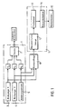
- Figure 1 shows in general at 1 a tunnel effect microscope.
- This microscope comprises a probe 2 constituted by a needle, the end of which forms a monoatomic point and the movements of which are ensured by means of piezoelectric crystals P X , P Y and P Z.
- the crystals P X and P Y are controlled respectively by a unit 3 for scanning in X, and a unit 4 for scanning in Y.
- the piezoelectric crystal P Z is controlled by a servo-control 5 tending to keep constant the current which circulates between the probe 2 and either the substrate or the polynucleotide 16 (FIG. 2) when a potential difference is applied between this probe and the substrate or the polynucleotide.
- the X, Y and Z coordinates of the end of the probe 2 are sampled in a sampler 10, the output of which is applied to the input of a processing unit 11.
- the processing unit 11 essentially consists of a module 12 for reconstituting the image, a module for processing images 13, and a recognition module 14.
- the module 12 makes it possible, from the sampled data, to reconstruct a plan image of the surface of the substrate and of the nucleic acid fragment which is deposited there.
- the processing unit 13 aims to eliminate from the image thus obtained, the image of the atoms of the substrate and of the atoms of the phosphoryl and ribose or deoxyribose groups so that only the purine or pyrimidine nuclei remain on the image as well as the atoms of the marker optionally used to distinguish the two bases of the purine type from one another as well as the two bases of the pyrimidine type.
- This can be achieved using an image processing program as there are in many technical fields.
- the recognition module 14 has the function of successively recognizing each base.
- the result thus obtained is stored in a storage unit 15 which therefore contains, at the end of the operation, the base sequence of the polynucleotide sequenced by the method according to the invention.
- the substrate 6 is constituted by a graphite crystal obtained for example by epitaxy, so that its surface 17 has no defect.
- a groove 18 is formed on the surface 17 of the substrate 6 by techniques known in microelectronics.
- marks 19 are engraved at regular intervals along the groove also by known means.
- the substrate also comprises a well (not shown) at each end of the groove 18 in order to immerse two electrodes making it possible to create an electric field in a solution bathing these wells and the groove.
- the groove may for example have a width of approximately 2 microns for a depth of approximately 100 nanometers.
- the bottom of the groove is doped in the form of a strip perpendicular to the groove and a width of about 100 nonometers, either of an electrostatic charge, or of functional groups having a high reactivity with the grafted functional groups. at the end of the polynucleotide.
- the electrostatic charge can be obtained either by removing electrons produced directly with a tunneling microscope, or by depositing a molecular layer of a molecule or depositing a charged macromolecule or a cluster of charged molecules.
- a monomolecular layer of general shape CH3 (CH2) n-2 COOH with n between 16 and 22 will be deposited, this monomolecular layer receiving functional groups inserted at the surface (Langmuir-Blotgett technique).
- a micro-drop 21 is deposited in the groove 18 upstream of the barrier 20 in the direction of migration, in a solution bathing the groove.
- This microdrop contains the polynucleotide to be sequenced 22 at one end of which has been synthesized a fragment 23 of known sequence, and a functional group 24 positively charged.
- the microdrop moves towards the barrier 20 which, in the present case is positively charged.
- the group 24 is therefore retained upstream of the barrier 20, the polynucleotide 22 continuing to stretch downstream of this barrier.
- the procedure is as described above, in order to separate the two strands, and the solution bathing the groove is evaporated.
- the end of the probe 2 is then brought in the vicinity of the bottom of the groove, and with this probe a primary scan in X is carried out over the width of the groove 18, and a secondary scan in Y while keeping the distance of the constant probe 2 tip at the surface.
- X, Y and Z are sampled as the scanning progresses with a precision which, in the current state of the art can be of the order of 0.1 angstroms, and the data thus obtained are entered into the processing unit.
- an image such as that shown in FIG. 5 is obtained and can be recognized by the shape recognition module 14.
- the method according to the invention therefore makes it possible to analyze extremely quickly a long fragment of nucleic acid.
- the processing of the data collected can be carried out in real time, in which case with the current shape recognition techniques the scanning must be relatively slow, but another possibility consists in carrying out the processing only in deferred time, in which case the analysis can be performed at high speed.
Landscapes
- Physics & Mathematics (AREA)
- Health & Medical Sciences (AREA)
- General Health & Medical Sciences (AREA)
- General Physics & Mathematics (AREA)
- Nuclear Medicine, Radiotherapy & Molecular Imaging (AREA)
- Radiology & Medical Imaging (AREA)
- Measuring Or Testing Involving Enzymes Or Micro-Organisms (AREA)
- Apparatus Associated With Microorganisms And Enzymes (AREA)
- Investigating Or Analysing Biological Materials (AREA)
Applications Claiming Priority (2)
| Application Number | Priority Date | Filing Date | Title |
|---|---|---|---|
| FR8908284A FR2648913B1 (fr) | 1989-06-21 | 1989-06-21 | Procede et dispositif de sequencage d'un polynucleotide |
| FR8908284 | 1989-06-21 |
Publications (3)
| Publication Number | Publication Date |
|---|---|
| EP0404682A2 true EP0404682A2 (de) | 1990-12-27 |
| EP0404682A3 EP0404682A3 (de) | 1991-07-24 |
| EP0404682B1 EP0404682B1 (de) | 1995-04-12 |
Family
ID=9382990
Family Applications (1)
| Application Number | Title | Priority Date | Filing Date |
|---|---|---|---|
| EP90401763A Expired - Lifetime EP0404682B1 (de) | 1989-06-21 | 1990-06-21 | Verfahren und Vorrichtung zur Sequenzierung eines Polynukleotids |
Country Status (5)
| Country | Link |
|---|---|
| EP (1) | EP0404682B1 (de) |
| JP (1) | JPH03128459A (de) |
| AT (1) | ATE121194T1 (de) |
| DE (1) | DE69018523T2 (de) |
| FR (1) | FR2648913B1 (de) |
Cited By (6)
| Publication number | Priority date | Publication date | Assignee | Title |
|---|---|---|---|---|
| EP0511662A1 (de) * | 1991-04-30 | 1992-11-04 | Matsushita Electric Industrial Co., Ltd. | Raster-Abtastmikroskop, molekulares Verarbeitungsverfahren unter Verwendung des Mikroskops und Verfahren zum Wahrnehmen der DNA-Basen-Anordnung |
| WO2004036591A1 (en) * | 2002-10-17 | 2004-04-29 | Intel Corporation | Model-based fusion of scanning probe microscopic images for detection and identification of molecular structures |
| US6931326B1 (en) | 2000-06-26 | 2005-08-16 | Genaissance Pharmaceuticals, Inc. | Methods for obtaining and using haplotype data |
| US7058517B1 (en) | 1999-06-25 | 2006-06-06 | Genaissance Pharmaceuticals, Inc. | Methods for obtaining and using haplotype data |
| US7606403B2 (en) | 2002-10-17 | 2009-10-20 | Intel Corporation | Model-based fusion of scanning probe microscopic images for detection and identification of molecular structures |
| US7736818B2 (en) | 2004-12-27 | 2010-06-15 | Inphase Technologies, Inc. | Holographic recording medium and method of making it |
Family Cites Families (1)
| Publication number | Priority date | Publication date | Assignee | Title |
|---|---|---|---|---|
| GB8910566D0 (en) * | 1989-05-08 | 1989-06-21 | Amersham Int Plc | Imaging apparatus and method |
-
1989
- 1989-06-21 FR FR8908284A patent/FR2648913B1/fr not_active Expired - Fee Related
-
1990
- 1990-06-21 JP JP2163980A patent/JPH03128459A/ja active Pending
- 1990-06-21 EP EP90401763A patent/EP0404682B1/de not_active Expired - Lifetime
- 1990-06-21 DE DE69018523T patent/DE69018523T2/de not_active Expired - Fee Related
- 1990-06-21 AT AT90401763T patent/ATE121194T1/de not_active IP Right Cessation
Cited By (9)
| Publication number | Priority date | Publication date | Assignee | Title |
|---|---|---|---|---|
| EP0511662A1 (de) * | 1991-04-30 | 1992-11-04 | Matsushita Electric Industrial Co., Ltd. | Raster-Abtastmikroskop, molekulares Verarbeitungsverfahren unter Verwendung des Mikroskops und Verfahren zum Wahrnehmen der DNA-Basen-Anordnung |
| US5363697A (en) * | 1991-04-30 | 1994-11-15 | Matsushita Electric Industrial Co., Ltd. | Scanning probe microscope, molecular processing method using the scanning probe microscope and DNA base arrangement detecting method |
| US5730940A (en) * | 1991-04-30 | 1998-03-24 | Matsushita Electric Industrial Co., Ltd. | Scanning probe microscope and molecular processing method using the scanning probe microscope |
| US7058517B1 (en) | 1999-06-25 | 2006-06-06 | Genaissance Pharmaceuticals, Inc. | Methods for obtaining and using haplotype data |
| US6931326B1 (en) | 2000-06-26 | 2005-08-16 | Genaissance Pharmaceuticals, Inc. | Methods for obtaining and using haplotype data |
| WO2004036591A1 (en) * | 2002-10-17 | 2004-04-29 | Intel Corporation | Model-based fusion of scanning probe microscopic images for detection and identification of molecular structures |
| US7606403B2 (en) | 2002-10-17 | 2009-10-20 | Intel Corporation | Model-based fusion of scanning probe microscopic images for detection and identification of molecular structures |
| US8934683B2 (en) | 2002-10-17 | 2015-01-13 | Intel Corporation | Model-based fusion of scanning probe microscopic images for detection and identification of molecular structures |
| US7736818B2 (en) | 2004-12-27 | 2010-06-15 | Inphase Technologies, Inc. | Holographic recording medium and method of making it |
Also Published As
| Publication number | Publication date |
|---|---|
| EP0404682A3 (de) | 1991-07-24 |
| DE69018523T2 (de) | 1996-01-11 |
| FR2648913B1 (fr) | 1991-10-04 |
| EP0404682B1 (de) | 1995-04-12 |
| ATE121194T1 (de) | 1995-04-15 |
| DE69018523D1 (de) | 1995-05-18 |
| FR2648913A1 (fr) | 1990-12-28 |
| JPH03128459A (ja) | 1991-05-31 |
Similar Documents
| Publication | Publication Date | Title |
|---|---|---|
| EP0693135B1 (de) | Schnelles verfahren fur die dns sequenzierung und diagnostische anwendungen | |
| US5612181A (en) | Method for arranging a polynucleotide on a substrate for point-by-point analysis of the bases thereof | |
| CN1159457C (zh) | Dna测序方法 | |
| US9400259B2 (en) | Method of making a microbead array with attached biomolecules | |
| US20070178507A1 (en) | Method and apparatus for detection of molecules using nanopores | |
| WO1995022056A1 (fr) | Surfaces hautement specifiques pour reactions biologiques, procede pour leur preparation et procede pour leur utilisation | |
| US5538898A (en) | Method suitable for identifying a code sequence of a biomolecule | |
| US20110100820A1 (en) | Triple function electrodes | |
| US20150159213A1 (en) | Nanopore sequencing using n-mers | |
| WO1996024689A1 (en) | Method and apparatus for determining the sequence of polynucleotides | |
| EP0404682B1 (de) | Verfahren und Vorrichtung zur Sequenzierung eines Polynukleotids | |
| US5624845A (en) | Assembly and a method suitable for identifying a code | |
| FR2737574A1 (fr) | Appareillage d'alignement parallele de macromolecules et utilisation | |
| EP1851331B1 (de) | Verfahren und vorrichtung zur trennung molekularer ziele in einem komplexen gemisch | |
| WO2021226291A1 (en) | Single-biomolecule-bridged sequencing biosensors and storage devices and methods for same | |
| KR100386606B1 (ko) | Dna 검출 방법 및 그 장치 | |
| Efcavitch et al. | Single-molecule DNA analysis | |
| FR2799988A1 (fr) | Procede de separation d'un compose chimique ou biologique dans un melange de composes similaires par diffusion dans un milieu tel qu'un gel | |
| WO1998037234A1 (fr) | Procede de caracterisation de duplex d'acide nucleique | |
| Li et al. | Docking and Activity of DNA Polymerase on Solid-State Nanopores | |
| FR2747786A1 (fr) | Dispositif integre pour la detection moleculaire optique et procede de cette detection | |
| Lanzavecchia | Customized 3D Nanopore Fabrication for Advanced Electrical and Optical Applications | |
| CA2239499C (fr) | Procede et dispositif de traitement par complexation d'un milieu liquide | |
| JP4045535B2 (ja) | ビーズを用いた生化学的分析方法 | |
| EP0478450A1 (de) | Verfahren zur schnellen Sequenzierung von linearen und geordneten, biologischen Sequenzen |
Legal Events
| Date | Code | Title | Description |
|---|---|---|---|
| PUAI | Public reference made under article 153(3) epc to a published international application that has entered the european phase |
Free format text: ORIGINAL CODE: 0009012 |
|
| AK | Designated contracting states |
Kind code of ref document: A2 Designated state(s): AT BE CH DE DK ES FR GB GR IT LI LU NL SE |
|
| PUAL | Search report despatched |
Free format text: ORIGINAL CODE: 0009013 |
|
| AK | Designated contracting states |
Kind code of ref document: A3 Designated state(s): AT BE CH DE DK ES FR GB GR IT LI LU NL SE |
|
| 17P | Request for examination filed |
Effective date: 19911018 |
|
| 17Q | First examination report despatched |
Effective date: 19931018 |
|
| GRAA | (expected) grant |
Free format text: ORIGINAL CODE: 0009210 |
|
| AK | Designated contracting states |
Kind code of ref document: B1 Designated state(s): AT BE CH DE DK ES FR GB GR IT LI LU NL SE |
|
| PG25 | Lapsed in a contracting state [announced via postgrant information from national office to epo] |
Ref country code: IT Free format text: LAPSE BECAUSE OF FAILURE TO SUBMIT A TRANSLATION OF THE DESCRIPTION OR TO PAY THE FEE WITHIN THE PRE;WARNING: LAPSES OF ITALIAN PATENTS WITH EFFECTIVE DATE BEFORE 2007 MAY HAVE OCCURRED AT ANY TIME BEFORE 2007. THE CORRECT EFFECTIVE DATE MAY BE DIFFERENT FROM THE ONE RECORDED.SCRIBED TIME-LIMIT Effective date: 19950412 Ref country code: ES Free format text: THE PATENT HAS BEEN ANNULLED BY A DECISION OF A NATIONAL AUTHORITY Effective date: 19950412 Ref country code: DK Effective date: 19950412 Ref country code: GR Free format text: LAPSE BECAUSE OF FAILURE TO SUBMIT A TRANSLATION OF THE DESCRIPTION OR TO PAY THE FEE WITHIN THE PRESCRIBED TIME-LIMIT Effective date: 19950412 Ref country code: AT Effective date: 19950412 |
|
| REF | Corresponds to: |
Ref document number: 121194 Country of ref document: AT Date of ref document: 19950415 Kind code of ref document: T |
|
| REF | Corresponds to: |
Ref document number: 69018523 Country of ref document: DE Date of ref document: 19950518 |
|
| PG25 | Lapsed in a contracting state [announced via postgrant information from national office to epo] |
Ref country code: LU Free format text: LAPSE BECAUSE OF NON-PAYMENT OF DUE FEES Effective date: 19950630 |
|
| GBT | Gb: translation of ep patent filed (gb section 77(6)(a)/1977) |
Effective date: 19950606 |
|
| PG25 | Lapsed in a contracting state [announced via postgrant information from national office to epo] |
Ref country code: SE Effective date: 19950712 |
|
| PLBE | No opposition filed within time limit |
Free format text: ORIGINAL CODE: 0009261 |
|
| STAA | Information on the status of an ep patent application or granted ep patent |
Free format text: STATUS: NO OPPOSITION FILED WITHIN TIME LIMIT |
|
| 26N | No opposition filed | ||
| PGFP | Annual fee paid to national office [announced via postgrant information from national office to epo] |
Ref country code: GB Payment date: 19960613 Year of fee payment: 7 |
|
| PGFP | Annual fee paid to national office [announced via postgrant information from national office to epo] |
Ref country code: FR Payment date: 19960616 Year of fee payment: 7 |
|
| PGFP | Annual fee paid to national office [announced via postgrant information from national office to epo] |
Ref country code: CH Payment date: 19960625 Year of fee payment: 7 |
|
| PGFP | Annual fee paid to national office [announced via postgrant information from national office to epo] |
Ref country code: NL Payment date: 19960630 Year of fee payment: 7 |
|
| PGFP | Annual fee paid to national office [announced via postgrant information from national office to epo] |
Ref country code: BE Payment date: 19960711 Year of fee payment: 7 |
|
| PGFP | Annual fee paid to national office [announced via postgrant information from national office to epo] |
Ref country code: DE Payment date: 19960726 Year of fee payment: 7 |
|
| PG25 | Lapsed in a contracting state [announced via postgrant information from national office to epo] |
Ref country code: GB Free format text: LAPSE BECAUSE OF NON-PAYMENT OF DUE FEES Effective date: 19970621 |
|
| PG25 | Lapsed in a contracting state [announced via postgrant information from national office to epo] |
Ref country code: CH Free format text: LAPSE BECAUSE OF NON-PAYMENT OF DUE FEES Effective date: 19970630 Ref country code: BE Effective date: 19970630 Ref country code: LI Free format text: LAPSE BECAUSE OF NON-PAYMENT OF DUE FEES Effective date: 19970630 |
|
| BERE | Be: lapsed |
Owner name: FOURMENTIN-GUILBERT JEAN ERNEST RAYMOND Effective date: 19970630 |
|
| PG25 | Lapsed in a contracting state [announced via postgrant information from national office to epo] |
Ref country code: NL Effective date: 19980101 |
|
| GBPC | Gb: european patent ceased through non-payment of renewal fee |
Effective date: 19970621 |
|
| REG | Reference to a national code |
Ref country code: CH Ref legal event code: PL |
|
| PG25 | Lapsed in a contracting state [announced via postgrant information from national office to epo] |
Ref country code: FR Free format text: LAPSE BECAUSE OF NON-PAYMENT OF DUE FEES Effective date: 19980227 |
|
| NLV4 | Nl: lapsed or anulled due to non-payment of the annual fee |
Effective date: 19980101 |
|
| PG25 | Lapsed in a contracting state [announced via postgrant information from national office to epo] |
Ref country code: DE Free format text: LAPSE BECAUSE OF NON-PAYMENT OF DUE FEES Effective date: 19980303 |
|
| REG | Reference to a national code |
Ref country code: FR Ref legal event code: ST |
|
| REG | Reference to a national code |
Ref country code: FR Ref legal event code: ST |