EP0429295A2 - Spectroscopie à résonance magnétique - Google Patents
Spectroscopie à résonance magnétique Download PDFInfo
- Publication number
- EP0429295A2 EP0429295A2 EP90312655A EP90312655A EP0429295A2 EP 0429295 A2 EP0429295 A2 EP 0429295A2 EP 90312655 A EP90312655 A EP 90312655A EP 90312655 A EP90312655 A EP 90312655A EP 0429295 A2 EP0429295 A2 EP 0429295A2
- Authority
- EP
- European Patent Office
- Prior art keywords
- resonance
- region
- pulse
- gradient
- interest
- Prior art date
- Legal status (The legal status is an assumption and is not a legal conclusion. Google has not performed a legal analysis and makes no representation as to the accuracy of the status listed.)
- Withdrawn
Links
Images


Classifications
-
- G—PHYSICS
- G01—MEASURING; TESTING
- G01R—MEASURING ELECTRIC VARIABLES; MEASURING MAGNETIC VARIABLES
- G01R33/00—Arrangements or instruments for measuring magnetic variables
- G01R33/20—Arrangements or instruments for measuring magnetic variables involving magnetic resonance
- G01R33/44—Arrangements or instruments for measuring magnetic variables involving magnetic resonance using nuclear magnetic resonance [NMR]
- G01R33/48—NMR imaging systems
- G01R33/483—NMR imaging systems with selection of signals or spectra from particular regions of the volume, e.g. in vivo spectroscopy
- G01R33/4838—NMR imaging systems with selection of signals or spectra from particular regions of the volume, e.g. in vivo spectroscopy using spatially selective suppression or saturation of MR signals
-
- G—PHYSICS
- G01—MEASURING; TESTING
- G01R—MEASURING ELECTRIC VARIABLES; MEASURING MAGNETIC VARIABLES
- G01R33/00—Arrangements or instruments for measuring magnetic variables
- G01R33/20—Arrangements or instruments for measuring magnetic variables involving magnetic resonance
- G01R33/44—Arrangements or instruments for measuring magnetic variables involving magnetic resonance using nuclear magnetic resonance [NMR]
- G01R33/48—NMR imaging systems
- G01R33/483—NMR imaging systems with selection of signals or spectra from particular regions of the volume, e.g. in vivo spectroscopy
- G01R33/485—NMR imaging systems with selection of signals or spectra from particular regions of the volume, e.g. in vivo spectroscopy based on chemical shift information [CSI] or spectroscopic imaging, e.g. to acquire the spatial distributions of metabolites
Definitions
- This invention relates to the art of magnetic resonance spectroscopy. It finds particular application in conjunction with in-vivo examinations and will be described with particular reference thereto. However, it is to be appreciated that the invention may find further application in conjunction with spectroscopy examination of localized regions for imaging, chemical shift analysis, quality assurance, drug flow tracing, and the like.
- phase encoding techniques phase encoding gradients are applied across the sample such that the resonance signals emanating from various locations across the sample are spatially encoded by this phase.
- voxel localization the signal is only recovered from a selected small volumetric element or voxel.
- phase encoding technigues typically provide spatial definition over a region whose size is defined by the sensitive volume of the receiver coil. Because the sensitive volume is determined by hardware geometry, it is substantially fixed from study to study with little aptitude for adjustment.
- One of the problems with phase encoding techniques is that they are susceptible to errors resulting from layers of different tissue types.
- the boundary layers between materials with different magnetic susceptibilities causes large magnetic field inhomogeneities that degrade water suppression and spatial resolution.
- fat layers that surround the skull and other regions of the body commonly produce much larger magnetic resonance signals than metabolites of interest.
- the fat signal response can obscure the spectra of voxels of interest due to leakage of the point-spread function from neighboring voxels. In this manner, the fat signals degrade the dynamic range of the resultant signals limiting the ability to detect low concentrations of metabolites.
- Voxel localization techniques such as the technique described in U.S. Patent No. 4,771,242, recover the signal from only a small voxel in the sample.
- To map or measure chemical concentrations over an extended region a plurality of single voxel experiments are conducted. In each repetition, the voxel is defined at different location within the sample. Although this technique is accurate, it tends to be time consuming.
- a volume is selected with refocused stimulated echoes.
- This volume selection technique utilizes a series of 90° pulses. Phase encoding for one or two dimensional spectroscopic imaging is combined with the volume selection in order to suppress unwanted lipid signals from surrounding tissue.
- subvoxels can be defined within a relative large voxel, this technique has several drawbacks. First, the 90° stimulated echo sequence recovers only half the signal. The other half is lost.
- these voxel techniques require three RF pulses in order to localize the voxel. Because the first RF pulse initially excites resonance, the time required for the following two pulses extends the duration between the first pulse excitation and data collection. Extending this duration limits the recovered signal to the contribution from dipoles with a longer T2 relaxation time.
- a method of magnetic resonance spectroscopy comprising: defining a region of interest by pre-saturating regions therearound; exciting resonance in the region of interest; encoding spatial locations along at least one direction by phase encoding the excited resonance along the selected direction; and collecting resonance data in the absence of a magnetic field gradient and Fourier transforming the collected data into an array of spectra, each spectrum corresponding to the resonance signal from a spatial section along the phase encode direction.
- a method of generating a magnetic resonance spectrum corresponding to each of an at least two dimensional array of subregions of a region of interest comprising applying radio frequency and gradient pulse combinations for saturating material on four sides of the region of interest along first and second axes; concurrently applying a radio frequency pulse for exciting magnetic resonance in the region of interest and a slice select gradient for limiting the excited region along a third axis; applying phase encode gradients for phase encoding the excited resonance along at least two directions; collecting resonance data in the absence of a magnetic field gradient; multi-dimensionally Fourier transforming the collected resonance data into an at least two dimensional array of multi-frequency spectra.
- an apparatus for magnetic resonance spectroscopy comprising means for concurrently applying combinations of radio frequency and gradient pulses for pre-saturating material around a region of interest; means for applying a radio frequency excitation pulse to excite magnetic resonance in the region of interest; means for applying a spatial position encoding gradient pulse for encoding position along a first direction; means for receiving a magnetic resonance signal in the absence of a magnetic field gradient; a Fourier transform means for Fourier transforming the received magnetic resonance signal into an array of spectra, each spectra corresponding to a different spatial location along the encoded direction.
- One advantage of the present invention resides in its shorter delay to acquisition.
- Another advantage of the present invention is its high signal-to-noise ratio.
- the full spin echo signal-to-noise ratio is attained.
- Another advantage of the present invention is that it employs a single spin echo method with only two RF pulses which affect the metabolites of interest.
- Still further advantages of the present invention include signal shot localization for shimming, refocusing of field homogeneity errors, and the elimination of selective refocusing pulses.
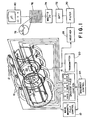
- a main magnetic field means including a main magnetic field control 10 and coils 12 generate a substantially uniform main magnetic field through an examination region 14.
- a gradient field means selectively causes magnetic field gradients across the main magnetic field in the examination region 14. In the preferred embodiment, the gradients are caused along three orthogonal axes, commonly denoted as x, y, and z.
- a magnetic field control 16 generates current pulses of an appropriate amplitude, duration, and configuration so as to cause gradient field coils 18 to generate the selected magnetic field gradients across the examination region.
- a radio frequency transmitter 20 transmits radio frequency signals into the examination region to pre-saturate dipoles in selected regions, to excite resonance, to refocus the resonance, and to perform other appropriate manipulations of the resonance.
- Radio frequency pulses from the transmitter 20 are applied to a radio frequency coil 22 that surrounds the examination region.
- a sequence control means 24 controls the gradient magnetic field control 16 and the radio frequency transmitter 20 to transmit selected pulse sequences.
- the sequence control 24 , the gradient field control 16 , and the RF transmitter 20 incorporates means for defining a selected volume of the examination region 14 for exciting resonance in a selected slab or slice, and phase encoding resonance received from the slab or slice by a radio frequency receiver 26 .
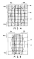
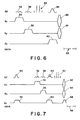
- a pre-saturation gradient pulse 30 and a first pre-saturation radio frequency pulse 32 are applied concurrently to saturate the spins in regions 34a and 34b of FIGURE 3.
- a gradient pulse 36 and radio frequency pulse configuration 38 are applied concurrently to pre-saturate spins in regions 40a and 40b of FIGURE 3.
- a voxel or columnar region of interest 42 is defined displaced away from a periphery 44 of a patient's head or other body part. This moves the imaged region away from the fat layer commonly found under the surface of the body portion.
- the radio frequency pulses 32 and 38 are preferably bimodal pulses as described in "Computer-Optimized Narrowband Pulses for Multislice Imaging" by J.B. Murdoch, A.H. Lent, and M.R. Kritzer, J. Mag. Res., Vol. 74, pp. 226-263 (1987), the text of which is incorporated herein by reference. Unimodal pulses may also be utilized.
- the spatial (non-frequency selective) pre-saturating pulses 32 and 38 remove the signal from the unwanted regions 34 and 40 on four sides of the region of interest 42 , e.g. from the fat and the scalp around the head.
- the pulses are applied with an appropriate frequency offset in order to saturate the desired regions on either side of the voxel in multiple tilted planes.
- a spin echo sequence with two or more dimensions of phase encoding is applied.
- the spin echo is formed by applying an RF excitation pulse 50 with a flip angle ⁇ followed by a selective binomial refocusing pulse 52 .
- the refocusing pulse is of the form 2 ⁇ -6 ⁇ -6 ⁇ -2 ⁇ .
- Dephasing gradient pulses 54 may be applied as necessary before and after the binomial pulse to dephase any unwanted transverse magnetization that might be generated.
- a slice select gradient 60 is applied concurrently with the resonance exciting RF pulse 50 .
- a first phase encoding gradient 62 and a second phase encoding gradient 64 apply phase encoding along two axes of the defined slice.
- the phase encode gradients may be applied either before or after the binomial refocusing pulse 52 .
- the phase encode gradients provide spatial encoding along the defined slice.
- a third phase encode gradient 66 may be applied to spatially encode the magnetic resonance symbol along the slice or slab to provide a resolvable three spatial dimensions of data.
- a magnetic resonance echo peak forms the same duration after the binomial pulse as the duration by which the binomial pulse followed the excitation pulse 50 .
- a data acquisition period 68 is defined to start collecting data at the center of the magnetic resonance echo and continue collecting data until the signal to noise ratio becomes unacceptably low.
- the magnetic resonance echo is picked up by the radio frequency coil 22 or other radio frequency reception coil, such as a surface coil (not shown).
- the radio frequency receiver 70 is controlled by the sequence control means 24 in order to define the data acquisition period 68 .
- a filter 70 smooths the data to improve the signal and remove noise artifacts.
- a one dimensional Fourier transform means 72 removes spatially encoded frequency data.
- a two dimensional Fourier transform 74 of the two dimensionally phase encoded data separates frequency spectra by spatial location. If the data is phase encoded in three dimensions, a three dimensional Fourier transform is performed.
- a memory means 76 defines a two dimensional matrix of memory addresses corresponding to the two dimensional array of subregions in the defined voxel or region of interest 42 . Each memory address stores an NMR frequency data set 78 representative of the frequency spectrum generated by the nuclei in the corresponding subregion. If it were desirable to form an image from this data, one would select the same part of each frequency spectrum 78 to define the gray scale for the corresponding element or pixel of the image.
- a display means 80 such as a video monitor, selectively displays the frequency data from the first Fourier transform means 72 , the frequency spectra 78 from one or more of the matrix elements, or a gray scale image corresponding to a selected part of the frequency spectrum.
- the spectra may be analyzed by computer to generate a direct display of one or more of the chemical constituents of the corresponding subregion. For example, an image of relative concentration of a selected constituent in each region may be displayed.
- the phase encoding may be limited to one axis. When phase encoded in the third direction, a three dimensional array of frequency spectra is defined corresponding to a plurality of effective slices as may be defined by the phase encode gradient 66 .
- phase encoded region relative to the pre-saturated/selected region or voxel is important in terms of the aliasing of the signal in the acquisition region.
- the phase encoded region is totally within the presaturated/selected region.
- the phase encoding is sized to extend over a region 82 that extends beyond the defined voxel or region of interest 42 .
- the phase encode gradient pulses 60 and 62 are enlarged such that each array element is larger causing the region to which phase encoding is applied to overlap a portion of the saturated regions 34 , 40 .
- the phase encoding may be enlarged along one axis to overlap a portion of periphery 44 .
- the phase encoding is utilized to identify and remove signals emanating from the pre-saturated region.
- the large signals from fat tend to bleed back into the voxel 42 because the pre-saturated dipoles can generate frequency components which are indistinguishable from the portions of the signals emanating from the selected voxel 42 .
- the phase encoding of these out-of voxel signals in FIGURES 4 and 5 enable them to be identified for removal.
- pre-saturation gradient and radio frequency pulse combinations 30 , 32 and 36 , 38 again pre-saturate the spins in regions 34 and 40 .
- a binomial excitation pulse 84 of the form ⁇ -3 ⁇ -3 ⁇ - ⁇ is followed by a bi- or unimodal refocusing pulse 86 applied concurrently with the slice select gradient 60 .
- the refocusing pulse is of amplitude ⁇ .
- the binomial pulse 84 is a 90° pulse, but may have smaller angles where different contrasts are desired.
- Spatial position encoding gradient pulses 62 and 64 are applied after, or before, the refocusing pulse 86 such that the magnetic resonance echo data received during the acquisition period 68 is phase encoded as an indicia of location. Dephasing gradients may again be applied as necessary to dephase any transverse magnetization generated by the refocusing pulse/slice select gradient pulse combination.
- an inversion pulse 90 such as a 180° pulse
- a slice select gradient 92 A duration later, which duration is calculated to be long enough for the T2 contribution to have attenuated significantly, the pre-saturation gradient and RF pulse combinations 30 , 32 and 36 , 38 are applied.
- the 90° RF pulse 50 is applied concurrently with the slice select gradient 60 followed by a binomial inversion pulse 52 to cause T1 weighted echo.
- Phase encode gradients 62 and 64 may be applied on either side of the binomial pulse 52 such that the echo which forms during the data acquisition period 68 is spatially encoded.
- the slice may be phase encoded in the third direction by phase encode gradient 66 for volumetric imaging.
- the present invention may also be applicable to multi-echo sequences.
- pre-saturation gradient and RF pulse pairs 30 , 32 and 36 , 38 are applied to pre-saturated regions 34 and 40 .
- the excitation pulse 50 applied in the presence of the slice select gradient 54 excites resonance in the selected slice or slab.
- the binomial inversion pulse 52 inverts the magnetization to refocus in an echo in the data acquisition period 68 .
- One or more of phase encode gradients 62 , 64 , and 66 are applied to encode spatial information in the resultant echo.
- another selective refocusing pulse 94 is applied to invert the magnetization to refocus in a second echo.
- the receiver is controlled to provide a second data acquisition period 96 for monitoring the second echo.
- the second echo tends to be more heavily weighted toward T2 spectra than the first echo.
- the present invention is also applicable to non-echo encoding techniques.
- pre-saturating RF and gradient pulse pairs 30 , 32 ; 36 , 38 ; and 100 pre-saturate regions of the patient surrounding a three dimensional voxel of interest.
- a 90° broadband excitation pulse 104 is applied to excite resonance within the voxel and a free induction decay resonance signal.
- the phase encoding gradients 62 , 64 , or 66 are applied along one or more of the axes in order to phase encode the resonance in up to three dimensions.
- phase encoded free induction decay signal from the resonating phosphorus 31 nuclei are monitored by the receiver 26 during the data acquisition period 68 .
- One advantage of this encoding scheme is that the region over which shimming occurs is limited resulting in higher homogeneity. Because no spin echo is formed, a linear phase shift is observed in the data that may be corrected using linear prediction methods.
- this non-echo technique it is preferred that the 90° excitation pulse have a binomial pulse configured to suppress water. This technique, again, has linear phase shift problems but is advantageous in that the data acquisition period follows the excitation even more closely than the echo sequences.
Landscapes
- Physics & Mathematics (AREA)
- Spectroscopy & Molecular Physics (AREA)
- Optics & Photonics (AREA)
- High Energy & Nuclear Physics (AREA)
- Condensed Matter Physics & Semiconductors (AREA)
- General Physics & Mathematics (AREA)
- Magnetic Resonance Imaging Apparatus (AREA)
Applications Claiming Priority (2)
| Application Number | Priority Date | Filing Date | Title |
|---|---|---|---|
| US440894 | 1989-11-22 | ||
| US07/440,894 US5010300A (en) | 1989-08-11 | 1989-11-22 | Multi-axis pre-saturated spin echo phase encoded spectroscopy |
Publications (2)
| Publication Number | Publication Date |
|---|---|
| EP0429295A2 true EP0429295A2 (fr) | 1991-05-29 |
| EP0429295A3 EP0429295A3 (en) | 1991-09-25 |
Family
ID=23750626
Family Applications (1)
| Application Number | Title | Priority Date | Filing Date |
|---|---|---|---|
| EP19900312655 Withdrawn EP0429295A3 (en) | 1989-11-22 | 1990-11-21 | Magnetic resonance spectroscopy |
Country Status (2)
| Country | Link |
|---|---|
| US (1) | US5010300A (fr) |
| EP (1) | EP0429295A3 (fr) |
Cited By (1)
| Publication number | Priority date | Publication date | Assignee | Title |
|---|---|---|---|---|
| GB2348708A (en) * | 1999-01-09 | 2000-10-11 | Univ Ludwigs Albert | MR spectroscopy |
Families Citing this family (14)
| Publication number | Priority date | Publication date | Assignee | Title |
|---|---|---|---|---|
| DE4018683A1 (de) * | 1989-06-23 | 1991-01-10 | Siemens Ag | Schichtprofiloptimierung fuer ein mit einer gradientenechosequenz betriebenes kernspin-tomographiegeraet |
| DE4024161A1 (de) * | 1989-08-11 | 1991-02-14 | Siemens Ag | Pulssequenz zur schnellen ermittlung von bildern der fett- und wasserverteilung in einem untersuchungsobjekt mittels der kernmagnetischen resonanz |
| GB9109592D0 (en) * | 1991-05-02 | 1991-07-17 | Nat Res Dev | Methods and apparatus for detecting substances |
| US5250899A (en) * | 1991-08-12 | 1993-10-05 | Trustees Of The University Of Pennsylvania | Method for fat suppression in magnetic resonance imaging |
| DE4405979C1 (de) * | 1994-02-24 | 1995-10-05 | Univ Ludwigs Albert | Verfahren der bildgebenden magnetischen Kernresonanz nach dem Mehrschichtverfahren |
| US5685305A (en) * | 1994-08-05 | 1997-11-11 | The United States Of America As Represented By The Department Of Health And Human Services | Method and system for MRI detection of abnormal blood flow |
| US6114853A (en) * | 1997-06-25 | 2000-09-05 | Toshiba America Mri, Inc. | NMR methods for qualification of sequence-induced B0 oscillation and correction of the resultant image artifacts in MRI |
| DK1042820T3 (da) * | 1997-12-19 | 2003-07-14 | Siemens Ag | Superlederkonstruktion med høj-Tc-superledermateriale, fremgangsmåde til fremstilling af konstruktionen samt strømbegrænserindretning med en sådan konstruktion |
| DE10108341C1 (de) * | 2001-02-21 | 2002-09-12 | Siemens Ag | Magnetresonanz-Spektroskopieverfahren mit einem Variieren von Phasen von HF-Pulsen |
| US6541971B1 (en) * | 2001-06-28 | 2003-04-01 | Koninklijke Philips Electronics, N.V. | Multi-dimensional spatial NMR excitation |
| US9360542B2 (en) * | 2012-04-20 | 2016-06-07 | Wisconsin Alumni Research Foundation | System and method for spectrally-resolved three-dimensional magnetic resonance imaging without frequency-encoding gradients |
| EP2976653A4 (fr) * | 2013-04-19 | 2017-02-08 | Halliburton Energy Services, Inc. | Procédés et systèmes de commande d'intervalle inter-écho de résonance magnétique nucléaire (rmn) |
| WO2017007578A1 (fr) | 2015-07-07 | 2017-01-12 | Kogent Surgical, LLC | Sonde micro-chirurgicale éclairée |
| CN117547246B (zh) * | 2024-01-12 | 2024-04-09 | 中国科学技术大学先进技术研究院 | 空间信号饱和方法、装置、设备及计算机可读存储介质 |
Family Cites Families (2)
| Publication number | Priority date | Publication date | Assignee | Title |
|---|---|---|---|---|
| US4506223A (en) * | 1982-11-22 | 1985-03-19 | General Electric Company | Method for performing two-dimensional and three-dimensional chemical shift imaging |
| US4742304A (en) * | 1986-05-02 | 1988-05-03 | Phospho-Energetics, Inc. | Multiple tuning NMR probe |
-
1989
- 1989-11-22 US US07/440,894 patent/US5010300A/en not_active Expired - Lifetime
-
1990
- 1990-11-21 EP EP19900312655 patent/EP0429295A3/en not_active Withdrawn
Cited By (3)
| Publication number | Priority date | Publication date | Assignee | Title |
|---|---|---|---|---|
| GB2348708A (en) * | 1999-01-09 | 2000-10-11 | Univ Ludwigs Albert | MR spectroscopy |
| US6320381B1 (en) | 1999-01-09 | 2001-11-20 | KLINIKUM DER ALBERT-LUDWIGS-UNIVERSITäT FREIBURG | Measuring method of spatially resolved magnetic resonance spectroscopy |
| GB2348708B (en) * | 1999-01-09 | 2003-07-23 | Univ Ludwigs Albert | Measuring method of spatially resolved magnetic resonence spectroscopy |
Also Published As
| Publication number | Publication date |
|---|---|
| US5010300A (en) | 1991-04-23 |
| EP0429295A3 (en) | 1991-09-25 |
Similar Documents
| Publication | Publication Date | Title |
|---|---|---|
| EP0091008B1 (fr) | Méthode de formation d'image RMN en trois dimensions utilisant une excitation sélective | |
| US5749834A (en) | Intersecting multislice MRI data acquistion method | |
| US9223001B2 (en) | MR imaging using navigators | |
| US5010300A (en) | Multi-axis pre-saturated spin echo phase encoded spectroscopy | |
| RU2683605C1 (ru) | Параллельная мр-томография с картированием чувствительности с помощью рч-катушки | |
| US4983920A (en) | NMR spectroscopy with phase encoding within a selected voxel | |
| US5537039A (en) | Virtual frequency encoding of acquired NMR image data | |
| JP2000135206A5 (ja) | 4重フィールドエコーシーケンスを用いて水と脂肪を定量的にmr撮影する装置 | |
| CN104685368A (zh) | 耐金属mr成像参考扫描 | |
| JP3866537B2 (ja) | 磁気共鳴撮影装置 | |
| EP0222325A2 (fr) | Méthode par laquelle on annule les moments du gradient de champ magnétique pour réduire dans une image RMN, les parasites occasionnés par les noyaux en déplacement | |
| EP0100183B1 (fr) | Procédé et appareil de résonance magnétique nucléaire | |
| JP6762284B2 (ja) | 磁気共鳴イメージング装置およびノイズ除去方法 | |
| US4771242A (en) | In-vivo spatially encoded magnetic resonance spectroscopy with solvent suppression | |
| US5079505A (en) | Method in the form of a pulse sequence for fast calculation of images of the fat and water distribution in an examination subject on the basis of nuclear magnetic resonance | |
| US6291996B1 (en) | Apparatus for and method of determining values of relaxation parameters | |
| US5283526A (en) | Method for performing single and multiple slice magnetic resonance spectroscopic imaging | |
| EP0390086B1 (fr) | Méthode d'imagerie par résonance magnétique | |
| US6891372B2 (en) | Imaging method | |
| US20030057946A1 (en) | Imaging method | |
| JP7645739B2 (ja) | 磁気共鳴イメージング装置およびその制御方法 | |
| US7620440B2 (en) | Direct temporal encoding of spatial information | |
| US6803762B2 (en) | Pulse train, nuclear magnetic resonance tomograph and imaging method | |
| KR100485197B1 (ko) | 이미지아티팩트저감을갖는볼륨분광법 | |
| US12216186B2 (en) | System and methods for ultra-fast multi-dimensional diffusion-relaxation MRI using time-division multiplexing sequences |
Legal Events
| Date | Code | Title | Description |
|---|---|---|---|
| PUAI | Public reference made under article 153(3) epc to a published international application that has entered the european phase |
Free format text: ORIGINAL CODE: 0009012 |
|
| AK | Designated contracting states |
Kind code of ref document: A2 Designated state(s): DE FR NL |
|
| PUAL | Search report despatched |
Free format text: ORIGINAL CODE: 0009013 |
|
| AK | Designated contracting states |
Kind code of ref document: A3 Designated state(s): DE FR NL |
|
| 17P | Request for examination filed |
Effective date: 19920319 |
|
| 17Q | First examination report despatched |
Effective date: 19950316 |
|
| STAA | Information on the status of an ep patent application or granted ep patent |
Free format text: STATUS: THE APPLICATION IS DEEMED TO BE WITHDRAWN |
|
| 18D | Application deemed to be withdrawn |
Effective date: 19960824 |