EP0460414A2 - Méthode à culture et détection de particule biologique stérile-filtrant de répliquer autonomique - Google Patents
Méthode à culture et détection de particule biologique stérile-filtrant de répliquer autonomique Download PDFInfo
- Publication number
- EP0460414A2 EP0460414A2 EP91107318A EP91107318A EP0460414A2 EP 0460414 A2 EP0460414 A2 EP 0460414A2 EP 91107318 A EP91107318 A EP 91107318A EP 91107318 A EP91107318 A EP 91107318A EP 0460414 A2 EP0460414 A2 EP 0460414A2
- Authority
- EP
- European Patent Office
- Prior art keywords
- nanobacteria
- culture
- nanobacterium
- bacteria
- serum
- Prior art date
- Legal status (The legal status is an assumption and is not a legal conclusion. Google has not performed a legal analysis and makes no representation as to the accuracy of the status listed.)
- Granted
Links
Images


Classifications
-
- C—CHEMISTRY; METALLURGY
- C12—BIOCHEMISTRY; BEER; SPIRITS; WINE; VINEGAR; MICROBIOLOGY; ENZYMOLOGY; MUTATION OR GENETIC ENGINEERING
- C12N—MICROORGANISMS OR ENZYMES; COMPOSITIONS THEREOF; PROPAGATING, PRESERVING, OR MAINTAINING MICROORGANISMS; MUTATION OR GENETIC ENGINEERING; CULTURE MEDIA
- C12N1/00—Microorganisms; Compositions thereof; Processes of propagating, maintaining or preserving microorganisms or compositions thereof; Processes of preparing or isolating a composition containing a microorganism; Culture media therefor
- C12N1/20—Bacteria; Culture media therefor
- C12N1/205—Bacterial isolates
-
- C—CHEMISTRY; METALLURGY
- C12—BIOCHEMISTRY; BEER; SPIRITS; WINE; VINEGAR; MICROBIOLOGY; ENZYMOLOGY; MUTATION OR GENETIC ENGINEERING
- C12N—MICROORGANISMS OR ENZYMES; COMPOSITIONS THEREOF; PROPAGATING, PRESERVING, OR MAINTAINING MICROORGANISMS; MUTATION OR GENETIC ENGINEERING; CULTURE MEDIA
- C12N1/00—Microorganisms; Compositions thereof; Processes of propagating, maintaining or preserving microorganisms or compositions thereof; Processes of preparing or isolating a composition containing a microorganism; Culture media therefor
- C12N1/20—Bacteria; Culture media therefor
-
- C—CHEMISTRY; METALLURGY
- C12—BIOCHEMISTRY; BEER; SPIRITS; WINE; VINEGAR; MICROBIOLOGY; ENZYMOLOGY; MUTATION OR GENETIC ENGINEERING
- C12Q—MEASURING OR TESTING PROCESSES INVOLVING ENZYMES, NUCLEIC ACIDS OR MICROORGANISMS; COMPOSITIONS OR TEST PAPERS THEREFOR; PROCESSES OF PREPARING SUCH COMPOSITIONS; CONDITION-RESPONSIVE CONTROL IN MICROBIOLOGICAL OR ENZYMOLOGICAL PROCESSES
- C12Q1/00—Measuring or testing processes involving enzymes, nucleic acids or microorganisms; Compositions therefor; Processes of preparing such compositions
- C12Q1/02—Measuring or testing processes involving enzymes, nucleic acids or microorganisms; Compositions therefor; Processes of preparing such compositions involving viable microorganisms
- C12Q1/04—Determining presence or kind of microorganism; Use of selective media for testing antibiotics or bacteriocides; Compositions containing a chemical indicator therefor
-
- C—CHEMISTRY; METALLURGY
- C12—BIOCHEMISTRY; BEER; SPIRITS; WINE; VINEGAR; MICROBIOLOGY; ENZYMOLOGY; MUTATION OR GENETIC ENGINEERING
- C12R—INDEXING SCHEME ASSOCIATED WITH SUBCLASSES C12C - C12Q, RELATING TO MICROORGANISMS
- C12R2001/00—Microorganisms ; Processes using microorganisms
- C12R2001/01—Bacteria or Actinomycetales ; using bacteria or Actinomycetales
-
- Y—GENERAL TAGGING OF NEW TECHNOLOGICAL DEVELOPMENTS; GENERAL TAGGING OF CROSS-SECTIONAL TECHNOLOGIES SPANNING OVER SEVERAL SECTIONS OF THE IPC; TECHNICAL SUBJECTS COVERED BY FORMER USPC CROSS-REFERENCE ART COLLECTIONS [XRACs] AND DIGESTS
- Y10—TECHNICAL SUBJECTS COVERED BY FORMER USPC
- Y10S—TECHNICAL SUBJECTS COVERED BY FORMER USPC CROSS-REFERENCE ART COLLECTIONS [XRACs] AND DIGESTS
- Y10S435/00—Chemistry: molecular biology and microbiology
- Y10S435/8215—Microorganisms
- Y10S435/822—Microorganisms using bacteria or actinomycetales
-
- Y—GENERAL TAGGING OF NEW TECHNOLOGICAL DEVELOPMENTS; GENERAL TAGGING OF CROSS-SECTIONAL TECHNOLOGIES SPANNING OVER SEVERAL SECTIONS OF THE IPC; TECHNICAL SUBJECTS COVERED BY FORMER USPC CROSS-REFERENCE ART COLLECTIONS [XRACs] AND DIGESTS
- Y10—TECHNICAL SUBJECTS COVERED BY FORMER USPC
- Y10S—TECHNICAL SUBJECTS COVERED BY FORMER USPC CROSS-REFERENCE ART COLLECTIONS [XRACs] AND DIGESTS
- Y10S435/00—Chemistry: molecular biology and microbiology
- Y10S435/975—Kit
Definitions
- the present invention is concerned with the identification, detection and isolation of a novel autonomously replicating biological particle.
- the biological particle is a bacteria-like organism that readily passes through sterile filters of pore size of 0.3 ⁇ m. It is a member of a novel genus hereby referred to as Nanobacterium and has been accorded a special name.
- the special bacterial L-form culture medium is typically a hyperosmolar medium containing a sugar such as sucrose, sodium chloride or polyvinylpyrrolidone (PVP) as a stabilizer; brain heart infusion broth or other bacteriological nutrient source; agar; protein such as horse serum; and an antibiotic such as penicillin. Penicillin and other antibiotics prevent peptidoglycan synthesis which both convert normal bacteria to L-forms and prevent the reversion of L-forms back to cell wall containing bacteria.
- a sugar such as sucrose, sodium chloride or polyvinylpyrrolidone (PVP) as a stabilizer
- PVP polyvinylpyrrolidone
- Penicillin and other antibiotics prevent peptidoglycan synthesis which both convert normal bacteria to L-forms and prevent the reversion of L-forms back to cell wall containing bacteria.
- Animal or human serum is widely utilized in cell culture as a growth supporter. Serum for cell culture is collected from animals at slaughterhouses. Animal serum has been recognized as a major source of contamination in cell culture. For instance, mycoplasma contamination was very common before commercial cell culture serum was screened for the presence of mycoplasma.
- Animal or human serum is sterilized using a step-wise sterile filtration procedure at high porosity.
- the final and smallest filter size is generally in the range of about 0.22 ⁇ m to 0.1 ⁇ m. Common bacteria are retained by 0.45 ⁇ m filters.
- the sterility of the final product is typically detected by taking samples from the serum lot and incubating them at 25°C and 37°C for one or two days and then performing standard microbiological culture assays on agar media or filters. The cultures are then examined at intervals up to two weeks.
- the conventional culture method will detect bacteria growing under culture conditions but will not show fastidious or noncolony forming bacteria.
- Bacterial L-forms or cell wall defective bacteria have been recognized for some time. Such bacteria have been found in many types of organisms including humans. Bacterial L-forms readily pass through sterile filtration. Thus, since bacterial L-forms may be present in animal sera and animal sera are often used in cell cultures and blood products, the bacterial L-forms remain in those compositions since they are usually sterilized by filtration.
- Bacteriological sterility of cell cultures is an absolute requirement for metabolic as well as other types of experiments in order to obtain valid results. Bacteria smaller than the porosity of filters used for sterilization or bacteria having elastic cell walls (L-forms) can readily pass through the filter and thus prevent adequate sterilization. If the contaminating organisms are not detected by the methods used for confirming bacteriological sterility, the product, e.g., the sera, is likely to form a very potent hazard for cell culture experiments. The available bacteriological methods are insufficient for the detection of such contamination.
- the present inventor has successfully isolated and cultivated a new bacterial contaminant in substantially pure form by developing new methods for cultivating and detecting such filterable bacteria discovered from commercial cell culture sera.
- Applicant has surprisingly isolated in substantially pure form a novel bacteria now designated by the genus Nanobacterium .
- the microorganism has been isolated by the inventor and three samples Nanobacterium sanguineum were deposited at the DSM * under the Budapest Treaty on the International Recognition of the Deposit of Microorganisms for the Purposes of Patent (Budapest Treaty) as Numbers 5819, 5820 and 5821 on March 2, 1990. It is possible that there are a number of different species of bacteria which have the characteristics of the genus Nanobacterium . * (Deutsche Sammlung von Mikroorganismen und Zellkulturen GmbH, Mascheroder Weg 1b, D-3300 Braunschweig, BRD)
- the present invention is related to a novel method for identifying or detecting bacteria of the genus Nanobacterium in a biological or commercial sample.
- Samples may include blood, serum, tissue fluids, tissues, cells or the like.
- the subject invention is also related to a process for the cultivation of bacteria of the genus Nanobacterium .
- the present invention is intended to provide a test kit for identifying the presence of bacteria of the genus Nanobacterium in biological samples such as cell culture sera, cell culture media and the like.
- the present invention is directed to a method for preparing and purifying Nanobacteria , e.g., for testing purposes.
- the subject invention is concerned with the diminution and elimination of Nanobacteria in serum.
- the present invention also includes the use of highly specific antibodies obtained using specifically purified Nanobacteria .
- the method combines simultaneous staining with labelled antibody and with a DNA stain using, e.g., 3T6 cells as an indicator cell culture for test purposes.
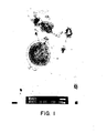
- FIG. 1 is a transmission electron micrograph of the coccoid form of a bacteria of the genus Nanobacterium where the cell membrane and cell wall are observed (bar 100 nm).
- FIG. 2 is a transmission electron micrograph of a bacteria of the genus Nanobacterium , and slime can be observed in the bottom of the micrograph.
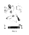
- FIG. 3 is a transmission electron micrograph of the bacillar form of a bacteria of the genus Nanobacterium .
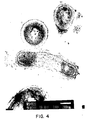
- FIG. 4 is a transmission electron micrograph of the coccoid form and the bacillar form of bacteria of the genus Nanobacterium .
- FIG. 5 is a transmission electron micrograph using negative staining of a bacteria of the genus Nanobacterium .
- FIG. 6 is a phase-contrast view (magnification 1600X) of bacteria of the genus Nanobacterium depicting the aggregation pattern seen using older cultures.
- FIG. 7 is a transmission electron micrograph of MG-63 cells (osteosarcoma) cultured on glass with a culture medium contaminated with Nanobacteria using Jones' silver staining where the black-stained Nanobacteria are located both extra cellularly and around the nuclear area.
- the applicant has succeeded in isolating unidentified bacteria-like particles present in biological samples such as commercial cell culture sera. Those cell culture sera are alleged to be sterile by the manufacturer.
- the unidentified bacteria now referred to as the genus Nanobacterium have the following characteristics: Shape : coccoid (most common) or bacillar. Degree of Aggregation : appears to depend on the culture medium and culture time; may be present alone (single), in pairs, in tetrads or in clusters. Size :
- the cell wall of Nanobacteria is resistant to proteolytic enzymes that digest the cell wall of micrococci or other gram-positive bacteria, e.g., lysozyme.
- the unidentified organisms do not grow detectably on any commonly used bacteriological culture media. In cell culture, the unidentified organisms do not turn the medium opaque nor do they change the color of the medium as do common bacteria.
- Nanobacteria differ from bacterial L-forms, in that they have a cell wall, although the thickness of the cell wall varies greatly. Nanobacteria do not require a hyperosmolar stabilized medium as do bacterial L-forms. However, osmotically stable L-form variants of common bacterial contaminants have been produced experimentally. Nanobacteria do not revert to normal bacteria. Bacterial L-forms usually revert back to a cell-walled (original) form. At the same time, bacterial L-forms also acquire their original growth properties and possibly their original pathogenicity.
- Nanobacteria never gain the ability to grow on common bacteriological media, and do not form colonies resembling any known bacterial species as do bacterial L-forms.
- Applicant has developed a culture and detection method that allows the propagation, microscopic detection and immunological verification of previously unidentified bacterial agents, i.e., bacteria of the genus Nanobacterium , which contaminates serum and serum products as well as other biological material.
- the described culture method supports the growth of common bacteria and osmotically stable bacterial variants, i.e., bacterial L-forms as well as enabling their simultaneous detection.
- all bacterial forms excluding strictly intracellular parasites that may contaminate the sample are detected.
- double staining method DNA stain plus immunostaining
- Nanobacteria can be specifically detected in spite of other bacteria being present in a sample.
- Nanobacteria alone may be selectively cultured and detected after filtration of the sample through 0.22 ⁇ m sterile filter available from commercial sources, e.g., Millipore.
- Nanobacteria cannot be grown on standard media for bacteria, and thus they escape detection when using standard culture methods.
- the detection of the extremely small unidentified bacteria is hampered by their size, which, e.g., in commercial cell culture isolates, is smaller than 0.5 ⁇ m.
- their detection via light microscopy is possible only with the best microscopes having maximum resolution. Tissue culture laboratories are seldom equipped with such microscopes.
- these bacteria are difficult to collect since centrifugation is difficult. They are also readily lost since they do not adhere to glass, and they cannot be stained with common bacteriological stains.
- the growth requirements of species of bacteria of the genus Nanobacterium are quite similar. The growth requirements can be met using standard tissue culture media. This is likely because these bacteria are adapted for living inside the mammalian body.
- the culture and detection method of the present invention rests on the principle of culturing the bacteria under conditions similar to tissue culture. Any standard tissue culture media is useful in the practice of the present invention. Cell or tissue culture media are generally used to culture mammalian cells. During culture, Nanobacteria become visible under light microscopy due to their multiplication, aggregation, secretion of slime and/or the thickening of their cell envelope. Their detection is aided by providing killed bacteria of the genus Nanobacterium for comparison as a control.
- Nanobacteria are pelleted by centrifuging the well-mixed culture medium at at least 14,000 g for at least 20 minutes. The pellet is then suspended in a drop of phosphate buffered saline or saline, and spread on a cover glass (or on an objective glass). An equal volume of commercial 25% glutaraldehyde solution (range 1-25% in phosphate buffered saline) is layered on the sample area. Glutaraldehyde functions as a fixative that aids in attaching the Nanobacteria on the glass.
- the glass is immersed into Carnoy's fixative (1:3 acetic acid:methanol) for another 30 minutes. After drying at room temperature, the glass is immersed into Hoechst stain solution (0.5 mg/l is the preferred concentration in phosphate buffered saline). After 30 minutes, the glass is washed by immersing twice in water, 1 minute for each wash. Thereafter, the glass is mounted with a suitable mounting medium such as 50% glycerol in phosphate buffered saline. (Sample side facing the mounting medium). The preparation can then be viewed by standard fluorescence microscopy using the commonly available filter sets intended for use with Hoechst 33258 stain as described by T. R. Chen, "In situ Detection of Mycoplasma Contamination in Cell Cultures by Fluorescent Hoechst 33258 Stain", Exp. Cell Res., Vol 104, pp. 255-262, (1977).
- Hoechst stain solution 0.5 mg/l is the preferred concentration in
- Suitable culture media include those identified below: DMEM/Ham's F-12 (1:1) Basal Media Eagle CMRL 1066 Media Dulbecco's Modified Eagle Media Fischer's Media Glasgow Minimum Essential Media (BHK-21) Hybridoma Media (Serum Free) Iscove's Modified Dulbecco's Media Leibovitz's L-15 Media McCoy's Media Media 199 Minimum Essential Media (MEM) NCTC Media Medium NCTC-135 F-10 Nutrient Mixture F-12 Nutrient Mixture Opti-MEM I Reduced Serum Medium RPMI Medium 1630 RPMI Medium 1640 Waymouth's Media William's Media E BGJb Media DMEM and its modifications where glucose levels vary between 1 to 4.5 g/liter
- All of the foregoing cell culture media are distributed by Gibco Co. and relate to the culture of mammalian cells.
- the media are marketed with modifications in buffering systems and contain Earle's or Hanks' salts with or without HEPES.
- a preferred embodiment of the present invention involves a synthetic medium suitable for the growth of Nanobacterium by fulfilling their growth requirements.
- the liquid medium comprises a standard tissue culture medium known as RPMI 1640.
- This medium is a standardized composition of amino acids, salts, etc. which can be obtained from Gibco (Uxbridge, Middlesex, U.K.).
- the components of the culture medium should be dissolved in essentially sterile water.
- the quality of water used is extremely important, since water can contain cytotoxic impurities for the unidentified agents. Care must be taken to avoid water as a source of contamination. Tap water, deionized water or sterile water for injection, for instance, may all be adequate if their sterility is checked in advance.
- the culture media can also be solidified using agar or agarose.
- agar or agarose may contain cytotoxic impurities.
- the presence of Nanobacteria is difficult to detect via microscopy when using agar or agarose since solid media have inferior microscopic properties.
- solid media are generally inferior to liquid media in the detection of the presence of Nanobacteria .
- the medium is preferably supplemented with a mixture (50-100x concentrate) prepared separately from D,L-selenomethionine, adenosine, thymidine, uracil, guanine and cytosine all of which can be obtained from Sigma Chemical Co., St. Louis, Missouri.
- a mixture 50-100x concentrate
- the 100x-concentrate contains 10mM DL-selenomethionine and 1mM by each of the following compounds: adenosine, thymidine, uracil, guanine and cytosine, dissolved in a solvent.
- the final medium is prepared by adding 1 ml of the dissolved 100x concentrate to 99 ml of the basal medium.
- a deionized distilled water is utilized in the preparation of the basal medium and of the supplement. Standard procedures in utilizing pharmaceutical grade components and biologically sterilized equipment are followed.
- Nanobacteria generally have a lag time for growth. This lag time typically varies anywhere from one day to one week.
- Gamma-irradiated sterile fetal bovine serum may preferably be added to a final concentration of 20%.
- Fetal bovine serum intended for use in sterility testing or culture of a certain species of the Nanobacteria bacteria is gamma-irradiated with a dose and for a time sufficient to kill Nanobacteria .
- the gamma-radiation is generally in the range of about 2.5 to 4.0 megarads, preferably 3 megarads.
- the serum may be in a frozen or melted state.
- the fetal bovine serum used for this purpose must not contain the Nanobacteria contaminants in detectable amounts, because the contaminant particles remain in the serum after irradiation.
- irradiation prevents their multiplication, killed contaminants may interfere with detection of the possible presence of Nanobacteria in the examined samples.
- the killed bacteria retain their shape and can be used as an aid in interpreting the microscopy during Nanobacteria testing (killed standard).
- Typical culture conditions may involve incubation at 37°C in an atmosphere of 5-10° CO2 and 95-90% air (moisture about 90%).
- the conditions are generally standard cell culture conditions. It may be advantageous to incubate the vial with the flat side facing down.
- Nanobacteria can be removed from serum by use of adsorption with specific antibodies recognizing Nanobacterial antigens.
- the antibodies are immobilized on a suitable surface, e.g., on a vinyl immunosorbent material or on Sepharose gel beads, preferably using well-documented covalent immobilization methods like coupling with either glutaraldehyde or with cyanogen bromide. Serum is then brought into a close contact with the immobilized antibody.
- the antibody coated material should preferably be packed in a compact form like a column or a pack of filters.
- the antibodies will then bind Nanobacteria which will then become immobilized. This takes place within minutes or a few hours. The procedure can be carried out at the temperature of 0 to 60 degrees centigrade. Nanobacteria -free serum can be recovered simply by eluting or collecting the serum. Removal of Nanobacteria with the adsorption method is preferably done before gamma-irradiation.
- Nanobacteria are preferably made by immunizing animals like rabbits with specifically purified Nanobacteria or with an extract obtained from purified Nanobacteria .
- the special purification of the Nanobacteria antigen is important because Nanobacterial preparations may contain adsorbed or precipitated impurities from their culture medium that typically contain highly immunogenic serum. Purification can utilize exceptional properties of Nanobacteria . They are highly resistant to the actions of practically all proteinases, like proteinase K, trypsin, papain, and the like. They can also be washed with organic solvents like chloroform, ether and alcohols. Furthermore, their structure endures extreme pH values like pH 1 to 14. Thus, washing with strong alkaline solutions may result in removal of impurities.
- a solubilized antigen offers many advantages for immunization and especially for detection of antibodies.
- Such a preparation can be achieved by incubating Nanobacteria with a strong acid. Typically, this can be done with 1 N hydrochloric acid (range 0.1 to 2.0 N HCl).
- Nanobacteria specific antiserum can be prepared in animals, e.g., in rabbits, as follows: Nanobacteria are cultured and thereafter harvested by centrifugation at least at 14,000 g for at least 20 minutes according to the conditions referred to in this application. The pellet is incubated with proteinase K added at 0.5 mg/ml (range 0.01 to 100 mg/ml) in a small volume of buffer like phosphate buffered saline. Typically, incubation is carried out at 37 degrees centigrate, but a broad range of temperatures are suitable as well, for 1 hour (range 0.1 to 48 hours). The temperature need only be sufficient to allow incubation. The incubation results in minimal loss of Nanobacteria .
- Nanobacteria may be used as an antigen or Nanobacteria are solubilized by incubation with 1 N HCI for 1 minute (range 0.1 to 10 minutes) in a suitable small volume at room temperature, but other temperatures can be used as well if incubation time is modified. Incubation is stopped by neutralizing the mixture with an alkaline solution or with buffer. Typically, 1 N NaOH or 1 N KOH is used in equal volume to the amount of HCI volume used.
- This treatment has now resulted in a solubilized antigenic preparation which is viscous like a gel.
- the present inventor has found that this kind of extract was highly immunogenic. Given preferably several times at about one week intervals intravenously, intraperitoneally or subcutaneously into a rabbit, it resulted in the formation of high titers of highly specific antibody binding specifically to Nanobacteria as detected by immunofluorescence microscopy.
- Nanobacteria could also be purified from serum and from Nanobacteria cultures by use of filters of small pore size.
- a sample of Nanobacteria culture was filtered with a commercial filter available from Millipore having a pore size of about 0.1 or preferably 0.05 microns.
- filter tests were performed using Swinnex Disc Filter Holder loaded with a Fluoropore, MF-Millipore or Durapore filter with appropriate pore size (all materials available from Millipore). If performed under relatively low pressure, most of the Nanobacteria present were retained in the filter.
- Nanobacteria can be washed and purified by incubating with a proteinase, e.g., with proteinase K under conditions described above for Nanobacteria purification but performed inside the filter holder. After washing degradation products away, Nanobacteria can be collected by changing the flow direction of the system to the opposite (backwards elution).
- a proteinase e.g., with proteinase K under conditions described above for Nanobacteria purification but performed inside the filter holder. After washing degradation products away, Nanobacteria can be collected by changing the flow direction of the system to the opposite (backwards elution).
- Nanobacteria and preparation of their solubilized antigens result in material that can be used as an antigen in immunization of animals or humans, and in construction of immunoassays for detection of Nanobacteria , or for detection of antibodies against Nanobacteria .
- antibodies against Nanobacteria can be bound and purified with them.
- Both Nanobacteria and the solubilized extract can be bound covalently to vinyl ELISA immunosorbent plates using well-known glutaraldehyde coupling to vinyl. Such plates performed excellently in Elisa detection of antibodies against Nanobacteria .
- Nanobacteria can bind Nanobacteria . They can be utilized to immobilize Nanobacteria or to remove Nanobacteria from a biological sample in the purposes of elimination, assay and purification of Nanobacteria .
- Antibodies can be used in many ways to construct an immunoassay for Nanobacteria . Also, the antibodies may find use in elimination of Nanobacteria from cell cultures or from animals or from humans. Further, the antibodies may be applicable for purification of products derived from cell cultures, animals or humans. These may include blood, serum and their products, or cells and organs, or in vitro cultured products including cells.
- Nanobacteria An important application of antibodies recognizing Nanobacteria is their use in immunofluorescence assays and stainings to detect specifically Nanobacteria .
- the antibodies may be coupled with any fluorescent label suitable for microscopy.
- the antibody bound to Nanobacteria antigen may be visualized with another antibody or protein A or protein G carrying a label.
- Immunofluorescent detection can be used in immunofluorescence microscopy in the presence of activated cell sorting and in fluorescence based immunosorbent type of assays.
- a method was developed combining staining with a fluorescent DNA stain and immunofluorescence with antibodies to Nanobacteria labelled with a fluorescent label, or visualized with another antibody, or protein A, or protein G labelled with a fluorescent label. Both stainings are preferably done simultaneously or one after the other to the same preparation. Staining with a DNA stain will reveal practically all microorganisms. Staining with specific antibodies will reveal only Nanobacteria . Both images or signals can be seen or analyzed simply by changing appropriate filter sets applicable for the used DNA stain and for the used fluorescent label of the antibody detection system. Such a double staining method is described in detail in Example II.
- 3T6 cells are exceptionally suitable for detection of Nanobacteria because they internalize Nanobacteria effectively allowing for the use of a mild fixation protocol described in Example II. Internalized Nanobacteria are not washed away from the sample in the necessary washing steps involved in an immunostaining. 3T6 cells also show minimal autofluorescence under the described conditions. However, other cell lines like 3T3, BHK, CHO and other adherent cell cultures may be used.
- 3T6 serves as an indicator cell culture into which a test sample is inoculated.
- 3T6 cells together with the sample are then cultured under cell culture conditions for a certain time, typically 3 days (range 1 hour to 10 days).
- microbes like mycoplasmas, common bacteria and Nanobacteria multiply in the culture.
- antibiotics are not included.
- the possible presence of microbes is detected by staining with a suitable DNA stain like Hoechst No. 33258 in combination with an immunofluorescent staining with an antibody detection system visualizing Nanobacteria .
- 3T6 cells are washed with a suitable buffer like phosphate buffered saline.
- Fixation time is typically minutes or hours and can be carried out at a temperature of about 0 to 40°C, preferably at room temperature.
- Glutaraldehyde solutions may also be used, but regretably they may cause strong autofluorescence in mammalian cells. Thereafter, the preparation is washed and then subjected to a brief permeabilization treatment, e.g., with Triton X-100 (0.5% for 1-20 minutes) in phosphate buffered saline.
- the preparation is stained with Hoechst 33258 at about 0.5 mg/l and with a suitable fluorescent antibody preparation, again in phosphate buffered saline. After sufficient labelling or staining, typically after 30 minutes (range 1 minute to 1000 minutes), excess label or stain is washed away and the preparation is analyzed by microscopy or by other suitable methods to visualize the fluorescence of both labels.
- the 3T6 indicator culture method together with the described double staining method serves as a test system to screen cell culture samples, sera, media or other biological samples to detect microorganisms, especially Nanobacteria .
- the staining method distinguishes Nanobacteria from other possible microbes. Practically all possible microbes are detected.
- the described double staining method can be used also to detect the presence of microbes on membrane filters, especially the presence of Nanobacteria .
- This application can be used to construct concentration-detection methods utilizing membrane filters with small pore sizes to collect Nanobacteria (see the purification method for Nanobacteria ). Nanobacteria are thereafter specifically detected by staining or by culture. After culture, the staining can be used to verify the result.
- the double staining may be used with fluorescent cell sorting systems to detect and even to isolate Nanobacteria .
- Culture of bacteria of the genus Nanobacterium is carried out by adding a biological or commercial sample to the culture medium typically contained in a vial using standard sterile technique. The cultures are then incubated in a standard tissue culture incubator at a suitable temperature, generally about 20 to 50°C, preferably about 37°C.
- tissue culture flasks should be used, e.g., from Nunc in Roskilde, Denmark.
- a 25 ml flask is suitable for the culture of the samples.
- a suitable amount preferably approximately eight ml of a tissue culture media such as an RPMI 1640 medium preferably containing a D,L-selenomethionine nucleotide precursor supplement at about 0.001x to 10x, preferably 1x is added to the flask.
- tissue culture media such as an RPMI 1640 medium preferably containing a D,L-selenomethionine nucleotide precursor supplement at about 0.001x to 10x, preferably 1x is added to the flask.
- approximately 2 ml of the tested serum sample is added.
- the flask is transferred to the incubator and cultured. Either the top of the flask is loosened or closed after flushing with approximately 5 to 10% CO2 typically in air. If dishes are used, the incubation should preferably be carried out in a humid incubator in an atmosphere of approximately 90 to 95% air and approximately 10 to 5% CO2. A range of about 80 to 99% air and 20 to 1% CO2 is suitable.
- the culture of the unidentified bacteria can be done from serum, body fluid or tissue, preferably after homogenization, even from samples containing common bacteria. Common bacteria are optionally eliminated by filtration through 0.22 ⁇ m filters. Thereafter, a culture is started from the filtered sample.
- the presence of bacteria of the genus Nanobacterium in cell cultures can be detected after centrifuging the cells at low g values (approximately 1 to 1000 g) and by using the supernatant as a sample.
- the cell pellet can be disrupted and the solution may be used as a sample (preferably filtered through 0.22 ⁇ m filter). This latter technique will generally bring intracellular forms to the sample.
- the criteria for a positive culture is detectable growth of organisms having the criteria of appearance, shape, size, filterability and lack of growth on common bacterial media like chocolate agar or blood agar as noted above. Further, verification can be done by immunofluorescence. Common bacteria will multiply very rapidly in the medium and they can be visually observed after culturing for one or two days.
- FIGS. 1 through 9 depict bacteria of the genus Nanobacterium .
- DNA stains are useful in the practice of the present invention. Such stains include Hoechst stain No. 33258. Hoechst stain No. 33342, Hoechst stain No. 2495, Dapi, Acridine Orange, and the like. Hoechst stain 33258 is preferred.
- control medium samples should be taken, handled, and incubated in the same way as the tested samples. No growth should take place in control cultures. If growth occurs in control cultures, the test is not valid, since this is an indication of outside contamination.
- a test kit is also provided which comprises tissue culture medium sufficient for allowing the culture of bacteria of the genus Nanobacterium gamma-irradiated bacteria of the genus Nanobacterium as a control and a nucleic acid stain suitable for staining bacteria of the genus Nanobacterium .
- tissue culture medium sufficient for allowing the culture of bacteria of the genus Nanobacterium gamma-irradiated bacteria of the genus Nanobacterium as a control and a nucleic acid stain suitable for staining bacteria of the genus Nanobacterium .
- specific antibodies recognizing Nanobacterial antigens are optionally provided.
- Nanobacteria -free serum may also be provided.
- an uncontaminated culture of 3T6 cells may be provided.
- a medium was prepared by mixing 5 ml of 100x D,L-selenomethionine supplement to 495 ml of RPMI 1640 as identified in Table I for a total volume of 500 ml.
- RPMI 1640 appears in Moore et al, "Culture of Normal Human Leucocytes", J.A.M.A. , 199 , 519-524 (1962).
- approximately 8 ml portions were then pipetted to 25 ml culture flasks and samples of 2 ml of the tested commercial cell culture sera were added. In the control experiments only medium was added. The flasks were incubated at 37°C in a humidified incubator containing 95% air and 5% CO2 for 15 days.
- the cultures were inspected daily using an Olympus CK2 inverted microscope (Olympus, Japan) at a magnification of 400x utilizing phase-contrast microscopy. Contaminants appeared barely visible under the microscope at various times.
- the sample was scored +++, if the criteria characteristic of Nanobacteria were observed within approximately 24 hours of the start of the culture. Those cultures scored ++, in which contaminants could be detected only after approximately 5 to 10 days culture, + was given to those cultures which showed visible contaminants approximately 10 to 15 days after the start of culturing. Cultures having no noticeable growth of bacteria after 15 days were considered negative. Contamination was very common, see Table II. These tests were carried out at least three separate times with essentially similar results. From several lots, samples were taken from many different serum bottles (maximum 12 bottles from a single lot). The results were again unanimous, although small variation of the degree of contamination was noticeable between bottles.
- 3T6 indicator cell culture method Possible presence of cell culture contaminants belonging to genus Nanobacterium or Mycoplasma in cultured cells was tested by the 3T6 indicator cell culture method. Samples of cell cultures to be tested (medium together with some cells) were taken using standard sterile techniques and stored at +4°C up to one week until tested. 3T6 cultures were prepared as follows: 3T6 cells (obtained from ATCC [code No. CCL 96] and tested to be free of contaminants) were cultured under sterile conditions in DMEM with 2mM L-glutamine and supplemented with 10% gamma-irradiated fetal bovine serum (tested to be free of Nanobacteria). Antibiotics were not used.
- Cultures were incubated at 37°C with an atmosphere of 10% CO2 - 90% air at 90% humidity. Cells near confluency were washed, trypsinized, collected and washed, and then counted. All of this was carried out using standard cell culture procedures. Approximately 3,000 cells were suspended in 0.5 ml of the culture medium (described above) and transferred to a special track bottle (Sterilin, Feltham, England, order No. 129AX/1). After incubation for 16 hours under cell culture conditions, 3T6 cells were attached to the cover glass on the bottom of the vessel. Then a sample of 100 ⁇ l to 500 ⁇ l of the material to be tested was added to the culture. Control cultures of 3T6 (sterile) were prepared by adding only culture medium, and contaminated control cultures were prepared by adding either Nanobacteria or Mycoplasma to cultures. All controls were incubated, treated and stained exactly as the test sample.
- 3T6 sterile
- Hoechst 33258 stain together with FITC coupled anti-Nanobacteria antibody was added in 0.5 ml of phosphate buffered saline. Final concentration of Hoechst 33258 was 0.5 mg/l.
- Anti- Nanobacteria serum was obtained by immunising rabbits with purified Nanobacteria . IgG-fraction of their serum was obtained using Protein A affinity chromatography (Pharmacia, Uppsala, Sweden). IgG was then labelled with fluorescein isothiocyanate (FITC) using the published standard coupling techniques. A preliminary test was constructed to determine the best dilution of FITC coupled anti- Nanobactria antibody in the staining procedure.
- cover slips were removed from the track bottle, washed thoroughly with phosphate buffered saline in a dish with 5 buffer exchanges (for 10 minutes) again protected from strong light.
- a cover slip was then mounted on an objective glass cell side facing the mounting medium.
- mounting medium 50% glycerol in phosphate buffered saline, preferably containing 0.5% n-propyl gallate can be used.
- suitable commercial mounting media can be used (e.g., Mount Quick "Aquous" Daido Sangyo Co, Tokyo, Japan).
- the preparations were then viewed by standard fluorescence microscopy using commonly available filter sets intended for use with the Hoechst 33258 stain and for the FITC label (different filters are used).
- Control 3T6 indicator cells No cytoplasmic or extracellular staining by Hoechst or by FITC.
- Mycoplasma added controls - Strongly blue cytoplasmic and extracellular fluorescent dots visualized by Hoechst filter set. No FITC fluorescence.
- Nanobacteria added controls - Cytoplasmic and extracellular fluorescent dots revealed by Hoechst filter set. Typically, cytoplasmic dots outnumber extracellular dots. The fluorescence intensity of Nanobacteria was slightly lower than that of mycoplasma. FITC filter set revealed strong yellow-greenish fluorescence. FITC positive dots were also Hoechst positive.
- 3T6 with the cell culture test samples - Some of the samples were identical to negative controls. These samples were also negative in Mycoplasmas and Nanobacteria testing by specific culture methods. Mycoplasmas were cultured on mycoplasma agar as described by Barile et al. (The identification and sources of mycoplasmas isolated from contaminated cell cultures, Annals of the New York Academy of Sciences , Vol. 225, 251-264 (1973)) and Nanobacteria with the method in Example I.
- Nanobacteria can be identified specifically with the specific FITC coupled antibody to Nanobacteria . Only Nanobacteria fluoresced. Both Mycoplasmas and Nanobacteria fluoresced in Hoechst staining. Thus, the present method could indicate rapidly and specifically whether Mycoplasmas or Nanobacteria were present.
Landscapes
- Chemical & Material Sciences (AREA)
- Life Sciences & Earth Sciences (AREA)
- Health & Medical Sciences (AREA)
- Engineering & Computer Science (AREA)
- Organic Chemistry (AREA)
- Zoology (AREA)
- Wood Science & Technology (AREA)
- Genetics & Genomics (AREA)
- Biotechnology (AREA)
- Bioinformatics & Cheminformatics (AREA)
- General Health & Medical Sciences (AREA)
- Microbiology (AREA)
- General Engineering & Computer Science (AREA)
- Biochemistry (AREA)
- Virology (AREA)
- Biomedical Technology (AREA)
- Tropical Medicine & Parasitology (AREA)
- Medicinal Chemistry (AREA)
- Proteomics, Peptides & Aminoacids (AREA)
- Biophysics (AREA)
- Analytical Chemistry (AREA)
- Physics & Mathematics (AREA)
- Toxicology (AREA)
- Immunology (AREA)
- Molecular Biology (AREA)
- Measuring Or Testing Involving Enzymes Or Micro-Organisms (AREA)
- Apparatus Associated With Microorganisms And Enzymes (AREA)
- Micro-Organisms Or Cultivation Processes Thereof (AREA)
Applications Claiming Priority (2)
| Application Number | Priority Date | Filing Date | Title |
|---|---|---|---|
| US07/520,443 US5135851A (en) | 1990-05-08 | 1990-05-08 | Culture and detection method for sterile-filterable autonomously replicating biological particles |
| US520443 | 1990-05-08 |
Publications (3)
| Publication Number | Publication Date |
|---|---|
| EP0460414A2 true EP0460414A2 (fr) | 1991-12-11 |
| EP0460414A3 EP0460414A3 (en) | 1993-03-03 |
| EP0460414B1 EP0460414B1 (fr) | 1995-05-17 |
Family
ID=24072615
Family Applications (1)
| Application Number | Title | Priority Date | Filing Date |
|---|---|---|---|
| EP91107318A Expired - Lifetime EP0460414B1 (fr) | 1990-05-08 | 1991-05-06 | Méthode à culture et détection de particule biologique stérile-filtrant de répliquer autonomique |
Country Status (7)
| Country | Link |
|---|---|
| US (1) | US5135851A (fr) |
| EP (1) | EP0460414B1 (fr) |
| AT (1) | ATE122725T1 (fr) |
| DE (1) | DE69109744T2 (fr) |
| DK (1) | DK0460414T3 (fr) |
| ES (1) | ES2072478T3 (fr) |
| GR (1) | GR3016991T3 (fr) |
Cited By (5)
| Publication number | Priority date | Publication date | Assignee | Title |
|---|---|---|---|---|
| EP0712494A4 (fr) * | 1993-08-06 | 1999-10-06 | Us Gov Sec Navy | Essai immunologique optiques pour des analytes microbiens, faisant appel a des colorants non specifiques |
| WO2000001238A1 (fr) * | 1998-07-06 | 2000-01-13 | Kajander E Olavi | Procedes relatifs a l'elimination des nanobacteries |
| EP1038951A1 (fr) * | 1999-03-23 | 2000-09-27 | Societe Des Produits Nestle S.A. | Milieu de synthèse pour la culture de Lactobacillus et/ou des Bifidobactéries |
| WO2003012058A3 (fr) * | 2001-08-02 | 2003-12-04 | Univ North Carolina State | Milieux et procedes pour cultiver des micro-organismes |
| US6706290B1 (en) | 1998-07-06 | 2004-03-16 | Olvai E. Kajander | Methods for eradication of nanobacteria |
Families Citing this family (14)
| Publication number | Priority date | Publication date | Assignee | Title |
|---|---|---|---|---|
| GB9001625D0 (en) * | 1990-01-24 | 1990-03-21 | Ciba Geigy Ag | A pharmaceutical preparation for maturation of prothymocytes |
| IES940182A2 (en) * | 1994-03-01 | 1995-11-29 | Teagasc Agric Food Dev Authori | "Rapid detection of bacteria in liquid cultures" |
| US5932624A (en) * | 1995-10-17 | 1999-08-03 | Herbert; Victor D. | Vitamin supplement composition |
| US6265391B1 (en) | 1995-10-17 | 2001-07-24 | Upsher-Smith Laboratories, Inc. | Method for preventing peripheral nerve damage |
| US6051395A (en) * | 1998-08-25 | 2000-04-18 | Biometric Imaging, Inc. | Method and compound for detecting low levels of microorganisms |
| US8043614B2 (en) * | 2004-03-09 | 2011-10-25 | Ahlfors Jan-Eric W | Autogenic living scaffolds and living tissue matrices: methods and uses thereof |
| US20050175630A1 (en) * | 2003-12-23 | 2005-08-11 | Eyal Raz | Immunogenic compositions and methods of use thereof |
| EP1836496A2 (fr) * | 2004-11-08 | 2007-09-26 | Nanobac Life Sciences | Procédés et compositions de complexes protéines-hydroxy-apatites et leur application pour tester et moduler un système immunologique contenant un nouveau test in vitro pour la détection d'anticorps contre les complexes proteines-hydroxy-apatites lian |
| US20060270571A1 (en) * | 2005-05-26 | 2006-11-30 | Burke Peter A | Deactivation of mineral encapsulated nanobacteria |
| EP1948259B1 (fr) * | 2005-10-26 | 2017-03-22 | Genesis Technologies Limited | Matrices de regeneration de tissus acellulaires bioabsorbables produites par incubation de produits sanguins acellulaires |
| US20070134814A1 (en) * | 2005-12-09 | 2007-06-14 | Kajander E O | Methods and compositions for the detection of calcifying nano-particles, identification and quantification of associated proteins thereon, and correlation to disease |
| US20110039282A1 (en) * | 2008-04-21 | 2011-02-17 | Florida Atlantic University | Method of detecting calcifying nanoparticles and susceptibility to calcifying nanoparticle information |
| US10584370B2 (en) | 2014-12-16 | 2020-03-10 | Soft Cell Biological Research, Llc | Screening for L-form bacteria |
| CN119643854B (zh) * | 2025-02-19 | 2025-05-27 | 中国人民解放军军事科学院军事医学研究院 | 一种结构完整的红细胞膜及其制备方法和应用 |
-
1990
- 1990-05-08 US US07/520,443 patent/US5135851A/en not_active Expired - Lifetime
-
1991
- 1991-05-06 EP EP91107318A patent/EP0460414B1/fr not_active Expired - Lifetime
- 1991-05-06 DK DK91107318.7T patent/DK0460414T3/da active
- 1991-05-06 ES ES91107318T patent/ES2072478T3/es not_active Expired - Lifetime
- 1991-05-06 AT AT91107318T patent/ATE122725T1/de not_active IP Right Cessation
- 1991-05-06 DE DE69109744T patent/DE69109744T2/de not_active Expired - Fee Related
-
1995
- 1995-08-02 GR GR950402107T patent/GR3016991T3/el unknown
Non-Patent Citations (4)
| Title |
|---|
| CLINICAL RESEARCH vol. 37, no. 2, 28 April 1989, WASHINGTON DC USA page 432A O. KAJANDR ET AL. 'Cell wall defective bacteria contaminate cell culture.' * |
| MICRON AND MICROSCOPICA ACTA vol. 21, no. 3, 10 June 1990, NEW YORK NY USA page 160 I. KURONEN ET AL. 'Rapid immunoelectron microscopic detection method for novel bacteria-like agents contaminating animal sera.' * |
| MICRON AND MICROSCOPICA ACTA vol. 21, no. 3, 10 June 1990, NEW YORK NY USA pages 180 - 181 K. AKERMAN ET AL. 'Study of mycoplasma and other bacteria-like contaminants in cell culture by scanning electron microscopy.' * |
| SCANDINAVIAN JOURNAL OF IMMUNOLOGY vol. 32, no. 4, 1 October 1990, OXFORD UK E.O. KAJANDER ET AL. 'Activation of mouse spleen lymphocytes by autonomously replicating bacteria-like particles contaminating cell culture serum.' * |
Cited By (7)
| Publication number | Priority date | Publication date | Assignee | Title |
|---|---|---|---|---|
| EP0712494A4 (fr) * | 1993-08-06 | 1999-10-06 | Us Gov Sec Navy | Essai immunologique optiques pour des analytes microbiens, faisant appel a des colorants non specifiques |
| WO2000001238A1 (fr) * | 1998-07-06 | 2000-01-13 | Kajander E Olavi | Procedes relatifs a l'elimination des nanobacteries |
| US6706290B1 (en) | 1998-07-06 | 2004-03-16 | Olvai E. Kajander | Methods for eradication of nanobacteria |
| EP1038951A1 (fr) * | 1999-03-23 | 2000-09-27 | Societe Des Produits Nestle S.A. | Milieu de synthèse pour la culture de Lactobacillus et/ou des Bifidobactéries |
| US6340585B1 (en) | 1999-03-23 | 2002-01-22 | Nestec S.A. | Synthetic medium for cultivating Lactobacillus and Bifidobacteria |
| WO2003012058A3 (fr) * | 2001-08-02 | 2003-12-04 | Univ North Carolina State | Milieux et procedes pour cultiver des micro-organismes |
| US7115385B2 (en) | 2001-08-02 | 2006-10-03 | North Carolina State University | Media and methods for cultivation of microorganisms |
Also Published As
| Publication number | Publication date |
|---|---|
| DE69109744T2 (de) | 1995-10-12 |
| ES2072478T3 (es) | 1995-07-16 |
| EP0460414B1 (fr) | 1995-05-17 |
| EP0460414A3 (en) | 1993-03-03 |
| DE69109744D1 (de) | 1995-06-22 |
| US5135851A (en) | 1992-08-04 |
| ATE122725T1 (de) | 1995-06-15 |
| DK0460414T3 (da) | 1995-07-10 |
| GR3016991T3 (en) | 1995-11-30 |
Similar Documents
| Publication | Publication Date | Title |
|---|---|---|
| EP0460414B1 (fr) | Méthode à culture et détection de particule biologique stérile-filtrant de répliquer autonomique | |
| Avery et al. | Studies on the chemical nature of the substance inducing transformation of pneumococcal types: induction of transformation by a desoxyribonucleic acid fraction isolated from pneumococcus type III | |
| Avery et al. | Studies on the chemical nature of the substance inducing transformation of pneumococcal types. Inductions of transformation by a desoxyribonucleic acid fraction isolated from pneumococcus type III. | |
| Rowbotham | Isolation of Legionella pneumophila from clinical specimens via amoebae, and the interaction of those and other isolates with amoebae. | |
| Salit et al. | Type I Escherichia coli pili: characterization of binding to monkey kidney cells. | |
| CA1086224A (fr) | Sorbants des antigenes de neisseria meningitides pour le depistage le neisseria gonorrhoeae | |
| Avery et al. | Studies on the Chemical Nature of the Substance Inducing Transformation of Pneumococcal Types: Induction of Transformation by a Desoxyribonucleic Acid Fraction Isolated from Pneumococcus Type III | |
| Lalonde | Immunological and ultrastructural demonstration of nodulation of the European Alnus glutinosa (L.) Gaertn. host plant by an actinomycetal isolate from the North American Comptonia peregrina (L.) Coult. root nodule | |
| EP0043272A1 (fr) | Production d'antigènes viraux | |
| Reitmeyer et al. | Salmonella cytotoxin: a component of the bacterial outer membrane | |
| Somerville et al. | Urea-mercaptoethanol-soluble protein from spores of Bacillus thuringiensis and other species | |
| IE49590B1 (en) | Babesiosis antigen and vaccine and diagnostic agent comprising it | |
| Bezuidenhout | The present state of Cowdria ruminantium cultivation in cell lines | |
| Danner et al. | In vitro studies on Borna virus: I. The use of cell cultures for the demonstration, titration and production of Borna virus | |
| Cummins | Some observations on the nature of the antigens in the cell wall of Corynebacterium diphtheriae | |
| Hieb et al. | Aging in the free-living nematode Turbatrix aceti. Techniques for synchronization and aging of large-scale axenic cultures | |
| Roberts et al. | Study on the growth of Coxiella burnetii in the L strain mouse fibroblast and the chick fibroblast | |
| Allen et al. | Method for the simultaneous establishment of many axenic cultures of Paramecium | |
| Wellings | Pathogenic Naegleria: distribution in nature | |
| Old et al. | Adherence of fimbriate and non‐fimbriate strains of Yersinia enterocolitica to human epithelial cells | |
| SU1701743A1 (ru) | Штамм бактерий BRUceLLa авоRтUS, используемый дл приготовлени вакцины против бруцеллеза крупного рогатого скота | |
| Fraser et al. | Mycoplasmas in cell cultures from rheumatoid synovial membranes | |
| EP0451217A1 (fr) | Production in vitro d'amebocytes de crabes des moluques | |
| JPS6384484A (ja) | 特殊なマイコプラズマ膜抗原および抗体とその臨床的応用 | |
| McCarty et al. | Studies on the chemical nature of the substance inducing transformation of pneumococcal types |
Legal Events
| Date | Code | Title | Description |
|---|---|---|---|
| PUAI | Public reference made under article 153(3) epc to a published international application that has entered the european phase |
Free format text: ORIGINAL CODE: 0009012 |
|
| AK | Designated contracting states |
Kind code of ref document: A2 Designated state(s): AT BE CH DE DK ES FR GB GR IT LI LU NL SE |
|
| PUAL | Search report despatched |
Free format text: ORIGINAL CODE: 0009013 |
|
| AK | Designated contracting states |
Kind code of ref document: A3 Designated state(s): AT BE CH DE DK ES FR GB GR IT LI LU NL SE |
|
| 17P | Request for examination filed |
Effective date: 19930713 |
|
| 17Q | First examination report despatched |
Effective date: 19930816 |
|
| GRAA | (expected) grant |
Free format text: ORIGINAL CODE: 0009210 |
|
| AK | Designated contracting states |
Kind code of ref document: B1 Designated state(s): AT BE CH DE DK ES FR GB GR IT LI LU NL SE |
|
| REF | Corresponds to: |
Ref document number: 122725 Country of ref document: AT Date of ref document: 19950615 Kind code of ref document: T |
|
| ITF | It: translation for a ep patent filed | ||
| REF | Corresponds to: |
Ref document number: 69109744 Country of ref document: DE Date of ref document: 19950622 |
|
| ET | Fr: translation filed | ||
| REG | Reference to a national code |
Ref country code: DK Ref legal event code: T3 |
|
| REG | Reference to a national code |
Ref country code: ES Ref legal event code: FG2A Ref document number: 2072478 Country of ref document: ES Kind code of ref document: T3 |
|
| REG | Reference to a national code |
Ref country code: GR Ref legal event code: FG4A Free format text: 3016991 |
|
| PLBE | No opposition filed within time limit |
Free format text: ORIGINAL CODE: 0009261 |
|
| STAA | Information on the status of an ep patent application or granted ep patent |
Free format text: STATUS: NO OPPOSITION FILED WITHIN TIME LIMIT |
|
| 26N | No opposition filed | ||
| REG | Reference to a national code |
Ref country code: GB Ref legal event code: IF02 |
|
| PGFP | Annual fee paid to national office [announced via postgrant information from national office to epo] |
Ref country code: GB Payment date: 20060503 Year of fee payment: 16 Ref country code: NL Payment date: 20060503 Year of fee payment: 16 |
|
| PGFP | Annual fee paid to national office [announced via postgrant information from national office to epo] |
Ref country code: GR Payment date: 20060511 Year of fee payment: 16 Ref country code: DE Payment date: 20060511 Year of fee payment: 16 Ref country code: AT Payment date: 20060511 Year of fee payment: 16 |
|
| PGFP | Annual fee paid to national office [announced via postgrant information from national office to epo] |
Ref country code: CH Payment date: 20060515 Year of fee payment: 16 Ref country code: FR Payment date: 20060515 Year of fee payment: 16 |
|
| PGFP | Annual fee paid to national office [announced via postgrant information from national office to epo] |
Ref country code: DK Payment date: 20060516 Year of fee payment: 16 |
|
| PGFP | Annual fee paid to national office [announced via postgrant information from national office to epo] |
Ref country code: SE Payment date: 20060526 Year of fee payment: 16 |
|
| PGFP | Annual fee paid to national office [announced via postgrant information from national office to epo] |
Ref country code: IT Payment date: 20060531 Year of fee payment: 16 |
|
| PGFP | Annual fee paid to national office [announced via postgrant information from national office to epo] |
Ref country code: LU Payment date: 20060607 Year of fee payment: 16 |
|
| PGFP | Annual fee paid to national office [announced via postgrant information from national office to epo] |
Ref country code: ES Payment date: 20060621 Year of fee payment: 16 |
|
| PGFP | Annual fee paid to national office [announced via postgrant information from national office to epo] |
Ref country code: BE Payment date: 20060712 Year of fee payment: 16 |
|
| REG | Reference to a national code |
Ref country code: CH Ref legal event code: PFA Owner name: E. OLAVI KAJANDER Free format text: E. OLAVI KAJANDER#SAVILANDENTIE 9F#KUOPIO (FI) -TRANSFER TO- E. OLAVI KAJANDER#SAVILANDENTIE 9F#KUOPIO (FI) |
|
| BERE | Be: lapsed |
Owner name: *KAJANDER E. OLAVI Effective date: 20070531 |
|
| EUG | Se: european patent has lapsed | ||
| REG | Reference to a national code |
Ref country code: CH Ref legal event code: PL |
|
| REG | Reference to a national code |
Ref country code: DK Ref legal event code: EBP |
|
| GBPC | Gb: european patent ceased through non-payment of renewal fee |
Effective date: 20070506 |
|
| PG25 | Lapsed in a contracting state [announced via postgrant information from national office to epo] |
Ref country code: NL Free format text: LAPSE BECAUSE OF NON-PAYMENT OF DUE FEES Effective date: 20071201 |
|
| NLV4 | Nl: lapsed or anulled due to non-payment of the annual fee |
Effective date: 20071201 |
|
| PG25 | Lapsed in a contracting state [announced via postgrant information from national office to epo] |
Ref country code: AT Free format text: LAPSE BECAUSE OF NON-PAYMENT OF DUE FEES Effective date: 20070506 Ref country code: CH Free format text: LAPSE BECAUSE OF NON-PAYMENT OF DUE FEES Effective date: 20070531 Ref country code: LI Free format text: LAPSE BECAUSE OF NON-PAYMENT OF DUE FEES Effective date: 20070531 |
|
| REG | Reference to a national code |
Ref country code: FR Ref legal event code: ST Effective date: 20080131 |
|
| PG25 | Lapsed in a contracting state [announced via postgrant information from national office to epo] |
Ref country code: BE Free format text: LAPSE BECAUSE OF NON-PAYMENT OF DUE FEES Effective date: 20070531 |
|
| PG25 | Lapsed in a contracting state [announced via postgrant information from national office to epo] |
Ref country code: DE Free format text: LAPSE BECAUSE OF NON-PAYMENT OF DUE FEES Effective date: 20071201 Ref country code: DK Free format text: LAPSE BECAUSE OF NON-PAYMENT OF DUE FEES Effective date: 20070531 |
|
| PG25 | Lapsed in a contracting state [announced via postgrant information from national office to epo] |
Ref country code: GB Free format text: LAPSE BECAUSE OF NON-PAYMENT OF DUE FEES Effective date: 20070506 |
|
| PG25 | Lapsed in a contracting state [announced via postgrant information from national office to epo] |
Ref country code: SE Free format text: LAPSE BECAUSE OF NON-PAYMENT OF DUE FEES Effective date: 20070507 |
|
| PG25 | Lapsed in a contracting state [announced via postgrant information from national office to epo] |
Ref country code: FR Free format text: LAPSE BECAUSE OF NON-PAYMENT OF DUE FEES Effective date: 20070531 |
|
| REG | Reference to a national code |
Ref country code: ES Ref legal event code: FD2A Effective date: 20070507 |
|
| PG25 | Lapsed in a contracting state [announced via postgrant information from national office to epo] |
Ref country code: GR Free format text: LAPSE BECAUSE OF NON-PAYMENT OF DUE FEES Effective date: 20071204 Ref country code: ES Free format text: LAPSE BECAUSE OF NON-PAYMENT OF DUE FEES Effective date: 20070507 |
|
| PG25 | Lapsed in a contracting state [announced via postgrant information from national office to epo] |
Ref country code: LU Free format text: LAPSE BECAUSE OF NON-PAYMENT OF DUE FEES Effective date: 20070506 |
|
| PG25 | Lapsed in a contracting state [announced via postgrant information from national office to epo] |
Ref country code: IT Free format text: LAPSE BECAUSE OF NON-PAYMENT OF DUE FEES Effective date: 20070506 |