EP0467087B1 - Verfahren zur Einstellung der Bedingung für ein Strahlungsaufzeichnungs- und -wiedergabesystem - Google Patents
Verfahren zur Einstellung der Bedingung für ein Strahlungsaufzeichnungs- und -wiedergabesystem Download PDFInfo
- Publication number
- EP0467087B1 EP0467087B1 EP91110076A EP91110076A EP0467087B1 EP 0467087 B1 EP0467087 B1 EP 0467087B1 EP 91110076 A EP91110076 A EP 91110076A EP 91110076 A EP91110076 A EP 91110076A EP 0467087 B1 EP0467087 B1 EP 0467087B1
- Authority
- EP
- European Patent Office
- Prior art keywords
- image
- read
- conditions
- radiation
- carried out
- Prior art date
- Legal status (The legal status is an assumption and is not a legal conclusion. Google has not performed a legal analysis and makes no representation as to the accuracy of the status listed.)
- Expired - Lifetime
Links
- 230000005855 radiation Effects 0.000 title claims description 159
- 238000000034 method Methods 0.000 title claims description 50
- OAICVXFJPJFONN-UHFFFAOYSA-N Phosphorus Chemical compound [P] OAICVXFJPJFONN-UHFFFAOYSA-N 0.000 claims description 111
- 230000004936 stimulating effect Effects 0.000 claims description 10
- 206010073306 Exposure to radiation Diseases 0.000 claims description 5
- 230000006870 function Effects 0.000 description 30
- 201000010099 disease Diseases 0.000 description 18
- 208000037265 diseases, disorders, signs and symptoms Diseases 0.000 description 18
- 238000013528 artificial neural network Methods 0.000 description 16
- 239000000463 material Substances 0.000 description 13
- 210000000988 bone and bone Anatomy 0.000 description 10
- 230000000638 stimulation Effects 0.000 description 8
- 230000035945 sensitivity Effects 0.000 description 7
- 238000010586 diagram Methods 0.000 description 4
- 230000003287 optical effect Effects 0.000 description 4
- 230000002159 abnormal effect Effects 0.000 description 3
- 238000003745 diagnosis Methods 0.000 description 3
- 238000003530 single readout Methods 0.000 description 3
- 230000003321 amplification Effects 0.000 description 2
- 239000002131 composite material Substances 0.000 description 2
- 238000003199 nucleic acid amplification method Methods 0.000 description 2
- 229910052709 silver Inorganic materials 0.000 description 2
- 239000004332 silver Substances 0.000 description 2
- -1 silver halide Chemical class 0.000 description 2
- 230000005856 abnormality Effects 0.000 description 1
- NIXOWILDQLNWCW-UHFFFAOYSA-N acrylic acid group Chemical group C(C=C)(=O)O NIXOWILDQLNWCW-UHFFFAOYSA-N 0.000 description 1
- 238000004458 analytical method Methods 0.000 description 1
- 238000006243 chemical reaction Methods 0.000 description 1
- 230000001747 exhibiting effect Effects 0.000 description 1
- 230000001537 neural effect Effects 0.000 description 1
- 210000000056 organ Anatomy 0.000 description 1
- 238000002360 preparation method Methods 0.000 description 1
- 238000002601 radiography Methods 0.000 description 1
- 238000001454 recorded image Methods 0.000 description 1
- 230000004044 response Effects 0.000 description 1
- 210000000225 synapse Anatomy 0.000 description 1
- 238000002834 transmittance Methods 0.000 description 1
Images


Classifications
-
- G—PHYSICS
- G01—MEASURING; TESTING
- G01T—MEASUREMENT OF NUCLEAR OR X-RADIATION
- G01T1/00—Measuring X-radiation, gamma radiation, corpuscular radiation, or cosmic radiation
- G01T1/16—Measuring radiation intensity
- G01T1/20—Measuring radiation intensity with scintillation detectors
- G01T1/2012—Measuring radiation intensity with scintillation detectors using stimulable phosphors, e.g. stimulable phosphor sheets
- G01T1/2014—Reading out of stimulable sheets, e.g. latent image
Definitions
- This invention relates to a method of radiation image recording, read-out, and reproducing according to the preamble of claim 1 and claim 5.
- phosphors when certain kinds of phosphors are exposed to radiation such as X-rays, ⁇ -rays, ⁇ -rays, ⁇ -rays, cathode rays or ultraviolet rays, they store part of the energy of the radiation. Then, when the phosphor which has been exposed to the radiation is exposed to stimulating rays such as visible light, light is emitted by the phosphor in proportion to the amount of energy stored thereon during its exposure to the radiation. A phosphor exhibiting such properties is referred to as a stimulable phosphor.
- a sheet provided with a layer of the stimulable phosphor (hereinafter referred to as a stimulable phosphor sheet) is first exposed to radiation which has passed through an object, such as the human body. A radiation image of the object is thereby stored on the stimulable phosphor sheet. The stimulable phosphor sheet is then scanned with stimulating rays, such as a laser beam, which cause it to emit light in proportion to the amount of energy stored thereon during its exposure to the radiation.
- the light emitted by the stimulable phosphor sheet, upon stimulation thereof, is photoelectrically detected and converted into an electric image signal.
- the image signal is then used during the reproduction of the radiation image of the object as a visible image on a recording material such as photographic film, on a display device such as a cathode ray tube (CRT) display device, or the like.
- a recording material such as photographic film
- a display device such as a cathode ray tube (CRT) display device, or the like.
- CTR cathode ray tube
- Radiation image recording and reproducing systems which use stimulable phosphor sheets are advantageous over conventional radiography using silver halide photographic materials, in that images can be recorded even when the energy intensity of the radiation to which the stimulable phosphor sheet is exposed varies over a wide range. More specifically, since the amount of light which the stimulable phosphor sheet emits when being stimulated varies over a wide range and is proportional to the amount of energy stored thereon during its exposure to the radiation, it is possible to obtain an image having a desirable density regardless of the energy intensity of the radiation to which the stimulable phosphor sheet was exposed.
- an appropriate read-out gain is set when the emitted light is being detected and converted into an electric signal to be used in the reproduction of a visible image on a recording material, such as photographic film, or on a display device, such as a CRT display device.
- a novel radiation image recording and reproducing system has been proposed in, for example, U.S. Patent No. 4,527,060.
- the proposed radiation image recording and reproducing system is constituted such that a preliminary read-out operation (hereinafter simply referred to as the "preliminary readout") is carried out.
- a preliminary read-out operation hereinafter simply referred to as the "preliminary readout”
- a stimulable phosphor sheet on which a radiation image has been stored is exposed to a light beam having a comparatively low energy level, which releases part of the energy stored on the stimulable phosphor sheet when it was exposed to radiation.
- a preliminary read-out image signal is obtained from the preliminary readout.
- the stimulable phosphor sheet is exposed to a light beam having an energy level higher than the energy level of the light beam used in the preliminary readout.
- an image signal is obtained, which image signal is to be used in the reproduction of a visible image.
- read-out conditions means a group of various factors, which are adjustable and which affect the values of the image signal obtained during the final readout.
- the image signal in turn affects the gradation and sensitivity of the visible image which is reproduced.
- the term “read-out conditions” may refer to a read-out gain, a scale factor, or the power of the source producing the light beam used during the final readout.
- image processing conditions as used herein means a group of various factors which are adjustable and which affect how an image is processed, which in turn affects the gradation and sensitivity of the reproduced visible image.
- image processing conditions may refer to the scale used in the conversion of an image signal.
- the term "energy level of a light beam” as used herein means the level of energy of the light beam to which the stimulable phosphor sheet is exposed per unit area.
- the term "energy level of a light beam” means the weighted energy level which is calculated by weighting the energy level of the light beam, to which the stimulable phosphor sheet is exposed per unit area, with the sensitivity of the stimulable phosphor sheet to the wavelength.
- light beams of different wavelengths may be used, the intensity of the light beam produced by a laser beam source or the like may be changed, or the intensity of the light beam may be changed by moving an ND filter or the like into and out of the optical path of the light beam.
- the diameter of the light beam may be changed in order to alter the scanning density, or the speed at which the stimulable phosphor sheet is scanned with the light beam may be changed.
- the preliminary readout it has also been proposed to analyze the image signal (or the preliminary read-out image signal) obtained and to adjust the image processing conditions, which are to be used when the image signal is processed, on the basis of the results of an analysis of the image signal.
- the proposed method is applicable to cases where an image signal is obtained from a radiation image recorded on a recording medium such as conventional X-ray film, as well as to systems using stimulable phosphor sheets.
- the read-out conditions for the final readout and/or the image processing conditions are adjusted such that each reproduced visible image has the best possible quality. Therefore, with the aforesaid radiation image recording and reproducing systems, each reproduced image is suitable for viewing.
- a change in the image density in, for example, an image of an object which was recorded in the past and an image which represents the current state of the same object cannot be ascertained accurately when the two images are compared with each other.
- Figure 3A is a schematic view showing a radiation image 6 of part (in this case, the frontal chest) of a human body, which image has been reproduced on a sheet of photographic film 5.
- a portion 6b in the radiation image 6 represents a part of the chest not affected by disease and has an approximately uniform level of image density
- a portion 6a represents a part of the chest affected by disease and has a level of image density lower than the image density at the portion 6b.
- Figure 3B is a graph showing the change in the abnormal level of image density at the portion 6a representing a part of the chest affected by disease, which change was investigated by recording and reproducing a plurality of radiation images of the object shown in Figure 3A over a period of time.
- the disease is becoming less severe as time passes.
- Curve A indicates the ideal levels of image density in the reproduced image of the portion 6a as the disease becomes less severe over time.
- curve B abnormal levels of image density in the reproduced image of the portion 6a are often represented by curve B.
- the severity of the disease is judged on the basis of the difference between the image density of the part of the reproduced image corresponding to the portion 6a and the image density of the part of the reproduced image corresponding to the portion 6b.
- the image density difference ⁇ D1 between the normal level of image density and the level of image density on curve A should be detected.
- the image density difference ⁇ D1 is large and indicates that the part of the chest represented by the portion 6a has been severely affected by disease.
- the image density difference ⁇ D1' is small and indicates that the disease is not very severe.
- the image density difference ⁇ D2 ( ⁇ D2 ⁇ ⁇ D1) between the normal level of image density and the level of image density on curve B should be detected.
- the image density difference ⁇ D2' ( ⁇ D2' > ⁇ D1') between the normal level of image density and the level of image density on curve B will be detected.
- the disease has actually become somewhat less severe than it was at time T1, i.e. ⁇ D2 ⁇ ⁇ D1.
- the disease will be judged as being more serious than it was at time T1, i.e. ⁇ D2' > ⁇ D1'.
- Figure 4A is a schematic view showing a radiation image 6' of part (in this case, the sides of the vertebrae) of a human body, which image has been reproduced on a sheet of photographic film 5'.
- a judgment about the progression of the disease must be made on the basis of the image density at the bone portions 6a', 6a', ... This is because, unlike the case shown in Figure 3A, the bone portions 6a', 6a', ... and a portion 6b' are images of different organs, and the difference in image density therebetween cannot be utilized to make a judgment.
- the read-out conditions for the final readout and/or the image processing conditions are generally adjusted so that the image density of the bone portions 6a', 6a', ... is constant in the reproduced images.
- Figure 4B is a graph showing the change in the abnormal levels of image density at the bone portions 6a', 6a', ..., which change was investigated by recording and reproducing a plurality of radiation images such as those shown in Figure 4A over a period of time.
- Curve A' indicates the ideal levels of image density in the reproduced images of the bone portions 6a', 6a', ... as the disease becomes less severe.
- the image readout and/or the image processing is carried out such that the levels of the image density in the reproduced images are identical at the bone portions 6a', 6a', ...
- the image density at the bone portions 6a', 6a', ... follows curve B' and is approximately the same as the level of image density in a reproduced image of bone portions not affected by disease. In this case, any abnormality in the image density in the reproduced images of the bone portions 6a', 6a', ... cannot be detected.
- the read-out conditions for the final readout and/or the image processing conditions are adjusted such that the image density of the part of a currently reproduced image corresponding to each step of the step wedge may become identical with the image density of the part of a past reproduced image corresponding to the corresponding step of the step wedge.
- the read-out conditions for the final readout and/or the image processing conditions are adjusted on the basis of the image signal (or the preliminary read-out image signal) detected from a radiation image
- image signal components of the image signal corresponding to the image of the step wedge must be extracted from the image signal.
- the read-out conditions for the final readout and/or the image processing conditions must be adjusted on the basis of the values of the image signal components corresponding to each step of the step wedge. Therefore, complicated operations are required to adjust the readout conditions for the final readout and/or the image processing conditions.
- US-A-4 861 993 discloses a method of radiation image recording, read-out and reproducing, wherein a plurality of phosphor sheets is exposed to radiation simultaneously or successivly, and when such stimulating phosphor sheet are to be read-out, only one preliminary read-out step is carried out for such plurality of phosphor sheets.
- EP-A-0 077 999 discloses a data-processing system for radiation image reproducing apparatuses including an information storage.
- the object of this prior art system is to facilitate the management of numerous kinds of a number of radiation image sheets.
- the primary object of the present invention is to provide a method according to the pre-characterizing part of claim 1 and of claim 5, in which necessary conditions can be adjusted such that a reproduced image capable of being appropriately compared with a past reproduced image may be obtained.
- a plurality of radiation images of an object which have been recorded at certain time intervals and which are to be used in finding a change in the state of the same object with the passage of time, can be reproduced as visible images such that a large amount of variation in image density may not occur between the visible images.
- a first method for adjusting conditions in accordance with the present invention is applicable when a stimulable phosphor sheet is used and the preliminary readout is carried out.
- a second method for adjusting conditions in accordance with the present invention is applicable when a recording medium, such as a stimulable phosphor sheet or X-ray film, which is capable of recording a radiation image thereon, is used.
- a recording medium such as a stimulable phosphor sheet or X-ray film, which is capable of recording a radiation image thereon, is used.
- the storage means may collectively store the information giving specifics about many objects.
- the storage means may be composed of a plurality of different and independent storage media.
- different storage media should preferably be prepared for different names of objects, which are one of the information giving specifics about objects.
- the image recording conditions and the read-out conditions for the final readout are adjusted such that the conditions coincide with those which were employed for a radiation image of the same object as the specific object, which image was obtained in the past.
- the image recording conditions and the normalizing conditions, which are to be employed for a specific object are adjusted such that the conditions coincide with those which were employed for a radiation image of the same object as the specific object, which image was obtained in the past. Therefore, with the first and second methods for adjusting conditions in accordance with the present invention, a step wedge need not be used for reference, but necessary conditions can be adjusted such that a reproduced image capable of being appropriately compared with a past reproduced image can be obtained.
- the storage means may be composed of a plurality of different and independent storage media, and information about past images corresponding to different names of objects may be stored on different storage media.
- the storage device need not have a large storage capacity, and the number of on-line signal transmitting operations can be kept small. Therefore, the configuration of the apparatus for carrying out the method for adjusting conditions can be kept simpler than when many pieces of information about past images are collectively stored on the storage means by using a hard disk drive unit, or the like. In cases where many pieces of information about past images are collectively stored on the storage means, the problem can be prevented from occurring in that, for example, one or some of a plurality of storage media are lost.
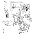
- Figure 1 is a schematic view showing an example of a radiation image recording and reproducing system, wherein an embodiment of the method for adjusting conditions in accordance with the present invention is employed.
- a stimulable phosphor sheet is used, and a preliminary readout is carried out.
- a memory card 51 is inserted into a card recording and reading device 50.
- a keyboard 43 of a computer system 40 By operating a keyboard 43 of a computer system 40, information, which has been recorded on the memory card 51 is read therefrom, or new information is recorded on the memory card 51.
- a plurality of of memory cards are prepared for a plurality of patients (or names of objects).
- a new memory card 51 is inserted into the card recording and reading device 50.
- Information giving specifics about an object such as the name of the object, the sex of the object, the name of disease of the object, and the portion of the object the image of which is recorded, is entered from the keyboard 43.
- the entered information is recorded on the memory card 51.
- image recording conditions under which an X-ray image is recorded in an X-ray image recording apparatus 1, are determined on the basis of the entered information.
- the image recording conditions include, for example, the tube voltage of an X-ray tube 2 (which tube voltage determines the quality of X-rays) and the time for which X-rays are irradiated (which determines the dose of X-rays).
- the corresponding memory card 51 is inserted into the card recording and reading device 50. From the memory card 51, it is found that an image recording operation was carried out for the patient.
- the same image recording conditions at those employed in the image recording operation carried out previously are set.
- information representing that a diagnosis is to be made for a different disease is entered from the keyboard 43, and new image recording conditions are determined.
- X-ray image of the object is recorded in the X-ray image recording apparatus 1.
- X-rays 3 are produced by an X-ray source 2 of the X-ray image recording apparatus 1 and irradiated to the chest 4a of a human body 4.
- X-rays 3a which have passed through the human body 4, impinge upon a stimulable phosphor sheet 11. In this manner, an X-ray image of the chest 4a of the human body 4 is stored on the stimulable phosphor sheet 11.
- the stimulable phosphor sheet 11, on which the X-ray image has been stored, is placed at a predetermined position in a preliminary read-out means 100 which carries out a preliminary readout by scanning the stimulable phosphor sheet 11 with a light beam having a low energy level, thereby releasing only part of the energy from the stimulable phosphor sheet 11, which energy was stored during its exposure to radiation.
- the stimulable phosphor sheet 11 is conveyed in a sub-scanning direction indicated by the arrow Y by a sheet conveyance means 13 which is constituted of an endless belt or the like and which is operated by a motor 12.
- a laser beam 15 which has a low energy level is produced by a laser beam source 14, and is reflected and deflected by a rotating polygon mirror 16 which is quickly rotated by a motor 23 in the direction indicated by the arrow.
- the laser beam 15 then passes through a converging lens 17 constituted of an f ⁇ lens or the like.
- the direction of the optical path of the laser beam 15 is then changed by a mirror 18, and the laser beam 15 impinges upon the stimulable phosphor sheet 11 and scans it in a main scanning direction indicated by the arrow X, which direction is approximately normal to the sub-scanning direction indicated by the arrow Y.
- the exposed portion of the stimulable phosphor sheet 11 emits light 19 in an amount proportional to the amount of energy stored thereon during its exposure to radiation.
- the emitted light 19 is guided by a light guide member 20 and photoelectrically detected by a photomultiplier 21.
- the light guide member 20 is made from a light guiding material such as an acrylic plate and has a linear light input face 20a, positioned so that it extends along the main scanning line on the stimulable phosphor sheet 11, and a ring-shaped light output face 20b, positioned so that it is in close contact with a light receiving face of the photomultiplier 21.
- An analog output signal S generated by the photomultiplier 21 is logarithmically amplified by a logarithmic amplifier 26, and digitized by an A/D converter 27 into a preliminary read-out image signal SP.
- the preliminary read-out image signal SP takes a value proportional to the logarithmic value of the amount of the light 19, which was emitted from each of picture elements in the X-ray image stored on the stimulable phosphor sheet 11.
- read-out conditions i.e. the voltage applied to the photomultiplier 21 and the amplification factor of the logarithmic amplifier 26, are adjusted so that image information can be detected accurately even if the amount of energy stored on the stimulable phosphor sheet 11 during its exposure to radiation varies over a wide range.
- the preliminary read-out image signal SP obtained in the manner described above is fed into a computer system 40.
- the computer system 40 comprises a main body 41 in which a CPU and an internal memory are incorporated, a disk drive unit 42 which operates a floppy disk serving as a subsidiary memory, a keyboard 43 from which necessary instructions, or the like, are fed into the computer system 40, and a CRT display device 44 which displays necessary information.
- the read-out conditions for the final readout i.e. the sensitivity and the contrast during the final readout, are determined in the manner described later.
- the voltage applied to a photomultiplier 21' and the amplification factor of a logarithmic amplifier 26' are controlled in accordance with the sensitivity and the contrast.
- the contrast corresponds to the ratio of the largest amount of emitted light, which is capable of being accurately converted into an image signal during the final readout, to the smallest amount of emitted light, which is capable of being accurately converted into an image signal during the final readout.
- the sensitivity corresponds to the photoelectric conversion factor, which represents to what image signal level a predetermined amount of emitted light is to be converted.
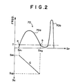
- Figure 2 is a graph showing an example of a probability density function of a preliminary read-out image signal SP.
- the values of the preliminary read-out image signal SP are plotted on the horizontal axis.
- the relative frequency of occurrence of the values of the preliminary read-out image signal SP is plotted on the vertical axis at the upper part of the graph, and the values of an image signal SQ, which is obtained during a final readout, are plotted on the vertical axis at the lower part of the graph.
- a probability density function 70 is composed of projecting parts 70a and 70b.
- the projecting part 70a represents the image signal components of the preliminary read-out image signal SP, which represent an object image.
- the projecting part 70b corresponds to a background region, upon which the X-rays impinged directly without passing through the object. Only the image signal components representing the object image need be obtained during the final readout. Therefore, the values of the probability density function 70 are compared to a threshold value T, starting with the value of the function at the minimum value of the preliminary read-out image signal SP (i.e. the left side of Figure 2) and working along the direction of increase of the image signal values (i.e. toward the right side of Figure 2).
- the read-out conditions for the final readout are adjusted such that, during the final readout, the image information represented by values of the emitted light signal falling within the range of SP1 to SP2 may be detected as an image signal with values lying on the straight line G shown in Figure 2.
- Information representing the read-out conditions for the final readout, which have thus been adjusted, is recorded on the memory card 51 in the card recording and reading device 50.
- the read-out conditions for the final readout are adjusted in the manner described above, or the information representing the read-out conditions for the final readout is read from the memory card 51. Thereafter, a stimulable phosphor sheet 11' on which the preliminary readout has been finished is placed at a predetermined position in the final read-out means 100' and scanned with a laser beam 15' having an energy level higher than that of the laser beam 15 used during the preliminary readout. In this manner, an image signal SQ is detected under the read-out conditions, which have been determined in the manner described above.
- the configuration of the final read-out means 100' is nearly the same as that of the preliminary read-out means 100, and therefore elements corresponding to those constituting the preliminary read-out means 100 are numbered with corresponding primed reference numerals in Figure 1.
- the resulting image signal SQ is fed into the computer system 40, which carries out appropriate image processing on the image signal SQ.
- the image signal is fed into a laser printer 90, which reproduces a visible image on film 91 from the image signal.
- memory cards 51, 51, ... are prepared, on which information about corresponding patients is recorded.
- the image recording and read-out operations are carried out under the same image recording conditions and the same read-out conditions for the final readout as those which were employed during the image recording and read-out operations carried out for the same old patient. Therefore, a reproduced image can be obtained which is capable of being appropriately compared with a previously reproduced image.
- the image recording conditions and the read-out conditions for the final readout are adjusted such that a reproduced image of the X-ray image of the new patient, which has best possible image quality, may be obtained.
- the storage device need not have a large storage capacity, and the number of on-line signal transmitting operations can be kept small. Therefore, the configuration of the apparatus for carrying out the method for adjusting conditions can be kept simple. However, in such cases, there is a risk that some of memory cards 51, 51, ... are lost. Therefore, a storage device 52 having a large capacity, such as a hard disk drive unit, may be used, and many pieces of information about a plurality of patients may be managed collectively.
- the preliminary read-out means 100 and the final read-out means 100' are separate from each other.
- a single read-out means may be utilized for performing both the preliminary readout and the final readout.
- the stimulable phosphor sheet 11 may be moved back to the position at which image readout is started. Thereafter, the final readout may be carried out.
- the read-out conditions for the final readout are adjusted.
- predetermined read-out conditions may be used when the final readout is carried out regardless of the characteristics of the preliminary read-out image signal SP and the past image recording and read-out operations.
- the computer system 40 may adjust the normalizing conditions. Under the normalizing conditions, a normalizing operation is carried out wherein only the image signal components representing a desired object image are extracted from the image signal SQ and the extracted image signal components are normalized such that they are suitable to be sent to the laser printer 90.
- information representing the corresponding normalizing conditions may be read from the corresponding memory card 51.
- the computer system 40 may manage both the read-out conditions and the normalizing conditions.
- the aforesaid embodiment is applied to the radiation image recording and reproducing system wherein the preliminary readout is carried out.
- the method for adjusting conditions in accordance with the present invention is also applicable when no preliminary read-out operations are carried out, and only the aforesaid final read-out operations are carried out.
- an image signal is obtained by use of predetermined read-out conditions.
- normalizing conditions are adjusted by the computer system 40. Also, information representing the normalizing conditions is recorded on a memory card 51 or read therefrom. A normalizing operation is carried out on the image signal under the adjusted normalizing conditions.
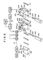
- Figure 5 shows a radiation image read-out and reproducing system, wherein an embodiment of the method for setting read-out conditions and/or image processing conditions for a radiation image in accordance with the present invention is employed.
- Figure 6 shows how a radiation image is recorded on a stimulable phosphor sheet.
- radiation 102 is produced by a radiation source 120, such as an X-ray tube.
- the radiation 102 is irradiated to an object 101.
- a stimulable phosphor sheet 103 is exposed to the radiation 102, which has passed through the object 101, and a radiation image of the object 101 is stored on the stimulable phosphor sheet 103.
- the radiation image is read out from the stimulable phosphor sheet 103.
- stimulable phosphors which may be employed to constitute the stimulable phosphor sheet 103, are described in detail in, for example, U.S. Patent No. 4,236,078 and European Patent No. 21,342.
- the radiation image read-out and reproducing system of Figure 5 comprises basically a preliminary read-out section 130, a final read-out section 140, and an image reproducing section 150.
- the stimulable phosphor sheet 103 on which the radiation image of the object 101 has been stored, is sent to the preliminary read-out section 130 by a sheet conveyance means 110, which may be constituted of a conveyor roller, or the like.
- a laser beam 202 is produced by a laser beam source 201.
- the laser beam 202 first passes through a filter 203, which filters out light having wavelengths within the range of wavelengths of the light emitted by the stimulable phosphor sheet 103 upon stimulation thereof by the laser beam 202.
- the laser beam 202 is one-dimensionally deflected by a light deflector 204, such as a galvanometer mirror, and directed onto the stimulable phosphor sheet 103 by a plane reflection mirror 205.
- the laser beam source 201 is selected so that the laser beam 202 produced thereby has a wavelength distribution different from and far apart from the wavelength distribution of the light emitted by the stimulable phosphor sheet 103 when it is stimulated.
- the stimulable phosphor sheet 103 While the laser beam 202 impinges upon the stimulable phosphor sheet 103, the stimulable phosphor sheet 103 is moved in the direction indicated by the arrow 206 (i.e. in the sub-scanning direction) by a sheet conveyance means 210, which may be constituted of conveyor rollers, or the like. In this manner, the overall surface of the stimulable phosphor sheet 103 is exposed to and scanned by the laser beam 202.
- the power of the laser beam source 201, the beam diameter of the laser beam 202, the speed with which the laser beam 202 scans, and the speed at which the stimulable phosphor sheet 103 moves are selected so that the level of the stimulation energy of the laser beam 202 used during the preliminary readout is lower than the level of the stimulation energy of the laser beam used during the final readout carried out in the final read-out section 140.
- the stimulable phosphor sheet 103 When it is exposed to the laser beam 202 in the manner described above, the stimulable phosphor sheet 103 emits light in an amount proportional to the amount of energy stored thereon during its exposure to the radiation.
- the emitted light enters a light guide member 207, which may be of the shape and material disclosed in U.S. Patent No. 4,346,295.
- the light is guided inside of the light guide member 207 through total reflection, emanates from a light output face of the light guide member 207 and is received by a photodetector 208, which may be constituted of a photomultiplier, or the like.
- the light receiving face of the photodetector 208 is positioned so that it is in close contact with a filter, which transmits only light having wavelengths within the range of wavelengths of light emitted by the stimulable phosphor sheet 103 and filters out light having wavelengths within the range of wavelengths of the stimulating rays. Therefore, the photodetector 208 detects only the light emitted by the stimulable phosphor sheet 103 upon stimulation thereof. The light detected by the photodetector 208 is converted into an electric signal carrying the image input information stored on the stimulable phosphor sheet 103, and amplified by an amplifier 209.
- the signal generated by the amplifier 209 is digitized by an A/D converter 211, and sent as a preliminary read-out image signal SP to a final read-out control means 314 in the final read-out section 140.
- the final read-out control means 314 adjusts a read-out gain setting value (a), a scale factor setting value (b), and an image processing condition setting value (c).
- the preliminary read-out image signal SP detected from the stimulable phosphor sheet 103 is stored on a recording medium, such as an optical disk or a magnetic disk, in an image filing apparatus 340.
- a laser beam 302 is produced by a laser beam source 301.
- the laser beam 302 first passes through a filter 303, which filters out light having wavelengths within the range of the wavelengths of light emitted by the stimulable phosphor sheet 103 upon stimulation thereof by the laser beam 302. Then, the beam diameter of the laser beam 302 is precisely adjusted by a beam expander 304.
- the laser beam 302 is then deflected by a light deflector 305, which may be formed of a galvanometer mirror, or the like.
- the laser beam 302 is then caused to impinge upon the stimulable phosphor sheet 103 by a plane reflection mirror 306. Between the light deflector 305 and the plane reflection mirror 306, an f ⁇ lens 307 is disposed for keeping the beam diameter of the laser beam 302 uniform as it scans the stimulable phosphor sheet 103. While the laser beam 302 impinges upon the stimulable phosphor sheet 103, the stimulable phosphor sheet 103 is moved in the direction indicated by the arrow 308 (i.e. in the sub-scanning direction) by a sheet conveyance means 320, which may be constituted of conveyor rollers, or the like. Consequently, the overall area of the stimulable phosphor sheet 103 is exposed to and scanned by the laser beam 302.
- the stimulable phosphor sheet 103 When the stimulable phosphor sheet 103 is exposed to the laser beam 302, it emits light in proportion to the amount of energy stored thereon during its exposure to the radiation.
- the light emitted enters a light guide member 309, which is made of the same material and has the same configuration as the light guide member 207 used for the preliminary readout.
- the light emitted by the stimulable phosphor sheet 103 is guided inside of the light guide member 309 through repeated total reflection, emanates from the light output face of the light guide member 309 and is received by a photodetector 310, which may be constituted of a photomultiplier, or the like.
- the light receiving face of the photodetector 310 is positioned so that it is in close contact with a filter, which selectively transmits only the light having wavelengths within the range of wavelengths of light emitted by the stimulable phosphor sheet 103. Therefore, the photodetector 310 detects only the light emitted by the stimulable phosphor sheet 103.
- the output of the photodetector 310 which photoelectrically detects the light emission representing the radiation image stored on the stimulable phosphor sheet 103, is amplified to an appropriate level by an amplifier 311.
- the gain of the amplifier 311 is adjusted on the basis of the read-out gain setting value (a) determined by the final read-out control means 314.
- the amplified electric signal is fed into an A/D converter 312, which converts the electric signal into a digital signal by use of a scale factor which is adjusted by the scale factor setting value (b) to suit the width in the fluctuation of the values of the signal.
- the digital signal thus obtained is fed into a signal processing circuit 313.
- the digital signal is subjected to signal processing (image processing), the nature of which signal processing is based on the image processing condition setting value (c).
- image processing image processing
- a visible radiation image is obtained which has good image quality and can serve as an effective tool in, particularly, the efficient and accurate diagnosis of an illness.
- the processed digital signal is output as a read-out image signal (a final read-out image signal) SO.
- the final read-out image signal SO generated by the signal processing circuit 313 is fed into a light modulator 401 in the image reproducing section 150.
- a laser beam 403 is produced by a reproducing laser beam source 402.
- the laser beam 403 is modulated by the light modulator 401 on the basis of the final read-out image signal SO received from the signal processing circuit 313.
- the laser beam 403 is then made to impinge upon a photosensitive material 405, such as photographic film, by a scanning mirror 404 which causes the laser beam 403 to scan the photosensitive material 405.
- the photosensitive material 405 is moved in a direction normal to the aforesaid scanning direction, i.e. in the direction indicated by the arrow 406. Accordingly, the radiation image represented by the final read-out image signal SO is recorded on the photosensitive material 405.
- any other appropriate method such as the aforesaid method using a CRT display device.
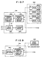
- Figure 7 shows the final read-out control means 314, which adjusts the setting values (a), (b), and (c).
- the final read-out control means 314 may be constituted of, for example, a known computer system.
- a radiation image of an object 101 which image is to be used in finding a change in the state of the same object with the passage of time
- a stimulable phosphor sheet 103 four radiation images of the object 101, which are to be used in finding a change in the state of the same object with the passage of time, were recorded and read out.
- a probability density function creating means 351 of the final read-out control means 314 receives a preliminary read-out image signal SP, which has currently been obtained from a preliminary readout.
- the probability density function creating means 351 also receives preliminary read-out image signals SP1, SP2, SP3, and SP4, which have been stored in four image files 350A, 350B, 350C, and 350D of an image filing apparatus 340.
- the preliminary read-out image signals SP1, SP2, SP3, and SP4 represent radiation images of the same object as the object the image of which was stored on the stimulable phosphor sheet 103 currently subjected to the preliminary readout. These radiation images are used in finding a change in the state of the same object with the passage of time.
- the probability density function creating means 351 creates the probability density function of all of the preliminary read-out image signals SP, SP1, SP2, SP3, and SP4.
- the probability density function basically has a pattern shown in Figure 8.
- the frequency of occurrence of each signal value takes a value approximately five times the frequency of occurrence of the corresponding signal value in a probability density function of only the preliminary read-out image signal SP detected from a single stimulable phosphor sheet 103.
- the probability density function creating means 351 normalizes the probability density function, which has been created in the manner described above, such that it may correspond to a probability density function of a single preliminary read-out image signal SP.
- Information H representing the probability density function, which has been created in the manner described above, is fed into a probability density function analyzing means 352.
- the probability density function analyzing means 352 calculates the maximum value of the signal, the minimum value of the signal, the signal value which occurs most frequently, i.e. the signal value corresponding to the maximum value of the probability density function, or the like, and feeds information Sr representing the calculated values into a reading means 353.
- a storage means 354 stores information representing the read-out gain setting value (a), the scale factor setting value (b), and the image processing condition setting value (c), which are suitable for the aforesaid maximum value, the minimum value, the signal value which occurs most frequently, or the like.
- the reading means 353 reads the pieces of the information representing the setting values (a), (b) and (c), which correspond to the information Sr, from the storage means 354. The reading means 353 then feeds these pieces of the information respectively into the amplifier 311, the A/D converter 312, and the signal processing circuit 313.
- the setting values (a), (b), and (c) are adjusted in the manner described above. Therefore, the setting values (a), (b), and (c) can be determined which are optimal for the radiation image currently stored on the stimulable phosphor sheet 103.
- the setting values (a), (b), and (c) are determined, the characteristics of the image input information on the four stimulable phosphor sheets, on which the radiation images were stored in the past, are taken into consideration. Therefore, the setting values (a), (b), and (c) thus determined become close to those which were employed when the radiation images to be used in finding a change in the state of the same object with the passage of time were reproduced as visible images.
- the setting values (a), (b), and (c) are determined, the characteristics of the image input information on the stimulable phosphor sheet 103, from which the preliminary readout is carried out currently, are taken into consideration. Therefore, unlike the cases where the setting values (a), (b), and (c) are fixed for the same object, a large amount of variation in image density does not occur between the visible radiation images even if the image recording conditions, under which the current radiation image was recorded on the stimulable phosphor sheet 103, are different from the image recording conditions, under which the past radiation images of the same object were recorded.
- the characteristics of the image input information on all of the plurality of the stimulable phosphor sheets, on which the image recording and read-out operations were carried out for the same object in the past, are utilized in setting the read-out conditions for the final readout and/or the image processing conditions with respect to the radiation image stored on the stimulable phosphor sheet 103, from which the image read-out operation is carried out currently.
- the characteristics of the image input information on some of the plurality of the stimulable phosphor sheets may be utilized in setting the read-out conditions for the final readout and/or the image processing conditions with respect to the radiation image stored on the stimulable phosphor sheet 103, from which the image read-out operation is carried out currently. This also applies to an embodiment described below.
- a different example of a final read-out control means 314, which may be employed in the method for setting read-out conditions and/or image processing conditions for a radiation image in accordance with the present invention, will be described hereinbelow with reference to Figure 9.
- the final read-out control means 314 shown in Figure 9 is provided with a neural network 360 in lieu of the probability density function analyzing means 352, the reading means 353, and the storage means 354 shown in Figure 7.
- the neural network 360 is constituted of a computer system.
- the neural network 360 is provided with a learning function by back propagation method. Specifically, when information (an instructor signal), which represents whether an output signal obtained when an input signal is given is or is not correct, is fed into the neural network, the weight of connections between units in the neural network (i.e. the weight of synapse connections) is corrected. By repeating the learning of the neural network, the probability that a correct answer will be obtained in response to a new input signal can be kept high. (Such functions are described in, for example, "Learning representations by back-propagating errors" by D. E. Rumelhart, G. E. Hinton and R. J.
- the neural network 360 receives the information H representing the probability density function of the preliminary read-out image signals SP, SP, ... obtained from five stimulable phosphor sheets 103, 103, ... In accordance with the information H, the neural network 360 feeds out information representing an appropriate read-out gain setting value (a), an appropriate scale factor setting value (b), and an appropriate image processing condition setting value (c).
- the read-out gain setting value (a), the scale factor setting value (b), and the image processing condition setting value (c) are determined on the basis of all of the characteristics of the image input information on the stimulable phosphor sheet 103, from which the preliminary readout is currently carried out, and the characteristics of the image input information on the four stimulable phosphor sheets, from which image read-out operations were carried out in the past.
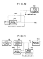
- a further different example of a final read-out control means 314, which may be employed in the method for setting read-out conditions and/or image processing conditions for a radiation image in accordance with the present invention, will be described hereinbelow with reference to Figure 10.
- the final read-out control means 314 shown in Figure 10 is provided with a neural network 370 in lieu of the probability density function creating means 351, the probability density function analyzing means 352, the reading means 353, and the storage means 354 shown in Figure 7.
- the neural network 370 receives the image signal components of the preliminary read-out image signals SP, SP, ... which represent picture elements in the radiation images stored on five stimulable phosphor sheets 103, 103, ... In accordance with the received signals, the neural network 360 feeds out information representing an appropriate read-out gain setting value (a), an appropriate scale factor setting value (b), and an appropriate image processing condition setting value (c).
- a an appropriate read-out gain setting value
- b an appropriate scale factor setting value
- c an appropriate image processing condition setting value
- a neural network for determining the read-out conditions for the final readout and/or the image processing conditions on the basis of a preliminary read-out image signal SP is described in detail in, for example, Japanese Patent Application No. 2(1990)-151040.
- the read-out gain setting value (a), the scale factor setting value (b), and the image processing condition setting value (c) are determined on the basis of all of the characteristics of the image input information on the stimulable phosphor sheet 103, from which the preliminary readout is currently carried out, and the characteristics of the image input information on the four stimulable phosphor sheets, from which image read-out operations were carried out in the past.
- the final read-out section and the preliminary read-out section are independent and separate from each other.
- a single read-out section may be utilized in common to carry out both the preliminary readout and the final readout.
- the stimulable phosphor sheet is returned by a sheet conveyance means to the read-out section.
- the final readout is then carried out.
- the stimulation energy of stimulating rays may be set by a stimulation energy adjusting means to a level lower than that employed during the final readout.
- the method for setting read-out conditions and/or image processing conditions for a radiation image in accordance with the present invention is also applicable in such cases.
- the read-out conditions for the final readout and the image processing conditions are set with the method in accordance with the present invention.
- a final readout may be carried out under predetermined read-out conditions regardless of the characteristics of a preliminary read-out image signal SP, and the image processing conditions, under which image processing is to be carried out on an image signal SO, may be determined on the basis of the preliminary read-out image signal SP.
- the image processing conditions may be fixed, and only the read-out conditions for the final readout may be set with the method in accordance with the present invention.
- a preliminary readout is carried out.
- the method for setting read-out conditions and/or image processing conditions for a radiation image in accordance with the present invention is also applicable when a preliminary readout is not carried out, but only a final readout is carried out.
- Figure 11 shows such a radiation image read-out and reproducing system.
- a read-out section 500 is basically constituted in the same manner as that in the final read-out section 140 of Figure 5.
- a radiation image is read out from a stimulable phosphor sheet.
- An image signal SO obtained thereby is subjected to image processing in the signal processing circuit 313.
- the image signal SO which has been obtained from the image processing, is sent to the image reproducing section 150 constituted in the same manner as that shown in Figure 5.
- the image reproducing section 150 a visible image is reproduced from the image signal SO.
- the image processing conditions are set on the basis of the image processing condition setting value (c), which has been determined by the neural network 370.
- the same means as that shown in Figure 10 is employed.
- the means shown in Figure 7 or Figure 9 may be employed for this purpose.
- the neural network 370 determines the image processing condition setting value (c) on the basis of only the image signal SO.
- image signals SO1, SO2, SO3, and SO4 corresponding to said object, which were obtained in the past from the read-out section 500 and stored in the image filing apparatus 340, are read from the image filing apparatus 340.
- the image signals SO1, SO2, SO3, and SO4 are fed into the neural network 370 together with the image signal SO.
- the neural network 370 determines the image processing condition setting value (c) on the basis of all of the image signals SO1, SO2, SO3, SO4, and SO.
- the image processing condition setting value (c) is determined on the basis of all of the characteristics of the image input information on the stimulable phosphor sheet 103, from which the preliminary readout is currently carried out, and the characteristics of the image input information on the four stimulable phosphor sheets, from which image read-out operations were carried out in the past. Accordingly, a large amount of variation in image density does not occur between the visible radiation image, which is to be used in finding a change in the state of the same object with the passage of time and which is reproduced currently, and the visible radiation images, which are to be used in finding a change in the state of the same object with the passage of time and which were reproduced in the past.
- the recording medium used is not limited to a stimulable phosphor sheet.
- the read-out section 500 of Figure 11 may be replaced by, for example, a means for reading out a radiation image from a sheet of silver halide photographic film or a storage means, such as an optical disk, for storing an image signal representing a radiation image.
- the method for setting read-out conditions and/or image processing conditions for a radiation image in accordance with the present invention is also applicable in such cases.
Landscapes
- Physics & Mathematics (AREA)
- Health & Medical Sciences (AREA)
- Life Sciences & Earth Sciences (AREA)
- General Physics & Mathematics (AREA)
- High Energy & Nuclear Physics (AREA)
- Molecular Biology (AREA)
- Spectroscopy & Molecular Physics (AREA)
- Apparatus For Radiation Diagnosis (AREA)
- Radiography Using Non-Light Waves (AREA)
- Transforming Light Signals Into Electric Signals (AREA)
Claims (11)
- Verfahren zum Aufzeichnen, Auslesen und Wiedergeben eines Strahlungsbildes, umfassend:i) Durchführen einer Strahlungsbildaufzeichnungsoperation, bei der Strahlung auf ein Objekt (4) gerichtet wird und ein anregbares Leuchtstoffblatt (11) der Strahlung ausgesetzt wird, die durch das Objekt (4) hindurchgegangen ist, wodurch auf dem anregbaren Leuchtstoffblatt (11) ein Strahlungsbild des Objekts (4) gespeichert wird,ii) Durchführen einer Bildleseoperation, bei der:ein erstes Bildsignal, repräsentativ für das Strahlungsbild des Objekts, mittels eines Vorab-Lesevorgangs erhalten wird, bei dem das anregbare Leuchtstoffblatt (11) mit dem darin gespeicherten Strahlungsbild Anregungsstrahlen ausgesetzt wird, die bewirken, daß das anregbare Leuchtstoffblatt (11) Licht im Verhältnis zu der in ihm bei der Strahlungsexposition gespeicherten Menge Energie emittiert wird und das emittierte Licht erfaßt wird, undein zweites Bildsignal, repräsentativ für das Strahlungsbild, im Anschluß gewonnen wird mit Hilfe eines endgültigen Auslesens, bei dem das anregbare Leuchtstoffblatt (11) erneut Anregungsstrahlen ausgesetzt wird und von dem anregbaren Leuchtstoffblatt emittiertes Licht erfaßt wird, wobei Auslesebedingungen für das endgültige Auslesen auf der Grundlage des ersten Bildsignals eingestellt werden,iii) falls notwendig, Unterziehen des durch das endgültige Auslesen gewonnenen zweiten Bildsignals einer Bildverarbeitung, undiv) Reproduzieren eines sichtbaren Bildes aus dem zweiten Bildsignal, welches gegebenenfalls einer Bildverarbeitung unterzogen wurde,wobei Bildaufkeichnungsbedingungen für den Strahlungsbildaufzeichnungsvorgang und die Auslesebedingungen für das endgültige Auslesen eingestellt werden,wobei ein Strahlungsbildaufzeichnungsvorgang und ein Bildauslesevorgang für das spezifische Objekt (4) unter den so eingestellten Bedingungen durchgeführt werden, wobei die Bedingungseinstellung folgende Schritte beinhaltet:a) Speichern von Information, repräsentativ für Bildaufzeichnungsbedingungen, unter denen die Strahlungsbild-Auheichnungsvorgänge in der Vergangenheit durchgeführt wurden, Information, die Spezifikationen über die entsprechenden Objekte, für die Strahlungsbild-Aufzeichnungsoperationen durchgeführt werden, und Information, die repräsentativ ist für die entsprechenden Auslesebedingungen beim endgültigen Auslesen im Zuge von Bildausleseoperationen, die in der Vergangenheit durchgeführt wurden, wobei diese Informationsstücke in einer Speichereinrichtung (51, 52) gespeichert sind;b) wenn eine Strahlungsbild-Aufzeichnungsoperation und eine Bildleseoperation für ein spezifisches Objekt (4) durchgeführt werden sollen, Überprüfen, ob in der Vergangenheit für dasselbe Objekt wie das spezifische Objekt (4) eine Strahlungsbild-Aufzeichnungsoperation und eine Bildleseoperation durchgeführt wurden oder nicht; undc) für die Fälle, in denen eine Strahlungsbild-Aufzeichnungsoperation und eine Bildleseoperation in der Vergangenheit für dasselbe Objekt wie das spezifische Objekt (4) durchgeführt wurden,Einstellen der Bildaufzeichnungsbedinungen für das spezifische Objekt (4) derart, daß die Bedingungen übereinstimmen mit den Bildaufzeichnungsbedingungen, unter denen die Strahlungsbild-Aufzeichnungsoperation für dasselbe Objekt wie das spezifische Objekt (4) durchgeführt wurde,anstatt Auslesebedingungen für das endgültige Auslesen, welches für das spezifische Objekt (4) zu erfolgen hat, auf der Grundlage eines ersten Bildsignals einzustellen, welches das Strahlungsbild des spezifischen Objekts (4) repräsentiert, Einstellen der Auslesebedingungen für das endgültige Auslesen, welches für das spezifische Objekt (4) durchgeführt werden soll, in der Weise, daß die Bedingungen übereinstimmen mit den Auslesebedingungen, unter denen das endgültige Auslesen eines Strahlungsbildes für dasselbe Objekt wie das spezifische Objekt (4) durchgeführt worden war.
- Verfahren nach Anspruch 1, bei dem die Speichereinrichtung kollektiv die Information bezüglich einer Mehrzahl von Objekten (4) speichert.
- Verfahren nach Anspruch 1 oder 2, bei dem die Information, die Spezifikationen über die entsprechenden Objekte (4) gibt, die Information enthält, welche die Namen der Objekte (4) repräsentieren, ferner die Information, die die Teile der Objekte repräsentiert, deren Bilder aufgezeichnet wurden, und
die Speichereinrichtung sich aus mehreren unabhängigen und verschiedenen Speichermedien (51) zusammensetzt, die für unterschiedliche Namen von Objekten vorbereitet wurden. - Verfahren nach Anspruch 1, 2 oder 3, bei dem die Anregungsstrahlen durch ein Laserstrahlbündel (15) gebildet werden.
- Verfahren zum Aufzeichnen, Auslesen und Wiedergeben eines Strahlungsbildes, umfassend die Schritte:Durchführen einer Strahlungsbild-Aufzeichnungsoperation, bei der Strahlung auf ein Objekt (101) gerichtet wird und ein Aufzeichnungsmedium (103) der Strahlung (102) ausgesetzt wird, die durch das Objekt (10) hindurchgegangen ist, wobei ein Strahlungsbild des Objekts (101) auf dem Aufzeichnungsmedium (103) aufgezeichnet wird,Durchführen einer Bildleseoperation, bei der das auf dem Aufzeichnungsmedium (103) aufgezeichnete Strahlungsbild photoelektrisch ausgelesen wird und ein Bildsignal, welches repräsentativ ist für das Strahlungsbild des Objekts (101), gewonnen wird, welches aus einer Reihe von Bildsignalkomponenten gebildet wird,Festlegen von Normierbedingungen für einen Normiervorgang auf der Grundlage des Bildsignals, welches durch die Bildleseoperation erhalten wurde, undDurchführen der Normieroperation, wobei nur Bildsignalkomponenten, die gewünschte Bildinformation darstellen, aus dem Bildsignal entnommen werden, welches durch die Bildleseoperation gewonnen wurde,falls erforderlich, Durchführen einer Bildverarbeitung an den Bildsignalkomponenten, die durch die Normieroperation gewonnen wurden, undReproduzieren eines sichtbaren Bildes aus den Bildsignalkomponenten, die gegebenenfalls einer Bildverarbeitung unterzogen wurden;wobei das Verfahren den Schritt beinhaltet:Einstellen der Bildaufzeichnungsbedingungen für die Strahlungsbild-Aufzeichnungsoperation sowie der Normierbedingungen für die Normieroperation, wobeidie Strahlungsbild-Aufzeichnungsoperation und die Bildleseoperation für das spezifische Objekt (101) unter denjenigen Bedingungen durchgeführt werden, die auf diese Weise eingestellt wurden, und wobei die Einstellungen der Bedingungen folgende Schritte beinhaltet:Speichern von Information betreffend die Bildaufzeichnungsbedingungen, unter denen Strahlungsbild-Aufzeichnungsoperationen in der Vergangenheit durchgeführt wurden, Information über Spezifikationen bezüglich der entsprechenden Objekte (101), für die die Strahlungsbild-Aufzeichnungsoperationen durchgeführt wurden, und Information über die entsprechenden Normierbedingungen für in der Vergangenheit ausgeführte Normieroperationen, wobei diese Informationsstücke in einer Speichereinrichtung (354, 340) gespeichert werden,wenn eine Strahlungsbild-Aufzeichnungsoperation, eine Bildleseoperation und eine Normieroperation für ein spezifisches Objekt (101) durchgeführt werden sollen, Überprüfen, ob in der Vergangenheit für dasselbe Objekt wie das spezifische Objekt bereits eine Strahlungsbild-Aufzeichnungsoperation, eine Bildleseoperation und eine Normieroperation durchgeführt wurden oder nicht, undin den Fällen, in denen für dasselbe Objekt wie das spezifische Objekt (101) eine Strahlungsbild-Aufzeichnungsoperation, eine Bildleseoperation und eine Normieroperation durchgeführt wurden,Einstellen der Bildaufzeichnungsbedingungen für das spezifische Objekt in der Weise, daß die Bedingungen übereinstimmen mit den Bildaufzeichnungsbedingungen, unter denen sie für dasselbe Objekt wie das spezifische Objekt (101) durchgeführt wurde,anstatt Normierbedingungen für eine Normieroperation, die für das spezifische Objekt (101) durchzuführen ist, auf der Grundlage eines Bildsignals einzustellen, welches das Strahlungsbild des spezifischen Objekts repräsentiert, Einstellen der Normierbedingungen für die Normieroperation, welche für das spezifische Objekt (101) durchzuführen ist, derart, daß die Bedingungen übereinstimmen mit den Normierbedingungen, unter denen der Normiervorgang für das Bildsignal (Spl-Sp4) durchgeführt wurde, welches das Strahlungsbild desselben Objekts (101) wie das spezifische Objekt (101) repräsentiert.
- Verfahren nach Anspruch 5, bei dem die Speichereinrichtung (340) kollektiv die Information über eine Mehrzahl von Objekten (101) speichert.
- Verfahren nach Anspruch 5 oder 6,bei dem die Information über Spezifikationen bezüglich entsprechender Objekte (101) die Information über die Namen der Objekte und die Informationen über die Teile von Objekten, deren Bilder aufgezeichnet wurden, enthält, unddie Speichereinrichtung sich zusammensetzt aus mehreren unabhängigen und verschiedenen Speichermedien, die für verschiedene Namen von Objekten (101) vorbereitet werden.
- Verfahren nach Anspruch 6 oder 7, bei dem das Aufzeichnungsmedium ein anregbares Leuchtstoffblatt (103) ist.
- Verfahren nach Anspruch 8, bei dem das Bildsignal dadurch gewonnen wird, daß das anregbare Leuchtstoffblatt (103) Anregungsstrahlen ausgesetzt wird, die veranlassen, daß das anregbare Leuchtstoffblatt (103) Licht im Verhältnis zu der Energiemenge emittiert, die während seiner Exposition mit Strahlung ausgesetzt wurde, und das emittierte Licht photoelektrisch nachgewiesen wird.
- Verfahren nach Anspruch 9, bei dem die Anregungsstrahlen durch ein Laserstrahlbündel gebildet werden.
- Verfahren nach Anspruch 5, bei dem das Aufzeichnungsmedium ein photographischer Film (405) ist.
Priority Applications (1)
| Application Number | Priority Date | Filing Date | Title |
|---|---|---|---|
| EP97106837A EP0802428A3 (de) | 1990-06-20 | 1991-06-19 | Verfahren zum Passen von Läsebedingungen und/oder Bildverarbeitungsbedingungen für Strahlungsbilder |
Applications Claiming Priority (4)
| Application Number | Priority Date | Filing Date | Title |
|---|---|---|---|
| JP2161594A JP2896804B2 (ja) | 1990-06-20 | 1990-06-20 | 放射線画像撮影読取再生方法における条件決定方法 |
| JP161594/90 | 1990-06-20 | ||
| JP2250881A JP2582668B2 (ja) | 1990-09-20 | 1990-09-20 | 放射線画像情報読取条件および/または画像処理条件設定方法 |
| JP250881/90 | 1990-09-20 |
Related Child Applications (1)
| Application Number | Title | Priority Date | Filing Date |
|---|---|---|---|
| EP97106837A Division EP0802428A3 (de) | 1990-06-20 | 1991-06-19 | Verfahren zum Passen von Läsebedingungen und/oder Bildverarbeitungsbedingungen für Strahlungsbilder |
Publications (3)
| Publication Number | Publication Date |
|---|---|
| EP0467087A2 EP0467087A2 (de) | 1992-01-22 |
| EP0467087A3 EP0467087A3 (de) | 1994-12-21 |
| EP0467087B1 true EP0467087B1 (de) | 1998-09-23 |
Family
ID=26487671
Family Applications (1)
| Application Number | Title | Priority Date | Filing Date |
|---|---|---|---|
| EP91110076A Expired - Lifetime EP0467087B1 (de) | 1990-06-20 | 1991-06-19 | Verfahren zur Einstellung der Bedingung für ein Strahlungsaufzeichnungs- und -wiedergabesystem |
Country Status (3)
| Country | Link |
|---|---|
| US (1) | US5272339A (de) |
| EP (1) | EP0467087B1 (de) |
| DE (1) | DE69130242T2 (de) |
Families Citing this family (11)
| Publication number | Priority date | Publication date | Assignee | Title |
|---|---|---|---|---|
| US5828775A (en) * | 1990-04-18 | 1998-10-27 | Fuji Photo Film Co., Ltd. | Method and apparatus for adjusting read-out conditions and/or image processing conditions for radiation images , radiation image read-out apparatus, and radiation image analyzing method and apparatus |
| EP0726542B1 (de) * | 1990-04-18 | 1999-09-15 | Fuji Photo Film Co., Ltd. | Verfahren und Vorrichtung zum Regeln von Lesebedingungen und/oder Bildverarbeitungsbedingungen für Strahlungsbilder, Strahlungsbildlesevorrichtung und Verfahren und Vorrichtung zur Strahlungsbildanalyse |
| JP2932020B2 (ja) * | 1992-10-15 | 1999-08-09 | 富士写真フイルム株式会社 | マーゲン画像読取条件および/または画像処理条件決定方法 |
| JP2934360B2 (ja) * | 1993-02-12 | 1999-08-16 | 三菱電機株式会社 | 映像信号処理装置 |
| JPH07128759A (ja) * | 1993-11-05 | 1995-05-19 | Fuji Photo Film Co Ltd | 放射線画像読取方法および装置 |
| JP3901770B2 (ja) * | 1996-10-21 | 2007-04-04 | 富士フイルム株式会社 | エネルギーサブトラクション処理方法および装置 |
| EP1329849A3 (de) | 1997-08-29 | 2004-08-04 | Fuji Photo Film Co., Ltd. | Bildverarbeitungssystem |
| US20090307931A1 (en) * | 2008-06-17 | 2009-12-17 | Frederick Robert May | Slip resistant ski boot protection apparatus |
| US20080035839A1 (en) * | 2006-08-10 | 2008-02-14 | Luc Struye | Radiation Image read-out method |
| FI20086241A7 (fi) | 2008-12-23 | 2010-06-24 | Palodex Group Oy | Kuvalevyn lukijalaite |
| FI20086240A7 (fi) * | 2008-12-23 | 2010-06-24 | Palodex Group Oy | Kuvalevyn lukijalaitteen puhdistusjärjestelmä |
Family Cites Families (20)
| Publication number | Priority date | Publication date | Assignee | Title |
|---|---|---|---|---|
| JPS5512429A (en) * | 1978-07-12 | 1980-01-29 | Fuji Photo Film Co Ltd | Radioactive image reader |
| NL7905433A (nl) * | 1978-07-12 | 1980-01-15 | Fuji Photo Film Co Ltd | Werkwijze en inrichting voor het registreren en weergeven van een stralingsbeeld. |
| US4346295A (en) * | 1978-12-26 | 1982-08-24 | Fuji Photo Film Co., Ltd. | Radiation image read out device |
| US4315318A (en) * | 1978-12-26 | 1982-02-09 | Fuji Photo Film Co., Ltd. | Method and apparatus for processing a radiation image |
| JPS55116340A (en) * | 1979-02-28 | 1980-09-06 | Fuji Photo Film Co Ltd | Method and device for processing gradation of radiation picture |
| DE3062728D1 (de) * | 1979-06-19 | 1983-05-19 | Kasei Optonix | Phosphor |
| JPS5611395A (en) * | 1979-07-11 | 1981-02-04 | Fuji Photo Film Co Ltd | Radiation image writeereading device |
| JPS56104645A (en) * | 1979-12-25 | 1981-08-20 | Fuji Photo Film Co Ltd | Radiation picture treating method and its device |
| EP0077677B1 (de) * | 1981-10-16 | 1986-02-12 | Fuji Photo Film Co., Ltd. | Verfahren und Einrichtung zur Wiedergabe einer Strahlungsabbildung |
| DE77999T1 (de) * | 1981-10-26 | 1983-09-01 | Fuji Photo Film Co., Ltd., Minami Ashigara, Kanagawa | Datenverarbeitungssystem fuer ein strahlenbild-wiedergabegeraet. |
| JPS615193A (ja) * | 1984-06-16 | 1986-01-10 | 星野 謙三 | 破砕法 |
| US4943723A (en) * | 1984-09-13 | 1990-07-24 | Fuji Photo Film Co., Ltd. | Radiation image read-out method |
| JPS6170547A (ja) * | 1984-09-13 | 1986-04-11 | Fuji Photo Film Co Ltd | 放射線画像情報読取方法 |
| US4914295A (en) * | 1985-06-25 | 1990-04-03 | Fuji Photo Film Co., Ltd. | Radiation image read-out and image signal storing apparatus |
| JPH083843B2 (ja) * | 1988-03-18 | 1996-01-17 | 富士写真フイルム株式会社 | 放射線画像読取装置 |
| JP2527361B2 (ja) * | 1988-03-19 | 1996-08-21 | 富士写真フイルム株式会社 | 放射線画像読取再生装置 |
| US4992663A (en) * | 1988-03-19 | 1991-02-12 | Fuji Photo Film Co., Ltd. | Method of judging the correctness or incorrectness of a prospective contour point of an irradiation field |
| DE68924085T2 (de) * | 1988-03-19 | 1996-02-15 | Fuji Photo Film Co Ltd | Verfahren zur Erkennung von möglichen Konturpunkten in einem Strahlungsfeld. |
| EP0360231B1 (de) * | 1988-09-19 | 1996-08-28 | Fuji Photo Film Co., Ltd. | Verfahren zur Bestimmung der gewünschten Bereiche eines Bildsignals und Verfahren zur Bestimmung der gewünschten Bildbereiche |
| US5157733A (en) * | 1990-06-08 | 1992-10-20 | Fuji Photo Film Co., Ltd. | Radiation image processing apparatus, determination apparatus, and radiation image read-out apparatus |
-
1991
- 1991-06-19 EP EP91110076A patent/EP0467087B1/de not_active Expired - Lifetime
- 1991-06-19 US US07/718,533 patent/US5272339A/en not_active Expired - Lifetime
- 1991-06-19 DE DE69130242T patent/DE69130242T2/de not_active Expired - Lifetime
Also Published As
| Publication number | Publication date |
|---|---|
| DE69130242T2 (de) | 1999-02-18 |
| EP0467087A3 (de) | 1994-12-21 |
| US5272339A (en) | 1993-12-21 |
| EP0467087A2 (de) | 1992-01-22 |
| DE69130242D1 (de) | 1998-10-29 |
Similar Documents
| Publication | Publication Date | Title |
|---|---|---|
| US4931644A (en) | Method of adjusting radiation image read-out conditions | |
| US4804842A (en) | Radiation image read-out method and apparatus | |
| US5067163A (en) | Method for determining a desired image signal range from an image having a single background | |
| EP0467087B1 (de) | Verfahren zur Einstellung der Bedingung für ein Strahlungsaufzeichnungs- und -wiedergabesystem | |
| US4999497A (en) | Radiation image read-out and reproducing method and apparatus | |
| US5502775A (en) | Method and apparatus for adjusting read-out and processing conditions for radiation images | |
| US5060081A (en) | Method of adjusting read-out condition and/or image processing condition for radiation image | |
| JPS63183435A (ja) | 画像処理条件決定方法 | |
| US4994662A (en) | Radiation image read-out apparatus and method for operating the same | |
| US5278755A (en) | Method for determining image points in object images using neural networks | |
| EP0340553A1 (de) | Verfahren zur Ermittlung eines Bildpunktes eines Objekts | |
| US5042074A (en) | Method for determining subdivision patterns of radiation images | |
| US5764791A (en) | Method for determining the shape and location of an irradiation field | |
| EP0252327B1 (de) | Vorrichtung und Verfahren zum Auslesen von Strahlungsbildern | |
| US5533142A (en) | Method for adjusting read-out and processing conditions for Magen images | |
| US4870277A (en) | Radiation image read-out method and apparatus, and radiation image read-out and reproducing method and apparatus | |
| US5553159A (en) | Radiation image processing method utilizing neural networks | |
| US4877958A (en) | Radiation image read-out method and apparatus | |
| EP0802428A2 (de) | Verfahren zum Passen von Läsebedingungen und/oder Bildverarbeitungsbedingungen für Strahlungsbilder | |
| EP0452915B1 (de) | Vorrichtung zum Einstellen von Auslese- und/oder Bildverarbeitungsbedingungen für Strahlungsbilder | |
| US5490164A (en) | Apparatus for classifying and storing connection coefficients for a multi-layer neural network | |
| US4810887A (en) | Radiation image read-out method and apparatus | |
| JP2739385B2 (ja) | 放射線画像読取条件及び/又は画像処理条件決定方法および装置 | |
| US4904867A (en) | Radiation image read-out method and apparatus | |
| US4864133A (en) | Method of adjusting radiation image read-out conditions |
Legal Events
| Date | Code | Title | Description |
|---|---|---|---|
| PUAI | Public reference made under article 153(3) epc to a published international application that has entered the european phase |
Free format text: ORIGINAL CODE: 0009012 |
|
| AK | Designated contracting states |
Kind code of ref document: A2 Designated state(s): DE FR NL |
|
| PUAL | Search report despatched |
Free format text: ORIGINAL CODE: 0009013 |
|
| AK | Designated contracting states |
Kind code of ref document: A3 Designated state(s): DE FR NL |
|
| 17P | Request for examination filed |
Effective date: 19950609 |
|
| 17Q | First examination report despatched |
Effective date: 19951208 |
|
| GRAG | Despatch of communication of intention to grant |
Free format text: ORIGINAL CODE: EPIDOS AGRA |
|
| GRAG | Despatch of communication of intention to grant |
Free format text: ORIGINAL CODE: EPIDOS AGRA |
|
| GRAG | Despatch of communication of intention to grant |
Free format text: ORIGINAL CODE: EPIDOS AGRA |
|
| GRAH | Despatch of communication of intention to grant a patent |
Free format text: ORIGINAL CODE: EPIDOS IGRA |
|
| GRAG | Despatch of communication of intention to grant |
Free format text: ORIGINAL CODE: EPIDOS AGRA |
|
| GRAH | Despatch of communication of intention to grant a patent |
Free format text: ORIGINAL CODE: EPIDOS IGRA |
|
| GRAH | Despatch of communication of intention to grant a patent |
Free format text: ORIGINAL CODE: EPIDOS IGRA |
|
| GRAA | (expected) grant |
Free format text: ORIGINAL CODE: 0009210 |
|
| AK | Designated contracting states |
Kind code of ref document: B1 Designated state(s): DE FR NL |
|
| DX | Miscellaneous (deleted) | ||
| REF | Corresponds to: |
Ref document number: 69130242 Country of ref document: DE Date of ref document: 19981029 |
|
| ET | Fr: translation filed | ||
| PLBE | No opposition filed within time limit |
Free format text: ORIGINAL CODE: 0009261 |
|
| STAA | Information on the status of an ep patent application or granted ep patent |
Free format text: STATUS: NO OPPOSITION FILED WITHIN TIME LIMIT |
|
| 26N | No opposition filed | ||
| REG | Reference to a national code |
Ref country code: FR Ref legal event code: CD Ref country code: FR Ref legal event code: TP |
|
| PGFP | Annual fee paid to national office [announced via postgrant information from national office to epo] |
Ref country code: NL Payment date: 20100616 Year of fee payment: 20 |
|
| PGFP | Annual fee paid to national office [announced via postgrant information from national office to epo] |
Ref country code: DE Payment date: 20100526 Year of fee payment: 20 Ref country code: FR Payment date: 20100729 Year of fee payment: 20 |
|
| REG | Reference to a national code |
Ref country code: DE Ref legal event code: R071 Ref document number: 69130242 Country of ref document: DE |
|
| REG | Reference to a national code |
Ref country code: DE Ref legal event code: R071 Ref document number: 69130242 Country of ref document: DE |
|
| REG | Reference to a national code |
Ref country code: NL Ref legal event code: V4 Effective date: 20110619 |
|
| PG25 | Lapsed in a contracting state [announced via postgrant information from national office to epo] |
Ref country code: NL Free format text: LAPSE BECAUSE OF EXPIRATION OF PROTECTION Effective date: 20110619 |
|
| PG25 | Lapsed in a contracting state [announced via postgrant information from national office to epo] |
Ref country code: DE Free format text: LAPSE BECAUSE OF EXPIRATION OF PROTECTION Effective date: 20110620 |