EP0488192A1 - Fantôme d'étalonnage destiné à la mesure du contenu de minéraux dans la partie lombaire de la colonne vertébrale - Google Patents
Fantôme d'étalonnage destiné à la mesure du contenu de minéraux dans la partie lombaire de la colonne vertébrale Download PDFInfo
- Publication number
- EP0488192A1 EP0488192A1 EP91120220A EP91120220A EP0488192A1 EP 0488192 A1 EP0488192 A1 EP 0488192A1 EP 91120220 A EP91120220 A EP 91120220A EP 91120220 A EP91120220 A EP 91120220A EP 0488192 A1 EP0488192 A1 EP 0488192A1
- Authority
- EP
- European Patent Office
- Prior art keywords
- phantom
- bone mineral
- determination
- mineral density
- lumbar region
- Prior art date
- Legal status (The legal status is an assumption and is not a legal conclusion. Google has not performed a legal analysis and makes no representation as to the accuracy of the status listed.)
- Granted
Links
- 210000000988 bone and bone Anatomy 0.000 title claims abstract description 9
- 229910052500 inorganic mineral Inorganic materials 0.000 title claims abstract description 7
- 239000011707 mineral Substances 0.000 title claims abstract description 7
- 210000004705 lumbosacral region Anatomy 0.000 title abstract 2
- 239000000463 material Substances 0.000 claims abstract description 11
- 210000001519 tissue Anatomy 0.000 claims description 4
- 239000004744 fabric Substances 0.000 claims description 2
- 238000009547 dual-energy X-ray absorptiometry Methods 0.000 description 5
- 230000001054 cortical effect Effects 0.000 description 3
- XLYOFNOQVPJJNP-UHFFFAOYSA-N water Substances O XLYOFNOQVPJJNP-UHFFFAOYSA-N 0.000 description 3
- 208000001132 Osteoporosis Diseases 0.000 description 2
- 238000005259 measurement Methods 0.000 description 2
- 238000000034 method Methods 0.000 description 2
- 239000000654 additive Substances 0.000 description 1
- 238000002591 computed tomography Methods 0.000 description 1
- 238000013399 early diagnosis Methods 0.000 description 1
- 239000003822 epoxy resin Substances 0.000 description 1
- 235000019625 fat content Nutrition 0.000 description 1
- LNEPOXFFQSENCJ-UHFFFAOYSA-N haloperidol Chemical compound C1CC(O)(C=2C=CC(Cl)=CC=2)CCN1CCCC(=O)C1=CC=C(F)C=C1 LNEPOXFFQSENCJ-UHFFFAOYSA-N 0.000 description 1
- 229920000647 polyepoxide Polymers 0.000 description 1
- 238000003908 quality control method Methods 0.000 description 1
- 230000010076 replication Effects 0.000 description 1
- 230000000717 retained effect Effects 0.000 description 1
- 210000004872 soft tissue Anatomy 0.000 description 1
- 239000007787 solid Substances 0.000 description 1
- 239000000126 substance Substances 0.000 description 1
- 238000002560 therapeutic procedure Methods 0.000 description 1
Images


Classifications
-
- A—HUMAN NECESSITIES
- A61—MEDICAL OR VETERINARY SCIENCE; HYGIENE
- A61B—DIAGNOSIS; SURGERY; IDENTIFICATION
- A61B6/00—Apparatus or devices for radiation diagnosis; Apparatus or devices for radiation diagnosis combined with radiation therapy equipment
- A61B6/58—Testing, adjusting or calibrating thereof
- A61B6/582—Calibration
- A61B6/583—Calibration using calibration phantoms
-
- G—PHYSICS
- G09—EDUCATION; CRYPTOGRAPHY; DISPLAY; ADVERTISING; SEALS
- G09B—EDUCATIONAL OR DEMONSTRATION APPLIANCES; APPLIANCES FOR TEACHING, OR COMMUNICATING WITH, THE BLIND, DEAF OR MUTE; MODELS; PLANETARIA; GLOBES; MAPS; DIAGRAMS
- G09B23/00—Models for scientific, medical, or mathematical purposes, e.g. full-sized devices for demonstration purposes
- G09B23/28—Models for scientific, medical, or mathematical purposes, e.g. full-sized devices for demonstration purposes for medicine
- G09B23/30—Anatomical models
Definitions
- the manufacturers of the QCT do not provide body-like phantoms. There are only a few smaller companies that offer such phantoms. However, these are not suitable for direct comparison with the DXA; nor do they offer cortical structures.
- the invention has for its object to provide a phantom for calibration and control of devices for determining the bone mineral content, which can be used in both types of device.
- a uniform phantom with a clearly defined, approximately anthropomorphic geometry and uniform materials should lead to standardization and in some cases eliminate unnecessary controversy.
- a base body simulating a vertebra was designed from tissue or water-equivalent material.
- the phantom also consists of tissue; or water equivalent materials.
- epoxy resins are used as the base material, to which different chemicals are mixed in a defined concentration in order to obtain solids that are equivalent in their properties with respect to the attenuation of X-rays, water, or soft and bone tissues.
- the phantom base body has an oval cross section of 18 cm x 28 cm and a depth of approx. 12 cm.
- Additives eg rings or plates
- the sides can be flattened slightly to allow stable lateral storage.
- the phantom is therefore manageable in size and mass ( ⁇ 5 kg). It can essentially consist of three sections, each containing a vertebral body with different dimensions and mineral densities.
- the DXA both a.p. (anterior-posterior) and lateral measurements possible.
- the area (cm2), the areal density (g / cm2), the mass (g) and the linearity of the measurement can be checked.
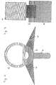
- 1 consists of three sections 1 to 3, each of which has a base body 4, 5, 6 made of fabric or water-equivalent material which simulates a vertebra. Additional bodies 7 to 18 made of tissue or water-equivalent material are attached to the base bodies 4, 5, 6 for better replication of the vertebrae.
- Components 4.7-10; 5.11-14; 6.15-18 each have different dimensions and densities to simulate different sized vertebrae.
- a special form of the phantom which is intended for exclusive use in QCT, consists only of the middle phantom section, e.g. with a depth of 3 cm.
- the cancellous bone is removable; inserts with different mineral densities and fat contents are available.
Landscapes
- Engineering & Computer Science (AREA)
- Health & Medical Sciences (AREA)
- Life Sciences & Earth Sciences (AREA)
- Medical Informatics (AREA)
- Physics & Mathematics (AREA)
- General Physics & Mathematics (AREA)
- General Health & Medical Sciences (AREA)
- Educational Technology (AREA)
- High Energy & Nuclear Physics (AREA)
- Algebra (AREA)
- Mathematical Analysis (AREA)
- Mathematical Optimization (AREA)
- Mathematical Physics (AREA)
- Pure & Applied Mathematics (AREA)
- Business, Economics & Management (AREA)
- Educational Administration (AREA)
- Medicinal Chemistry (AREA)
- Theoretical Computer Science (AREA)
- Chemical & Material Sciences (AREA)
- Biophysics (AREA)
- Computational Mathematics (AREA)
- Nuclear Medicine, Radiotherapy & Molecular Imaging (AREA)
- Optics & Photonics (AREA)
- Pathology (AREA)
- Radiology & Medical Imaging (AREA)
- Biomedical Technology (AREA)
- Heart & Thoracic Surgery (AREA)
- Molecular Biology (AREA)
- Surgery (AREA)
- Animal Behavior & Ethology (AREA)
- Public Health (AREA)
- Veterinary Medicine (AREA)
- Apparatus For Radiation Diagnosis (AREA)
Applications Claiming Priority (2)
| Application Number | Priority Date | Filing Date | Title |
|---|---|---|---|
| DE9016046U | 1990-11-26 | ||
| DE9016046U DE9016046U1 (de) | 1990-11-26 | 1990-11-26 | Kalibrierphantom für Knochenmineralmessungen an der Lendenwirbelsäule |
Publications (2)
| Publication Number | Publication Date |
|---|---|
| EP0488192A1 true EP0488192A1 (fr) | 1992-06-03 |
| EP0488192B1 EP0488192B1 (fr) | 1995-08-30 |
Family
ID=6859683
Family Applications (1)
| Application Number | Title | Priority Date | Filing Date |
|---|---|---|---|
| EP91120220A Expired - Lifetime EP0488192B1 (fr) | 1990-11-26 | 1991-11-26 | Fantôme d'étalonnage destiné à la mesure du contenu de minéraux dans la partie lombaire de la colonne vertébrale |
Country Status (3)
| Country | Link |
|---|---|
| US (1) | US5235628A (fr) |
| EP (1) | EP0488192B1 (fr) |
| DE (2) | DE9016046U1 (fr) |
Families Citing this family (39)
| Publication number | Priority date | Publication date | Assignee | Title |
|---|---|---|---|---|
| US5533084A (en) * | 1991-02-13 | 1996-07-02 | Lunar Corporation | Bone densitometer with improved vertebral characterization |
| US5481587A (en) * | 1994-05-09 | 1996-01-02 | Lunar Corporation | Radiographic phantom for vertebral morphometry |
| US6115487A (en) * | 1998-01-08 | 2000-09-05 | General Electric Company | Correction algorithm for bone-induced spectral artifacts in computed tomograph imaging |
| US6302582B1 (en) * | 1998-12-22 | 2001-10-16 | Bio-Imaging Technologies, Inc. | Spine phantom simulating cortical and trabecular bone for calibration of dual energy x-ray bone densitometers |
| US6510197B1 (en) | 2000-01-11 | 2003-01-21 | Alara, Inc. | Method and apparatus for osteoporosis screening |
| US6662148B1 (en) * | 2000-01-28 | 2003-12-09 | International Business Machines Corporation | Computation of shapes of three-dimensional linkage structures based on optimization techniques |
| US7467892B2 (en) | 2000-08-29 | 2008-12-23 | Imaging Therapeutics, Inc. | Calibration devices and methods of use thereof |
| US20020186818A1 (en) * | 2000-08-29 | 2002-12-12 | Osteonet, Inc. | System and method for building and manipulating a centralized measurement value database |
| US6904123B2 (en) | 2000-08-29 | 2005-06-07 | Imaging Therapeutics, Inc. | Methods and devices for quantitative analysis of x-ray images |
| CN1498385A (zh) | 2000-08-29 | 2004-05-19 | X图像量析的方法与装置 | |
| US7050534B2 (en) * | 2000-08-29 | 2006-05-23 | Imaging Therapeutics, Inc. | Methods and devices for quantitative analysis of x-ray images |
| US8639009B2 (en) * | 2000-10-11 | 2014-01-28 | Imatx, Inc. | Methods and devices for evaluating and treating a bone condition based on x-ray image analysis |
| CN1469721A (zh) | 2000-10-11 | 2004-01-21 | �����ɷ� | X射线图像的分析方法和装置 |
| US20070047794A1 (en) * | 2000-10-11 | 2007-03-01 | Philipp Lang | Methods and devices for analysis of x-ray images |
| US7660453B2 (en) * | 2000-10-11 | 2010-02-09 | Imaging Therapeutics, Inc. | Methods and devices for analysis of x-ray images |
| EP1389947B1 (fr) * | 2001-05-25 | 2009-08-26 | Imaging Therapeutics, Inc. | Procedes pour le diagnostic, le traitement et la prevention de perte osseuse |
| US8649843B2 (en) * | 2001-11-24 | 2014-02-11 | Ben A. Arnold | Automated calcium scoring of the aorta |
| AU2002348241A1 (en) * | 2001-11-24 | 2003-06-10 | Image Analysis, Inc. | Automatic detection and quantification of coronary and aortic calcium |
| US7295691B2 (en) * | 2002-05-15 | 2007-11-13 | Ge Medical Systems Global Technology Company, Llc | Computer aided diagnosis of an image set |
| US8965075B2 (en) | 2002-09-16 | 2015-02-24 | Imatx, Inc. | System and method for predicting future fractures |
| US7840247B2 (en) | 2002-09-16 | 2010-11-23 | Imatx, Inc. | Methods of predicting musculoskeletal disease |
| EP1605824A2 (fr) | 2003-03-25 | 2005-12-21 | Imaging Therapeutics, Inc. | Procedes de compensation de technique d'imagerie dans le traitement d'images radiographiques |
| US8290564B2 (en) * | 2003-09-19 | 2012-10-16 | Imatx, Inc. | Method for bone structure prognosis and simulated bone remodeling |
| US8073521B2 (en) | 2003-09-19 | 2011-12-06 | Imatx, Inc. | Method for bone structure prognosis and simulated bone remodeling |
| JP4284411B2 (ja) * | 2003-12-24 | 2009-06-24 | 独立行政法人放射線医学総合研究所 | ファントム及びファントム集合体 |
| CA2580726A1 (fr) | 2004-09-16 | 2006-03-30 | Imaging Therapeutics, Inc. | Systeme et procede de prediction de futures fractures |
| US8417010B1 (en) | 2006-01-12 | 2013-04-09 | Diagnoscan, LLC | Digital x-ray diagnosis and evaluation of dental disease |
| US7959742B2 (en) * | 2007-07-11 | 2011-06-14 | Whirlpool Corporation | Outer support body for a drawer-type dishwasher |
| WO2009102996A2 (fr) * | 2008-02-15 | 2009-08-20 | Mayo Foundation For Medical Education And Research | Système et méthode d'imagerie quantitative d'une composition chimique utiles pour décomposer plus de deux matières |
| EP2243021B1 (fr) * | 2008-02-15 | 2018-01-24 | Mayo Foundation For Medical Education And Research | Système et méthode d'imagerie quantitative d'une composition chimique pour décomposer plusieurs matières |
| US8186880B1 (en) * | 2008-11-27 | 2012-05-29 | Arnold Ben A | Extended and fixed INTable simultaneously imaged calibration and correction methods and references for 3-D imaging devices |
| US8649577B1 (en) | 2008-11-30 | 2014-02-11 | Image Analysis, Inc. | Automatic method and system for measurements of bone density and structure of the hip from 3-D X-ray imaging devices |
| US8939917B2 (en) | 2009-02-13 | 2015-01-27 | Imatx, Inc. | Methods and devices for quantitative analysis of bone and cartilage |
| US20140072108A1 (en) * | 2010-07-16 | 2014-03-13 | David P. Rohler | Methods and apparatus for extended low contrast detectability for radiographic imaging systems |
| WO2012161852A2 (fr) * | 2011-03-07 | 2012-11-29 | Loma Linda University Medical Center | Systèmes, dispositifs et procédés relatifs à l'étalonnage d'un scanner de tomographie par émission de protons calculée par ordinateur |
| US8517608B1 (en) | 2011-08-03 | 2013-08-27 | Ben A. Arnold | System and method for calibration of CT scanners and display of images in density units without the use of water phantoms |
| RU2663933C1 (ru) * | 2017-03-22 | 2018-08-13 | Юрий Иванович Колягин | Манекен-тренажер позвоночника компьютеризированный |
| CN113423342B (zh) * | 2019-02-14 | 2024-09-10 | 棱镜传感器公司 | x射线成像系统的校准 |
| US11172907B2 (en) | 2020-02-24 | 2021-11-16 | GE Precision Healthcare LLC | Systems and methods for cross calibration in dual energy x-ray absorptiometry |
Citations (3)
| Publication number | Priority date | Publication date | Assignee | Title |
|---|---|---|---|---|
| EP0218367A1 (fr) * | 1985-09-30 | 1987-04-15 | Picker International, Inc. | Fantôme pour analyse minérale d'os |
| US4873707A (en) * | 1987-09-11 | 1989-10-10 | Brigham & Women's Hospital | X-ray tomography phantoms, method and system |
| EP0409698A1 (fr) * | 1989-07-20 | 1991-01-23 | General Electric Cgr S.A. | Procédé de correction de la mesure de la densité osseuse dans un scannner |
Family Cites Families (6)
| Publication number | Priority date | Publication date | Assignee | Title |
|---|---|---|---|---|
| US4126789A (en) * | 1977-06-06 | 1978-11-21 | Vogl Thomas M | X-ray phantom |
| US4646334A (en) * | 1982-11-30 | 1987-02-24 | Zerhouni Elias A | Radiographic test phantom for computed tomographic lung nodule analysis |
| US4638502A (en) * | 1985-07-08 | 1987-01-20 | The Ontario Cancer Institute | Anthropomorphic phantoms |
| US4870666A (en) * | 1986-08-07 | 1989-09-26 | General Electric Company | Computer tomographic phantom |
| US4985906A (en) * | 1987-02-17 | 1991-01-15 | Arnold Ben A | Calibration phantom for computer tomography system |
| US5122664A (en) * | 1990-04-27 | 1992-06-16 | Fuji Photo Film Co., Ltd. | Method and apparatus for quantitatively analyzing bone calcium |
-
1990
- 1990-11-26 DE DE9016046U patent/DE9016046U1/de not_active Expired - Lifetime
-
1991
- 1991-11-25 US US07/797,722 patent/US5235628A/en not_active Expired - Fee Related
- 1991-11-26 DE DE59106369T patent/DE59106369D1/de not_active Expired - Fee Related
- 1991-11-26 EP EP91120220A patent/EP0488192B1/fr not_active Expired - Lifetime
Patent Citations (3)
| Publication number | Priority date | Publication date | Assignee | Title |
|---|---|---|---|---|
| EP0218367A1 (fr) * | 1985-09-30 | 1987-04-15 | Picker International, Inc. | Fantôme pour analyse minérale d'os |
| US4873707A (en) * | 1987-09-11 | 1989-10-10 | Brigham & Women's Hospital | X-ray tomography phantoms, method and system |
| EP0409698A1 (fr) * | 1989-07-20 | 1991-01-23 | General Electric Cgr S.A. | Procédé de correction de la mesure de la densité osseuse dans un scannner |
Also Published As
| Publication number | Publication date |
|---|---|
| DE59106369D1 (de) | 1995-10-05 |
| EP0488192B1 (fr) | 1995-08-30 |
| DE9016046U1 (de) | 1991-02-14 |
| US5235628A (en) | 1993-08-10 |
Similar Documents
| Publication | Publication Date | Title |
|---|---|---|
| EP0488192B1 (fr) | Fantôme d'étalonnage destiné à la mesure du contenu de minéraux dans la partie lombaire de la colonne vertébrale | |
| DE69329950T2 (de) | Vorrichtung und verfahren zur bestimmung der kalziumkonzentration | |
| Sorenson et al. | A reliable in vivo measurement of bone-mineral content | |
| DE102006009222B4 (de) | Verfahren und Vorrichtung zur Bestimmung der Konzentration einer Substanz in einem Körpermaterial mittels Mehr-Energie-Computertomographie | |
| Kelly et al. | Calibration and standardization of bone mineral densitometers | |
| DE10311628B4 (de) | Bildgebungsverfahren | |
| WO2003024331A2 (fr) | Procede pour determiner des distributions en densite et en nombre atomique dans des procedes d'examen radiographique | |
| DE3725373A1 (de) | Unterlagekoerper fuer computer-tomographie | |
| DE69501082T2 (de) | Radiographisches phantom für morphometrie der wirbelsäule | |
| DE10035984C1 (de) | Röntgen-Computertomographieeinrichtung | |
| DE3726456A1 (de) | Verfahren und einrichtung zur messung der knochenmineraldichte | |
| DE10160613A1 (de) | Röhrenseitig modifiziertes bildgebendes Röntgengerät | |
| EP2889002A1 (fr) | Procédé et unité de calcul de mesure et de représentation de la densité osseuse d'un patient | |
| DE10305105A1 (de) | Eichung der Transformation spektraler Röntgenschwächungswerte in Dichte- und Ordnungszahlinformation | |
| DE10352013B4 (de) | Verfahren und Vorrichtung zur ortsaufgelösten Bestimmung der Elementkonzentrationen in Untersuchungsobjekten | |
| DE102004033989B4 (de) | Verfahren zur Messung der dreidimensionalen Dichteverteilung in Knochen | |
| DE2609226A1 (de) | Anordnung zur untersuchung eines koerpers mit ionisierender strahlung | |
| EP1052937A1 (fr) | Appareil d'examen aux rayons x | |
| DE602004006736T2 (de) | Phantom für die qualitätskontrolle eines systems zur virtuellen simulation einer radiotherapeutischen behandlung | |
| DE102008049604A1 (de) | Verfahren zur Bestimmung einer Temperatur oder Temperaturverteilung in einem Objektbereich | |
| Stern et al. | Nationwide evaluation of X-ray trends (NEXT) 2000–01: Survey of patient radiation exposure from computed tomographic (CT) examinations in the United States | |
| Bennemann et al. | Evaluating miniscrew position using orthopantomograms compared to cone-beam computed tomography | |
| Smith et al. | Comparison between 153Gd and 241Am, 137Cs for dual-photon absorptiometry of the spine | |
| DE10018769A1 (de) | Verfahren und Vorrichtung zur intraoperativen Bestimmung der Knochenqualität | |
| Engelke | Quantitative Computertomographie |
Legal Events
| Date | Code | Title | Description |
|---|---|---|---|
| PUAI | Public reference made under article 153(3) epc to a published international application that has entered the european phase |
Free format text: ORIGINAL CODE: 0009012 |
|
| AK | Designated contracting states |
Kind code of ref document: A1 Designated state(s): CH DE FR GB LI |
|
| 17P | Request for examination filed |
Effective date: 19921113 |
|
| 17Q | First examination report despatched |
Effective date: 19940405 |
|
| GRAA | (expected) grant |
Free format text: ORIGINAL CODE: 0009210 |
|
| AK | Designated contracting states |
Kind code of ref document: B1 Designated state(s): CH DE FR GB LI |
|
| PG25 | Lapsed in a contracting state [announced via postgrant information from national office to epo] |
Ref country code: GB Effective date: 19950830 Ref country code: FR Free format text: THE PATENT HAS BEEN ANNULLED BY A DECISION OF A NATIONAL AUTHORITY Effective date: 19950830 |
|
| REF | Corresponds to: |
Ref document number: 59106369 Country of ref document: DE Date of ref document: 19951005 |
|
| PG25 | Lapsed in a contracting state [announced via postgrant information from national office to epo] |
Ref country code: LI Effective date: 19951130 Ref country code: CH Effective date: 19951130 |
|
| EN | Fr: translation not filed | ||
| GBV | Gb: ep patent (uk) treated as always having been void in accordance with gb section 77(7)/1977 [no translation filed] |
Effective date: 19950830 |
|
| PLBE | No opposition filed within time limit |
Free format text: ORIGINAL CODE: 0009261 |
|
| STAA | Information on the status of an ep patent application or granted ep patent |
Free format text: STATUS: NO OPPOSITION FILED WITHIN TIME LIMIT |
|
| REG | Reference to a national code |
Ref country code: CH Ref legal event code: PL |
|
| 26N | No opposition filed | ||
| PGFP | Annual fee paid to national office [announced via postgrant information from national office to epo] |
Ref country code: DE Payment date: 19980326 Year of fee payment: 7 |
|
| PG25 | Lapsed in a contracting state [announced via postgrant information from national office to epo] |
Ref country code: DE Free format text: LAPSE BECAUSE OF NON-PAYMENT OF DUE FEES Effective date: 19990901 |