EP0490428A2 - Spectromètre à détection multicanale - Google Patents
Spectromètre à détection multicanale Download PDFInfo
- Publication number
- EP0490428A2 EP0490428A2 EP91203155A EP91203155A EP0490428A2 EP 0490428 A2 EP0490428 A2 EP 0490428A2 EP 91203155 A EP91203155 A EP 91203155A EP 91203155 A EP91203155 A EP 91203155A EP 0490428 A2 EP0490428 A2 EP 0490428A2
- Authority
- EP
- European Patent Office
- Prior art keywords
- skin surface
- slit
- spectrometer
- line
- white standard
- Prior art date
- Legal status (The legal status is an assumption and is not a legal conclusion. Google has not performed a legal analysis and makes no representation as to the accuracy of the status listed.)
- Granted
Links
Images
Classifications
-
- A—HUMAN NECESSITIES
- A61—MEDICAL OR VETERINARY SCIENCE; HYGIENE
- A61B—DIAGNOSIS; SURGERY; IDENTIFICATION
- A61B5/00—Measuring for diagnostic purposes; Identification of persons
- A61B5/44—Detecting, measuring or recording for evaluating the integumentary system, e.g. skin, hair or nails
- A61B5/441—Skin evaluation, e.g. for skin disorder diagnosis
- A61B5/444—Evaluating skin marks, e.g. mole, nevi, tumour, scar
-
- A—HUMAN NECESSITIES
- A61—MEDICAL OR VETERINARY SCIENCE; HYGIENE
- A61B—DIAGNOSIS; SURGERY; IDENTIFICATION
- A61B5/00—Measuring for diagnostic purposes; Identification of persons
- A61B5/0059—Measuring for diagnostic purposes; Identification of persons using light, e.g. diagnosis by transillumination, diascopy, fluorescence
- A61B5/0062—Arrangements for scanning
- A61B5/0064—Body surface scanning
-
- A—HUMAN NECESSITIES
- A61—MEDICAL OR VETERINARY SCIENCE; HYGIENE
- A61B—DIAGNOSIS; SURGERY; IDENTIFICATION
- A61B5/00—Measuring for diagnostic purposes; Identification of persons
- A61B5/0059—Measuring for diagnostic purposes; Identification of persons using light, e.g. diagnosis by transillumination, diascopy, fluorescence
- A61B5/0075—Measuring for diagnostic purposes; Identification of persons using light, e.g. diagnosis by transillumination, diascopy, fluorescence by spectroscopy, i.e. measuring spectra, e.g. Raman spectroscopy, infrared absorption spectroscopy
-
- G—PHYSICS
- G01—MEASURING; TESTING
- G01J—MEASUREMENT OF INTENSITY, VELOCITY, SPECTRAL CONTENT, POLARISATION, PHASE OR PULSE CHARACTERISTICS OF INFRARED, VISIBLE OR ULTRAVIOLET LIGHT; COLORIMETRY; RADIATION PYROMETRY
- G01J3/00—Spectrometry; Spectrophotometry; Monochromators; Measuring colours
- G01J3/28—Investigating the spectrum
- G01J3/2823—Imaging spectrometer
-
- G—PHYSICS
- G01—MEASURING; TESTING
- G01J—MEASUREMENT OF INTENSITY, VELOCITY, SPECTRAL CONTENT, POLARISATION, PHASE OR PULSE CHARACTERISTICS OF INFRARED, VISIBLE OR ULTRAVIOLET LIGHT; COLORIMETRY; RADIATION PYROMETRY
- G01J3/00—Spectrometry; Spectrophotometry; Monochromators; Measuring colours
- G01J3/28—Investigating the spectrum
- G01J3/2803—Investigating the spectrum using photoelectric array detector
- G01J2003/2813—2D-array
-
- G—PHYSICS
- G01—MEASURING; TESTING
- G01N—INVESTIGATING OR ANALYSING MATERIALS BY DETERMINING THEIR CHEMICAL OR PHYSICAL PROPERTIES
- G01N21/00—Investigating or analysing materials by the use of optical means, i.e. using sub-millimetre waves, infrared, visible or ultraviolet light
- G01N21/17—Systems in which incident light is modified in accordance with the properties of the material investigated
- G01N21/47—Scattering, i.e. diffuse reflection
- G01N21/4738—Diffuse reflection, e.g. also for testing fluids, fibrous materials
Definitions
- the invention relates to a multichannel spectrometer, in particular for examining pigment marks, with an evaluation and display device and optical means for determining and imaging reflectance spectra of a skin surface on an evaluable image plane, taking into account white standards.
- Body color measurements are becoming increasingly important in many areas of technology.
- one is interested in obtaining objective measures for body colors, e.g. to gain diagnostic information about the skin surface.
- objective measures for body colors e.g. to gain diagnostic information about the skin surface.
- the light that is emitted from the skin surface of a body point is broken down into its spectrum with the aid of a monochromator and then calculated with predetermined standard spectral value curves to determine the color coordinates.
- predetermined standard spectral value curves to determine the color coordinates.
- a certain predetermined type of lighting must be assumed or the primary light spectrum must also be recorded and the reflectance spectrum of the body point corrected thereon.
- Such a spectrometer is known from DE-OS 27 26 606. This is essentially a so-called medical spectrophotometer with a spacer ring at the lens end for color measurement using the spectrometry of reflected light, i.e. the measurement of the reflectance of a skin surface, for example, in which a white standard alternates as a reference is used. Such a spectrometer makes it possible to determine the reflectance spectrum from a point on the surface.
- the object of the present invention is to provide an optical multichannel spectrometer for the contactless determination of the different reflectance spectra of the points of at least one line of a skin surface to be examined while continuously taking into account a primary light correction.
- the optical means a lens system directed towards the skin surface and the white standard and a slit diaphragm with a downstream one corresponding to a slit of the Slit diaphragm aligned diffraction grids, for the simultaneous generation of the reflectance spectrum of the points of a determinable line of the skin surface and the white standard on the image plane downstream of the diffraction grating and connected to the evaluation and display device.
- the lens system has an objective lens on an optical axis with means for sharp imaging of the white standard and at the same time the skin surface on the slit diaphragm.
- the slit diaphragm only allows a line-shaped image section to pass.
- the gratings of the diffraction grating are aligned parallel to the direction of the slit of the slit diaphragm.
- the white standard consists of, for example, pressed barium sulfate and can be attached in a small section in the area of the observed line of the skin surface to the pigment mole itself or next to it, e.g. be glued on.
- the multichannel spectrometer according to the invention is arranged such that it can be displaced and adjusted orthogonally to the slit direction and to the optical axis of the lens system, so that reflectance spectra according to the invention can be generated from virtually every line of the pigment mark.
- the multi-channel spectrometer according to the invention can have a housing and can be mounted in front of a low-light camera instead of a lens.
- the means for optically sharp imaging are advantageously formed by a linear displacement unit of the objective lens. It can also be provided that the objective lens is integrated in a zoom lens with a variable focal length.
- the arrangement of the lens system, the slit diaphragm and the diffraction grating according to the invention has the result that the diffraction of the parallel light beam at the diffraction grating in the image plane in a direction perpendicular to the direction of the slit results in the spectra of the individual slit image points and also perpendicular to the slit image and twice from the direct one Extending the zero-order slit image in mirror image to each other in two opposite directions.
- the correction i.e. the normalization of the reflectance spectra of e.g. Pigmentmal with regard to the spectral distribution of the primary light occurs via the reflectance spectrum of the white standard, which is also recorded at the same time, which is why practically any primary light can be used as long as the line being viewed is illuminated.
- a two-dimensional detector is arranged in the image plane, for the detection of the spectra of the individual slit image points of the skin surface and at the same time the white standard, in order to ultimately add the color coordinates for each point of the line under consideration via a subordinate evaluation and display device calculate and display.
- the calculation is carried out for each point of the line from the standardized reflectance spectra and tabulated standard spectral value curves, which can be stored in a memory of the evaluation device.
- a line-shaped section from the skin surface for example of a pigment mark
- To scan the complete Area only requires the corresponding repeated shifting of the spectrometer.
- it is often sufficient for diagnostic purposes if the color profile of a line is known, for example.
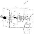
- FIG. 1 shows the basic structure and the mode of operation of the multi-channel spectrometer 10 according to the invention, which is arranged in a housing 11 and contains a two-dimensional detector 12, which is connected via a connecting line 13 to an evaluation and display device, for example arranged outside (dashed line) of the housing 11 14 is connected.
- the evaluation and display device 14 (solid line) can also be arranged in the housing 11.
- the optical axis 15 of the multichannel spectrometer 10 is directed at a skin surface outside the housing 11 and particularly at a pigment mark 16 located on the skin surface.
- a sample of pressed barium sulfate for example in tablet form, made of pressed barium sulfate as white standard is on the skin surface, not shown 17 attached. It can be provided that the white standard 17 is also arranged on or partially on the pigment 16, but does not substantially cover it.
- the pigment mark 16 and also the white standard 17 on the surface of the skin are illuminated in a spectrally broadband manner. However, it is also possible to provide spectrally narrowband lighting. The illumination of the respectively evaluated section or the evaluated line 25 must be even across the section.
- the pigment mark 16 and the white standard 17 are simultaneously imaged sharply into the slit region of a slit aperture 19.
- the distance of the objective lens 18 from the slit 20 can be variable in the direction of the optical axis 15 via a linear displacement unit, not shown.
- a zoom object can also be provided as an objective lens 18. Different magnifications can then advantageously be set without having to change the distance between the spectrometer and the skin surface.
- the slit aperture 19 has the gap 20 running horizontally in the plane of the drawing, which only has a line-shaped cutout 25 of the skin surface, i.e. of the pigment mark 16 and the white standard 18.
- the line-shaped intermediate image is projected into an evaluable image plane x, y via a system of two lenses 21 and 22 arranged downstream of the slit aperture 19.
- the correspondingly sensitive two-dimensional optical detector 12 is arranged in this image plane x, y, which is arranged downstream of the double lens system comprising the lenses 21 and 22.
- a diffraction grating 23 is arranged between the lenses 21 and 22, the grating 24 of said grating 24 running parallel to the direction of the slit 20 of the slit diaphragm 19.
- the double lens system comprising the lenses 21 and 22 is arranged between the slit aperture 19 and the detector 12 in such a way that the front focal point of the lens 21 lies in the slit plane 19 and the rear focal point of the lens 22 lies in the image plane x, y.
- the parallelism of the light beam in the double lens system is therefore determined by the distance between the lens 21 and the slit diaphragm 19. That is, it is necessary that the focal point of lens 21 facing the slit 20 lies exactly in the plane of the slit 20.
- the detector-side focal point of the lens 22 lies exactly in the image plane x, y, that is to say the detector 12.
- the diffraction of the parallel light beam of the intermediate image takes place at the diffraction grating 23 in a direction orthogonal to the direction of the slit 20 and the optical axis 15.
- This state of affairs is hatched in each case in FIG. 1 on the detector 12 and indicated in a diagram 26. The latter is shown in the correct position next to the detector 12 for better understanding.
- the correction i.e. Standardization of the reflectance spectra of the pigment 16 with respect to the spectral distribution of the primary light takes place via the reflectance spectrum of the white standard 17, which is also recorded, as is also shown hatched on the detector 12.
- the calculation of the color coordinates is carried out in the evaluation device of the evaluation and display device 14 for each point of the line 25 of the pigment mark 16 just under consideration from the standardized reflectance spectra and tabulated standard spectral value curves, which are stored in a memory of the evaluation device.
- the evaluation device can be a microprocessor.
- the housing 11 can advantageously be designed similar to a camera lens, as a result of which the multichannel spectrometer can be mounted in front of a low-light camera instead of a lens, with the aid of which the spectra can then be detected and transmitted to the evaluation and display device 14.
- a device for moving, ie raising or lowering the multichannel spectrometer in order to evaluate several lines 15 of a pigment mark is not shown in FIG. 1.
- a normal camera tripod can be used.
Landscapes
- Health & Medical Sciences (AREA)
- Life Sciences & Earth Sciences (AREA)
- Physics & Mathematics (AREA)
- Molecular Biology (AREA)
- Animal Behavior & Ethology (AREA)
- Pathology (AREA)
- Engineering & Computer Science (AREA)
- Biomedical Technology (AREA)
- Heart & Thoracic Surgery (AREA)
- Medical Informatics (AREA)
- Spectroscopy & Molecular Physics (AREA)
- Surgery (AREA)
- Biophysics (AREA)
- General Health & Medical Sciences (AREA)
- Public Health (AREA)
- Veterinary Medicine (AREA)
- Dermatology (AREA)
- General Physics & Mathematics (AREA)
- Nuclear Medicine, Radiotherapy & Molecular Imaging (AREA)
- Radiology & Medical Imaging (AREA)
- Spectrometry And Color Measurement (AREA)
- Investigating Or Analysing Materials By Optical Means (AREA)
- Measuring And Recording Apparatus For Diagnosis (AREA)
Applications Claiming Priority (2)
| Application Number | Priority Date | Filing Date | Title |
|---|---|---|---|
| DE4039070 | 1990-12-07 | ||
| DE4039070A DE4039070A1 (de) | 1990-12-07 | 1990-12-07 | Vielkanalspektrometer |
Publications (3)
| Publication Number | Publication Date |
|---|---|
| EP0490428A2 true EP0490428A2 (fr) | 1992-06-17 |
| EP0490428A3 EP0490428A3 (en) | 1992-08-26 |
| EP0490428B1 EP0490428B1 (fr) | 1996-03-13 |
Family
ID=6419805
Family Applications (1)
| Application Number | Title | Priority Date | Filing Date |
|---|---|---|---|
| EP91203155A Expired - Lifetime EP0490428B1 (fr) | 1990-12-07 | 1991-12-03 | Spectromètre à détection multicanale |
Country Status (4)
| Country | Link |
|---|---|
| US (1) | US5297555A (fr) |
| EP (1) | EP0490428B1 (fr) |
| JP (1) | JPH04294224A (fr) |
| DE (2) | DE4039070A1 (fr) |
Cited By (2)
| Publication number | Priority date | Publication date | Assignee | Title |
|---|---|---|---|---|
| WO1996011386A1 (fr) * | 1994-10-10 | 1996-04-18 | Soliton Elektronik Ab | Procede de representation a base spectrale concernant des elements d'image disposes sur une ligne |
| WO2002065069A3 (fr) * | 2000-11-07 | 2004-02-19 | Hypermed Inc | Dispositif d'etalonnage d'imagerie en hyperespace spectral |
Families Citing this family (14)
| Publication number | Priority date | Publication date | Assignee | Title |
|---|---|---|---|---|
| US5708504A (en) * | 1996-10-25 | 1998-01-13 | The United States Of America As Represented By The Secretary Of The Air Force | Interfering imaging spectrometer |
| US7202949B2 (en) * | 2003-09-30 | 2007-04-10 | The Trustees Of Boston University | Binocular spectrometer |
| US7315371B2 (en) * | 2004-01-23 | 2008-01-01 | P&P Optica Inc. | Multi-channel spectrum analyzer |
| JP4499476B2 (ja) * | 2004-05-25 | 2010-07-07 | オリンパス株式会社 | 分光画像入力装置及びそれを備えた光学装置 |
| TWI245114B (en) * | 2004-10-11 | 2005-12-11 | Ind Tech Res Inst | Apparatus for measuring imaging spectrograph |
| GB0504279D0 (en) * | 2005-03-02 | 2005-04-06 | Carroll Charles C | Skin colour matching system and method |
| US7564547B2 (en) * | 2005-11-07 | 2009-07-21 | Wafermasters, Inc. | Spectroscopy system |
| TWI305832B (en) | 2006-12-13 | 2009-02-01 | Ind Tech Res Inst | Multi-channel imaging spectrometer |
| US8315692B2 (en) * | 2007-02-22 | 2012-11-20 | Sheinis Andrew I | Multi-spectral imaging spectrometer for early detection of skin cancer |
| JP5655437B2 (ja) * | 2009-09-14 | 2015-01-21 | 株式会社リコー | 分光特性取得装置 |
| US20130341509A1 (en) * | 2010-06-11 | 2013-12-26 | Chemimage Corporation | Portable system for detecting explosive materials using near infrared hyperspectral imaging and method for using thereof |
| US10123738B1 (en) | 2017-07-03 | 2018-11-13 | Spyros Kokolis | Methods and apparatus for skin color patient monitoring |
| US10154813B1 (en) | 2017-07-03 | 2018-12-18 | Spyros Kokolis | Method and apparatus for patient skin color monitoring and drug efficacy measurement |
| US12569190B2 (en) | 2022-05-13 | 2026-03-10 | Maria SFINAROLAKIS-KOKOLIS | Method for measurement of vascular endothelial response to stimuli |
Family Cites Families (7)
| Publication number | Priority date | Publication date | Assignee | Title |
|---|---|---|---|---|
| US3874799A (en) * | 1973-06-01 | 1975-04-01 | Color Control Inc | Method and apparatus for color spectrophotometry |
| DE2726606A1 (de) * | 1977-06-13 | 1978-12-21 | Max Planck Gesellschaft | Medizinisches spektralfotometer |
| US4170987A (en) * | 1977-11-28 | 1979-10-16 | California Institute Of Technology | Medical diagnosis system and method with multispectral imaging |
| DE2758141C2 (de) * | 1977-12-27 | 1982-11-18 | Ibm Deutschland Gmbh, 7000 Stuttgart | Spektrophotometer |
| US4729372A (en) * | 1983-11-17 | 1988-03-08 | Lri L.P. | Apparatus for performing ophthalmic laser surgery |
| DE3815743A1 (de) * | 1988-05-07 | 1989-11-16 | Zeiss Carl Fa | Vorrichtung zur messung und auswertung von eigenfluoreszenzspektren organischer gewebeflaechen |
| US5014709A (en) * | 1989-06-13 | 1991-05-14 | Biologic Systems Corp. | Method and apparatus for high resolution holographic imaging of biological tissue |
-
1990
- 1990-12-07 DE DE4039070A patent/DE4039070A1/de not_active Withdrawn
-
1991
- 1991-12-02 US US07/803,313 patent/US5297555A/en not_active Expired - Fee Related
- 1991-12-03 EP EP91203155A patent/EP0490428B1/fr not_active Expired - Lifetime
- 1991-12-03 DE DE59107547T patent/DE59107547D1/de not_active Expired - Fee Related
- 1991-12-05 JP JP3322038A patent/JPH04294224A/ja active Pending
Cited By (4)
| Publication number | Priority date | Publication date | Assignee | Title |
|---|---|---|---|---|
| WO1996011386A1 (fr) * | 1994-10-10 | 1996-04-18 | Soliton Elektronik Ab | Procede de representation a base spectrale concernant des elements d'image disposes sur une ligne |
| WO2002065069A3 (fr) * | 2000-11-07 | 2004-02-19 | Hypermed Inc | Dispositif d'etalonnage d'imagerie en hyperespace spectral |
| US6810279B2 (en) | 2000-11-07 | 2004-10-26 | Hypermed, Inc. | Hyperspectral imaging calibration device |
| US7013172B2 (en) | 2000-11-07 | 2006-03-14 | Hypermed, Inc. | Hyperspectral imaging calibration device |
Also Published As
| Publication number | Publication date |
|---|---|
| US5297555A (en) | 1994-03-29 |
| DE4039070A1 (de) | 1992-06-11 |
| EP0490428B1 (fr) | 1996-03-13 |
| EP0490428A3 (en) | 1992-08-26 |
| JPH04294224A (ja) | 1992-10-19 |
| DE59107547D1 (de) | 1996-04-18 |
Similar Documents
| Publication | Publication Date | Title |
|---|---|---|
| EP3891465B1 (fr) | Dispositif de mesure optique | |
| EP0490428B1 (fr) | Spectromètre à détection multicanale | |
| EP0116321B1 (fr) | Spectromètre infrarouge | |
| EP0279191B1 (fr) | Dispositif pour la mesure sans contact de la rémission | |
| DE69314377T2 (de) | Tragbares Spektrophotometer | |
| EP0778459B1 (fr) | Appareil de spectrométrie | |
| DE3713149A1 (de) | Fernmess-spektrophotometer | |
| DE60129247T2 (de) | Optische sensoreinrichtung und verfahren zur spektralanalyse | |
| DE3843876C2 (fr) | ||
| EP0209860A2 (fr) | Appareil de mesure de la réflexion pour mesure sans contact | |
| DE68921249T2 (de) | Mikroskop-Spektralgerät. | |
| DE102016212507A1 (de) | Chromatisch-konfokaler Bereichssensor mit Kamerateil | |
| DE19961481B4 (de) | Zentriertes Sphärenspektrometer | |
| EP1754032A1 (fr) | Spectrometre-echelle a mise en valeur amelioree du detecteur par l'utilisation de deux ensembles spectrometres | |
| DE10159234A1 (de) | Vorrichtung zur Untersuchung von Dokumenten | |
| EP1273951B1 (fr) | Microscope à balayage et méthode de detection dependant de la longueur d'onde | |
| DE69005681T2 (de) | Transmissions-/Reflektionsdensitometer mit zwei Messköpfen. | |
| DE102016008884A1 (de) | Spektroskopievorrichtung und -verfahren | |
| DE102005024271B4 (de) | Gitterspektrometersystem und Verfahren zur Messwerterfassung | |
| DE102020107457B4 (de) | Messvorrichtung zur Vermessung von optischen Strahlungsquellen und Verfahren zur Durchführung einer Vermessung | |
| EP1588135B1 (fr) | Dispositif et procede de detection a resolution spectrale d'un echantillon | |
| DE102019107963B4 (de) | Messlichtquelle sowie Messanordnung zum Erfassen eines Reflexionsspektrums | |
| DE102022207661A1 (de) | EUV-Reflektometer und Messverfahren | |
| DE10356729B4 (de) | Farbsensor | |
| EP1353210A2 (fr) | Microscope à dispositif pour déterminer l'intensité de la lumière d'un faisceau d'illumination |
Legal Events
| Date | Code | Title | Description |
|---|---|---|---|
| PUAI | Public reference made under article 153(3) epc to a published international application that has entered the european phase |
Free format text: ORIGINAL CODE: 0009012 |
|
| AK | Designated contracting states |
Kind code of ref document: A2 Designated state(s): DE FR GB |
|
| PUAL | Search report despatched |
Free format text: ORIGINAL CODE: 0009013 |
|
| AK | Designated contracting states |
Kind code of ref document: A3 Designated state(s): DE FR GB |
|
| 17P | Request for examination filed |
Effective date: 19930223 |
|
| 17Q | First examination report despatched |
Effective date: 19941128 |
|
| GRAA | (expected) grant |
Free format text: ORIGINAL CODE: 0009210 |
|
| AK | Designated contracting states |
Kind code of ref document: B1 Designated state(s): DE FR GB |
|
| REF | Corresponds to: |
Ref document number: 59107547 Country of ref document: DE Date of ref document: 19960418 |
|
| ET | Fr: translation filed | ||
| GBT | Gb: translation of ep patent filed (gb section 77(6)(a)/1977) |
Effective date: 19960528 |
|
| PGFP | Annual fee paid to national office [announced via postgrant information from national office to epo] |
Ref country code: GB Payment date: 19961202 Year of fee payment: 6 |
|
| PGFP | Annual fee paid to national office [announced via postgrant information from national office to epo] |
Ref country code: FR Payment date: 19961217 Year of fee payment: 6 |
|
| PLBE | No opposition filed within time limit |
Free format text: ORIGINAL CODE: 0009261 |
|
| STAA | Information on the status of an ep patent application or granted ep patent |
Free format text: STATUS: NO OPPOSITION FILED WITHIN TIME LIMIT |
|
| PGFP | Annual fee paid to national office [announced via postgrant information from national office to epo] |
Ref country code: DE Payment date: 19970221 Year of fee payment: 6 |
|
| 26N | No opposition filed | ||
| PG25 | Lapsed in a contracting state [announced via postgrant information from national office to epo] |
Ref country code: GB Free format text: LAPSE BECAUSE OF NON-PAYMENT OF DUE FEES Effective date: 19971203 |
|
| PG25 | Lapsed in a contracting state [announced via postgrant information from national office to epo] |
Ref country code: FR Free format text: THE PATENT HAS BEEN ANNULLED BY A DECISION OF A NATIONAL AUTHORITY Effective date: 19971231 |
|
| GBPC | Gb: european patent ceased through non-payment of renewal fee |
Effective date: 19971203 |
|
| PG25 | Lapsed in a contracting state [announced via postgrant information from national office to epo] |
Ref country code: DE Free format text: LAPSE BECAUSE OF NON-PAYMENT OF DUE FEES Effective date: 19980901 |
|
| REG | Reference to a national code |
Ref country code: FR Ref legal event code: ST |