EP0497399A1 - Procédé pour évalver l'irritation de la peau - Google Patents
Procédé pour évalver l'irritation de la peau Download PDFInfo
- Publication number
- EP0497399A1 EP0497399A1 EP92200099A EP92200099A EP0497399A1 EP 0497399 A1 EP0497399 A1 EP 0497399A1 EP 92200099 A EP92200099 A EP 92200099A EP 92200099 A EP92200099 A EP 92200099A EP 0497399 A1 EP0497399 A1 EP 0497399A1
- Authority
- EP
- European Patent Office
- Prior art keywords
- skin
- culture
- cells
- cultures
- cell
- Prior art date
- Legal status (The legal status is an assumption and is not a legal conclusion. Google has not performed a legal analysis and makes no representation as to the accuracy of the status listed.)
- Withdrawn
Links
Images


Classifications
-
- G—PHYSICS
- G01—MEASURING; TESTING
- G01N—INVESTIGATING OR ANALYSING MATERIALS BY DETERMINING THEIR CHEMICAL OR PHYSICAL PROPERTIES
- G01N33/00—Investigating or analysing materials by specific methods not covered by groups G01N1/00 - G01N31/00
- G01N33/48—Biological material, e.g. blood, urine; Haemocytometers
- G01N33/50—Chemical analysis of biological material, e.g. blood, urine; Testing involving biospecific ligand binding methods; Immunological testing
- G01N33/5005—Chemical analysis of biological material, e.g. blood, urine; Testing involving biospecific ligand binding methods; Immunological testing involving human or animal cells
- G01N33/5008—Chemical analysis of biological material, e.g. blood, urine; Testing involving biospecific ligand binding methods; Immunological testing involving human or animal cells for testing or evaluating the effect of chemical or biological compounds, e.g. drugs, cosmetics
- G01N33/5014—Chemical analysis of biological material, e.g. blood, urine; Testing involving biospecific ligand binding methods; Immunological testing involving human or animal cells for testing or evaluating the effect of chemical or biological compounds, e.g. drugs, cosmetics for testing toxicity
Definitions
- the use of skin cultures for evaluating skin irritancy of ingredients and of commercial products provides a viable alternative method to animal testing.
- the irritancy is measured in co-cultures of human keratinocytes and fibroblasts using tests of cell viability, cytotoxicity, glucose utilization and release of pro-inflammatory mediator PGE2 (prostaglandin E2).
- a commercial culture system has been developed which is the closest in vitro analog to human skin available. These cultures contain a multi-layered dermis with mitotically and metabolically active dermal fibroblasts, naturally secreted collagen Types I and III, and a basement membrane zone containing laminin and collagen Type IV. A multi-layered epidermis is present in the co-cultures.
- the development of a battery of objective quantitative biomarkers of skin irritation utilizing cytotoxicity and irritancy endpoints in human skin cell monolayers and co-culture "skin equivalents" is the focus of the present invention.
- the predictive endpoints can be used to establish rank orders of agents with known toxicity within a chemical class in order to establish a baseline for assessing and predicting toxicity of related novel agents.
- the culture system has the potential to predict absolute levels of human skin irritation.
- the endpoints can also be used to evaluate the utility of commercial sources of human skin cultures.
- Skin equivalents provide skin toxicity information related to mechanisms of erythema and edema, e.g. PGE2 (prostaglandin E2) release as a proinflammatory mediator. It is likely that irritancy data from these systems will be acceptable to regulatory agencies.
- the present invention includes the evaluation of cell viability by two techniques: 1) incorporation of the vital dye neutral red into lysosomes of viable cells; and 2) reduction of a tetrazolium salt (MTT) to formazan dye by the respiratory electron transport chain of cells with competent mitochondria.
- TMT tetrazolium salt
- Cytotoxicity endpoints which measure the irritancy potential of surfactants and surfactant-containing products and other materials which come into contact with skin include release of the cytoplasmic enzyme lactate dehydrogenase (LDH) and release of the lysosomal enzyme N-acetylglucosaminidase (NAGS) from damaged cells into culture medium.
- LDH lactate dehydrogenase
- NAGS N-acetylglucosaminidase
- PGE2 Prostaglandin E2
- Glucose utilization by skin cells is used as an indicator of metabolic activity. Skin sensitivity as well as the effects of ultraviolet light on skin can be measured by the method herein.
- Human skin cell co-cultures consisting of human neonatal foreskin fibroblasts and keratinocytes contain a substratum of fibroblasts grown on a nylon mesh.
- a view (250x magnification) of the surface of a culture indicates the large number of cells that grow in the spaces between the interwoven nylon mesh filaments. Phase contrast microscopy of the mesh surface, as in Fig. 1 was routinely used to confirm the cellularity of the cultures.
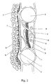
- FIG. 2 A schematic diagram of a commercial (Marrow-Tech) culture in cross-section (Fig. 2) shows fibroblast (1) attachment to the nylon filaments (2), the presence of collagen (3) and extracellular matrix components (4) secreted from the cells, and the epidermal keratinocytes (5) growing in layers above the fibroblast/nylon substratum. Although adipocytes (6) and melanocytes (7) are included in this diagram, they were not components of the cultures used.
- An histological cross-section (400x magnification) of a commercial (Marrow-Tech) culture of the type used in the examples (Fig. 3) shows approximately 5 cell layers of human neonatal foreskin fibroblasts, one layer of basal keratinocytes, and 2-3 layers of flattened stratified keratinocytes on top.
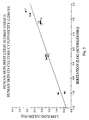
- Figure 4 and 5 shows a comparison of the results of human studies with the results of the same materials tested by the method herein.
- the surfactants tested were:
- Human skin cell co-cultures consisting of human neonatal foreskin fibroblasts and keratinocytes seeded onto an inert medical grade nylon mesh are used. These are available from Marrow-Tech (10933 North Torrey Pines Road, LaJolla, California 92037). A similar culture grown in a collagen matrix on a filter is available from Organogenesis (83 Rogers Street, Cambridge, Massachusetts 02142). Clonetics (9620 Chesapeake Drive, San Diego, California 92123) has human skin keratinocyte and dermal fibroblast monolayer cell culture models.
- the cell co-cultures if air-interfaced can contain a stratum corneum layer which resembles skin. This layer has differentiated keratinocyte cells and is a skin equivalent.
- the skin equivalent cultures are most preferred because they can be used as a model for percutaneous absorption of the challenging surfactant or product containing it.
- HumanK/F refers to these human skin neonatal foreskin keratinocyte and fibroblast co-cultures.
- Human refers to cell cultures of human keratinocytes.
- Human refers to cull cultures of human fibroblasts.
- the human skin co-culture system has advantages over other commercially available skin toxicity testing systems in that it is morphologically similar to human skin. This is particularly true of those which contain a stratum corneum layer.
- One problem inherent in in vitro models is that cell cultures present physical problems regarding the solubility, stability and biophysical effects of the test compound in the aqueous culture medium in which the cells are grown and treated. Stratum corneum-containing skin cultures allow application of test material to the surface of the cell layer.
- Capillary channels containing cultured endothelial cells for feeding the cells and for sampling release of biomarkers could also be incorporated into the culture.
- Cells that contribute to the inflammatory process such as melanocytes and mast cells could be incorporated into the cultures for better response.
- the human skin irritation data is based on a subjective endpoint (i.e., visual skin grading for erythema and edema, 0-4 grades).
- the data used for our comparisons are from a variety of sources including, Nixon, G.A., Bannon, E.A., Gaynor, T.W., Johnston, D.H. and Griffith, J.F., Evaluation of Modified Methods for Determining Skin Irritation in Animals , Regulatory Toxicol. Pharmacol., 1990, in press, and non-published data from Procter & Gamble.
- the human scores have been normalized for dose concentration.
- the correlation between the cytotoxicity endpoints measured in HuK/F cultures and human skin response shows promise for prediction of human skin responses to the class of materials known as surfactants. See Figures 4 and 5.
- the cell cultures can be maintained as follows: The cultures are placed aseptically into 24 well plates (Corning Catalog #25820) in a medium (2 ml/well) consisting of Dulbecco's Modified Eagle's Medium (Cellgro, Catalog #15-013-LM) to which is added Modified Eagle's Medium with Non-Essential Amino Acids (Gibco Catalog #320-114AG, 1x final concentration), L-Glutamine (Cellgro Catalog #25 -005-L1, 1x final concentration), Antibiotic/Antimycotic (Gibco Catalog #600-5240-AG, 1 x final concentration), and Ultroser-G serum substitute (IBF, Catalog #259501, 1-ten ml bottle/500 ml medium).
- a medium (2 ml/well) consisting of Dulbecco's Modified Eagle's Medium (Cellgro, Catalog #15-013-LM) to which is added Modified Eagle's Medium with Non-Essential Amino Acids (Gib
- the medium contains essential cell nutrients.
- fibroblasts Human neonatal fibroblasts are grown in Dulbecco's Modified Eagles Medium (DMEM) supplemented with 10% Fetal Bovine Serum (FBS) (provided by Marrow-Tech). This maintenance medium is the same formulation as described for the above co-cultures with the substitution of 10% FBS for the 2% Ultroser-G medium. Twenty-four hours prior to treatment fibroblasts are trypsinized (Gibco, Trypsin EDTA, Catalog #610-5200AG) by placing 2 ml of 0.25% trypsin over PBS washed cells. After about 1 minute the trypsin is aspirated and cells observed microscopically for rounding up and release from the culture dish.
- DMEM Dulbecco's Modified Eagles Medium
- FBS Fetal Bovine Serum
- Released cells are washed into a 50 ml conical tube with 10% FBS/DMEM to neutralize the trypsin, and centrifuged at 200 g for 10 minutes. The medium is aspirated and the cell pellet resuspended for counting on the Coulter Multisizer. Cells are diluted further with 10% FBS-DMEM and plated in a 24 well Corning culture plate (about 33,000 cells/test well).
- Human neonatal foreskin keratinocytes are grown in keratinocyte growth medium-KGM complete with epidermal growth factor, insulin, hydrocortisone, anti-microbial agents and supplemented with bovine pituitary extract. (This medium is available from Clonetics). Cultures are maintained in a humidified atmosphere at 370C and 5% carbon dioxide throughout the experiment. Four days prior to treatments keratinocytes are trypsinized and subcultured. KGM medium is used and Clonetics reagents and methods used as per their standard protocol. Cells are plated at a density of about 33,000/ well in a 24 well Corning culture plate.
- PGE2 means prostaglandin E2.
- MTT assay for cell viability (based on the reduction of a tetrazolium dye by functional mitochondria) is preferred to the neutral red assay (hereinafter referred to as, "NR") which is based on incorporation of NR dye into the lysosomes of viable cells.
- NR neutral red assay
- HuK/F cultures treated for 48 hours there are dose dependent changes in cell viability (MTT incorporation), cytotoxicity (lactate dehydrogenase-LDH and N-acetylglucosaminidase-NAGS release), glucose utilization and prostaglandin E2 (PGE2) generation in response to the materials tested.
- MTT incorporation cytotoxicity
- PGE2 prostaglandin E2
- HuK and HuF cells responded to sodium dodecyl sulfate with dose-dependent decreases in cell viability although these cells were approximately 10-fold more sensitive than the HuK/F cultures.
- the method described herein can be used to test a variety of materials which come into contact with the skin.
- materials which come into contact with the skin include surfactants (anionic, cationic, or nonionic), and products containing these surfactants, e.g. shampoos, detergents, fabric softeners, conditioners, dishwashing liquids, skin cleansers, cleaning agents and skin care items.
- Other materials and products that come into contact with the skin can also be tested. These include permanent waving solutions, hair straighteners, hair dyes, cosmetics, moisturizers, colors and dyes used in cosmetics, cleaning agents, sunscreens and tanning agents. These materials all have a potential to cause skin irritation.
- a positive control of 0.01% sodium dodecyl sulfate salt (sodium dodecyl sulfate, Sigma #L-4509) is included as a positive internal control for each test plate.
- sodium dodecyl sulfate salt sodium dodecyl sulfate, Sigma #L-4509
- One hundred milligrams of sodium dodecyl sulfate is dissolved, with vortexing, in 10 ml of sterile deionized distilled water (Lab-5-Technic) and sterile filtered using a 0.2 micron filter (Nalgene, 150 ml).
- the 1% stock is diluted (1:100) with the appropriate amount of medium to prepare a 0.01% sodium dodecyl sulfate treatment solution.
- Sterile stock solutions of each test material are prepared in culture medium. Samples are vortexed at high speed or sonicated, as required, to dissolve the test materials, and then pre-warmed to 370C before
- Test solution dilutions are prepared in Marrow-Tech or similar cell medium from a sterile stock solution at the appropriate dilutions.
- the pH (Orion Model EA920 pH electrode) and osmolarity (measured with Wescor 5500 vapor pressure osmometer) of the highest concentration of test solution is recorded.
- test material For each HuK/F experiment, on a 24 well plate, 1 test material is evaluated at 4 concentrations (in quadruplicate wells per each concentration). In addition, 4 wells serve as medium control, and 4 wells as sodium dodecyl sulfate positive control.
- a dose-range finding study is performed at 0.001, 0.01, 0.1 and 1.0% (w/v) concentrations to determine the concentration eliciting minimum and maximum cytotoxicity in the MTT assay of cell viability. Based on the dose-range finding study, a range of test material concentrations is selected to determine the concentration eliciting minimum and maximum cytotoxicity in the MTT assay of cell viability.
- concentrations are selected at the maximal cytoxicity level and minimum level and at two 1/3 log intervals between these concentrations (e.g., if the maximum cytoxicity was at 1% with no cytotoxicity or cell death at 0.1% the concentrations evaluated subsequently would be 0.1, 0.3, 0.6 and 1%).
- the medium samples are collected (2/ml per well) and split into two one ml aliquots in individually labeled cryotubes (available from Corning Catalog #25702).
- a one ml sample is analyzed for lactate dehydrogenase, N-acetylglucosaminidase, and glucose using Boehringer-Mannheim biochemicals automated on an Hitachi 705 autoanalyzer.
- a second one ml sample is purged with nitrogen, immediately placed on dry ice and stored frozen at -700C. These samples are subsequently analyzed for Prostaglandin E2 by radioimmunoassay (Advanced Magnetics kit #6001).
- the Neutral Red Assay measures the uptake of neutral red into lysosomes of viable cells.
- the MTT assay measures the reduction of a tetrazolium dye by electron transport in mitochondria of viable cells, and subsequent intracellular trapping of the formazan product.
- Neutral Red incorporation was compared to MTT in control cultures and cultures treated with 0.01% sodium dodecyl sulfate or 1% concentrations of benzalkonium chloride, benzethonium chloride, and Tween 20. These concentrations were selected as maximally cytotoxic. Under the conditions used (linear for both assays) comparisons of dye incorporation based on absorbance readings indicated MTT incorporation was 5-6 fold greater than NR in control cultures. In surfactant-treated cultures, NR incorporation was 2.2 fold greater than MTT; this level of NR or MTT incorporation was equivalent to background binding of dye to mesh alone (without cells). The MTT assay is preferred to the NR assay because of the greater dynamic range of the assay (due to higher maximal incorporation of dye and lower non-specific binding to the nylon mesh substrate).
- the human keratinocyte monolayer cultures (available from Clonetics) were characterized using a fluorescein-labeled anti-cytokeratin antibody that stained the cytoskeleton of keratinocytes preferentially. This stain confirms that the cells are epithelial in nature.
- HuK and HuF cells responded to sodium dodecyl sulfate with a dose-dependent decrease in cell viability as demonstrated by the NR assay, and are about 10-fold more sensitive than HuK/F cultures.
- PGE2 release in HuK cells was significantly increased about 2.5 fold over control. These cells were approximately 10-fold more sensitive than the HuK/F cultures for PGE2 release.
- Tables 4 through Table 7 demonstrate the use of these measurements on various commercial surfactant containing compositions. All analyses were done on 4 samples, the values represent averages of the 4 samples. Sodium dodecyl sulfate was included as a positive control.
- MTT-50 and NR-50 Calculations.
- the endpoint of the MTT and NR cytotoxicity assays used for reporting the toxicity of test agents is the concentration of test agent which results in a 50% decrease in either MTT or NR dye uptake (i.e., MTT-50 or NR-50) when compared to untreated control values. Calculations were performed as follows:
- the MTT and NR assays measure the number of cells which are viable.
- the cells are treated with a number of doses of challenging agent, i.e. a surfactant, to get a range of responses, from little or no effect to killing of all the cells.
- challenging agent i.e. a surfactant
- the MTT-50s for test agents within a chemical class may be used to rank order their relative toxicities.
- lactate dehydrogenase N-acytylglucoseaminidase
- glucose from the cell co-cultures
- this system can be used for measuring the effect of UV (ultraviolet) radiation, the effect of UV light as a non-chemical irritant, or for phototoxicity screening.
- the system can also be used to study skin sensitization, age sensitivity, or body site differences in responses of human skin to irritants.
- Percutaneous absorption studies and cell proliferation studies can also be studied using this method.
- the cell proliferation studies would be a short term assay predictive of skin carcinogenicity.
Landscapes
- Health & Medical Sciences (AREA)
- Engineering & Computer Science (AREA)
- Life Sciences & Earth Sciences (AREA)
- Biomedical Technology (AREA)
- Immunology (AREA)
- Hematology (AREA)
- Toxicology (AREA)
- Chemical & Material Sciences (AREA)
- Urology & Nephrology (AREA)
- Molecular Biology (AREA)
- Tropical Medicine & Parasitology (AREA)
- Physics & Mathematics (AREA)
- Cell Biology (AREA)
- Biotechnology (AREA)
- Bioinformatics & Cheminformatics (AREA)
- Food Science & Technology (AREA)
- Medicinal Chemistry (AREA)
- Microbiology (AREA)
- Analytical Chemistry (AREA)
- Biochemistry (AREA)
- General Health & Medical Sciences (AREA)
- General Physics & Mathematics (AREA)
- Pathology (AREA)
- Measuring Or Testing Involving Enzymes Or Micro-Organisms (AREA)
- Investigating Or Analysing Biological Materials (AREA)
Applications Claiming Priority (2)
| Application Number | Priority Date | Filing Date | Title |
|---|---|---|---|
| US64737991A | 1991-01-28 | 1991-01-28 | |
| US647379 | 1991-01-28 |
Publications (1)
| Publication Number | Publication Date |
|---|---|
| EP0497399A1 true EP0497399A1 (fr) | 1992-08-05 |
Family
ID=24596762
Family Applications (1)
| Application Number | Title | Priority Date | Filing Date |
|---|---|---|---|
| EP92200099A Withdrawn EP0497399A1 (fr) | 1991-01-28 | 1992-01-15 | Procédé pour évalver l'irritation de la peau |
Country Status (2)
| Country | Link |
|---|---|
| EP (1) | EP0497399A1 (fr) |
| JP (1) | JPH06180311A (fr) |
Cited By (11)
| Publication number | Priority date | Publication date | Assignee | Title |
|---|---|---|---|---|
| WO1993017336A1 (fr) * | 1992-02-19 | 1993-09-02 | The Procter & Gamble Company | Technique de test in vitro des irritations de l'×il et de la peau |
| FR2738914A1 (fr) * | 1995-09-20 | 1997-03-21 | Biopredic | Procede et kit pour la mesure de la phototoxicite d'un produit in vitro |
| DE19606207A1 (de) * | 1996-02-21 | 1997-08-28 | Univ Duesseldorf H Heine | Verfahren zur Bestimmung der Phototoxizität und/oder Photosensibilität von Stoffen oder Stoffgemischen sowie dessen Verwendung |
| EP0857971A1 (fr) * | 1997-02-11 | 1998-08-12 | L'oreal | Procédé d'évaluation du potentiel sensibilisant et/ou irritant et/ou allergène d'un produit |
| US6410333B1 (en) * | 1997-06-17 | 2002-06-25 | UNIVERSITé LAVAL | Assessment of human skin damage following exposure to harmful agents |
| WO2003005023A3 (fr) * | 2001-07-02 | 2003-10-23 | Cognis France Sa | Procede pour determiner des interactions entre des keratinocytes et des neurones |
| EP1343014A3 (fr) * | 2002-03-06 | 2004-01-07 | JOHNSON & JOHNSON CONSUMER COMPANIES, INC. | Procédé et trousse pour détecter des maladies d'inflammation ou d'irritation de la peau |
| WO2006090259A3 (fr) * | 2005-02-24 | 2007-07-12 | Pfizer Japan Inc | Procede de criblage d'un compose photosensible |
| CN110628866A (zh) * | 2019-08-27 | 2019-12-31 | 广州市华代生物科技有限公司 | 一种基于双细胞水平的皮肤光刺激性评价方法 |
| CN112961899A (zh) * | 2021-02-23 | 2021-06-15 | 云南贝泰妮生物科技集团股份有限公司 | 一种化妆品原料体外巨噬细胞联合3d皮肤模型抗炎功效筛选方法 |
| CN120761595A (zh) * | 2025-09-09 | 2025-10-10 | 中检华通威国际检验(苏州)有限公司 | 基于人工皮肤模型的医疗器械皮肤刺激试验数据处理方法 |
Families Citing this family (4)
| Publication number | Priority date | Publication date | Assignee | Title |
|---|---|---|---|---|
| JPWO2005080977A1 (ja) * | 2004-02-24 | 2007-10-25 | ロート製薬株式会社 | 皮膚刺激性評価方法 |
| JP2007127444A (ja) * | 2005-11-01 | 2007-05-24 | Toyobo Co Ltd | 保湿性評価方法 |
| JP2018183105A (ja) * | 2017-04-27 | 2018-11-22 | 日光ケミカルズ株式会社 | 皮膚刺激評価方法 |
| US11486880B2 (en) * | 2018-06-28 | 2022-11-01 | Conopco, Inc. | Method of measuring harshness of a surfactant |
Citations (2)
| Publication number | Priority date | Publication date | Assignee | Title |
|---|---|---|---|---|
| US4016036A (en) * | 1975-11-14 | 1977-04-05 | Massachusetts Institute Of Technology | Process for serially culturing keratinocytes |
| DE3737652A1 (de) * | 1987-11-06 | 1989-07-20 | Battelle Institut E V | Verfahren zur pruefung auf hautreizende eigenschaften |
-
1992
- 1992-01-15 EP EP92200099A patent/EP0497399A1/fr not_active Withdrawn
- 1992-01-28 JP JP3721192A patent/JPH06180311A/ja not_active Withdrawn
Patent Citations (2)
| Publication number | Priority date | Publication date | Assignee | Title |
|---|---|---|---|---|
| US4016036A (en) * | 1975-11-14 | 1977-04-05 | Massachusetts Institute Of Technology | Process for serially culturing keratinocytes |
| DE3737652A1 (de) * | 1987-11-06 | 1989-07-20 | Battelle Institut E V | Verfahren zur pruefung auf hautreizende eigenschaften |
Non-Patent Citations (2)
| Title |
|---|
| CLINICAL RESEARCH vol. 38, no. 2, 1 February 1990, NEW YORK NY USA page 616; R. OSBORNE ET AL.: 'Evaluation of surfactant-induced toxicity in cultured skin cells.' * |
| TOXICOLOGY IN VITRO vol. 5, no. 5-6, 1 May 1991, LONDON UK pages 563 - 567; R. OSBORNE ET AL: 'In vitro skin irritation testing with human skin cell cultures.' * |
Cited By (15)
| Publication number | Priority date | Publication date | Assignee | Title |
|---|---|---|---|---|
| WO1993017336A1 (fr) * | 1992-02-19 | 1993-09-02 | The Procter & Gamble Company | Technique de test in vitro des irritations de l'×il et de la peau |
| FR2738914A1 (fr) * | 1995-09-20 | 1997-03-21 | Biopredic | Procede et kit pour la mesure de la phototoxicite d'un produit in vitro |
| US6171858B1 (en) | 1996-02-12 | 2001-01-09 | Norbert J. Neumann | Proscess for determining the phototoxicity and/or photosensitivity of substances or mixtures thereof, and uses thereof |
| DE19606207A1 (de) * | 1996-02-21 | 1997-08-28 | Univ Duesseldorf H Heine | Verfahren zur Bestimmung der Phototoxizität und/oder Photosensibilität von Stoffen oder Stoffgemischen sowie dessen Verwendung |
| EP0857971A1 (fr) * | 1997-02-11 | 1998-08-12 | L'oreal | Procédé d'évaluation du potentiel sensibilisant et/ou irritant et/ou allergène d'un produit |
| US6103482A (en) * | 1997-02-11 | 2000-08-15 | Societe L'oreal S.A. | Evaluating the sensitizing/irritant/allergenic potential of a given substrate |
| FR2759381A1 (fr) * | 1997-02-11 | 1998-08-14 | Oreal | Procede d'evaluation du potentiel sensibilisant et/ou irritant et/ou allergene d'un produit |
| US6410333B1 (en) * | 1997-06-17 | 2002-06-25 | UNIVERSITé LAVAL | Assessment of human skin damage following exposure to harmful agents |
| WO2003005023A3 (fr) * | 2001-07-02 | 2003-10-23 | Cognis France Sa | Procede pour determiner des interactions entre des keratinocytes et des neurones |
| EP1343014A3 (fr) * | 2002-03-06 | 2004-01-07 | JOHNSON & JOHNSON CONSUMER COMPANIES, INC. | Procédé et trousse pour détecter des maladies d'inflammation ou d'irritation de la peau |
| WO2006090259A3 (fr) * | 2005-02-24 | 2007-07-12 | Pfizer Japan Inc | Procede de criblage d'un compose photosensible |
| CN110628866A (zh) * | 2019-08-27 | 2019-12-31 | 广州市华代生物科技有限公司 | 一种基于双细胞水平的皮肤光刺激性评价方法 |
| CN112961899A (zh) * | 2021-02-23 | 2021-06-15 | 云南贝泰妮生物科技集团股份有限公司 | 一种化妆品原料体外巨噬细胞联合3d皮肤模型抗炎功效筛选方法 |
| CN112961899B (zh) * | 2021-02-23 | 2023-06-09 | 云南贝泰妮生物科技集团股份有限公司 | 一种化妆品原料体外巨噬细胞联合3d皮肤模型抗炎功效筛选方法 |
| CN120761595A (zh) * | 2025-09-09 | 2025-10-10 | 中检华通威国际检验(苏州)有限公司 | 基于人工皮肤模型的医疗器械皮肤刺激试验数据处理方法 |
Also Published As
| Publication number | Publication date |
|---|---|
| JPH06180311A (ja) | 1994-06-28 |
Similar Documents
| Publication | Publication Date | Title |
|---|---|---|
| EP0497399A1 (fr) | Procédé pour évalver l'irritation de la peau | |
| Osborne et al. | An approach for development of alternative test methods based on mechanisms of skin irritation | |
| PONEC | In vitro cultured human skin cells as alternatives to animals for skin irritancy screening | |
| Deisenroth et al. | Development of an in vitro human thyroid microtissue model for chemical screening | |
| WO2011087524A1 (fr) | Procedes a base de biomarqueurs pour formuler des compositions qui ameliorent la qualite de la peau et reduisent les signes visibles du vieillissement de la peau | |
| Wolff-Schreiner | Ultrastructural cytochemistry of the epidermis. | |
| Spooner | The expression of differentiation by chick embryo thyroid in cell culture. I. Functional and fine structural stability in mass and clonal culture | |
| JPH06505636A (ja) | 皮膚の未変性状態組織培養法 | |
| Chamberlain et al. | Irag Working Group 1: Organotypic models for the assessment/prediction of ocular irritation | |
| US6020148A (en) | In vitro method for eye and skin irritation testing | |
| JP6194003B2 (ja) | 有益剤及びこれを含む組成物の特定又は評価方法 | |
| US11365397B2 (en) | Methods for screening personal care products | |
| Cingolani et al. | Morphology of sweat glands in determining time of death | |
| Gay et al. | The living dermal equivalent as an in vitro model for predicting ocular irritation | |
| US11137387B2 (en) | Method of identifying or evaluating synergistic combinations of actives and compositions containing the same | |
| Clothier et al. | The development and evaluation of in vitro tests by the FRAME Alternatives Laboratory | |
| WO1993017336A1 (fr) | Technique de test in vitro des irritations de l'×il et de la peau | |
| FR2879443A1 (fr) | Utilisation d'une base nutritive complexe dans le domaine cosmetique, en particulier capillaire | |
| Varani et al. | Effects of sodium lauryl sulfate on human skin in organ culture: comparison with all-trans-retinoic acid and epidermal growth factor | |
| Miyamoto et al. | Disseminated epidermolytic acanthoma | |
| Pappinen et al. | Rat epidermal keratinocyte organotypic culture (ROC) as a model for chemically induced skin irritation testing | |
| JPH10229978A (ja) | 育毛検定方法 | |
| Gettings et al. | Development of potential alternatives to the Draize eye test: The CTFA Evaluation of Alternatives Program. Phase II: Review of materials and methods | |
| Rougier et al. | The use of in vitro methods in the ocular irritation assessment of cosmetic products | |
| Carrara et al. | An in vitro method for assessing potential toxicity of cosmetic products |
Legal Events
| Date | Code | Title | Description |
|---|---|---|---|
| PUAI | Public reference made under article 153(3) epc to a published international application that has entered the european phase |
Free format text: ORIGINAL CODE: 0009012 |
|
| AK | Designated contracting states |
Kind code of ref document: A1 Designated state(s): AT BE CH DE DK ES FR GB GR IT LI LU NL PT SE |
|
| 17P | Request for examination filed |
Effective date: 19930202 |
|
| 17Q | First examination report despatched |
Effective date: 19950213 |
|
| STAA | Information on the status of an ep patent application or granted ep patent |
Free format text: STATUS: THE APPLICATION HAS BEEN WITHDRAWN |
|
| 18W | Application withdrawn |
Withdrawal date: 19950801 |