EP0504027A2 - Methode und Vorrichtung für die dreidimensionale Tomographie der Aktivität und des Ausmasses der Verbindungen der elektromagnetischen Generatoren im Hirn und im Herzen - Google Patents
Methode und Vorrichtung für die dreidimensionale Tomographie der Aktivität und des Ausmasses der Verbindungen der elektromagnetischen Generatoren im Hirn und im Herzen Download PDFInfo
- Publication number
- EP0504027A2 EP0504027A2 EP19920400595 EP92400595A EP0504027A2 EP 0504027 A2 EP0504027 A2 EP 0504027A2 EP 19920400595 EP19920400595 EP 19920400595 EP 92400595 A EP92400595 A EP 92400595A EP 0504027 A2 EP0504027 A2 EP 0504027A2
- Authority
- EP
- European Patent Office
- Prior art keywords
- generators
- eeg
- meg
- ercs
- anatomical
- Prior art date
- Legal status (The legal status is an assumption and is not a legal conclusion. Google has not performed a legal analysis and makes no representation as to the accuracy of the status listed.)
- Withdrawn
Links
Images


Classifications
-
- A—HUMAN NECESSITIES
- A61—MEDICAL OR VETERINARY SCIENCE; HYGIENE
- A61B—DIAGNOSIS; SURGERY; IDENTIFICATION
- A61B5/00—Measuring for diagnostic purposes; Identification of persons
- A61B5/0059—Measuring for diagnostic purposes; Identification of persons using light, e.g. diagnosis by transillumination, diascopy, fluorescence
- A61B5/0073—Measuring for diagnostic purposes; Identification of persons using light, e.g. diagnosis by transillumination, diascopy, fluorescence by tomography, i.e. reconstruction of 3D images from 2D projections
-
- A—HUMAN NECESSITIES
- A61—MEDICAL OR VETERINARY SCIENCE; HYGIENE
- A61B—DIAGNOSIS; SURGERY; IDENTIFICATION
- A61B5/00—Measuring for diagnostic purposes; Identification of persons
- A61B5/24—Detecting, measuring or recording bioelectric or biomagnetic signals of the body or parts thereof
- A61B5/242—Detecting biomagnetic fields, e.g. magnetic fields produced by bioelectric currents
- A61B5/245—Detecting biomagnetic fields, e.g. magnetic fields produced by bioelectric currents specially adapted for magnetoencephalographic [MEG] signals
- A61B5/246—Detecting biomagnetic fields, e.g. magnetic fields produced by bioelectric currents specially adapted for magnetoencephalographic [MEG] signals using evoked responses
Definitions
- the present invention relates to electronic computerized medical instruments and more particularly to the localization and characterization of the generators of brain and heart electric and magnetic activity by a non-invasive computerized method and system.
- EEG electroencephalogram
- MEG magnetoencephalogram
- CNS central nervous system
- the total current density vector field is determined by the vectorial additive combination of all of the elementary currents.
- the effects of the volume conductor properties of the different tissues composing the head must be taken into account : brain, meninges, cerebral spinal fluid, skull, and scalp.
- the resulting measured fields have the characteristics of a stochastic process, which can be described either in the frequency domain or in the temporal domain, as a function of the statistical moments.
- a stochastic process which can be described either in the frequency domain or in the temporal domain, as a function of the statistical moments.
- first and second order moments give an exhaustive description.
- the neural elements which generate a given EEG or MEG component may be localized on a small cortical area ("concentrated generator”) or may, on the other hand, be widely distributed in different parts of the CNS ("diffuse generator").
- concentration generator or may, on the other hand, be widely distributed in different parts of the CNS (“diffuse generator”).
- diffuse generator The determination of the spatial distribution of the generators and of the multivariate statistical moments describing their interactions is very important.
- the objective of the present invention is a method and system for the characterization of both concentrated and diffuse generators of the EEG and MEG, based on all the statistical information available in these signals, in the form of statistical moments of all orders, in the time or frequency domain.
- the invention will allow the detection and estimation of the effect of the diffuse generators on the EEG and MEG. Also, it will allow the estimation of an increased number of concentrated generators, together with their linear and non-linear interactions.
- a method for the three dimensional tomography of activity and connectivity of brain electromagnetic waves generators including :
- a system for the three dimensional tomography of activity and connectivity of brain electromagnetic waves generators including :
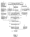
- Figure 1 is a schematic drawing of one embodiment of the present invention.
- Figure 2 illustrates an example of a visual display of the characteristics of diffuse and concentrated generators in a normal experimental subject.
- Figure 3 illustrates an example of a visual display of the characteristics of diffuse and concentrated generators in a patient with a cerebrovascular accident.
- a plurality of sensors (1) are placed on or in proximity to the experimental subject's scalp, for the detection of brain electromagnetic signals, which are generated due to neuronal sources.
- the sensors are placed according to a pre-defined plan in order to maximize the amount of information about the generators. Due to variability of head shape and size in the human population, measurements of the exact positions of the sensors are required, with respect to a reference coordinate system determined by certain anatomical landmarks of the individual subject's head.
- the electromagnetic signals are amplified (2) to the dynamic range of an analog to digital converter (3), which converts the signals into numbers that are stored in the memory of a digital computer.
- the recording of electromagnetic signals is carried out under the control of a central experimental program.
- the experimental subject can optionally be presented with visual, auditory, and somato-sensorial stimulation (4), introducing fiducial markers in the recording for later identification. Stimulation can be presented in the form of video-games.
- responses produced by the experimental subject (5) in the form of vocalizations or body movements can be recorded and identified, and can modify dynamically the central experimental program.
- real-time detection of spontaneous events in the EEG and MEG produced by the experimental subject during recording (6) is provided, allowing the dynamic modification of the central experimental program.
- Anatomical and functional information about the head including aspects such as geometry of the different constituent tissues (e.g., brain, skull, scalp), location and orientation of the cortex and of other neuronal aggregates, is summarized in what is here termed descriptive parametric geometry (8).
- anatomical images e. g., CAT images or NMR images
- functional images e.g., PET images or SPECT images
- M m is the number of sampled points on the m-th boundary.
- the descriptive parametric geometry previously explained can be used for computing the anatomical deconvolution operator (10), which is defined in the following equations : where are the electric potential and magnetic field component measurement vectors (the vector elements correspond to measurements made at different sensor positions), respectively; correspond to electric potential and magnetic field component values at the same sensor positions, and due to the same neuronal generators, in an infinite homogeneous medium; and are transfer coefficient matrices, which define the anatomical deconvolution operator.
- the anatomical deconvolution operator can be computed by the method and system described in CUBAN patent application 4/91, or alternatively by the new method described in the present invention.
- the descriptive parametric geometry is used for constructing a head phantom (9) with all the volume conductor properties of the experimental subject's head, said volume conductor properties consisting of the geometry and conductivity profile of the different constituent tissues (e.g., brain, skull, scalp).
- Electric potential ( V ) and magnetic field component ( B ) measurements are performed on the head phantom due to a plurality of implanted current dipoles (one at a time) with known locations and moments (dipoles located in the corresponding neural tissue volume).
- a further important use of the descriptive parametric geometry is the determination of anatomical and functional constraints for the localizations, orientations, activities, and connectivities of the brain electromagnetic waves generators (11) (generator constraints).
- Generator constraints are necessary for obtaining a unique inverse solution (14). For example, if the measured EEG and MEG activity is known beforehand to be generated only by cortical sources, then the generators can be located only on the cortical surface with orthogonal orientations.
- the sufficient statistics can consist of cumulants of any order, or multiple time series parametric models, either in the time, frequency, or time-frequency domain.
- Karhunen-Loeve type representations can be used for fitting the sufficient statistics in stationary or non-stationary, linear or non-linear models.
- This model has the following characteristics :
- Inverse solutions (14) are computed for the source model in infinite homogeneous medium, based on the sufficient statistics of the measured data transformed to infinite homogeneous medium by means of the anatomical deconvolution operator (12), and taking into account the constraints imposed on the generators (11).
- Inverse solutions can be obtained by a least squares criterion in which tr( ⁇ V - ⁇ V )2 is minimized with respect to the dipole location parameters in matrix ⁇ , the dipole orientation parameters in matrix , the generators cross spectral matrix ⁇ g , and the coefficients a k of the noise cross spectral matrix, where ⁇ v is the sample electric potential cross spectral matrix.
- inverse solutions can be obtained by maximizing the likelihood function, which is equivalent to minimizing tr( ⁇ -1 V ⁇ V )-det( ⁇ -1 V ⁇ V ). Independently of the estimation method used, the inverse solutions must be obtained under the generator constraints.
- G * t (g 1t g 2t .. g Ndt ) can be estimated at each time instant as : which is obtained by either maximum likelihood or weighted least squares methods.
- the estimated localizations, orientations, activities, and connectivities of the ERCs generators of an experimental subject are compared with those of a normal population (17), by means of multivariate metrics for measuring distances between estimators and normative data of a sample from the normal population, taking properly into account the effect of covariables such as age in order to decrease metric variability.
- visual displays are presented in the form of three dimensional and two dimensional images of the head, where the localizations, orientations, activities, and connectivities of the ERCs generators are displayed by coding their numerical values in terms of color, intensity, and graphical icons, and where optionally, the multivariate metrics corresponding to comparison with norms can also be displayed by superposition.
- Figure 2 illustrates a visual display of an inverse solution obtained from a normal experimental subject, based on a spontaneous EEG recording under eyes closed, awake, conditions.
- Two dipoles were fitted, together with additive uncorrelated homogeneous isotropic spatial noise.
- Generators are represented as arrows in the three head views (back, top, and left views).
- Generator localization and moments are given in cartesian coordinates referred to a unit radius sphere, with z axis coming out through the vertex, x axis coming out through nasion, and y axis coming out through the left ear (T3 electrode position).
- Connectivities are given in terms of the generator correlation matrix.
- Noise characteristics are illustrated as "BASE ACTIVITY", giving the values of the expansion coefficients of the homogeneous isotropic process. Note the alpha rhythm generators located in the occipital cortex.
- Figure 3 illustrates a visual display of an inverse solution obtained from an experimental subject with a lateralized right cerebrovascular accident, based on a spontaneous EEG recording under eyes closed, awake, conditions. Analysis procedures were the same to those used for the normal subject of Figure 2. Note that only one alpha generator lies in the normal position (left occipital cortex).
Landscapes
- Health & Medical Sciences (AREA)
- Life Sciences & Earth Sciences (AREA)
- Biomedical Technology (AREA)
- Molecular Biology (AREA)
- Veterinary Medicine (AREA)
- Biophysics (AREA)
- Pathology (AREA)
- Engineering & Computer Science (AREA)
- Public Health (AREA)
- Heart & Thoracic Surgery (AREA)
- Medical Informatics (AREA)
- Physics & Mathematics (AREA)
- Surgery (AREA)
- Animal Behavior & Ethology (AREA)
- General Health & Medical Sciences (AREA)
- Nuclear Medicine, Radiotherapy & Molecular Imaging (AREA)
- Radiology & Medical Imaging (AREA)
- Measurement And Recording Of Electrical Phenomena And Electrical Characteristics Of The Living Body (AREA)
- Magnetic Resonance Imaging Apparatus (AREA)
Applications Claiming Priority (2)
| Application Number | Priority Date | Filing Date | Title |
|---|---|---|---|
| CU4491 | 1991-03-15 | ||
| CU1991044 | 1991-03-15 |
Publications (2)
| Publication Number | Publication Date |
|---|---|
| EP0504027A2 true EP0504027A2 (de) | 1992-09-16 |
| EP0504027A3 EP0504027A3 (en) | 1993-04-21 |
Family
ID=5459256
Family Applications (1)
| Application Number | Title | Priority Date | Filing Date |
|---|---|---|---|
| EP19920400595 Withdrawn EP0504027A3 (en) | 1991-03-15 | 1992-03-06 | Method and system for three-dimensional tomography of activity and connectivity of brain and heart electromagnetic waves generators |
Country Status (2)
| Country | Link |
|---|---|
| US (1) | US5307807A (de) |
| EP (1) | EP0504027A3 (de) |
Cited By (10)
| Publication number | Priority date | Publication date | Assignee | Title |
|---|---|---|---|---|
| WO1994012100A1 (en) * | 1992-11-30 | 1994-06-09 | Risto Ilmoniemi | Method and apparatus for separating the different components of evoked response and spontaneous activity brain signals as well as of signals measured from the heart |
| WO1998018384A1 (en) * | 1996-10-30 | 1998-05-07 | Risto Ilmoniemi | Method and apparatus for mapping cortical connections |
| WO2000010454A1 (en) * | 1998-08-24 | 2000-03-02 | Ctf Systems Inc. | Functional brain imaging from magnetoencephalographic data |
| US6370414B1 (en) | 1998-01-23 | 2002-04-09 | Ctf Systems, Inc. | System and method for measuring, estimating and displaying RMS current density maps |
| EP0869736A4 (de) * | 1995-01-27 | 2004-03-31 | David Eidelberg | Markierer für das durchleuchten von patienten |
| EP1579799A1 (de) * | 2004-03-25 | 2005-09-28 | Sei Matsuoka | Vorrichtung und Verfahren zur Messung des quantitativen Wertes von Nachrichten |
| ES2319080A1 (es) * | 2008-09-26 | 2009-05-01 | Universidad Politecnica De Madrid | Fantoma multicanal de dipolos magneticos orientables para magnetoencefalografia. |
| EP2130489A1 (de) * | 2008-06-06 | 2009-12-09 | Electrical Geodesics Inc. | Verfahren zur Ortung von Bereichen mit elektrischer Hirnaktivität |
| WO2011051807A1 (en) * | 2009-10-27 | 2011-05-05 | Cerebral Diagnostics Canada Incorporated | Spectral decomposition and display of three-dimensional electrical activity in the cerebral cortex |
| CN104755026A (zh) * | 2012-11-26 | 2015-07-01 | 珀西斯特发展公司 | 用于显示存在于eeg记录中的伪影量的方法和系统 |
Families Citing this family (35)
| Publication number | Priority date | Publication date | Assignee | Title |
|---|---|---|---|---|
| US6006126A (en) | 1991-01-28 | 1999-12-21 | Cosman; Eric R. | System and method for stereotactic registration of image scan data |
| US6405072B1 (en) * | 1991-01-28 | 2002-06-11 | Sherwood Services Ag | Apparatus and method for determining a location of an anatomical target with reference to a medical apparatus |
| DE69227463T2 (de) * | 1991-12-17 | 1999-06-10 | Dynamics Imaging, Inc., Devon, Penn. | Verfahren und vorrichtung zur diagnose von lebenden organismen |
| US5699797A (en) * | 1992-10-05 | 1997-12-23 | Dynamics Imaging, Inc. | Method of investigation of microcirculation functional dynamics of physiological liquids in skin and apparatus for its realization |
| US6002958A (en) * | 1992-12-24 | 1999-12-14 | Dynamics Imaging, Inc. | Method and apparatus for diagnostics of internal organs |
| US5458142A (en) * | 1993-03-19 | 1995-10-17 | Farmer; Edward J. | Device for monitoring a magnetic field emanating from an organism |
| JP2739804B2 (ja) * | 1993-05-14 | 1998-04-15 | 日本電気株式会社 | 双極子推定装置 |
| US5687724A (en) * | 1993-10-26 | 1997-11-18 | Abratech Corporation | Apparatus and method for determining variations in measured physical parameters of signal-generators |
| US5357965A (en) * | 1993-11-24 | 1994-10-25 | General Electric Company | Method for controlling adaptive color flow processing using fuzzy logic |
| US5747789A (en) * | 1993-12-01 | 1998-05-05 | Dynamics Imaging, Inc. | Method for investigation of distribution of physiological components in human body tissues and apparatus for its realization |
| US6192262B1 (en) | 1994-02-23 | 2001-02-20 | Dobi Medical Systems, Llc | Method of living organism multimodal functional mapping |
| US5865743A (en) * | 1994-02-23 | 1999-02-02 | Dynamics Imaging, Inc. | Method of living organism multimodal functional mapping |
| US5730133A (en) * | 1994-05-20 | 1998-03-24 | Dynamics Imaging, Inc. | Optical functional mamoscope |
| JP3724014B2 (ja) * | 1994-08-25 | 2005-12-07 | ソニー株式会社 | 画像認識装置および画像認識方法 |
| JP2735158B2 (ja) * | 1996-03-18 | 1998-04-02 | 工業技術院長 | 磁場源可動式ファントムヘッド |
| GB2321363A (en) * | 1997-01-21 | 1998-07-22 | Northern Telecom Ltd | Telecommunications |
| US6697660B1 (en) | 1998-01-23 | 2004-02-24 | Ctf Systems, Inc. | Method for functional brain imaging from magnetoencephalographic data by estimation of source signal-to-noise ratio |
| JP3246433B2 (ja) * | 1998-01-27 | 2002-01-15 | 日本電気株式会社 | 暗号強度評価支援装置及びプログラムを記録した機械読み取り可能な記録媒体 |
| US6195576B1 (en) * | 1998-03-09 | 2001-02-27 | New York University | Quantitative magnetoencephalogram system and method |
| US6309361B1 (en) | 1998-05-04 | 2001-10-30 | Kirtley E. Thornton | Method for improving memory by identifying and using QEEG parameters correlated to specific cognitive functioning |
| US7092748B2 (en) * | 2000-02-18 | 2006-08-15 | Centro Nacional De Investigaciones Cientificas (Cnic) | System and method for the tomography of the primary electric current of the brain and of the heart |
| US6856830B2 (en) * | 2001-07-19 | 2005-02-15 | Bin He | Method and apparatus of three dimension electrocardiographic imaging |
| US7840250B2 (en) * | 2001-11-13 | 2010-11-23 | Electrical Geodesics, Inc. | Method for neural current imaging |
| CA2657087A1 (en) * | 2008-03-06 | 2009-09-06 | David N. Fernandes | Normative database system and method |
| US9576107B2 (en) * | 2013-07-09 | 2017-02-21 | Biosense Webster (Israel) Ltd. | Model based reconstruction of the heart from sparse samples |
| JP6996203B2 (ja) * | 2017-03-17 | 2022-01-17 | 株式会社リコー | 情報処理装置、情報処理方法、プログラムおよび生体信号計測システム |
| EP3684463B1 (de) | 2017-09-19 | 2025-05-14 | Neuroenhancement Lab, LLC | Verfahren und vorrichtung für neuro-enhancement |
| US11717686B2 (en) | 2017-12-04 | 2023-08-08 | Neuroenhancement Lab, LLC | Method and apparatus for neuroenhancement to facilitate learning and performance |
| US12280219B2 (en) | 2017-12-31 | 2025-04-22 | NeuroLight, Inc. | Method and apparatus for neuroenhancement to enhance emotional response |
| US11478603B2 (en) | 2017-12-31 | 2022-10-25 | Neuroenhancement Lab, LLC | Method and apparatus for neuroenhancement to enhance emotional response |
| US11364361B2 (en) | 2018-04-20 | 2022-06-21 | Neuroenhancement Lab, LLC | System and method for inducing sleep by transplanting mental states |
| CN113382683A (zh) | 2018-09-14 | 2021-09-10 | 纽罗因恒思蒙特实验有限责任公司 | 改善睡眠的系统和方法 |
| US11786694B2 (en) | 2019-05-24 | 2023-10-17 | NeuroLight, Inc. | Device, method, and app for facilitating sleep |
| CN110584898B (zh) * | 2019-10-08 | 2020-08-14 | 南京邮电大学 | 一种基于多传感器的脑控轮椅自动避障方法 |
| CN114631830B (zh) * | 2022-03-12 | 2024-08-09 | 北京工业大学 | 基于d-k分区的简化分布式偶极子模型建立与识别方法 |
Family Cites Families (14)
| Publication number | Priority date | Publication date | Assignee | Title |
|---|---|---|---|---|
| US4201224A (en) * | 1978-12-29 | 1980-05-06 | Roy John E | Electroencephalographic method and system for the quantitative description of patient brain states |
| US4416288A (en) * | 1980-08-14 | 1983-11-22 | The Regents Of The University Of California | Apparatus and method for reconstructing subsurface electrophysiological patterns |
| US4417592A (en) * | 1981-05-11 | 1983-11-29 | Roy John E | Digital electroencephalographic instrument and method |
| US4408616A (en) * | 1981-05-15 | 1983-10-11 | The Children's Medical Center Corporation | Brain electricaL activity mapping |
| DE3247585A1 (de) * | 1982-12-22 | 1984-06-28 | Siemens AG, 1000 Berlin und 8000 München | Mehrkanalige vorrichtung zur messung von verschiedenen feldquellen hervorgerufener schwacher magnetfelder |
| US4753246A (en) * | 1986-03-28 | 1988-06-28 | The Regents Of The University Of California | EEG spatial filter and method |
| US4736751A (en) * | 1986-12-16 | 1988-04-12 | Eeg Systems Laboratory | Brain wave source network location scanning method and system |
| US4913160A (en) * | 1987-09-30 | 1990-04-03 | New York University | Electroencephalographic system and method using factor structure of the evoked potentials |
| FR2622990B1 (fr) * | 1987-11-05 | 1990-03-30 | Centre Nat Rech Scient | Dispositif de stereolocalisation spatiale de sources d'activites cerebrales |
| US4991579A (en) * | 1987-11-10 | 1991-02-12 | Allen George S | Method and apparatus for providing related images over time of a portion of the anatomy using fiducial implants |
| US4949725A (en) * | 1988-07-01 | 1990-08-21 | Bio-Logic Systems Corporation | Apparatus and method for displaying electrical activity generated within a living body |
| EP0355506B1 (de) * | 1988-08-16 | 1994-12-14 | Siemens Aktiengesellschaft | Anordnung zum Messen lokaler bioelektrischer Ströme in biologischen Gewebekomplexen |
| FI83266C (fi) * | 1988-09-12 | 1991-06-10 | Teknillinen Korkeakoulu | Foerfarande och anordning foer lokalisering av elektroder faestade vid kroppen av en maenniska, i synnerhet huvudet. |
| US4977896A (en) * | 1989-05-26 | 1990-12-18 | Biomagnetic Technologies, Inc. | Analysis of biological signals using data from arrays of sensors |
-
1992
- 1992-03-06 EP EP19920400595 patent/EP0504027A3/xx not_active Withdrawn
- 1992-03-11 US US07/849,594 patent/US5307807A/en not_active Expired - Lifetime
Cited By (14)
| Publication number | Priority date | Publication date | Assignee | Title |
|---|---|---|---|---|
| WO1994012100A1 (en) * | 1992-11-30 | 1994-06-09 | Risto Ilmoniemi | Method and apparatus for separating the different components of evoked response and spontaneous activity brain signals as well as of signals measured from the heart |
| EP0869736A4 (de) * | 1995-01-27 | 2004-03-31 | David Eidelberg | Markierer für das durchleuchten von patienten |
| WO1998018384A1 (en) * | 1996-10-30 | 1998-05-07 | Risto Ilmoniemi | Method and apparatus for mapping cortical connections |
| US6256531B1 (en) | 1996-10-30 | 2001-07-03 | Risto Ilmoniemi | Method and apparatus for mapping cortical connections |
| US6370414B1 (en) | 1998-01-23 | 2002-04-09 | Ctf Systems, Inc. | System and method for measuring, estimating and displaying RMS current density maps |
| WO2000010454A1 (en) * | 1998-08-24 | 2000-03-02 | Ctf Systems Inc. | Functional brain imaging from magnetoencephalographic data |
| EP1579799A1 (de) * | 2004-03-25 | 2005-09-28 | Sei Matsuoka | Vorrichtung und Verfahren zur Messung des quantitativen Wertes von Nachrichten |
| EP2130489A1 (de) * | 2008-06-06 | 2009-12-09 | Electrical Geodesics Inc. | Verfahren zur Ortung von Bereichen mit elektrischer Hirnaktivität |
| EP2478837A1 (de) * | 2008-06-06 | 2012-07-25 | Electrical Geodesics Inc. | Verfahren zum Auffinden von Wegen elektrischer Hirnaktivität |
| ES2319080A1 (es) * | 2008-09-26 | 2009-05-01 | Universidad Politecnica De Madrid | Fantoma multicanal de dipolos magneticos orientables para magnetoencefalografia. |
| ES2319080B2 (es) * | 2008-09-26 | 2009-09-22 | Universidad Politecnica De Madrid | Fantoma multicanal de dipolos magneticos orientables para magnetoencefalografia. |
| WO2011051807A1 (en) * | 2009-10-27 | 2011-05-05 | Cerebral Diagnostics Canada Incorporated | Spectral decomposition and display of three-dimensional electrical activity in the cerebral cortex |
| CN104755026A (zh) * | 2012-11-26 | 2015-07-01 | 珀西斯特发展公司 | 用于显示存在于eeg记录中的伪影量的方法和系统 |
| EP2922466A4 (de) * | 2012-11-26 | 2016-07-20 | Persyst Dev Corp | Verfahren und system zur anzeige der menge von artefakten in einer eeg-aufzeichnung |
Also Published As
| Publication number | Publication date |
|---|---|
| US5307807A (en) | 1994-05-03 |
| EP0504027A3 (en) | 1993-04-21 |
Similar Documents
| Publication | Publication Date | Title |
|---|---|---|
| US5307807A (en) | Method and system for three dimensional tomography of activity and connectivity of brain and heart electromagnetic waves generators | |
| Samuelsson et al. | Spatial fidelity of MEG/EEG source estimates: A general evaluation approach | |
| Van Veen et al. | Localization of brain electrical activity via linearly constrained minimum variance spatial filtering | |
| EP1049402B1 (de) | Verfahren zur messung, bestimmung und anzeige von effektivwerten der stromdichteverteilung | |
| Wikswo Jr et al. | The future of the EEG and MEG | |
| Liu et al. | Detecting large‐scale networks in the human brain using high‐density electroencephalography | |
| Mutanen et al. | Recovering TMS-evoked EEG responses masked by muscle artifacts | |
| Hämäläinen et al. | Magnetoencephalographic (MEG) characterization of dynamic brain activation | |
| Brookes et al. | Beamformer reconstruction of correlated sources using a modified source model | |
| Vrba et al. | Fetal MEG redistribution by projection operators | |
| Zumer et al. | A probabilistic algorithm integrating source localization and noise suppression of MEG and EEG data | |
| Schulz et al. | An integrative MEG–fMRI study of the primary somatosensory cortex using cross-modal correspondence analysis | |
| Im et al. | Spatial resolution of EEG cortical source imaging revealed by localization of retinotopic organization in human primary visual cortex | |
| Van Hoey et al. | Influence of measurement noise and electrode mislocalisation on EEG dipole-source localisation | |
| Goebel et al. | The added value of EEG-fMRI in imaging neuroscience | |
| Fender | Models of the human brain and the surrounding media: their influence on the reliability of source localization | |
| Pascual-Marqui et al. | Imaging the electric neuronal generators of EEG/MEG | |
| Halchenko et al. | Multimodal integration: fMRI, mri, EEG, MEG | |
| Bauer | Slow potential topography | |
| Ustinin et al. | Functional tomography of complex systems using spectral analysis of multichannel measurement data | |
| Gutiérrez et al. | Ellipsoidal head model for fetal magnetoencephalography: forward and inverse solutions | |
| Ramírez | Source localization | |
| Liu | Spatiotemporal brain imaging | |
| Makhortykh | Generalized spectral-analytical method for biomedical data processing | |
| Acar et al. | Patch-basis electrocortical source imaging in epilepsy |
Legal Events
| Date | Code | Title | Description |
|---|---|---|---|
| PUAI | Public reference made under article 153(3) epc to a published international application that has entered the european phase |
Free format text: ORIGINAL CODE: 0009012 |
|
| AK | Designated contracting states |
Kind code of ref document: A2 Designated state(s): AT BE CH DE DK ES FR GB GR IT LI LU NL SE |
|
| RIN1 | Information on inventor provided before grant (corrected) |
Inventor name: PASCUAL MARQUI, ROBERTO D. Inventor name: GRAVE DE PERALTA MENENDEZ, ROLANDO Inventor name: GONZALEZ ANDINO, SARA L. Inventor name: BISCAY LIRIO, ROLANDO Inventor name: BOSCH BAYARD, JORGE Inventor name: RIERA DIAZ, JORGE J. Inventor name: VALDES SOSA, PEDRO A. |
|
| PUAL | Search report despatched |
Free format text: ORIGINAL CODE: 0009013 |
|
| AK | Designated contracting states |
Kind code of ref document: A3 Designated state(s): AT BE CH DE DK ES FR GB GR IT LI LU NL SE |
|
| 17P | Request for examination filed |
Effective date: 19930930 |
|
| 17Q | First examination report despatched |
Effective date: 19950127 |
|
| STAA | Information on the status of an ep patent application or granted ep patent |
Free format text: STATUS: THE APPLICATION IS DEEMED TO BE WITHDRAWN |
|
| 18D | Application deemed to be withdrawn |
Effective date: 19950607 |