EP0515099B1 - Dispositif pour analyser des cellules dans l'urine - Google Patents
Dispositif pour analyser des cellules dans l'urine Download PDFInfo
- Publication number
- EP0515099B1 EP0515099B1 EP92304368A EP92304368A EP0515099B1 EP 0515099 B1 EP0515099 B1 EP 0515099B1 EP 92304368 A EP92304368 A EP 92304368A EP 92304368 A EP92304368 A EP 92304368A EP 0515099 B1 EP0515099 B1 EP 0515099B1
- Authority
- EP
- European Patent Office
- Prior art keywords
- cell
- light intensity
- data
- scattered
- cells
- Prior art date
- Legal status (The legal status is an assumption and is not a legal conclusion. Google has not performed a legal analysis and makes no representation as to the accuracy of the status listed.)
- Expired - Lifetime
Links
- 210000002700 urine Anatomy 0.000 title claims description 49
- 210000004027 cell Anatomy 0.000 claims description 168
- 210000003743 erythrocyte Anatomy 0.000 claims description 29
- 241000894006 Bacteria Species 0.000 claims description 27
- 238000000034 method Methods 0.000 claims description 15
- 238000005259 measurement Methods 0.000 claims description 9
- 230000001678 irradiating effect Effects 0.000 claims description 5
- 210000002919 epithelial cell Anatomy 0.000 description 26
- 238000010586 diagram Methods 0.000 description 23
- 210000000265 leukocyte Anatomy 0.000 description 23
- 239000002245 particle Substances 0.000 description 13
- 238000012545 processing Methods 0.000 description 12
- 238000006243 chemical reaction Methods 0.000 description 5
- 238000000684 flow cytometry Methods 0.000 description 5
- 239000013049 sediment Substances 0.000 description 5
- 108020004414 DNA Proteins 0.000 description 4
- 230000002159 abnormal effect Effects 0.000 description 3
- 238000004458 analytical method Methods 0.000 description 3
- 239000003153 chemical reaction reagent Substances 0.000 description 3
- 230000003287 optical effect Effects 0.000 description 3
- 206010061218 Inflammation Diseases 0.000 description 2
- 210000000805 cytoplasm Anatomy 0.000 description 2
- 238000001514 detection method Methods 0.000 description 2
- 239000000284 extract Substances 0.000 description 2
- 208000015181 infectious disease Diseases 0.000 description 2
- 230000004054 inflammatory process Effects 0.000 description 2
- 201000006370 kidney failure Diseases 0.000 description 2
- 239000004816 latex Substances 0.000 description 2
- 229920000126 latex Polymers 0.000 description 2
- 238000002562 urinalysis Methods 0.000 description 2
- 208000032843 Hemorrhage Diseases 0.000 description 1
- 108091093105 Nuclear DNA Proteins 0.000 description 1
- 206010037596 Pyelonephritis Diseases 0.000 description 1
- 208000001647 Renal Insufficiency Diseases 0.000 description 1
- 210000000601 blood cell Anatomy 0.000 description 1
- 230000005779 cell damage Effects 0.000 description 1
- 208000037887 cell injury Diseases 0.000 description 1
- 210000003850 cellular structure Anatomy 0.000 description 1
- 239000000356 contaminant Substances 0.000 description 1
- 238000007796 conventional method Methods 0.000 description 1
- 239000000428 dust Substances 0.000 description 1
- 230000000694 effects Effects 0.000 description 1
- 238000005516 engineering process Methods 0.000 description 1
- 239000012530 fluid Substances 0.000 description 1
- MHMNJMPURVTYEJ-UHFFFAOYSA-N fluorescein-5-isothiocyanate Chemical compound O1C(=O)C2=CC(N=C=S)=CC=C2C21C1=CC=C(O)C=C1OC1=CC(O)=CC=C21 MHMNJMPURVTYEJ-UHFFFAOYSA-N 0.000 description 1
- 239000011521 glass Substances 0.000 description 1
- 210000004276 hyalin Anatomy 0.000 description 1
- 238000005286 illumination Methods 0.000 description 1
- 210000003734 kidney Anatomy 0.000 description 1
- 208000017169 kidney disease Diseases 0.000 description 1
- 238000000386 microscopy Methods 0.000 description 1
- 239000000203 mixture Substances 0.000 description 1
- 230000003204 osmotic effect Effects 0.000 description 1
- 238000002203 pretreatment Methods 0.000 description 1
- ZDWVWKDAWBGPDN-UHFFFAOYSA-O propidium Chemical compound C12=CC(N)=CC=C2C2=CC=C(N)C=C2[N+](CCC[N+](C)(CC)CC)=C1C1=CC=CC=C1 ZDWVWKDAWBGPDN-UHFFFAOYSA-O 0.000 description 1
- 238000012552 review Methods 0.000 description 1
- 238000012216 screening Methods 0.000 description 1
- 238000000926 separation method Methods 0.000 description 1
- 230000000087 stabilizing effect Effects 0.000 description 1
- 238000010186 staining Methods 0.000 description 1
- 238000012360 testing method Methods 0.000 description 1
- 210000003708 urethra Anatomy 0.000 description 1
- 230000000007 visual effect Effects 0.000 description 1
Images




Classifications
-
- G—PHYSICS
- G01—MEASURING; TESTING
- G01N—INVESTIGATING OR ANALYSING MATERIALS BY DETERMINING THEIR CHEMICAL OR PHYSICAL PROPERTIES
- G01N15/00—Investigating characteristics of particles; Investigating permeability, pore-volume or surface-area of porous materials
- G01N15/10—Investigating individual particles
- G01N15/14—Optical investigation techniques, e.g. flow cytometry
- G01N15/1456—Optical investigation techniques, e.g. flow cytometry without spatial resolution of the texture or inner structure of the particle, e.g. processing of pulse signals
- G01N15/1459—Optical investigation techniques, e.g. flow cytometry without spatial resolution of the texture or inner structure of the particle, e.g. processing of pulse signals the analysis being performed on a sample stream
-
- G—PHYSICS
- G01—MEASURING; TESTING
- G01N—INVESTIGATING OR ANALYSING MATERIALS BY DETERMINING THEIR CHEMICAL OR PHYSICAL PROPERTIES
- G01N15/00—Investigating characteristics of particles; Investigating permeability, pore-volume or surface-area of porous materials
- G01N15/10—Investigating individual particles
- G01N15/14—Optical investigation techniques, e.g. flow cytometry
- G01N2015/1486—Counting the particles
-
- G—PHYSICS
- G01—MEASURING; TESTING
- G01N—INVESTIGATING OR ANALYSING MATERIALS BY DETERMINING THEIR CHEMICAL OR PHYSICAL PROPERTIES
- G01N21/00—Investigating or analysing materials by the use of optical means, i.e. using sub-millimetre waves, infrared, visible or ultraviolet light
- G01N21/17—Systems in which incident light is modified in accordance with the properties of the material investigated
- G01N21/47—Scattering, i.e. diffuse reflection
- G01N2021/4704—Angular selective
- G01N2021/4707—Forward scatter; Low angle scatter
-
- G—PHYSICS
- G01—MEASURING; TESTING
- G01N—INVESTIGATING OR ANALYSING MATERIALS BY DETERMINING THEIR CHEMICAL OR PHYSICAL PROPERTIES
- G01N21/00—Investigating or analysing materials by the use of optical means, i.e. using sub-millimetre waves, infrared, visible or ultraviolet light
- G01N21/62—Systems in which the material investigated is excited whereby it emits light or causes a change in wavelength of the incident light
- G01N21/63—Systems in which the material investigated is excited whereby it emits light or causes a change in wavelength of the incident light optically excited
- G01N21/64—Fluorescence; Phosphorescence
-
- G—PHYSICS
- G01—MEASURING; TESTING
- G01N—INVESTIGATING OR ANALYSING MATERIALS BY DETERMINING THEIR CHEMICAL OR PHYSICAL PROPERTIES
- G01N33/00—Investigating or analysing materials by specific methods not covered by groups G01N1/00 - G01N31/00
- G01N33/48—Biological material, e.g. blood, urine; Haemocytometers
- G01N33/483—Physical analysis of biological material
- G01N33/487—Physical analysis of biological material of liquid biological material
- G01N33/493—Physical analysis of biological material of liquid biological material urine
-
- Y—GENERAL TAGGING OF NEW TECHNOLOGICAL DEVELOPMENTS; GENERAL TAGGING OF CROSS-SECTIONAL TECHNOLOGIES SPANNING OVER SEVERAL SECTIONS OF THE IPC; TECHNICAL SUBJECTS COVERED BY FORMER USPC CROSS-REFERENCE ART COLLECTIONS [XRACs] AND DIGESTS
- Y10—TECHNICAL SUBJECTS COVERED BY FORMER USPC
- Y10S—TECHNICAL SUBJECTS COVERED BY FORMER USPC CROSS-REFERENCE ART COLLECTIONS [XRACs] AND DIGESTS
- Y10S209/00—Classifying, separating, and assorting solids
- Y10S209/939—Video scanning
Definitions
- This invention relates to an apparatus which uses flow cytometry to classify and enumerate cells such as leukocytes, erythrocytes, epithelial cells, casts and bacteria contained in urine.
- a screening test for kidney failure can be conducted based upon the presence of erythrocytes, leukocytes, epithelial cells, casts and bacteria in urine.
- Measurement of erythrocytes is important in terms of determining whether hemorrhage has occurred in the tract from the slomerulus to the urethra of the kidney.
- the appearance of leukocytes is considered to be a possible indication of a kidney disorder such as pyelonephritis, and detection thereof is important in early discovery of inflammation and infection.
- cast and erythrocyte morphology the origin of such inflammation and infection, namely the abnormal parts of the body, can be surmised.
- cell shall be used as a generic term for an erythrocyte, epithelial cell, cast and bacterium.
- Conventional methods of analyzing cells in urine include (a) visual examination based upon microscopy and (b) automatic measurement using a combination of a flat sheath flow and image processing technology.
- Method (a) involves centrifuging a urine specimen, preparing a slide sample of the matter of sediment and observing, classifying and counting cells under a microscope.
- Method (b) involves using a video camera to capture an image of a urine specimen made to flow as an extremely flat stream within a sheathing solution employed as an outer layer, and subjecting the still picture obtained to image processing, whereby the images of the cells in the specimen are extracted and displayed.
- the apparatus which uses method (b) is itself high in cost owing to reliance upon image processing, and the processing speed is low. Furthermore, the advantage of automation afforded by the apparatus of method (b) merely displays the images upon roughly classifying the imaged components based upon their size, and it is required that classification process be performed by a human being while the display is observed. Thus, the automatic classification and enumeration of cell components is not possible.
- Another problem is that since the types of components in urine sediment are numerous and differ widely in size from one specimen to another, and in view of the fact that the degree of cell damage can be considered to be large, it is understood that analysis of urine sediment is not possible using flow cytometry.
- US Patent No. 4661913 discloses flow apparatus and a method for the detection of particles in a sample. Particles are moved, substantially one at a time, in a fluid flow stream. A beam of illumination is directed at particles in the stream and data associated with each particle is detected as they pass through the beam. The data is compared to stored data detected from a class of particles. A determination is made that particles from an unknown class belong to the established class as a result of matching respective data.
- Bacteria classification in human urine is exemplified using a PCM (Principal Component Method).
- the present invention seeks to improve upon the foregoing shortcomings and its object is to provide an apparatus for analyzing cells in urine, in which a large quantity of a urine specimen can be analyzed using flow cytometry, the number of various cells (erythrocytes, leukocytes, epithelial cells, casts and bacteria, etc.) detected in the specimen can be greatly increased to enable more precise analysis of cells in urine, the process from drawing of the urine specimen into the apparatus to display of the analytical results can be fully automated to eliminate the need for any human intervention, the processing speed can be raised and the cost of the apparatus can be kept low.
- the foregoing object is attained by providing an apparatus for analyzing cells in urine as defined in claims 1,3.
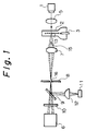
- Fig. 1 is a block diagram showing the arrangement of the principal elements of an optical system embodying the present invention.
- the optical system includes a light source 1 constituted by an argon-ion laser at one end of the system, a flow cell 2, a condenser lens 5 provided between the light source 1 and the flow cell 2, a photomultiplier 6 at the other end of the system, a collector lens 7, a light shield 8, a dichroic mirror 9 and a filter 10 provided between the flow cell 2 and the photomultiplier 6, and a lens 12 provided between the dichroic mirror 9 and a photodiode 11.
- a urine specimen flows into the flow cell 2 from a nozzle 3 attached to the flow cell.
- Reference numeral 15 denotes a beam stopper.
- Fig. 2 is a block diagram illustrating the principal components of an electric circuit embodying the present invention.
- the circuitry of Fig. 2 is divided into a signal processing portion 19 and data processing portion 20.
- An output signal from the photodiode 11 is connected to an amplifier 21 in the signal processing portion 19, and the output of the amplifier 21 is connected to a DC (direct-current) restorer 22.
- the output of the DC restorer 22 is connected to a peak-hold circuit 23, the output of which is connected to an analog/digital converter circuit (hereinafter referred to simply as an "A/D converter") 24.
- A/D converter analog/digital converter circuit
- the output of the photomultiplier 6 is connected to an amplifier 26 in the signal processing portion 19, the output of which is connected to a DC (direct-current) restorer 27.
- the output of the latter is connected to a peak-hold circuit 28, the output of which is connected to an A/D (analog/digital) converter 29.
- a threshold circuit 30 is connected to a digital/analog converter circuit (hereinafter referred to simply as a "D/A converter") 31, the output of which is connected to the reference input of a comparator 32.
- the output of the DC restorer 22 is connected to the comparator 32 at its other input terminal, namely the terminal whose input is to be compared with the reference.
- the output of the comparator 32 is connected to a pulse-width counter 33, to which a clock signal output from a clock signal generating circuit 34 is applied as an input.
- the output of the DC restorer 22 is further connected to a control circuit 35. The latter produces a control signal output connected to the A/D converters 24, 29 and to the pulse-width counter 33.
- the outputs of the A/D converters 24, 29 and the output of the pulse-width counter 33 are connected to a control circuit 40 for controlling a read/write operation.
- a trigger signal T is connected to the control circuit 40 and a counter 41.
- a memory circuit 42 for storing data indicative of individual cells is connected to the control circuit 40.
- the memory circuit 42 is connected, via the control circuit 40, to a data analyzing circuit 43 which classifies and enumerates cells.
- the output of the counter 41 also is connected to the data analyzing circuit 43.
- a memory circuit 45 which stores the control program of the apparatus, cell-diameter conversion values, cell judgment values, etc., and a counter 46, which counts the number of each type of cell, are connected to the data analyzing circuit 43.
- Figs. 3 through 8 are flowcharts illustrating the operation of the electrical circuitry according to this embodiment. These flowcharts will be referred to later.
- the original solution of the urine specimen contains in admixture a first reagent containing a specific stain and a second reagent for stabilizing pH and osmotic pressure.
- the resulting urine specimen mixture containing the reagent is discharged from the nozzle 9, and a sheathed flow is formed by causing a sheathing solution to flow along the periphery of the urine stream.
- cells 13 erythrocytes, leukocytes, epithelial cells, casts and bacteria, etc.
- in the urine specimen flow in an ordered array such as in single file through a narrow zone at the central portion of the flow cell 2, as shown in Fig. 1.
- the laser light from the light source 1 is condensed by the condenser lens 5 so as to irradiate the narrow flow zone of the flow cell with an elliptical beam spot that is slender in the direction of flow and broad in the direction perpendicular to the flow direction.
- the present invention is directed toward measurement of cells in urine, namely components of urine sediment.
- the thickness of the narrow flow zone should be set to be comparatively small in comparison with the sizes of the cells.
- a suitable value for the minor axis of the ellipse is 1 - 20 ⁇ m. It will suffice to make the major axis of the ellipse large enough to fully extend across the width of the slender specimen stream in the narrow flow zone.
- the cells 13 in the slender specimen stream are irradiated with the laser light.
- Transmitted laser light which has passed through the flow cell intact without striking the cells is blocked by a beam stopper 15.
- Forward-scattered light and forward fluorescent light emitted from an irradiated cell at a narrow angle is condensed by the collector lens 7, and the condensed light passes through a pin hole 16 of the shield 8. Almost all of the forward-scattered light and forward fluorescent light thus emitted from the cell 13 arrives at the dichroic mirror 9.
- the fluorescent light the wavelength of which is greater than that of the scattered light, is transmitted intact by the dichroic mirror 9 at a high rate and stray light is removed by the filter 10, after which the fluorescent light is detected and converted into an electric signal by the photomultiplier 6, from which a forward fluorescent light signal is outputted. Meanwhile, the scattered light is reflected by the dichroic mirror 9 at a high rate, after which the light is condensed by the lens 12 and converted into an electric signal by the photodiode 11, from which a forward-scattered light signal is outputted.
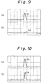
- Output waveforms of the forward-scattered light signal from the photodiode 11 and output waveforms of the forward fluorescent-light signal from the photomultiplier 6 are as illustrated in the waveform diagrams of Figs. 9 through 13, in which time is plotted along the horizontal axis and voltage along the vertical axis.
- FIG. 9 shows the output waveform of a forward-scattered light signal from an erythrocyte and (b) shows an output waveform of a forward fluorescent-light signal from the erythrocyte. Since erythrocytes are small in size and regular in shape, a forward-scattered light signal S 1 having a single peak is obtained. However, since an erythrocyte does not possess a nucleus, it cannot be stained by a stain and therefore fluorescent light S 2 is not detected.
- Fig. 10 shows the output waveform of a forward-scattered light signal from a leukocyte and (b) shows an output waveform of a forward fluorescent-light signal from the leukocyte.
- Leukocytes are large but are of the same size or slightly larger than erythrocytes, and a forward-scattered light signal S 3 similar to the forward-scattered light signal S 1 is detected. However, a forward fluorescent-light signal S 4 also is detected owing to the presence of a nucleus.
- Fig. 11 shows the output waveform of a forward-scattered light signal from an epithelial cell and (b) shows an output waveform of a forward fluorescent-light signal from the epithelial cell.
- Epithelial cells exist in a wide variety of sizes from large to small, but they are small in thickness and possess a complicated shape and internal structure.
- a forward-scattered light signal S 5 obtained exhibits a large width and a complicated waveform. Since the minor axis of the beam spot of the irradiating light is smaller than the diameter of an epithelial cell, the signal waveform obtained reflects the size, shape and internal structure of the cell.
- the forward-scattered light signal S 5 of the epithelial cell does not have a peak value which is that high in proportion to the large width of the signal.
- the reason for this is understood to be that since the cell thickness is small, all of the irradiating light is not scattered; i.e., some of it transmitted through the cell.
- a forward fluorescent-light signal S 6 obtained also has a large width and a complicated waveform. The portion of the waveform at which signal strength is very high represents the location of the nucleus.
- Epithelial cells have a large amount of cytoplasm, which also emits a certain degree of fluorescence.
- FIG. 12 shows the output waveform of a forward-scattered light signal from a bacterium and (b) shows an output waveform of a forward fluorescent-light signal from the bacterium. Since bacteria are small in comparison with blood cells and the like, a small forward-scattered light signal S 7 and forward fluorescent-light signal S 8 are detected.
- FIG. 13 shows the output waveform of a forward-scattered light signal from a cast and (b) shows an output waveform of a forward fluorescent-light signal from the cast. Since a cast has a size on the order of one hundred to several hundred microns, a forward-scattered light signal S 9 and a forward fluorescent-light signal S 10 having a very large width are obtained.
- the photomultiplier 6 produces a forward fluorescent-light signal for each of the variety of cells contained in the urine specimen.
- Each forward fluorescent-light signal is amplified by the amplifier 26 in the signal processing portion 19, and the amplified signal is applied to the DC restorer 27, which removes DC components and extracts the amplitude portion (namely the portion of the forward fluorescent-light signal).
- the resulting forward fluorescent-light signal from which the DC components have been removed is applied to the peak-hold circuit 28, which detects the peak value of the signal.
- the detected peak value is converted into a digital value by A/D converter 29, and this value is outputted as data indicative of the intensity of forward fluorescent light.
- the photodiode 11 produces a forward-scattered light signal for each of the variety of cells contained in the urine specimen.
- Each forward-scattered light signal is amplified by the amplifier 21 in the signal processing portion 19, and the amplified signal is applied to the DC restorer 22, which removes DC components and extracts the amplitude portion (namely the portion of the forward-scattered light signal).
- the resulting forward-scattered light signal from which the DC components have been removed is applied to the peak-hold circuit 23, which detects the peak value of the signal.
- the detected peak value is converted into a digital value by the A/D converter 24, and this value is outputted as data indicative of the intensity of forward-scattered light.
- the resulting forward-scattered light signal from which the DC components have been removed is applied to the comparison input terminal of the comparator 32.
- the threshold circuit 30 is set beforehand to an appropriate threshold value. This value is converted into an analog value by the D/A converter 31, and a voltage indicative of the analog value is applied to the reference input terminal of the comparator 32. Only a forward-scattered light signal which exceeds the threshold value is outputted by the comparator 32 in the form of square wave.
- the square-wave signal enters the pulse-width counter 33, which counts the clock signal from the clock signal generator 34 for the duration of the square wave-input. As a result, the forward-scattered light signal is converted into pulse-width data, which is outputted by the pulse-width counter 33.
- each item of pulse-width data is data which corresponds to the cell diameter of each detected cell.
- the above-mentioned control signal from the control circuit 35 is applied also to the A/D converters 24 and 29, and the A/D converters 24, 29 respectively output data indicative of the forward-scattered light intensity and data indicative of the forward fluorescent-light intensity for each and every cell, as described above.
- a measurement is taken using latex particles of a known particle diameter before a urine specimen is measured. Then, from the pulse-width data acquired by this preliminary measurement of the latex particles and the known diameter of these particles, conversion values for the purpose of converting the above-mentioned pulse-width data into particle diameters are calculated and the conversion values are then stored in the memory circuit 45 beforehand as conversion values of cell diameter.
- the cell diameters are values decided statistically upon actually measuring the erythrocytes, leukocytes, epithelial cells, casts and bacteria in a urine specimen using a microscope.
- the intensity of forward-scattered light reflects a plurality of items of information, such as cell size, density and surface configuration. There are cells (epithelial cells) for which the intensity of scattered light is high, cells (erythrocytes, leukocytes, casts and some epithelial cells) for which the intensity is medium, and cells (bacteria) for which the intensity is low.
- the intensity of the fluorescent light is information proportional to the amount of DNA.
- the items of forward-scattered light intensity data, forward fluorescent-light intensity data and pulse-width data associated with each cell are stored, on a cell-by-cell basis, in the memory circuit 42 whenever the trigger signal T, which is generated at each cell flow-by, enters the data processing portion 20.
- the pulse-width data is converted into cell-diameter data by the data analyzer 43 based upon the aforementioned cell diameter conversion values, and the cell-diameter data obtained is stored in the memory circuit 42.
- the counter 41 counts the cells in successive fashion. This operation is carried out until the entirety of the urine specimen has passed through the flow cell 2.
- the storage and counting operations are executed at step 51 in the flowchart of Fig. 3.
- the total number of cells detected in the urine specimen is held in the counter 41, and the items of forward-scattered light intensity data, forward fluorescent-light intensity data and cell-diameter data associated with each cell detected in the urine specimen are stored in the memory circuit 42.
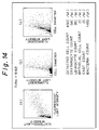
- the data analyzer 43 reads the data for all cells out of the memory circuit 42 and then creates and displays various scatter diagrams (a), (b) and (c) shown in the upper part of Fig. 14 (step 52). Observing the scatter diagrams of Fig. 14 makes it possible for the user to judge at a glance whether the results of urinalysis are normal or abnormal. In addition, the user can easily verify whether the scatter diagrams of Fig. 14 are indicative of patterns of an abnormal specimen. if the specimen is normal, almost no cells will appear in the scatter diagram.
- (a) is a scatter diagram of scattered light intensity and fluorescent light intensity
- (b) a scatter diagram of fluorescent light intensity and cell diameter
- (c) a scatter diagram of scattered-light intensity and cell diameter is a scatter diagram of scattered-light intensity and cell diameter.
- Each item of cell data is read out of the memory circuit 42 (step 53), and each cell is analyzed by the data analyzer 43 in accordance with the cell judgment data in the memory 45. More specifically, if the data indicative of the diameter of the read cell is 3 - 10 ⁇ m, the scattered-light intensity data is medium and the fluorescence intensity data is low, then the cell is judged to be an erythrocyte (steps 54 - 58). When a cell is judged to be an erythrocyte, the counter 46 increments the erythrocyte count (step 59). If the cell is not an erythrocyte, it is judged to be another cell (step 50).
- the cell is judged to be a leukocyte (steps 60 - 64).
- the counter 46 increments the leukocyte count (step 65). If the cell is not a leukocyte, it is judged to be another cell (step 66).
- the cell is judged to be an epithelial cell (steps 68 - 72).
- the counter 46 increments the epithelial cell count (step 73). If the cell is not an epithelial cell, it is judged to be another cell (step 74).
- the cell is judged to be a cast (steps 75 - 79).
- the counter 46 increments the cast count (step 80). If the cell is not a cast, it is judged to be another cell (step 81).
- the cell is judged to be a bacterium (steps 82 - 86).
- the counter 46 increments the bacteria count (step 87).
- the cell is not a bacterium, it is judged to be another cell (step 88).
- Cell analysis is carried out for all detected cells (step 89), the erythrocyte count, leukocyte count, epithelial cell count, cast count and bacteria count per microliter of the urine specimen are calculated, and the calculated numerical values are displayed together with the cell count below the scatter diagrams of Fig. 14 (step 90).
- the apparatus of the invention described above is so adapted that scattered light and fluorescent light from individual cells in a urine specimen is detected by flow cytometry, data indicative of scattered-light intensity and data indicative of fluorescent light intensity is obtained from the scattered light and fluorescent light, the scattered light is converted into pulse signals, and the pulse widths is converted into data indicative of cell diameter in accordance with known cell diameters. Furthermore, actual-measurement data indicating the characteristics of each cell is stored in memory beforehand as cell judgement values, and the data indicative of the detected cells is analyzed in accordance with these cell judgment values.
- the cells contained in a urine specimen can be classified into erythrocytes, leukocytes, epithelial cells, casts and bacteria, etc., automatically.
- all cells detected in the urine specimen can be enumerated and displayed as well as the counts of individual cell types in the urine specimen.
Landscapes
- Chemical & Material Sciences (AREA)
- Dispersion Chemistry (AREA)
- Physics & Mathematics (AREA)
- Health & Medical Sciences (AREA)
- Life Sciences & Earth Sciences (AREA)
- Analytical Chemistry (AREA)
- Biochemistry (AREA)
- General Health & Medical Sciences (AREA)
- General Physics & Mathematics (AREA)
- Immunology (AREA)
- Pathology (AREA)
- Investigating Or Analysing Biological Materials (AREA)
Claims (4)
- Dispositif pour analyser des cellules (13) dans l'urine, comprenant :des premiers moyens (1-12) pour irradier avec une lumière une zone rétrécie (2), à travers laquelle différentes cellules (13) contenues sans un échantillon d'urine circulent en une seule file, et pour détecter la lumière dispersée et la lumière fluorescente émise par des cellules individuelles (13), lesdites cellules individuelles (13) ayant été colorées par un colorant tel que l'ADN émettant de façon spécifique une fluorescence;un premier circuit de conversion photoélectrique (21,22) pour convertir la lumière dispersée détectée par lesdits premiers moyens en un signal de sortie électrique;un second circuit de conversion photoélectrique (26,27) pour convertir la lumière fluorescente détectée par lesdits premiers moyens en un signal de sortie électrique;des seconds moyens (23,24) pour produire des données d'intensité de lumière dispersée sur la base du signal de sortie électrique délivré par ledit premier circuit de conversion photoélectrique;des troisièmes moyens (28,29) pour produire des données de l'intensité de lumière fluorescente sur la base du signal de sortie électrique délivré par ledit second circuit de conversion photoélectrique;des quatrièmes moyens (31-34) pour convertir le signal de sortie électrique délivré par ledit premier circuit de conversion photoélectrique en des données de largeur d'impulsions;des cinquièmes moyens (40-43) pour convertir les données de largeur d'impulsions obtenues par lesdits quatrièmes moyens en des données de diamètre des cellules;des moyens de mémoire (45), dans lesquels une information de diamètre de cellules, une information d'intensité de la lumière dispersée et une information d'intensité de la lumière fluorescente sont mémorisées par avance sous la forme de valeurs d'évaluation des cellules, lesdites valeurs d'évaluation des cellules étant indicatives de caractéristiques de différents types de cellules et obtenues statistiquement lors de l'exécution de mesures réelles de différentes cellules dans l'urine, lesdites valeurs d'évaluation de cellules établissant des niveaux faibles, moyens et élevés pour l'intensité de la lumière dispersée et l'intensité de la lumière fluorescente desdits différents types de cellules; etdes sixièmes moyens (40-43) pour classer et dénombrer des cellules (13) dans l'échantillon d'urine sur la base des valeurs d'évaluation des cellules, de telle sorte que :(a) une cellule (13) est classée comme étant un érythrocyte lorsque la donnée d'intensité de la lumière dispersée desdits seconds moyens est moyenne, que la donnée de l'intensité de la lumière fluorescente est faible et que la donnée de diamètre de cellule est de 3-10 µm;(b) une cellule (13) est classée comme étant un leucocyte lorsque la donnée d'intensité de la lumière dispersée desdits seconds moyens est moyenne, que la donnée d'intensité de la lumière fluorescente est élevée et que la donnée de diamètre de cellule est égale à 3-15 µm;(c) une cellule (13) est classée comme étant une cellule épithéliale lorsque la donnée d'intensité de la lumière dispersée desdits seconds moyens est moyenne ou élevée, que la donnée d'intensité de la lumière fluorescente est élevée et que la donnée de diamètre de la cellule est de 15-150 µm;(d) une cellule (13) est classée comme étant un cylindre lorsque la donnée d'intensité de la lumière dispersée desdits seconds moyens est moyenne, que la donnée d'intensité de la lumière fluorescente est faible ou élevée et que la donnée du diamètre de cellule est supérieure à 100 µm; et(e) une cellule (13) est classée comme étant une bactérie lorsque la donnée d'intensité de la lumière dispersée desdits seconds moyens est faible, que la donnée d'intensité de la lumière fluorescente est faible ou moyenne et que la donnée de diamètre de cellule est de 1-3 µm.
- Dispositif selon la revendication 1, comprenant en outre des septièmes moyens (46) pour afficher des classements de cellules et des comptages de cellules obtenus par lesdits sixièmes moyens.
- Procédé pour analyser des cellules (13) dans l'urine, comprenant les étapes consistant à :irradier avec une lumière une zone rétrécie (2), dans laquelle différentes cellules (13) contenues dans un échantillon d'urine circulent en une seule file;détecter une lumière dispersée et une lumière fluorescente émise par les cellules individuelles (13), lesdites cellules individuelles (13) ayant été colorées par un colorant tel que l'ADN émettant de façon spécifique une fluorescence;convertir la lumière dispersée détectée lors de l'étape de détection en un signal de sortie électrique en utilisant un premier circuit de conversion photoélectrique (21, 22);convertir la lumière fluorescente détectée lors de l'étape de détection en un signal de sortie électrique en utilisant un second circuit de conversion photoélectrique (26, 27);produire des données d'intensité de lumière dispersée sur la base du signal de sortie électrique délivré par ledit premier circuit de conversion photoélectrique;produire une donnée d'intensité de lumière de fluorescence sur la base du signal de sortie électrique délivré par ledit second circuit de conversion photoélectrique;produire des données de largeur d'impulsions à partir du signal électrique de sortie délivré par le premier circuit de conversion photoélectrique;convertir les données de largeur d'impulsions en des données de diamètre de cellules;prévoir un dispositif de mémoire (45), dans lequel des valeurs d'évaluation de cellules ont été mémorisées par avance, lesdites valeurs d'évaluation de cellules comprenant une information sur le diamètre de la cellule, une information d'intensité de la lumière dispersée et une information d'intensité de la lumière fluorescente, lesdites valeurs d'évaluation de cellules étant indicatives de caractéristiques de différents types de cellules (13) et obtenues statistiquement par exécution d'une mesure effective de différentes cellules dans l'urine, lesdites valeurs d'évaluation des cellules établissant des niveaux bas, moyen et élevé pour l'intensité de la lumière dispersée et l'intensité de la lumière fluorescente desdits différents types de cellules (13), etclasser et dénombrer chaque cellule (13) dans l'échantillon d'urine sur la base des valeurs d'évaluation de cellules de telle sorte que :(a) une cellule (13) est classée comme étant un érythrocyte lorsque la donnée d'intensité de la lumière dispersée est moyenne, que la donnée de l'intensité de la lumière fluorescente est faible et que la donnée de diamètre de cellule est de 3-10 µm;(b) une cellule (13) est classée comme étant un leucocyte lorsque la donnée d'intensité de la lumière dispersée est moyenne, que la donnée d'intensité de la lumière fluorescente est élevée et que la donnée de diamètre de cellule est égale à 3-15 µm;(c) une cellule (13) est classée comme étant une cellule épithéliale lorsque la donnée d'intensité de la lumière dispersée desdits seconds moyens est moyenne ou élevée, que la donnée d'intensité de la lumière fluorescente est élevée et que la donnée de diamètre de la cellule est de 15-150 µm;(d) la cellule (13) est classée comme étant un cylindre lorsque la donnée d'intensité de la lumière dispersée est moyenne, que la donnée d'intensité de la lumière fluorescente est faible ou élevée et que la donnée du diamètre de cellule est supérieure à 100 µm; et(e) une cellule (13) est classée comme étant une bactérie lorsque la donnée d'intensité de la lumière dispersée est faible, que la donnée d'intensité de la lumière fluorescente est faible ou moyenne et que la donnée de diamètre de cellule est de 1-3 µm.
- Procédé selon la revendication 3, comprenant en outre l'étape consistant à afficher des classements de cellules obtenus lors de ladite étape de classement et des comptages de cellules obtenus lors de ladite étape de dénombrement.
Applications Claiming Priority (2)
| Application Number | Priority Date | Filing Date | Title |
|---|---|---|---|
| JP10804591A JP3213334B2 (ja) | 1991-05-14 | 1991-05-14 | 尿中の細胞分析装置 |
| JP108045/91 | 1991-05-14 |
Publications (2)
| Publication Number | Publication Date |
|---|---|
| EP0515099A1 EP0515099A1 (fr) | 1992-11-25 |
| EP0515099B1 true EP0515099B1 (fr) | 1997-08-20 |
Family
ID=14474541
Family Applications (1)
| Application Number | Title | Priority Date | Filing Date |
|---|---|---|---|
| EP92304368A Expired - Lifetime EP0515099B1 (fr) | 1991-05-14 | 1992-05-14 | Dispositif pour analyser des cellules dans l'urine |
Country Status (6)
| Country | Link |
|---|---|
| US (1) | US5325168A (fr) |
| EP (1) | EP0515099B1 (fr) |
| JP (1) | JP3213334B2 (fr) |
| AU (1) | AU1622592A (fr) |
| CA (1) | CA2068480A1 (fr) |
| DE (1) | DE69221668T2 (fr) |
Families Citing this family (36)
| Publication number | Priority date | Publication date | Assignee | Title |
|---|---|---|---|---|
| JP3070968B2 (ja) * | 1991-05-14 | 2000-07-31 | シスメックス株式会社 | 尿中の細胞分析用試薬及び方法 |
| US5540494A (en) * | 1994-06-03 | 1996-07-30 | Purvis, Jr.; Norman B. | Method and apparatus for determining absolute particle size, surface area and volume normalized fluorescence using forward angle light scatter intensity in flow cytometry |
| JP3050046B2 (ja) * | 1994-07-18 | 2000-06-05 | 株式会社日立製作所 | 粒子自動分類システム |
| US5891734A (en) * | 1994-08-01 | 1999-04-06 | Abbott Laboratories | Method for performing automated analysis |
| US5631165A (en) * | 1994-08-01 | 1997-05-20 | Abbott Laboratories | Method for performing automated hematology and cytometry analysis |
| AU3235095A (en) * | 1994-08-01 | 1996-03-04 | Abbott Laboratories | Pseudo telecentric optical design for flow cytometric blood cell analyzer |
| US5656499A (en) * | 1994-08-01 | 1997-08-12 | Abbott Laboratories | Method for performing automated hematology and cytometry analysis |
| JP3347495B2 (ja) * | 1994-11-14 | 2002-11-20 | シスメックス株式会社 | 粒子分析装置 |
| EP0750045A1 (fr) * | 1995-06-22 | 1996-12-27 | Chemunex | Procédé pour détecter des infections de l'urètre |
| KR100461987B1 (ko) * | 1995-12-06 | 2005-07-01 | 시스멕스 가부시키가이샤 | 데이터체크장치 |
| JP3308441B2 (ja) * | 1995-12-19 | 2002-07-29 | シスメックス株式会社 | 尿中有形成分分析装置 |
| TW438973B (en) * | 1995-12-19 | 2001-06-07 | Sysmex Corp | Apparatus and method for analyzing solid components in urine |
| JP3305181B2 (ja) * | 1995-12-19 | 2002-07-22 | シスメックス株式会社 | 尿中有形成分分析装置 |
| US5872111A (en) * | 1997-05-19 | 1999-02-16 | Lever Brothers Company, Division Of Conopco, Inc. | Compositions comprising glycosylamide surfactants |
| JP3867880B2 (ja) * | 1998-04-08 | 2007-01-17 | シスメックス株式会社 | 尿中赤血球の鑑別装置および方法 |
| US6087182A (en) * | 1998-08-27 | 2000-07-11 | Abbott Laboratories | Reagentless analysis of biological samples |
| JP2002202241A (ja) | 2000-10-30 | 2002-07-19 | Sysmex Corp | 粒子計測装置用電解液 |
| JP2002310886A (ja) * | 2001-04-11 | 2002-10-23 | Canon Inc | ディスクサイトメトリーによる分析方法及び装置 |
| EP1586903A1 (fr) * | 2004-03-30 | 2005-10-19 | Sysmex Corporation | Méthode de dépistage du cancer du col de l'utérus |
| CN100595564C (zh) | 2004-07-30 | 2010-03-24 | 百维吉伦特系统有限公司 | 病原体和粒子检测器系统和方法 |
| JP4436741B2 (ja) * | 2004-09-29 | 2010-03-24 | シスメックス株式会社 | 測定結果チェック方法、測定結果チェックシステム、測定結果チェック装置、及びコンピュータプログラム |
| JP4436742B2 (ja) * | 2004-09-29 | 2010-03-24 | シスメックス株式会社 | 尿分析システム、尿分析装置、及びコンピュータプログラム |
| JP4555664B2 (ja) * | 2004-11-01 | 2010-10-06 | 神栄株式会社 | 粒子計数装置 |
| JP2006138654A (ja) * | 2004-11-10 | 2006-06-01 | A & T Corp | 有形成分分析装置および有形成分分析方法 |
| JP4759438B2 (ja) | 2006-05-17 | 2011-08-31 | シスメックス株式会社 | 尿中有形成分分析装置 |
| JP4918281B2 (ja) | 2006-05-18 | 2012-04-18 | シスメックス株式会社 | 尿中有形成分分析装置 |
| US8148101B2 (en) * | 2008-07-22 | 2012-04-03 | Abbott Laboratories | Method for classifying and counting bacteria in body fluids |
| JP5334643B2 (ja) * | 2009-03-30 | 2013-11-06 | シスメックス株式会社 | 尿試料分析装置 |
| JP5351585B2 (ja) | 2009-03-31 | 2013-11-27 | シスメックス株式会社 | 腎疾患診断支援装置およびコンピュータプログラム |
| EP2963418B1 (fr) * | 2013-02-28 | 2023-04-12 | Sysmex Corporation | Dispositif et procédé d'analyse d'échantillons d'urine |
| EP2811032B1 (fr) | 2013-06-03 | 2018-04-04 | Sysmex Corporation | Procédé d'analyse de bactéries et analyseur d'échantillon |
| JP6116502B2 (ja) * | 2014-02-28 | 2017-04-19 | シスメックス株式会社 | 検体分析装置および検体分析方法 |
| JP6317976B2 (ja) | 2014-03-31 | 2018-04-25 | シスメックス株式会社 | 尿検体分析方法及び尿検体分析装置 |
| JP6238856B2 (ja) * | 2014-08-25 | 2017-11-29 | シスメックス株式会社 | 尿中異型細胞の分析方法、尿分析装置および体液中異型細胞の分析方法 |
| JP6680492B2 (ja) * | 2015-09-11 | 2020-04-15 | シスメックス株式会社 | 細胞分析装置および細胞分析方法 |
| CN105973811A (zh) * | 2016-04-28 | 2016-09-28 | 江苏英诺华医疗技术有限公司 | 具有血液分析和生化分析功能的分析仪及方法 |
Family Cites Families (11)
| Publication number | Priority date | Publication date | Assignee | Title |
|---|---|---|---|---|
| US3675768A (en) * | 1969-03-17 | 1972-07-11 | Gildardo Legorreta Sanchez | Method and apparatus for classifying and segregating particles with electrical and optical means |
| US3824402A (en) * | 1973-06-04 | 1974-07-16 | Energy Commission | Dual parameter flow photometric apparatus and method |
| US3916197A (en) * | 1973-11-28 | 1975-10-28 | Particle Technology Inc | Method and apparatus for classifying biological cells |
| US4263508A (en) * | 1979-04-20 | 1981-04-21 | Research Corporation | Pulse edge measurement for determining particle dimensional characteristics |
| US4338024A (en) * | 1980-05-02 | 1982-07-06 | International Remote Imaging Systems, Inc. | Flow analyzer and system for analysis of fluids with particles |
| US4661913A (en) * | 1984-09-11 | 1987-04-28 | Becton, Dickinson And Company | Apparatus and method for the detection and classification of articles using flow cytometry techniques |
| US4662742A (en) * | 1985-05-10 | 1987-05-05 | Becton, Dickinson And Company | Scatter/fluorescene beam splitter in a flow cytometry apparatus |
| US4765737A (en) * | 1987-03-30 | 1988-08-23 | Cornell Research Foundation | Cell size measurements using light in flow cytometry and cell sorting |
| JPS6435345A (en) * | 1987-07-31 | 1989-02-06 | Canon Kk | Particle analyzing device |
| JP2827300B2 (ja) * | 1989-07-18 | 1998-11-25 | アイシン精機株式会社 | 超音波モータ |
| JP3049254B2 (ja) * | 1990-02-08 | 2000-06-05 | シスメックス株式会社 | 2種類の光源を備えた光学式粒子分析装置 |
-
1991
- 1991-05-14 JP JP10804591A patent/JP3213334B2/ja not_active Expired - Lifetime
-
1992
- 1992-05-12 CA CA002068480A patent/CA2068480A1/fr not_active Abandoned
- 1992-05-13 US US07/882,305 patent/US5325168A/en not_active Expired - Lifetime
- 1992-05-13 AU AU16225/92A patent/AU1622592A/en not_active Abandoned
- 1992-05-14 EP EP92304368A patent/EP0515099B1/fr not_active Expired - Lifetime
- 1992-05-14 DE DE69221668T patent/DE69221668T2/de not_active Expired - Lifetime
Also Published As
| Publication number | Publication date |
|---|---|
| EP0515099A1 (fr) | 1992-11-25 |
| AU1622592A (en) | 1992-11-19 |
| JPH05322885A (ja) | 1993-12-07 |
| US5325168A (en) | 1994-06-28 |
| JP3213334B2 (ja) | 2001-10-02 |
| DE69221668D1 (de) | 1997-09-25 |
| CA2068480A1 (fr) | 1992-11-15 |
| DE69221668T2 (de) | 1998-01-15 |
Similar Documents
| Publication | Publication Date | Title |
|---|---|---|
| US5325168A (en) | Apparatus and method for analyzing cells in urine | |
| EP0514178B1 (fr) | Appareil pour analyser des cellules dans l'urine | |
| US4727020A (en) | Method for analysis of subpopulations of blood cells | |
| US7390662B2 (en) | Method and apparatus for performing platelet measurement | |
| EP0022670B1 (fr) | Procédé et appareil pour l'identification et l'énumération automatisées de sousclasses spécifiées de globules sanguins | |
| US5559037A (en) | Method for rapid and simultaneous analysis of nucleated red blood cells | |
| JP2635126B2 (ja) | 核の分葉指数を求めるための粒子分析装置及び方法 | |
| CA1309328C (fr) | Methode de classification des leucocytes par cytometrie de flux, et reactifs utilises dans le cadre de cette methode | |
| JP2004347608A (ja) | フロー蛍光法及び装置 | |
| US5684584A (en) | Apparatus for analyzing cells in urine | |
| JP4279900B2 (ja) | 細胞生存率、有核赤血球、および白血球分類の同時分析方法 | |
| JPS6132182A (ja) | 細胞分類装置 | |
| Gray et al. | A new method for cell volume measurement based on volume exclusion of a fluorescent dye | |
| JPH05322882A (ja) | 血液分析装置 | |
| JP2002277381A (ja) | 粒子分析装置 | |
| CN111954802B (zh) | 用于分析包含生物细胞的生物样本的方法以及用于实施该分析方法的分析设备 | |
| CA2104156A1 (fr) | Methode et dispositif de mesure de la taille des cellules a partir du sang total par detection de fluorescence | |
| Persaud | Development of a helium-neon laser based flow cytometer for evaluation of particulate matter | |
| JPS63195548A (ja) | 粒子解析装置 | |
| Visser | AUTOMATED DETECTION OF RARE HEMOPOIETIC CELL TYPES BY FLOW CYTOMETRY AND |
Legal Events
| Date | Code | Title | Description |
|---|---|---|---|
| PUAI | Public reference made under article 153(3) epc to a published international application that has entered the european phase |
Free format text: ORIGINAL CODE: 0009012 |
|
| AK | Designated contracting states |
Kind code of ref document: A1 Designated state(s): DE FR GB IT NL |
|
| 17P | Request for examination filed |
Effective date: 19930201 |
|
| 17Q | First examination report despatched |
Effective date: 19950511 |
|
| GRAG | Despatch of communication of intention to grant |
Free format text: ORIGINAL CODE: EPIDOS AGRA |
|
| GRAH | Despatch of communication of intention to grant a patent |
Free format text: ORIGINAL CODE: EPIDOS IGRA |
|
| GRAH | Despatch of communication of intention to grant a patent |
Free format text: ORIGINAL CODE: EPIDOS IGRA |
|
| GRAA | (expected) grant |
Free format text: ORIGINAL CODE: 0009210 |
|
| AK | Designated contracting states |
Kind code of ref document: B1 Designated state(s): DE FR GB IT NL |
|
| ITF | It: translation for a ep patent filed | ||
| REF | Corresponds to: |
Ref document number: 69221668 Country of ref document: DE Date of ref document: 19970925 |
|
| ET | Fr: translation filed | ||
| PLBE | No opposition filed within time limit |
Free format text: ORIGINAL CODE: 0009261 |
|
| STAA | Information on the status of an ep patent application or granted ep patent |
Free format text: STATUS: NO OPPOSITION FILED WITHIN TIME LIMIT |
|
| 26N | No opposition filed | ||
| PGFP | Annual fee paid to national office [announced via postgrant information from national office to epo] |
Ref country code: NL Payment date: 20000531 Year of fee payment: 9 |
|
| PG25 | Lapsed in a contracting state [announced via postgrant information from national office to epo] |
Ref country code: NL Free format text: LAPSE BECAUSE OF NON-PAYMENT OF DUE FEES Effective date: 20011201 |
|
| REG | Reference to a national code |
Ref country code: GB Ref legal event code: IF02 |
|
| NLV4 | Nl: lapsed or anulled due to non-payment of the annual fee |
Effective date: 20011201 |
|
| PGFP | Annual fee paid to national office [announced via postgrant information from national office to epo] |
Ref country code: IT Payment date: 20100525 Year of fee payment: 19 |
|
| PGFP | Annual fee paid to national office [announced via postgrant information from national office to epo] |
Ref country code: FR Payment date: 20110523 Year of fee payment: 20 |
|
| PGFP | Annual fee paid to national office [announced via postgrant information from national office to epo] |
Ref country code: GB Payment date: 20110511 Year of fee payment: 20 |
|
| PGFP | Annual fee paid to national office [announced via postgrant information from national office to epo] |
Ref country code: DE Payment date: 20110511 Year of fee payment: 20 |
|
| PG25 | Lapsed in a contracting state [announced via postgrant information from national office to epo] |
Ref country code: IT Free format text: LAPSE BECAUSE OF NON-PAYMENT OF DUE FEES Effective date: 20110514 |
|
| REG | Reference to a national code |
Ref country code: DE Ref legal event code: R071 Ref document number: 69221668 Country of ref document: DE |
|
| REG | Reference to a national code |
Ref country code: DE Ref legal event code: R071 Ref document number: 69221668 Country of ref document: DE |
|
| REG | Reference to a national code |
Ref country code: GB Ref legal event code: PE20 Expiry date: 20120513 |
|
| PG25 | Lapsed in a contracting state [announced via postgrant information from national office to epo] |
Ref country code: DE Free format text: LAPSE BECAUSE OF EXPIRATION OF PROTECTION Effective date: 20120515 |
|
| PG25 | Lapsed in a contracting state [announced via postgrant information from national office to epo] |
Ref country code: GB Free format text: LAPSE BECAUSE OF EXPIRATION OF PROTECTION Effective date: 20120513 |