EP0518650A2 - Fixation de P53 sur l'ADN au moyen d'une séquence spécifique - Google Patents
Fixation de P53 sur l'ADN au moyen d'une séquence spécifique Download PDFInfo
- Publication number
- EP0518650A2 EP0518650A2 EP92305333A EP92305333A EP0518650A2 EP 0518650 A2 EP0518650 A2 EP 0518650A2 EP 92305333 A EP92305333 A EP 92305333A EP 92305333 A EP92305333 A EP 92305333A EP 0518650 A2 EP0518650 A2 EP 0518650A2
- Authority
- EP
- European Patent Office
- Prior art keywords
- dna
- binding
- wild
- protein
- type
- Prior art date
- Legal status (The legal status is an assumption and is not a legal conclusion. Google has not performed a legal analysis and makes no representation as to the accuracy of the status listed.)
- Granted
Links
Images











Classifications
-
- G—PHYSICS
- G01—MEASURING; TESTING
- G01N—INVESTIGATING OR ANALYSING MATERIALS BY DETERMINING THEIR CHEMICAL OR PHYSICAL PROPERTIES
- G01N33/00—Investigating or analysing materials by specific methods not covered by groups G01N1/00 - G01N31/00
- G01N33/48—Biological material, e.g. blood, urine; Haemocytometers
- G01N33/50—Chemical analysis of biological material, e.g. blood, urine; Testing involving biospecific ligand binding methods; Immunological testing
- G01N33/5005—Chemical analysis of biological material, e.g. blood, urine; Testing involving biospecific ligand binding methods; Immunological testing involving human or animal cells
- G01N33/5008—Chemical analysis of biological material, e.g. blood, urine; Testing involving biospecific ligand binding methods; Immunological testing involving human or animal cells for testing or evaluating the effect of chemical or biological compounds, e.g. drugs, cosmetics
-
- C—CHEMISTRY; METALLURGY
- C07—ORGANIC CHEMISTRY
- C07K—PEPTIDES
- C07K14/00—Peptides having more than 20 amino acids; Gastrins; Somatostatins; Melanotropins; Derivatives thereof
- C07K14/435—Peptides having more than 20 amino acids; Gastrins; Somatostatins; Melanotropins; Derivatives thereof from animals; from humans
- C07K14/46—Peptides having more than 20 amino acids; Gastrins; Somatostatins; Melanotropins; Derivatives thereof from animals; from humans from vertebrates
- C07K14/47—Peptides having more than 20 amino acids; Gastrins; Somatostatins; Melanotropins; Derivatives thereof from animals; from humans from vertebrates from mammals
- C07K14/4701—Peptides having more than 20 amino acids; Gastrins; Somatostatins; Melanotropins; Derivatives thereof from animals; from humans from vertebrates from mammals not used
- C07K14/4746—Peptides having more than 20 amino acids; Gastrins; Somatostatins; Melanotropins; Derivatives thereof from animals; from humans from vertebrates from mammals not used p53
-
- C—CHEMISTRY; METALLURGY
- C12—BIOCHEMISTRY; BEER; SPIRITS; WINE; VINEGAR; MICROBIOLOGY; ENZYMOLOGY; MUTATION OR GENETIC ENGINEERING
- C12Q—MEASURING OR TESTING PROCESSES INVOLVING ENZYMES, NUCLEIC ACIDS OR MICROORGANISMS; COMPOSITIONS OR TEST PAPERS THEREFOR; PROCESSES OF PREPARING SUCH COMPOSITIONS; CONDITION-RESPONSIVE CONTROL IN MICROBIOLOGICAL OR ENZYMOLOGICAL PROCESSES
- C12Q1/00—Measuring or testing processes involving enzymes, nucleic acids or microorganisms; Compositions therefor; Processes of preparing such compositions
- C12Q1/68—Measuring or testing processes involving enzymes, nucleic acids or microorganisms; Compositions therefor; Processes of preparing such compositions involving nucleic acids
- C12Q1/6811—Selection methods for production or design of target specific oligonucleotides or binding molecules
-
- C—CHEMISTRY; METALLURGY
- C12—BIOCHEMISTRY; BEER; SPIRITS; WINE; VINEGAR; MICROBIOLOGY; ENZYMOLOGY; MUTATION OR GENETIC ENGINEERING
- C12Q—MEASURING OR TESTING PROCESSES INVOLVING ENZYMES, NUCLEIC ACIDS OR MICROORGANISMS; COMPOSITIONS OR TEST PAPERS THEREFOR; PROCESSES OF PREPARING SUCH COMPOSITIONS; CONDITION-RESPONSIVE CONTROL IN MICROBIOLOGICAL OR ENZYMOLOGICAL PROCESSES
- C12Q1/00—Measuring or testing processes involving enzymes, nucleic acids or microorganisms; Compositions therefor; Processes of preparing such compositions
- C12Q1/68—Measuring or testing processes involving enzymes, nucleic acids or microorganisms; Compositions therefor; Processes of preparing such compositions involving nucleic acids
- C12Q1/6876—Nucleic acid products used in the analysis of nucleic acids, e.g. primers or probes
- C12Q1/6883—Nucleic acid products used in the analysis of nucleic acids, e.g. primers or probes for diseases caused by alterations of genetic material
- C12Q1/6886—Nucleic acid products used in the analysis of nucleic acids, e.g. primers or probes for diseases caused by alterations of genetic material for cancer
-
- G—PHYSICS
- G01—MEASURING; TESTING
- G01N—INVESTIGATING OR ANALYSING MATERIALS BY DETERMINING THEIR CHEMICAL OR PHYSICAL PROPERTIES
- G01N33/00—Investigating or analysing materials by specific methods not covered by groups G01N1/00 - G01N31/00
- G01N33/48—Biological material, e.g. blood, urine; Haemocytometers
- G01N33/50—Chemical analysis of biological material, e.g. blood, urine; Testing involving biospecific ligand binding methods; Immunological testing
- G01N33/5005—Chemical analysis of biological material, e.g. blood, urine; Testing involving biospecific ligand binding methods; Immunological testing involving human or animal cells
- G01N33/5008—Chemical analysis of biological material, e.g. blood, urine; Testing involving biospecific ligand binding methods; Immunological testing involving human or animal cells for testing or evaluating the effect of chemical or biological compounds, e.g. drugs, cosmetics
- G01N33/5011—Chemical analysis of biological material, e.g. blood, urine; Testing involving biospecific ligand binding methods; Immunological testing involving human or animal cells for testing or evaluating the effect of chemical or biological compounds, e.g. drugs, cosmetics for testing antineoplastic activity
-
- G—PHYSICS
- G01—MEASURING; TESTING
- G01N—INVESTIGATING OR ANALYSING MATERIALS BY DETERMINING THEIR CHEMICAL OR PHYSICAL PROPERTIES
- G01N33/00—Investigating or analysing materials by specific methods not covered by groups G01N1/00 - G01N31/00
- G01N33/48—Biological material, e.g. blood, urine; Haemocytometers
- G01N33/50—Chemical analysis of biological material, e.g. blood, urine; Testing involving biospecific ligand binding methods; Immunological testing
- G01N33/53—Immunoassay; Biospecific binding assay; Materials therefor
- G01N33/575—Immunoassay; Biospecific binding assay; Materials therefor for cancer
- G01N33/5758—Immunoassay; Biospecific binding assay; Materials therefor for cancer involving compounds serving as markers for tumours, cancers or neoplasias, e.g. cellular determinants, receptors, heat shock/stress proteins, A-protein, oligosaccharides or metabolites
- G01N33/57595—Immunoassay; Biospecific binding assay; Materials therefor for cancer involving compounds serving as markers for tumours, cancers or neoplasias, e.g. cellular determinants, receptors, heat shock/stress proteins, A-protein, oligosaccharides or metabolites involving intracellular compounds
-
- G—PHYSICS
- G01—MEASURING; TESTING
- G01N—INVESTIGATING OR ANALYSING MATERIALS BY DETERMINING THEIR CHEMICAL OR PHYSICAL PROPERTIES
- G01N33/00—Investigating or analysing materials by specific methods not covered by groups G01N1/00 - G01N31/00
- G01N33/48—Biological material, e.g. blood, urine; Haemocytometers
- G01N33/50—Chemical analysis of biological material, e.g. blood, urine; Testing involving biospecific ligand binding methods; Immunological testing
- G01N33/68—Chemical analysis of biological material, e.g. blood, urine; Testing involving biospecific ligand binding methods; Immunological testing involving proteins, peptides or amino acids
-
- C—CHEMISTRY; METALLURGY
- C12—BIOCHEMISTRY; BEER; SPIRITS; WINE; VINEGAR; MICROBIOLOGY; ENZYMOLOGY; MUTATION OR GENETIC ENGINEERING
- C12Q—MEASURING OR TESTING PROCESSES INVOLVING ENZYMES, NUCLEIC ACIDS OR MICROORGANISMS; COMPOSITIONS OR TEST PAPERS THEREFOR; PROCESSES OF PREPARING SUCH COMPOSITIONS; CONDITION-RESPONSIVE CONTROL IN MICROBIOLOGICAL OR ENZYMOLOGICAL PROCESSES
- C12Q2600/00—Oligonucleotides characterized by their use
- C12Q2600/106—Pharmacogenomics, i.e. genetic variability in individual responses to drugs and drug metabolism
-
- C—CHEMISTRY; METALLURGY
- C12—BIOCHEMISTRY; BEER; SPIRITS; WINE; VINEGAR; MICROBIOLOGY; ENZYMOLOGY; MUTATION OR GENETIC ENGINEERING
- C12Q—MEASURING OR TESTING PROCESSES INVOLVING ENZYMES, NUCLEIC ACIDS OR MICROORGANISMS; COMPOSITIONS OR TEST PAPERS THEREFOR; PROCESSES OF PREPARING SUCH COMPOSITIONS; CONDITION-RESPONSIVE CONTROL IN MICROBIOLOGICAL OR ENZYMOLOGICAL PROCESSES
- C12Q2600/00—Oligonucleotides characterized by their use
- C12Q2600/136—Screening for pharmacological compounds
-
- C—CHEMISTRY; METALLURGY
- C12—BIOCHEMISTRY; BEER; SPIRITS; WINE; VINEGAR; MICROBIOLOGY; ENZYMOLOGY; MUTATION OR GENETIC ENGINEERING
- C12Q—MEASURING OR TESTING PROCESSES INVOLVING ENZYMES, NUCLEIC ACIDS OR MICROORGANISMS; COMPOSITIONS OR TEST PAPERS THEREFOR; PROCESSES OF PREPARING SUCH COMPOSITIONS; CONDITION-RESPONSIVE CONTROL IN MICROBIOLOGICAL OR ENZYMOLOGICAL PROCESSES
- C12Q2600/00—Oligonucleotides characterized by their use
- C12Q2600/154—Methylation markers
Definitions
- This invention relates to diagnostic and therapeutic methods for detecting and alleviating cancers. Specifically it relates to the identification of DNA sequences specifically bound by tumor suppressor p53.
- the gene for the nuclear phosphoprotein p53 is the most commonly mutated gene yet identified in human cancers (Vogelstein, B., Nature , 348 :681 (1990)). Missense mutations occur in tumors of the colon, lung, breast, ovary, bladder, and several other organs (S.J. Baker, et al., Science , 244 :217 (1989); J.M. Nigro, et al., Nature , 342 :705 (1989); T. Takahashi, et al., Science , 246 :491 (1989); Romano, et al., Oncogene , 4 :1483 (1989), Menon, Proc. Natl Acad. Sci.
- a method for detecting the presence of wild-type p53 protein in a cell comprising the steps of: contacting a p53-specific-binding DNA fragment with a cell lysate from a tissue of a human to bind the DNA fragment to wild-type p53 present in the cell lysate; and detecting the binding of the p53-specific-binding DNA fragment to wild-type p53.
- a method for providing the physiological effect of wild-type p53 protein to cell comprising the steps of: providing to a cell a compound which is able to specifically complex with a p53-specific binding site.
- a double-stranded DNA fragment which comprises a p53-specific-DNA binding site, wherein the fragment comprises more than one monomer repeat of the sequence 5′-RRRCWWGYYY-3′ and wherein the fragment is covalently attached to an insoluble polymeric support.
- a single-stranded oligonucleotide containing natural nucleotides and/or nucleotide analogs is provided which is able to complex specifically with a p53-specific binding site, said binding site comprising more than one monomer of the sequence 5′-RRRCWWGYYY-3′.
- a method for identifying compounds which specifically bind to p53-specific DNA binding sequences comprising the steps of: contacting a p53-specific DNA binding fragment immobilized on a solid support with a test compound to bind the test compound to the DNA fragment; and determining the amount of test compound which is bound to the DNA fragment.
- a method for identifying compounds which specifically bind to p53-specific-DNA binding sequences comprising the steps of: contacting a p53-binding DNA fragment immobilized on a solid support with both a test compound and wild-type p53 protein to bind the wild-type p53 protein to the DNA fragment; determining the amount of wild-type p53 protein which is bound to the DNA fragment, inhibition of binding of wild-type p53 protein by the test compound suggesting binding of the test compound to the p53-specific DNA binding sequences.
- a method of prescreening agents for use in cancer therapy comprising: measuring the amount of binding of a p53 protein which is encoded by a mutant gene found in cancer cells of a patient to a DNA molecule which comprises more than one monomer of RRRCWWGYYY; measuring the amount of binding of said p53 protein to said DNA molecule in the presence of a test substance; and comparing the amount of binding of the p53 protein in the presence of said test substance to the amount of binding of the p53 protein in the absence of said test substance, a test substance which increases the amount of binding being a candidate for use in cancer therapy.
- a method for prescreening agents for use in cancer therapy comprising: contacting a transfected cell with a test substance, said transfected cell containing a p53 protein which is encoded by a mutant gene found in cancer cells of a patient and a reporter gene construct comprising a reporter gene which encodes an assayable product and a sequence which conforms to the p53 consensus binding site having more than one monomer of RRRCWWGYYY, wherein said sequence is upstream from and adjacent to said reporter gene; and determining whether the amount of expression of said reporter gene is altered by the test substance, a test substance which alters the amount of expression of said reporter gene being a candidate for use in cancer therapy.
- a method of prescreening agents for use in cancer therapy comprising: adding RNA polymerase and ribonucleotides to a transcription construct, said transcription construct comprising a reporter gene which encodes an assayable product and a sequence which conforms to the p53 consensus binding site having more than one monomer of RRRCWWGYYY, said sequence being upstream from and adjacent to said reporter gene, said step of adding being effected in the presence and absence of a test substance; determining whether the amount of transcription of said reporter gene is altered by the presence of said test substance, a test substance which alters the amount of transcription of said reporter gene being a candidate for use in cancer therapy.
- a DNA construct comprising: a reporter gene which encodes an assayable product; and a sequence which conforms to the p53 consensus binding site having more than one monomer of RRRCWWGYYY upstream from and adjacent to said reporter gene; wherein said DNA construct is selected from the group consisting of a recombinant plasmid, a viral vector or an isolated molecule of DNA.
- a method is provided of diagnosing tumor-inducing or hyperplastia-inducing strains of human papilloma virus (HPV) comprising: contacting cells or cell extracts of patients suspected of being infected by HPV with a p53-specific binding DNA fragment; and detecting the amount of wild-type p53 in said cells or cell extract which binds to said DNA fragment, absence of bound p53 indicating infection by strains of HPV which sequester p53.
- HPV human papilloma virus
- Figure 1A Screening for fragments bound by p53 using an immunoprecipitation assay.
- Panel 1 contains the hFosAva2 clone; panel 2, 772 C BE ; panel 3, Lambda 5R; panel 4, a pool of clones with inserts of randomly cloned human genomic sequences.
- 772 C BE and Lambda 5R contain HinfI fragments (259 and 190 bp, respectively) which bound p53 relatively strongly (arrowheads).
- "C” - control lane containing 2% of the labelled DNA used in the binding reactions.
- Figure 1B Tests for dependence on p53 and specific antibody.
- Cell lysates were produced by infection with vaccinia virus that did (+) or did not (-) contain an insert of wild-type p53 cDNA. Immunoprecipitation was performed with anti-p53 monoclonal antibodies (+) or normal mouse IgG (-).
- FIG. 2 Relative abilities of wild-type and mutant p53 to precipitate fragment A.
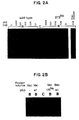
- Figure 2A Increasing quantities of wild-type and mutant 273 his p53, affinity-purified from a baculovirus expression system, were used to precipitate labelled C BE fragments.
- Figure 2B Lysates from a vaccinia virus system (Vac) producing the wild-type (wt), mutant (175 his ), or no p53 protein (-), were used to immunoprecipitate labelled C BE fragments.
- Vac vaccinia virus system
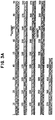
- FIG. 3A Fragment A and adjacent vector sequences in the 10d subclone of 772 C BE . This differs from the published 772 C BE sequence (Genbank M25718) in the number of CTT repeats (bp 173-229) and in the presence of A instead of C at bp 116.
- Figure 3B Fragment B and adjacent vector sequences in the 8a subclone of Lambda 5R. A related sequence on file (Genbank X05913) varied somewhat from the Lambda 5R subclone studied.
- Figure 4 Binding of various subfragments of fragments A and B to p53 from vaccinia-infected cell lysates.
- Fragment 5mut1 had a G to T transversion at bp 120; 5mut2 had G to T transversions at bp 120 to 122.
- Figure 4B The fragment A (panels 1-4) and fragment B (panel 5) subfragments illustrated in Figure 4A are labelled to the left of the bands.
- the "v" band in panel 4 corresponds to the 2.9 kb vector into which subfragments 6 was cloned.
- Subfragment 8 (panel 5) contained bp 104-238 of fragment B (see Fig. 3B).
- Control lanes (C) contained 2% of the labelled fragments used in the binding assays (B).
- Figure 5A Methylation interference assay.
- B bound DNA recovered from the immunoprecipitate.
- C equivalent amount of control DNA fragment, not subject to binding reaction. Bound and control DNA samples were cleaved at methylated guanines and equal amount separated by electrophoresis on a 6% denaturing gel. Dots represent methylation-sensitive sites (open for partial, solid for strong interference); some variation in band intensities occurred between assays, and only the reproducible changes are marked.
- Figure 5B Methylation interference assay.
- Binding of "mutant" subfragments of fragment A (5mut1 and 5mut2) to purified baculovirus-produced p53 is compared to that of the normal subfragment 5 sequence.
- 5mut1 contains a T instead of G at bp 120
- 5mut2 contains Ts in place of Gs at bp 120, 121, and 122.
- Figure 6A shows RNase protection analysis of transfected clonal lines.
- a labeled antisense p53 probe was hybridized with total cellular RNA, and digested with RNase A. Endogenous RNA included all sequences represented in the labeled probe. Exogenous p53 RNA produced from the expression vectors extended only about 2/3 of the length of the probe.
- Figure 6B shows Southern blot analysis of transfected clonal lines.
- the exogenous p53 gene was present on a 1.8 kb BamHI fragment.
- the endogenous p53 gene gave rise to a 7.8 kb BamHI fragment.
- Other sized fragments presumably arose by rearrangements.
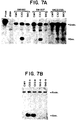
- Figure 7A shows expression analysis of pooled clones; the analysis was as described in Figure 6A.
- Figure 7B shows a Southern blot analysis of SW480 pooled clones.
- Figure 8 Isolation of human genomic sequences which bound to p53.
- Figure 8A Experimental strategy used for isolation and analysis of human genomic DNA fragments which bound to p53.
- FIG. 8B Immunoprecipitation (IP) assays of cloned fragments.
- Clones of amplified and selected (AS) DNA were tested for the presence of p53-binding fragments by IP.
- the bound DNA is shown in the B lane, adjacent to a control (c) lane containing 2% of the total end-labeled DNA used in the binding assay.
- eight binding fragments were identified, representing six unique genomic fragments.
- the inserts from the clones in lanes labeled, 2, 3, 5, 9, 10, and 11 contained p53-binding fragment, while the other lanes contained none.
- the clones in lanes 2 and 5 each contained two binding fragments.
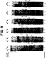
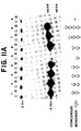
- Figure 9 Mapping of p53-binding sites by DNase I protection (DP) and methylation interference (MI). For each footprint, the first and fourth lanes contain control samples of the total labeled DNA, whereas the middle two lanes contain an equivalent amount of p53-bound DNA. DNA sequences corresponding to the p53-binding regions on strand 1 are shown in Fig. 10.
- Figure 10 Definition of a consensus binding site for p53.
- the p53 binding site of 18 cloned human genomic DNA fragments, determined by footprinting methods are displayed along the central axis of symmetry which separates the two 10 bp consensus monomers. Nucleotides in capital letters represent identity of a genomic sequence to the consensus, whereas lower case letters identify disparity with the consensus. Sequences surrounding the consensus or separating the two 10 bp monomers are also shown in lower case.
- the ten synthetic oligonucleotides investigated for the ability to be bound by p53 are shown at the bottom. Oligonucleotides No. 6 to 10 were tested after cloning into plasmid vectors. Lower case letters represent vector-derived sequences. Combined nucleotide usage (%) within the two monomers of the consensus binding site is shown in the middle.
- FIG 11A The 10 bp consensus monomer was insufficient for binding, whereas dimers in various orientations or multimers of the 10 bp consensus bound strongly to p53.
- the control lane (C) contained 2% of the total DNA used in the binding reaction, and was composed of two fragments: 2.9 kb vector DNA fragment, and a fragment of 40 to 80 bp containing no insert (lane 9; XhoI plus Pst1 digest of pBluescript II SK+), the 10 bp consensus monomer sequence 5′-AGGCATGTCT-3′(lane 5), or multimers of this sequence arranged as indicated (lanes 1 to 4, 6, to 8). Bound DNA from the IP is shown in the B lanes.
- FIG 11B Comparison of the ability of wild-type and mutant p53 to bind to the consensus dimer.
- In vitro translated p53 proteins were tested for the ability to bind the consensus dimer by IP. Two percent of the total DNA used for binding is shown in lane 1.
- Lane 7 shows binding to baculovirus-produced human wild-type p53 protein.
- Lanes 2 to 6 show binding of in vitro translated wild-type and mutant p53 proteins.
- the mutant p53 proteins contained changes at codon 143 (val to ala), 175 (arg to his), 248 (arg to trp), and 273 (arg to his).
- Figure 12A Reporter constructs in pBluescript II SK+ vector (Stratagene).
- PG n concatemers of n copies of the p53 binding sequence PG.
- MG n concatemers of the mutated sequence, which does not bind p53.
- CAT the chloramphenicol acetyltransferase coding sequence.
- LacZ the ⁇ -galactosidase coding sequence.
- Py the early gene promoter from polyomavirus.
- CYC the yeast cytochrome c gene promoter.
- FIG. 12B Expression vectors.
- CMV the cytomegalovirus promoter from the parent vector pCMVneoBam.
- Gal the galactose-inducible promoter from yeast.
- Figure 13A Relative DNA-binding abilities of various length concatemers of a p53-binding sequence (PG n series), using an immunoprecipitation assay. Clones were cleaved by restriction endonucleases to extricate the concatemers, end-labelled, incubated with purified baculovirus-produced wild-type human p53, immunoprecipitated with anti-p53 and protein A-Sepharose, and bound fragments recovered and separated on a nondenaturing polyacrylamide gel. C, control lane, containing 2% of the labeled DNA used in the binding reactions. B, bound DNA recovered from the binding reactions.
- PG n series p53-binding sequence
- FIG. 13B Transactivation efficiencies of reporters containing the various PG n concatemers compared by chloramphenicol acetyltransferase (CAT) assay.
- CAT chloramphenicol acetyltransferase
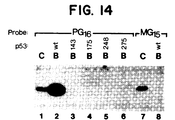
- FIG. 14 Comparison of the ability of wild-type and mutant p53 to bind to a concatemerized binding sequence (PG16), and lack of DNA-binding ability to MG15 are shown.
- Each p53 form (as a rabbit reticulocyte preparation (Promega) made essentially as described in Kern, et al., Oncogene , 6 :131-136 (1991); equalized for p53 quantity by immunoblot analysis, data not shown) was used to immunoprecipitate end-labeled DNA.
- C control lanes, containing 2% of the labeled DNA used in the binding reaction.
- B bound DNA recovered from the immunoprecipitate.
- FIG. 15A Efficiencies of reporters with PG13 separated by varying distances from the polyomavirus promoter. Various lengths of the non-binding sequence (MG n series) were used to provide inert spacers of defined length (see Fig. 8A). CAT assays are shown.
- FIG. 15B Transactivation efficiency of a reporter with PG16 downstream of CAT (CAT-PG16). 1.7 ⁇ g each of expression and reporter construct were transfected. The MG15-CAT served as a negative control. CAT assays are shown.
- FIG. 16A Representative CAT assays (Exp. 2 from Table 1).
- FIG 16B Western blot analysis of p53 expression in lysates of transfected cells, showing levels of expression at least as high as that of wild-type p53 were obtained with the mutant p53 clones.
- the level of expression of p53 (lane 3) was slightly lower than with the other constructs, for unknown reasons.
- the level produced by the p53-143 vector (lane 7) was at least as high as that for the p53-wt vector, but no transactivation was observed (Table I).
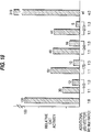
- FIG. 17 Effect of co-expressed 175 his mutant p53 on transactivation by wild-type p53 is shown.
- 1.7 ⁇ g of PG13-CAT reporter was used with the stated amount of p53 expression constructs.
- the balance of transfected plasmid was pCMVneoBam (for expressor) and pBluescript II SK+ (for reporter) to a total of 5.1 ⁇ g.
- CAT assays are shown.
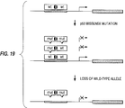
- FIG. 19 A biochemical model for the effect of p53 mutations is depicted.
- Cellular p53 exists as oligomers. Oligomeric p53 binds its recognition sequence of DNA (shaded thick line) and activates the transcription of adjacent genes (filled box). The depiction of p53 dimers is for illustrative purposes only: tetramers and other forms likely occur.
- Hetero-oligomerization with mutant proteins inactivates the transactivation function of the participating wild-type p53 molecules, producing the dominant-negative effect of missense p53 mutations. The remaining activity provided by the residual wild-type homo-oligomers is lost upon deletion of the remaining wild-type allele, a frequent event in the progression of human neoplasms.
- wild-type p53 protein binds specific fragment of human chromosomal DNA.
- Each of the fragment contains more than one monomer of the double-stranded motif 5′-RRRCWWGYYY-3′ separated by 0 to 13 bp.
- Some of these sequences are found near origins of replication of certain animal viruses and animal cells. See Jelinek et al, Proc. Natl. Acad. Sci. USA, vol. 77, pp. 1398-1402 (1980).
- Four mutant forms of p53 protein which are commonly found in human tumors do not have the ability to bind to these sequences.
- a function of p53 may be mediated by its ability to bind to specific DNA sequences in the human genome.
- Wild-type p53 protein binds specifically to certain DNA sequences.
- Fragment A contains sequences near a putative replication origin of the ribosomal gene cluster, while fragment B contains sequences that may allow adjacent sequences to replicate as extrachromosomal circles in HeLa cells (Sylvester, et al., Gene 84 , 193, 1989).
- the TGCCT repeat present in the DNA binding region of both of these fragments has been observed in other potential replication origins.
- p53 may be involved in the regulation of initiation of DNA synthesis by virtue of its binding.
- the consensus dimers contain a striking symmetry, with four 5′-RRRCW-3′units oriented in alternating directions.
- a synthetic monomer containing the 10 bp consensus sequence is insufficient for binding, while the combination of two or more monomers bind strongly to wt p53, but negligibly to p53 mutants. Thus, more than one monomer appears to be required for binding.
- the spacing between monomers may be from 0 to 40 nucleotides, although all natural binding sites isolated have spacings of less than 15 nucleotides.
- the symmetry of the four half-sites within the consensus dimers suggests that p53 interacts with DNA as a tetrameric protein.
- the eighteen unique clones shown in Figure 10 allow the identification of adjacent genes which may be regulated by p53 and may mediate its growth-suppressive action.
- a sequence which conforms to the consensus sequence need not have the exact sequence of the dimer.
- p53-binding sequences can have nucleotides which are different than those designated. There can also be extra nucleotides inserted within the dimer. Nucleotides can also be missing from the dimer. Typically p53-binding sequences will vary from the consensus sequence at no more than 5 nucleotide positions.
- wild-type p53 can activate the expression of genes adjacent to a specific binding site. Moreover, the level of in vivo transactivation is proportional to the in vitro strength of DNA-binding. Mutant p53 encoded by oncogenic p53 genes (i.e., those found in cancer cells of human patients), completely lose the ability to transactivate. In addition the mutant p53 proteins exert a dominant-negative effect, dramatically reducing the transactivating activity of wild-type p53.
- cell lysates are tested for the presence or absence of wild-type p53 by virtue of its specific DNA binding ability.
- testing for the presence or absence of wild-type p53 protein can provide diagnostic and prognostic information regarding a tumor and the patient.
- the cells to be tested are typically isolated from a tissue suspected of being neoplastic. Preferably the tissues are carefully prepared and isolated so that non-neoplastic tissues are not mixed with the neoplastic tissues, which can confound the analysis.
- Means for separating neoplastic tissues from non-neoplastic tissues are known in the art and include dissection of paraffin or cryostat sections, as well as use of flow cytometry.
- a cell lysate can be prepared from the tumor tissue according to any method known in the art. The cell lysate is then incubated with DNA fragments which are known to bind the wild-type p53 protein, under conditions which are conducive to such DNA/protein interactions. Alternatively, a histological sample can be analyzed by incubation with DNA fragments, as described for cell lysates.
- p53 also binds non-specifically to DNA.
- Specific binding can be distinguished from non-specific binding by any means known in the art. For example, specific binding interactions are stronger than non-specific binding interactions.
- the incubation mixture can be subjected to any agent or condition which destabilizes protein/DNA interactions such that the specific binding reaction is the predominant one detected.
- a non-specific competitor such as dI-dC, can be added to the incubation mixture. If the DNA containing the specific binding sites is labelled and the competitor is unlabeled, then the specific binding reactions will be the ones predominantly detected upon measuring labelled DNA.
- the DNA which is bound to p53 is separated from unbound DNA by immunoprecipitation with antibodies which are specific for p53.
- Use of two different monoclonal anti-p53 antibodies may result in more complete immunoprecipitation than either one alone. Unbound DNA remains in suspension.
- the amount of DNA which is in the immunoprecipitate can be quantitated by any means known in the art.
- the DNA fragment is labelled with a detectable moiety, such as a radioactive moiety, a colorimetric moiety or a fluorescent moiety. Techniques for so labelling DNA are well known in the art.
- p53 which binds to the specific DNA sequence of the present invention can be detected by gel shift assays (Tan, Cell , 62 :367, 1990), nuclease protection assays (see Example 9, infra ), or methylase interference assays (see Example 9, infra ).
- p53 after incubation of p53 with specific binding DNA fragments all components of the cell lysate which do not bind to the DNA fragments are removed. This can be accomplished, among other ways, by employing DNA fragment which are attached to an insoluble polymeric support such as agarose, cellulose and the like. After binding, all non-binding components can be washed away, leaving p53 bound to the DNA/solid support.
- the p53 can be quantitated by any means known in the art. It can be determined using an immunological assay, such as an ELISA, RIA or Western blotting.
- the diagnostic assay of the present invention has applicability not only with regard to cancers which are known to involve mutation of p53, but also with regard to human viruses such as human papilloma virus (HPV).
- HPV protein E6 binds tightly to wild-type but not mutant p53. See Werness et al., Science, 248 , 76-69 (1990). This tight binding is likely to block the interaction of p53 with it specific DNA binding sequences.
- infected cells By testing cells or cell extracts suspected of being infected with potentially tumor-inducing or hyperplastia-inducing strains of HPV or possibly other viruses, infected cells can be identified, because the E6 protein of the infected cells will have sequestered the wild-type p53, rendering it unable to bind to its specific DNA binding sequences.
- assays may be performed on cell extracts or on histological specimens.
- a method is also provided of supplying wild-type p53 function to a cell which carries mutant p53 alleles.
- the wild-type p53 gene or a part of the gene may be introduced into the cell in a vector such that the gene remains extrachromosomal. In such a situation the gene will be expressed by the cell from the extrachromosomal location. If the mutant p53 genes present in the cell are expressed, then the wild-type p53 gene or gene portion should be expressed to a higher level than that of the mutant gene. This is because the mutant forms of the protein are thought to oligomerize with wild-type forms of the protein. (Eliyahu et al., Oncogene, vol. 3, p.
- a gene portion is introduced and expressed in a cell carrying a mutant p53 allele, the gene portion should encode a part of the p53 protein which is required for non-neoplastic growth of the cell. More preferred is the situation where the wild-type p53 gene or a part of it is introduced into the mutant cell in such a way that it recombines with the endogenous mutant p53 gene present in the cell. Such recombination would require a double recombination event which would result in the correction of the p53 gene mutation.
- Vectors for introduction of genes both for recombination and for extrachromosomal maintenance are known in the art and any suitable vector may be used.
- Polypeptides or other molecules which have p53 activity may be supplied to cells which carry mutant p53 alleles.
- the active molecules can be introduced into the cell by microinjection or by liposomes, for example. Alternatively, some such active molecules may be taken up by the cells, actively or by diffusion. Supply of such active molecules will effect a less aggressive state of neoplasia.
- compounds which have p53 activity are those which specifically complex with a p53-specific DNA binding site.
- Wild-type p53 is one such compound, but portions of p53 which retain the ability to bind to p53-specific binding sites may also be used.
- Oligonucleotides and oligonucleotide containing nucleotide analogs are also contemplated among those compounds which are able to complex with a p53-specific DNA binding site. Although applicants do not wish to be bound by any particular theory, it is believed that oligonucleotides bind double-stranded DNA to form triplexes.
- triplexes have been shown to block transcription of certain genes, as well as protect the DNA binding sites from the action of enzymes such as DNA methylases.
- oligonucleotides were thought to require only or predominantly pyrimidines (cytosine and thymine), purines have also successfully been incorporated into triplex forming oligonucleotides.
- Particular oligonucleotides which may be used include: nucleotides 140-162 of SEQ ID NO:2, nucleotides 128-158 of SEQ ID NO: 1, nucleotides 114-123 of SEQ IQ NO: 1, or portions thereof having at least ten nucleotides.
- Oligonucleotides containing nucleotide analogs or otherwise modified oligonucleotides may also be useful in the formation of complexes with double-stranded DNA.
- certain modifications to the 3′-terminus can be employed to reduce the susceptibility of the oligonucleotide to nuclease degradation.
- Moieties which may be appended to the 3′- or 5′-termini include a substituted or unsubstituted amino group, polyethylene glycol, polylysine, acridine, dodecanol, and cholesterol.
- Oligonucleotides and oligonucleotides containing nucleotide analogs which may be used include methylphosphonates, aminomethylphosphonates, phosphorothioates, phosphorodithioates, substituted or unsubstituted phosphoramidates, oligoribonucleotides, oligodeoxyribonucleotides, alpha-oligonucleotides and mixtures thereof.
- Other modifications to oligonucleotides may be desirable to increase the uptake by cells or nuclei of the oligonucleotides or to decrease nuclease sensitivity. All such modifications are within the contemplation of the invention.
- Switchback linkers may also be incorporated into the midst of an oligonucleotide or analog. Such linkers are taught by Riordan and Martin (Nature, 350 , 452, 1991). They are designed by molecular modeling to provide the proper spacing between portions of an oligonucleotide which are to interact with different strands of a double-stranded DNA molecule. Examples of oligonucleotides having such linkers include the following: TTGCCTTGCCT-switchback linker-CCT-switchback linker-CTTGCCT (corresponding to nucleotides 105-125 of the double-stranded sequence represented by SEQ ID NO:1) or portions thereof.
- Single-tranded, linear or circular oligonucleotides containing nucleotide analogs which are able to complex specifically with all or part of a p53-specific binding site as described above are also contemplated as part of the invention.
- Such oligonucleotides containing nucleotide analogs should comprise at least about ten nucleotides in length in order to have the requisite specificity with respect to the entire human genome.
- Oligonucleotides or oligonucleotides containing nucleotide analogs which are able to complex specifically with part of a p53 binding site as well as with adjacent sequences are also contemplated as part of the invention.
- the oligonucleotides or oligonucleotides containing nucleotide analogs will preferably bind to the identified p53 binding regions.
- other binding regions in the human genome may well be found which are also suitable target for the oligonucleotides or analogs of the present invention. These other binding sites may well be the primary targets of p53; complexation of these sites may inhibit the unregulated growth which characterizes neoplastic cells.
- Double-stranded DNA fragments which comprise a p53-specific DNA binding site and are attached to an insoluble polymeric support are also contemplated by this invention.
- the support may be agarose, cellulose, polycarbonate, polystyrene and the like.
- Such supported fragments may be used in screens to identify compounds which bind to p53-specific DNA binding sites.
- Such supported fragments may be used to perform diagnostic tests on cell lysates from suspected tumor tissues. They may also be used in assays used to screen potential chemotherapeutic agents, as discussed infra .
- test compound is incubated with a supported DNA fragment, as described above.
- the amount of test compound which binds to the supported DNA fragment is determined. This determination can be performed according to any means which is convenient. For example, the amount of a compound which can be removed after incubation with the supported fragment can be compared to the amount originally applied. Alternatively, the test compound can be labelled and the amount which binds to the supported fragment can be assayed directly.
- soluble DNA fragments which do not contain the p53 DNA binding sequence can be added to the incubation mixture. The soluble fragment would not have the ability to specifically bind to p53 wild-type protein.
- test compounds are incubated with supported DNA fragments as described above.
- wild-type p53 protein is also added to the incubation mixture.
- the amount of p53 protein which binds to the DNA fragment is measured using methods as described above.
- the amount of p53 protein bound is compared to the amount which binds in the absence of the test compound. Any diminution of p53 binding which result from the presence of the test compound is presumptively due to the competition of the test compound with p53 for the specific DNA binding sites of the supported fragments. Direct binding of the test compound to the binding site fragments can be confirmed using the assay described above.
- the presence or absence of p53 genes or gene products can also be detected in body samples, such as, serum, stool, or other body fluids, such as urine and sputum.
- body samples such as, serum, stool, or other body fluids, such as urine and sputum.
- the same techniques discussed above for detection of the presence or absence of gene products in tissues can be applied to other body samples. By screening such body samples, a simple early diagnosis can be achieved for many types of cancers. In addition, the progress of chemotherapy or radiotherapy can be monitored more easily by testing such body samples for the presence or absence p53 genes or gene products.
- the methods of the present invention for diagnosis and therapy of neoplastic tissue is applicable across a broad range of tumors. These include lung, breast, brain, colorectal, bladder, mesenchyme, prostate, liver as well as stomach tumors. In addition the method may be used in leukemias and osteosarcomas. It thus appears that the p53 gene has a role in the development of a broad range of tumors.
- the methods of diagnosis and therapy of the present invention are applicable to any tumor in which p53 has a role in tumorigenesis.
- the diagnostic method of the present invention is useful for clinicians so that they can decide upon an appropriate course of treatment. For example, a tumor displaying loss of both p53 alleles suggests a more aggressive therapeutic regimen than a tumor displaying loss of only one p53 allele.
- Compounds which have p53-specific DNA-binding activity can also be administered to humans and animals as a pharmaceutical and therapeutic composition. Effective amounts will be administered to cause neoplastic cells to become less aggressively neoplastic or even to stop the growth of the neoplastic cells entirely. Generally, such amounts will be in the range of 10 ng to 10 ⁇ g per dose per person or other animal.
- the therapeutic compounds can be prepared in any conventional pharmaceutical excipient, such as physiological saline or other physiologically compatible aqueous buffer. Typically, the compounds will be administered by injection, either intravenous or intramuscular. However, other administration methods as are known in the art and may be used to administer the compounds of the present invention.
- screening methods can be devised to isolate chemical agents which may have use in cancer therapy. Specifically, agents can be screened for the ability to affect the structure of mutant p53 molecules so that their ability to bind and/or transactivate at specific-DNA-binding sites is restored.
- the necessary components for such a screening method are provided by this invention and include DNA molecules which contain more than one monomer of the sequence RRRCWWGYYY, and mutant p53 proteins which are found in tumors. 1 / 1 / Not all mutations in p53 destroy specific-DNA-binding ability. For example, mutations in phosphorylation sites of p53 have been made and tested; they retain binding activity. Such mutations have never been found in tumors. Mutations in p53 which are found in tumors are termed oncogenic herein.
- One such prescreening method is a binding assay in which the amount of binding of a p53 mutant protein to a DNA molecule which comprises the consensus binding site (or a conforming sequence) is measured. The amount of binding is also measured for a p53 mutant protein in the presence of a test substance. If the test substance increases the amount of p53 binding, then the test substance is a candidate for use in anti-tumor therapy. Further testing will be desirable before use in humans is attempted.
- Methods for measuring the amount of binding can be any which are known in the art. See, e.g., Tan, et al., Cell , 62 :367-377 (1990).
- One particular method employs immunoprecipitation. Briefly, purified p53 or a lysate of a cell expressing p53 is incubated with radiolabeled DNA and anti-p53 antibodies under conditions where proteins bind to DNA. Protein A-Sepharose and poly-dIdC-poly-dIdC are then added for an additional incubation. A pellet is formed and washed and the proteins are removed by digestion with a protease, and DNA is obtained by phenol extraction. The extracted DNA is then analyzed by electrophoresis and quantified. Quantitation of the DNA can be by autoradiography, for example. The amount of DNA immunoprecipitated is proportional to the amount of binding of the p53 protein to the DNA.
- the ability of a mutant p53 protein from a tumor to transactivate transcription in vitro is assessed with and without a test substance. If the test substance increases the amount of transcription activated by the p53, then the test substance is a candidate for use in anti-tumor therapy. Transcriptional activation is measured using a transcription construct which comprises a reporter gene encoding a convenient assayable enzyme activity, such as chloramphenicol acetyltransferase or ⁇ -galactosidase, and an upstream p53 consensus binding site (or a conforming sequence). The binding site must be upstream, although the distance from the start of transcription is not critical.
- a convenient assayable enzyme activity such as chloramphenicol acetyltransferase or ⁇ -galactosidase
- the binding site which is adjacent to the reporter gene, may be from 0 to 1 kb upstream.
- In vitro transcription assay systems are well known in the art. See, e.g., Lue, Science , 246 , 661-664 (1989).
- transcriptional activation can be measured in a cell containing a mutant p53 protein which has been transfected with a reporter gene construct comprising a gene encoding an assayable product, such as an enzyme or antigen, with a p53 consensus binding site (or conforming sequence) adjacent and upstream therefrom.
- the transfected cells are treated with a test substance. If the amount of transactivation caused by the mutant p53 is enhanced by the test substance, then the substance is a candidate for anti-tumor therapy presumably due to its interaction with the mutant protein.
- a particular transactivation assay transient expression assay
- Particular reporter constructs are shown in Figure 12A. Others may be used within the scope of the invention.
- the transfected cell contains wild-type p53 diminished activation would indicate that the test substance competes with p53 for binding sites.
- oligonucleotides can be isolated which restore to mutant p53 proteins the ability to bind to the consensus binding sequence or conforming sequences. Mutant p53 protein and random oligonucleotides are added to a solid support on which p53-specific-binding DNA fragments are immobilized. Oligonucleotides which bind to the solid support are recovered and analyzed. Those whose binding to the solid support is dependent on the presence of the mutant p53 protein are presumptively binding the support by binding to and restoring the conformation of the mutant protein.
- Transient expression constructs are conveniently made on plasmids and viral vectors, so that they can be propagated. These can also be used in vitro for transcription assays in the presence of RNA polymerase, ribonucleotides, and other cofactors.
- This example demonstrates the screening methods used to identify a p53 sequence-specific binding site.
- Binding reactions included 5 ⁇ l of vaccinia-infected cell lysate or purified baculovirus p53 preparations, 95 ⁇ l binding buffer (20 mM Tris pH 7.2, 100 mM NaCl, 10% glycerol, 1% NP-40, 5 mM EDTA), 6-20 x 104 dpm 32P-labelled DNA, 4 ⁇ l (0.4 ⁇ g) pAb421 and 4 ⁇ l (0.4 ⁇ g) pAb1801 anti-p53 purified monoclonal antibodies (Oncogene Science), at 4°C for 30 min.
- Recombinant vaccinia viruses expressing p53 from the 40K promoter were isolated and purified as described in J. Lyons et al., Infec. and Immun. 58 , 4089 (1990).
- Vaccinia-infected cell lysates were prepared as follows: the indicated cells (about 2.5 x 10⁷) were infected (MOI 2), harvested at 24 hrs and lysed in 2 ml lysis buffer (PBS containing 5mM EDTA, 0.5% NP-40, 0.5 mM PMSF, 10 ⁇ g/ml TPCK, 1 ug/ml aprotinin, 10 ⁇ g/ml trypsin inhibitor, 1 ⁇ g/ml leupeptin). Cell debris was pelleted at 16,000xg and the supernatant UV-irradiated (0.5 joules). The supernatant was frozen in aliquots at -80°C and found to be stable for at least 6 months.
- Baculovirus-produced p53 was purified as described in P.N. Friedman, et al., Proc. Natl. Acad. Sci. USA, 87 , 9275 (1990); while the presence of an accessory cofactor in the purified preparation cannot be excluded, SDS-polyacrylamide gel electrophoresis of the preparation revealed only a single polypeptide migrating at 53 kD upon silver-staining.
- the first class consisted of 400 clones containing inserts of 300 to 1000 base-pairs obtained randomly from the human genome.
- the second class consisted of cosmid and plasmid clones chosen because they contained sequences which might be important in normal growth control.
- Random human genomic clones were prepared from a partial MboI digest of human DNA; 300-1000 bp fragments were purified and ligated to EcoRI linkers, then cloned into the EcoRI site of pBluescript II (Stratagene). Clones selected because they might be important in normal growth included SP65hFosAva2, containing regulatory sequences from the fos gene, obtained from T Curran; 772 C BE (J.E. Sylvester, R.
- Each clone was digested with an appropriate restriction endonuclease, end-labelled with 32P, and incubated with p53 protein from a lysate of cells infected with a recombinant vaccinia virus expressing p53 protein. Labelled DNA fragments which bound to p53 were then recovered by immunoprecipitation with monoclonal antibodies against p53.
- a 259 basepair HinfI fragment (fragment A) of clone 772 C BE Panel 2, Fig.
- fragment B of clone Lambda 5R (Panel 3, Fig. 1A); these fragments bound to a far greater extent than any of the other labelled fragments of larger or smaller size present in the same assay mixes.
- the immunoprecipitation assay was performed on fragment A as described in Example 1.
- Cell lysates were produced by infection with vaccinia virus that did or did not contain an insert of wild-type p53 DNA. Either anti-p53 antibodies were used or normal mouse IgG was used.
- Lysates from cells infected with wild-type vaccinia virus were not able to specifically immunoprecipitate fragment A (Fig. 1B).
- the detection of the precipitation of fragment A was dependent on the presence of anti-p53 antibodies (Fig. 1B).
- the binding was evident in lysates prepared from either human HeLa cells or monkey BSC40 cells infected with vaccinia virus and expressing wild-type p53 (Fig. 1B).
- Affinity-purified baculovirus-produced wild-type p53 protein was substituted for the vaccinia-infected cell lysates in the immunoprecipitation assay and found to bind fragment A strongly (Fig. 2A). This suggested that the binding to fragment A was an intrinsic property of the p53 polypeptide and not dependent on other factors present in the vaccinia virus-infected cell lysates.
- the example demonstrates that p53 mutant proteins found in human tumors fail to bind to fragment A.
- This example defines the particular sequences within fragment A that allow it to bind to wild-type p53 protein.
- Fragment A was subcloned, and the 259 bp insert from the subclone (10d, SEQ ID: 1 and Fig. 3A) bound to p53 as expected.
- a strategy based on the polymerase chain reaction (PCR) and restriction endonuclease digestion was used to generate subfragments of this clone.
- PCR contained 350 ng of each of the appropriate primers and approximately 50 ng plasmid template in a 50 ⁇ l reaction, using 25 cycles and the PCR conditions specified in Baker SJ, et al., Cancer. Res., 50 :7717 (1990). The products were extracted with phenol and chloroform, ethanol-precipitated, and dissolved in 3mM Tris, 0.2 mM EDTA prior to binding.
- Subfragment 1 contained bp 1 to 425 of subclone 10d of fragment A (Fig.
- subfragments 1a, 1b, 1c, 1d, and 1e were generated by digestion of subfragment 1 with BamHI, MboI, HindIII, HindIII, and BamHI, respectively, from fragment 1.
- Subfragment 2 contained bp 283 to 425.
- Subfragment 3a was generated by digestion of subfragment 3 (bp 106 to 294) with Hae III.
- Subfragment 4a was produced from subfragment 4 (gp 1 to 141) by Hind III digestion.
- Subfragments 5a and 5b were products of the HaeIII digestion of subfragment 5 (bp 87 to 141).
- “Mutant” subfragments 5mut1 and 5 mut2 were produced using primers P3ml (5′-GAAAGAAAAGGCAAGGCCAGG A AAGT-3′) and P3mut2 (5′-GAAAGAAAAGGCAAGGCCA TTA AAGT-3′) and were identical to subfragment 5 except for the positions underlined in the primers.
- Subfragment 6 contained bp 106 to 138, and the insert was excised by restriction with HindIII and BamHI to generate 6a or with HindIII and EcoRI to generate 6b.
- Subfragment 3, including basepairs 106 to 294 (Fig. 4B, panel 2) bound well to p53 as did subfragment 4, containing basepairs 1 to 141 (Fig. 4B, panel 3).
- PCR products labelled at one end were generated using primers labelled with T4 polynucleotide kinase (US Biochemicals).
- the product of each PCR reaction was purified by polyacrylamide gel electrophoresis, eluted from a crushed gel slice in 500 mM ammonium acetate, extracted with phenol and chloroform, and precipitated with ethanol. 2 x 10⁶ dpm of DNA was methylated at guanine residues using dimethylsulfate as described (T. Maniatis, E.F. Fritsch, J.
- Partial interference was also produced by methylation at nearby G sites (bp 110-112, 114, and 115).
- the methylation interference assay pinpointed one of the central TGCCT repeats (centered at basepair 121) and adjacent basepairs as critical for binding.
- This example defines the region of fragment B which is important for p53 binding.
- Fragment B was subcloned (sequence in Fig. 3B and SEQ ID NO:2). Interestingly, this fragment had two repeats of the TGCCT motif (centered at bp 135 and 152). A PCR strategy similar to that used for fragment A was used to demonstrate that a 95 basepair subfragment (bp 104 to 198), which contained both of these repeats, was sufficient for binding (Fig. 4B, panel 5).
- the colorectal carcinoma lines SW480 and SW837 which are representative of 75% of colon carcinomas, have each lost one copy of chromosome 17p (including the p53 gene) and the remaining p53 allele is mutated (Baker, et al., Science 244, 217 (1989); Nigro et al, Nature 342, 705 (1989)).
- the SW837 line contains an arginine to tryptophan mutation at codon 248 (Nigro, supra ).
- the SW480 line contains two point mutations, arginine to histidine at codon 273 and proline to serine at codon 309 (Nigro, supra .).
- the substitutions at codon 248 and 273 are typical of those observed in human tumors, occurring within two of the four mutation "hot spots" (Nigro, supra ).
- the expression vector pCMV-Neo-Bam was derived from plasmid BCMGNeo-mIL2 (Karasuyama, et al., J. Exp. Med. 169, 13 (1989) by excision of the human beta globin sequences and bovine papilloma virus sequences with Bam HI and Not I.
- BCMGNeo-mIL2 plasmid BCMGNeo-mIL2
- bovine papilloma virus sequences with Bam HI and Not I.
- the interleukin 2 (IL-2) sequences present at the unique Xho I site were removed, and the Xho I site was changed to a Bam HI site by linker addition.
- the vector included CMV promoter/enhancer sequences, which could drive expression of the insert at the Bam HI site, and splicing and polyadenylation sites derived from the rabbit beta globin gene, which ensured proper processing of the transcribed insert in the cells.
- a pBR322 origin of replication and ⁇ -lactamase gene facilitated growth of the plasmid in E. coli .
- the plasmid conferred geneticin resistance through expression of the neomycin resistance gene under separate control of an HSV thymidine kinase promoter.
- the first transcription unit comprised a cytomegalovirus (CMV) promoter/enhancer upstream of a site for insertion of the cDNA sequences to be expressed, and splice and polyadenylation sites to ensure appropriate processing.
- the second transcription unit included a herpes simplex virus (HSV) thymidine kinase promoter/enhancer upstream of the neomycin resistance gene, allowing for selection of transfected cells in geneticin.
- HSV herpes simplex virus
- a wild-type p53 cDNA was inserted into pCMV-Neo-Bam to produce pC53-SN3.
- the only difference between pC53-SN3 and pC53-SCX3 was a single nucleotide (C to T) resulting in a substitution of alanine for valine at p53 codon 143 in pC53-SCX3.
- the two constructs were made as follows: a 1.8-kb Xba I fragment, extending from nucleotide -130 to 1671 relative to the translation initiation site, was isolated from wild-type or CX3 cDNA clones. The fragment was blunt-ended with the Klenow fragment of DNA polymerase, ligated to Bam HI linkers, and cloned into the unique Bam HI site in the expression vector pCMV-Neo-Bam.
- the constructs were transfected into SW837 and SW480 cells (obtained from the American Type Culture Collection, Rockville, Maryland), and geneticin-resistant colonies were counted 3 weeks later.
- Cells transfected with pC53-SN3 formed five-to tenfold fewer colonies than those transfected with pC53-SCX3 in both recipient cell types (Table 1).
- the number of colonies produced by the expression vector pCMV-Neo-Bam was similar to that induced by the pC53-SCX3 construct.
- RKO cells were obtained through the generosity of M. Brattain. Although RKO cells did not contain a mutation within the susceptible p53 coding sequences, i.e., exons 5-9, they expressed low concentrations of p53 mRNA compared to normal colorectal mucosa or the other lines studied and did not produce detectable amount of protein.
- Results of colony formation assays in transfected RKO cells were similar to those in SW480 and SW837 cells. Colony formation by wild-type p53 gene transfectants occurred with a tenfold decrease in efficiency compared to the mutant p53 construct (Table 1). Immunocytochemical detection of p53 protein in transfected RKO cells was done as follows: approximately 5 x 10⁴ cells were cytocentrifuged onto polylysine-coated slides, fixed for 10 min in formalin, and permeabilized for 5 min in 0.5% Triton X-100.
- transient mRNA expression was also significantly lower in the SN3 transfectants compared to the SCX3 transfectants at 48 and 96 hours, supporting the idea that RKO cells expressing wild-type p53 were at a selective disadvantage compared to those producing mutant p53 products.
- the p53 alleles of the VACO 235 cell line were sequenced (exons 5-9) and found to be wild-type and the expression of p53 mRNA was found to be similar to that of normal colorectal mucosa.
- the pC53-SN3 and pC53-SCX3 constructs produced similar numbers of geneticin-resistant colonies after transfection of the VACO 235 line (Table 1).
- the most definitive test for differential growth inhibition by wild-type versus mutant p53 genes involved analysis of exogenous p53 expression in pooled transfectants.
- This sensitivity may only be relative: expression of the wild-type gene at high concentrations might inhibit the growth of any cell type, including non-neoplastic cells, by overwhelming normal regulatory processes such as phosphorylation.
- Genetic alterations that occur during the progression of colorectal tumors may increase the sensitivity of cells to p53 inhibition, making wild-type p53 expression a key, rate-limiting factor for further tumor growth and expansion.
- mutations in the p53 gene would confer a selective growth advantage to cells in vivo , which would explain the frequent occurrence of p53 gene mutations and allelic loss only in the more advanced stages of colorectal tumorigenesis.
- This example demonstrates the identification of human genomic fragments that can bond to wt p53 protein in vitro .
- DNA-binding buffer containing 100 mM NaCl, 20 mM Tris, pH 7.0, 10% glycerol, 1 % NP40, and 5 mM DTT.
- DNA fragments bound to p53 were complexed to antibodies by the addition of 8 ⁇ l containing 400 ng each of anti-p53 antibodies pAb421 and pAb1801, both obtained from Oncogene Science, and incubated for 30 minutes at 4°C.
- DNA-binding buffer containing 1.5 mg protein A precipitated following the addition of 26 ⁇ l of DNA-binding buffer containing 1.5 mg protein A Sepharose and 10 ⁇ g of poly dI-dC and mixing at 4°C for 30 minutes. After removal of the supernatant, the immunoprecipitate was washed twice with 1 ml of DNA-binding buffer. Bound DNA was purified by treatment with SDS and proteinase K at 48°C for 30 minutes, extracted with phenol and chloroform, precipitated with ethanol, separated by electrophoresis on a 10% nondenaturing polyacrylamide gel, and autoradiographed.
- This example demonstrates the localization of p53 contacts with bound DNA fragments.
- DP labelled DNA fragments were first subject to IP, then incubated with various amounts of DNase I.
- p53 binding provided protection against DNase I cleavage at residues 187 to 211 (Fig. 9, footprint 9). MI showed interference by G residues only within the region protected by DNase I (Fig. 9, footprint 10).
- Other examples of DP and MI mapping are shown in Figure 9. p53-binding DNA fragments were subcloned and labeled on one end, gel-purified and subjected to DP or MI mapping.
- the samples were incubated with 100 ⁇ l of 1 M piperidine at 90°C for 30 minutes. The samples were then dried under vacuum and separated electrophoretically on a 6% polyacrylamide sequencing gel. The control DNA samples were carried through all incubations except no p53 was added. For these control samples, the protein A Sepharose pellets were treated with SDS and proteinase K without removal of the supernatants (which contained the labeled DNA in the absence of p53).
- DP assays For DP assays, end-labeled DNA fragments were immunoprecipitated as described in the legend to Figure 8. The protein A Sepharose pellet were incubated for two minutes at 25°C with 200 ng DNase I in 5 mM MgCl2. After purification of the DNA, as described above, samples were separated by electrophoresis on sequencing gels and loaded as described above for MI. MI was performed on all 18 genomic DNA fragments which bound to p53. DP assays were performed on 13 fragments and the regions of protection uniformly coincided with those indicated by the MI assays.
- This example analyzes the sequences of p53-bound DNA regions.
- the average insert sequence was 307 bp (range 139-470).
- ten of the twenty-three clones were not unique, showing at least one hundred contiguous nucleotides identical to one other clone.
- the twenty-three clones represented only eighteen independent genomic DNA fragment.
- a striking and consistent feature of the clones became apparent (Fig. 10).
- Each of the binding sites contained two copies of the 10 bp motif 5′-RRRCWWGYYY-3′, separated by 0 to 13 bp.
- One clone (S592) contained two separate areas of footprinting, and both regions contained a dimer of the 10 bp motif (Fig. 10). In all clones, the regions displaying DP and MI were always centered within the dimers, and G residues within the 10 bp motif strongly interfered with binding to p53 (examples in Fig. 9).
- the 10 bp consensus monomer contained an internal symmetry, with two oppositely oriented half-sites of the form 5′-RRRCW-3′.
- a synthetic oligonucleotide (5′-AGGCATGTCT-3′) containing the consensus sequence was studied. Oligonucleotide duplexes were tested either directly or after cloning into plasmid vectors. The monomer was found not to bind to p53, either alone (not shown) or flanked by 43 nucleotides of plasmid sequences (Fig. 11A, lane 5). In contrast, the dimers (composed of two copies of the monomer arranged in head-to-head, tail-to-tail, or head-to-tail orientation), each bound strongly to p53 protein (Fig. 11A, lanes 1 to 4, 6).
- This example demonstrates that intact p53 can activate expression in human cells.
- reporter plasmids containing part of the polyomavirus early promoter and the CAT gene located downstream of DNA sequences which could bind to p53 in vitro ( Figure 8).
- CAT reporters concatemers of the p53-binding region of C BE were formed by ligation of complementary oligonucleotides, ligated into the EcoRV site of pBluescript II SK+ (Stratagene) to form the PG n and MG n series.
- the BglII-BamHI fragment of pPyOICAT (Murakami, et al.
- Oncogene , 5 :5) containing the polyomavirus early promoter and the CAT gene coding region, was ligated into the BamHI site of the PG n and MG n series clones to form the PG n -CAT and MG n -CAT series, and the orientation of the inserts characterized by restriction enzyme analysis.
- PG and MG sequences were ligated as SalI-SmaI fragments to the SalI and filled-in XhoI sites of pCZ (Buchanan, et al. (1988), Mol. Cell Biol. , 8 :50806).
- the construction of the p53-wt expression construct has been described (Baker, et al. (1990), Science , 249 :912); the mutant expression plasmids were constructed similarly from the previously described cDNA plasmids (Nigro, et al. (1989), Nature , 342 :705, and Kern, et al. (1991), Oncogene , 6 :131), or in the case of the engineered phosphorylation site mutants, by in vitro mutagenesis (Altered Sites, Promega) with verification by sequencing.
- the construction of the yeast p53 expression vectors based on pRS314 has been described (Nigro, et al., Mol. Cell Biol. (in press)).
- p53 binding sequences we used a series of concatemers of the oligonucleotide PG (5′-CCTGCCTGGACTTGCCTGG-3′). This contained the binding region of plasmid C BE , previously shown to bind p53 in vitro .
- the reporter and an expression vector coding for the intact human wild-type protein (p53-wt) were transfected together into the human colorectal cancer cell line HCT 116. This line makes low amount of apparently wild-type p53 protein.
- Exons 5-8 of the p53 genes from HCT 116 cells were amplified by PCR and sequenced as described in Sidransky et al. (1991), Science , 252 :706. Previously, these exons had been shown to contain over 90% of the mutations observed in human tumors. No mutations were observed from HCT 116 cells. Small amount of apparently wild-type protein could be detected in Western blots of HCT 116 protein (See Fig. 16B).
- the intact wild-type p53 protein was indeed able to activate transcription (Fig. 13).
- the level of transactivation of the CAT gene depended on the strength of binding to p53 of the upstream sequences.
- the longer the number of PG repeats the greater the binding to p53 in vitro (Fig. 13A) and the higher the CAT expression in vivo (Fig. 13B, lanes 1-7).
- the level of transactivation also increased with the amount of p53 expression vector transfected (Table 3). Specificity for the binding sequences was tested by replacing six GC basepairs in the 18-basepair oligonucleotide to generate a mutant form of PG, termed MG (5′-CCTTAATGGACTTTAATGG-3′).
- CAT assay Cultures of HCT 116 cells at 50-80% confluence in 25 cm2 flasks were transfected using Lipofectin (BRL, Gaithersburg, MD) according to the manufacturer's instructions. All flasks within an experiment were transfected with the same total amount of plasmid using pCMVneoBam or pBluescript II SK+ as "filler". Cells were harvested at 20-24 hr. and the CAT activity of the lysates measured by acetylation of 14C-labeled chloramphenicol (ICN) as previously described (Gorman, et al. (1982), Mol. Cell Biol. , 2 :1044).
- ICN 14C-labeled chloramphenicol
- Bio-Rad protein assay was used to assure equivalence of lysate protein. Percent conversion to the acetylated form of chloramphenicol was calculated after quantitation by scintillation counting of excised chromatographic spot. Results reported are representative of at least two transfections done on separate days.
- This example demonstrates the dominant-negative effect of p53 mutations on wild-type p53's ability to bind DNA and enhance transcription.
- a three-fold ratio of mutant p53 to wild-type p53 produced an 84-95% reduction.
- the CAT expression increased rather than decreased, as expected (Fig. 18, "wt" lane).
- expression of the CAT reporter was dependent on the ratio of wild-type to mutant p53 in the cell.
- the dominant-negative effect could be caused by a failure of the mutant/wild-type complexes to bind to DNA or to a failure to activate transcription once bound.
- the mutant p53 protein inhibited the ability of wild-type protein to bind DNA by over 90% when co-expressed at a 3:1 ratio.
Landscapes
- Health & Medical Sciences (AREA)
- Life Sciences & Earth Sciences (AREA)
- Chemical & Material Sciences (AREA)
- Engineering & Computer Science (AREA)
- Immunology (AREA)
- Molecular Biology (AREA)
- Biomedical Technology (AREA)
- Urology & Nephrology (AREA)
- Organic Chemistry (AREA)
- Hematology (AREA)
- Analytical Chemistry (AREA)
- Biochemistry (AREA)
- General Health & Medical Sciences (AREA)
- Proteomics, Peptides & Aminoacids (AREA)
- Microbiology (AREA)
- Physics & Mathematics (AREA)
- Biotechnology (AREA)
- Pathology (AREA)
- Zoology (AREA)
- Medicinal Chemistry (AREA)
- Food Science & Technology (AREA)
- General Physics & Mathematics (AREA)
- Genetics & Genomics (AREA)
- Wood Science & Technology (AREA)
- Cell Biology (AREA)
- Bioinformatics & Cheminformatics (AREA)
- Biophysics (AREA)
- Toxicology (AREA)
- Tropical Medicine & Parasitology (AREA)
- General Engineering & Computer Science (AREA)
- Hospice & Palliative Care (AREA)
- Oncology (AREA)
- Gastroenterology & Hepatology (AREA)
- Measuring Or Testing Involving Enzymes Or Micro-Organisms (AREA)
- Saccharide Compounds (AREA)
- Medicines That Contain Protein Lipid Enzymes And Other Medicines (AREA)
- Micro-Organisms Or Cultivation Processes Thereof (AREA)
- Peptides Or Proteins (AREA)
- Preparation Of Compounds By Using Micro-Organisms (AREA)
- Pharmaceuticals Containing Other Organic And Inorganic Compounds (AREA)
Applications Claiming Priority (4)
| Application Number | Priority Date | Filing Date | Title |
|---|---|---|---|
| US71518291A | 1991-06-14 | 1991-06-14 | |
| US07/860,758 US5362623A (en) | 1991-06-14 | 1992-03-31 | Sequence specific DNA binding by p53 |
| US860758 | 1992-03-31 | ||
| US715182 | 1992-03-31 |
Publications (3)
| Publication Number | Publication Date |
|---|---|
| EP0518650A2 true EP0518650A2 (fr) | 1992-12-16 |
| EP0518650A3 EP0518650A3 (en) | 1993-01-20 |
| EP0518650B1 EP0518650B1 (fr) | 1997-01-08 |
Family
ID=27109289
Family Applications (1)
| Application Number | Title | Priority Date | Filing Date |
|---|---|---|---|
| EP92305333A Expired - Lifetime EP0518650B1 (fr) | 1991-06-14 | 1992-06-10 | Fixation de P53 sur l'ADN au moyen d'une séquence spécifique |
Country Status (10)
| Country | Link |
|---|---|
| US (4) | US5362623A (fr) |
| EP (1) | EP0518650B1 (fr) |
| JP (1) | JPH0678798A (fr) |
| AT (1) | ATE147512T1 (fr) |
| AU (1) | AU666479B2 (fr) |
| CA (1) | CA2070979C (fr) |
| DE (1) | DE69216478T2 (fr) |
| DK (1) | DK0518650T3 (fr) |
| ES (1) | ES2097878T3 (fr) |
| GR (1) | GR3022473T3 (fr) |
Cited By (22)
| Publication number | Priority date | Publication date | Assignee | Title |
|---|---|---|---|---|
| WO1996005308A1 (fr) * | 1994-08-12 | 1996-02-22 | Myriad Genetics, Inc. | Procede pour diagnostiquer une predisposition au cancer du sein et des ovaires |
| US5532220A (en) * | 1987-08-31 | 1996-07-02 | The Regents Of The University Of California | Genetic mechanisms of tumor suppression |
| WO1996025434A1 (fr) * | 1995-02-16 | 1996-08-22 | The Wistar Institute | PEPTIDES ET MOLECULES A COMPORTEMENT MIMETIQUE DES PEPTIDES AYANT UNE SIMILARITE STRUCTURELLE AVEC LA PROTEINE p53 HUMAINE ACTIVANT LA FONCTION p53 |
| US5573925A (en) * | 1994-11-28 | 1996-11-12 | The Wistar Institute Of Anatomy And Biology | P53 proteins with altered tetramerization domains |
| WO1997007244A1 (fr) * | 1995-08-11 | 1997-02-27 | The United States Of America, Represented By The | SEPARATION DE GENES AMPLIFIES, PAR HYBRIDATION SOUSTRACTIVE D'ADNc |
| WO1997011367A1 (fr) * | 1995-09-18 | 1997-03-27 | Novartis Ag | DOSAGE DESTINE A IDENTIFIER DES INHIBITEURS DE L'INTERACTION ENTRE DES PROTEINES p53 ET dm2 |
| EP0633268A3 (fr) * | 1993-05-14 | 1997-10-22 | Cancer Inst | Protéines MDC et ADNs les encodant. |
| US5693473A (en) * | 1994-08-12 | 1997-12-02 | Myriad Genetics, Inc. | Linked breast and ovarian cancer susceptibility gene |
| US5709999A (en) * | 1994-08-12 | 1998-01-20 | Myriad Genetics Inc. | Linked breast and ovarian cancer susceptibility gene |
| US5721340A (en) * | 1994-11-28 | 1998-02-24 | The Wistar Institute Of Anatomy & Biology | p53 proteins with altered tetramerization domains |
| US5753441A (en) * | 1994-08-12 | 1998-05-19 | Myriad Genetics, Inc. | 170-linked breast and ovarian cancer susceptibility gene |
| US5827658A (en) * | 1996-08-09 | 1998-10-27 | The United States Of America As Reprsented By The Department Of Health And Human Services | Isolation of amplified genes via cDNA subtractive hybridization |
| US5847083A (en) * | 1996-08-21 | 1998-12-08 | The Wistar Institute Of Anatomy And Biology | Modified p53 constructs which enhance DNA binding |
| WO1999011817A1 (fr) * | 1997-09-02 | 1999-03-11 | Mcgill University | Procede de depistage permettant de determiner les sujets qui presentent un risque de developper des maladies associees a differentes formes polymorphes de p53 de type sauvage |
| WO1999001763A3 (fr) * | 1997-07-02 | 1999-03-25 | Univ Texas | p53 COMME REGULATEUR DE DIFFERENCIATION CELLULAIRE |
| WO2000023583A3 (fr) * | 1998-10-16 | 2000-08-10 | Deutsches Krebsforsch | ZONES DE LIAISON DE p53 |
| WO2000022115A3 (fr) * | 1998-10-13 | 2000-09-21 | Univ Texas | Dosages permettant d'identifier des alterations fonctionnelles dans le gene suppresseur de tumeur p53 |
| US6162897A (en) * | 1994-08-12 | 2000-12-19 | Myriad Genetics, Inc. | 17q-linked breast and ovarian cancer susceptibility gene |
| WO2001000817A1 (fr) * | 1999-06-24 | 2001-01-04 | Dnab Diagnostics, Inc. | Compositions et methodes permettant de determiner l'activite de proteines se liant a l'adn et l'activite d'initiation de la transcription |
| EP0965636A4 (fr) * | 1996-03-26 | 2002-10-23 | Nippon Suisan Kaisha Ltd | Oligonucleotides utilises pour detecter vibrio parahaemolyticus et procede de detection |
| US6569833B1 (en) | 1995-09-21 | 2003-05-27 | Cyclacel Limited | Cyclin dependent kinase binding peptides |
| CN107656059A (zh) * | 2017-09-26 | 2018-02-02 | 中南大学 | 一种用于p53蛋白的荧光检测剂及其制备方法和应用 |
Families Citing this family (89)
| Publication number | Priority date | Publication date | Assignee | Title |
|---|---|---|---|---|
| NO312681B1 (no) * | 1990-08-24 | 2002-06-17 | Univ California | Fremgangsmåte for fremstilling av en farmasöytisk blanding med suppresiv virkning/aktivitet |
| US5747469A (en) * | 1991-03-06 | 1998-05-05 | Board Of Regents, The University Of Texas System | Methods and compositions comprising DNA damaging agents and p53 |
| AU663702B2 (en) | 1991-03-06 | 1995-10-19 | Board Of Regents, The University Of Texas System | Methods and compositions for the selective inhibition of gene expression |
| US6410010B1 (en) * | 1992-10-13 | 2002-06-25 | Board Of Regents, The University Of Texas System | Recombinant P53 adenovirus compositions |
| WO1993020235A1 (fr) * | 1992-04-01 | 1993-10-14 | The Johns Hopkins University School Of Medicine | Procedes de detection d'acides nucleiques de mammifere isoles a partir d'un echantillon de selles, et reactifs utilises a cet effet |
| US5670330A (en) * | 1992-09-29 | 1997-09-23 | Mcgill University | Anti-tumor agent assay using PKR |
| EP0675967A4 (fr) * | 1992-10-01 | 1999-04-28 | Gen Hospital Corp | Procede fonctionnel de determination de genes suppresseurs de tumeurs. |
| US5659024A (en) * | 1994-01-14 | 1997-08-19 | The Burnham Institute | Promotors that regulate the expression of genes involved in cell death |
| WO1997010843A1 (fr) * | 1995-09-22 | 1997-03-27 | The Wistar Institute Of Anatomy And Biology | Constructions de p53 modifiees et utilisations leur convenant |
| ES2264145T3 (es) * | 1995-11-30 | 2006-12-16 | The Board Of Regents, The University Of Texas System | Metodos y composiciones para el tratamiento de cancer. |
| US5670325A (en) * | 1996-08-14 | 1997-09-23 | Exact Laboratories, Inc. | Method for the detection of clonal populations of transformed cells in a genomically heterogeneous cellular sample |
| US5741650A (en) * | 1996-01-30 | 1998-04-21 | Exact Laboratories, Inc. | Methods for detecting colon cancer from stool samples |
| US6242212B1 (en) * | 1996-02-09 | 2001-06-05 | Thomas Jefferson University | Fragile histidine triad (FHIT) nucleic acids and methods of producing FHIT proteins |
| US5861249A (en) * | 1996-04-23 | 1999-01-19 | Cold Spring Harbor Laboratory | Assays and reagents for identifying modulators of cdc25-mediated mitotic activation |
| US5830751A (en) * | 1996-05-01 | 1998-11-03 | The Johns Hopkins University | Genetic assays and strains using human TP53 |
| US5876711A (en) * | 1996-05-31 | 1999-03-02 | Onyx Pharmaceuticals, Inc. | Methods and compositions for determining the tumor suppressor status of cells |
| US5858673A (en) * | 1996-06-24 | 1999-01-12 | Charlotte-Mecklenburg Hospital Authority | Method for detecting prostate cells |
| US7083983B2 (en) | 1996-07-05 | 2006-08-01 | Cancer Research Campaign Technology Limited | Inhibitors of the interaction between P53 and MDM2 |
| US5744310A (en) * | 1996-07-29 | 1998-04-28 | The Burnham Institute | Bax promoter sequence and screening assays for indentifying agents that regulate bax gene expression |
| US5952178A (en) | 1996-08-14 | 1999-09-14 | Exact Laboratories | Methods for disease diagnosis from stool samples |
| US6020137A (en) * | 1996-08-14 | 2000-02-01 | Exact Laboratories, Inc. | Methods for the detection of loss of heterozygosity |
| US6300077B1 (en) | 1996-08-14 | 2001-10-09 | Exact Sciences Corporation | Methods for the detection of nucleic acids |
| US5928870A (en) * | 1997-06-16 | 1999-07-27 | Exact Laboratories, Inc. | Methods for the detection of loss of heterozygosity |
| US6146828A (en) * | 1996-08-14 | 2000-11-14 | Exact Laboratories, Inc. | Methods for detecting differences in RNA expression levels and uses therefor |
| US6203993B1 (en) | 1996-08-14 | 2001-03-20 | Exact Science Corp. | Methods for the detection of nucleic acids |
| US6100029A (en) * | 1996-08-14 | 2000-08-08 | Exact Laboratories, Inc. | Methods for the detection of chromosomal aberrations |
| US6406857B1 (en) | 1997-06-16 | 2002-06-18 | Exact Sciences Corporation | Methods for stool sample preparation |
| US6268136B1 (en) | 1997-06-16 | 2001-07-31 | Exact Science Corporation | Methods for stool sample preparation |
| US20020064792A1 (en) * | 1997-11-13 | 2002-05-30 | Lincoln Stephen E. | Database for storage and analysis of full-length sequences |
| US6388062B1 (en) | 1998-05-08 | 2002-05-14 | The Wistar Institute Of Anatomy And Biology | Modified p53 tetramerization domains having hydrophobic amino acid substitutions |
| US6911307B1 (en) * | 1998-12-08 | 2005-06-28 | Proteus S.A. | Method of detection in vitro of a target substance in a sample comprising the labelling of said substance with a reporter gene and with the sequences necessary for the expression of said reporter gene in vitro |
| US6280947B1 (en) | 1999-08-11 | 2001-08-28 | Exact Sciences Corporation | Methods for detecting nucleotide insertion or deletion using primer extension |
| US6503718B2 (en) | 1999-01-10 | 2003-01-07 | Exact Sciences Corporation | Methods for detecting mutations using primer extension for detecting disease |
| CA2362251C (fr) | 1999-02-25 | 2008-06-17 | Exact Sciences Corporation | Methodes de conservation de l'integrite de l'adn |
| DE60029092T2 (de) | 1999-04-09 | 2007-02-01 | Exact Sciences Corp., Marlborough | Verfahren zur detektion von nukleinsäuren, welche auf krebs hinweisen |
| US6849403B1 (en) | 1999-09-08 | 2005-02-01 | Exact Sciences Corporation | Apparatus and method for drug screening |
| US6586177B1 (en) | 1999-09-08 | 2003-07-01 | Exact Sciences Corporation | Methods for disease detection |
| DE60043896D1 (de) * | 1999-12-07 | 2010-04-08 | Exact Sciences Corp | Verfahren zum nachweis von lungenneoplasmen in fäkalen proben |
| US6919174B1 (en) | 1999-12-07 | 2005-07-19 | Exact Sciences Corporation | Methods for disease detection |
| US6831155B2 (en) * | 1999-12-08 | 2004-12-14 | President And Fellows Of Harvard College | Inhibition of p53 degradation |
| US6989239B2 (en) * | 2001-06-20 | 2006-01-24 | R.E.D. Laboratories, N.V./S.A. | Methods for diagnosis and treatment of chronic immune diseases |
| US20040241653A1 (en) * | 2001-12-31 | 2004-12-02 | Elena Feinstein | Methods for identifying marker genes for cancer |
| WO2003071252A2 (fr) | 2002-02-15 | 2003-08-28 | Exact Sciences Corporation | Procedes d'analyse d'evenements moleculaires |
| JP4051327B2 (ja) * | 2003-09-19 | 2008-02-20 | 新科實業有限公司 | 垂直磁気記録ヘッドおよびその製造方法、ならびに磁気記録装置 |
| PL1799230T3 (pl) | 2004-08-27 | 2013-05-31 | Cyclacel Ltd | Purynowe i pirymidynowe inhibitory CDK i ich zastosowanie do leczenia chorób autoimmunologicznych |
| US7981607B2 (en) | 2004-08-27 | 2011-07-19 | Esoterix Genetic Laboratories LLC | Method for detecting recombinant event |
| US9109256B2 (en) | 2004-10-27 | 2015-08-18 | Esoterix Genetic Laboratories, Llc | Method for monitoring disease progression or recurrence |
| WO2007044071A2 (fr) | 2005-04-21 | 2007-04-19 | Exact Sciences Corporation | Analyse d'echantillons d'acide nucleique heterogenes |
| US20110236916A1 (en) | 2010-03-26 | 2011-09-29 | Mayo Foundation For Medical Education And Research | Methods and materials for detecting colorectal neoplasm |
| US8470967B2 (en) * | 2010-09-24 | 2013-06-25 | Duke University | Phase transition biopolymers and methods of use |
| JP2014519310A (ja) | 2011-02-02 | 2014-08-14 | エクザクト サイエンシーズ コーポレイション | Dnaメチル化のデジタル配列分析 |
| ES2859645T3 (es) | 2013-03-14 | 2021-10-04 | Mayo Found Medical Education & Res | Detección de neoplasia |
| US10364451B2 (en) | 2013-05-30 | 2019-07-30 | Duke University | Polymer conjugates having reduced antigenicity and methods of using the same |
| US10392611B2 (en) | 2013-05-30 | 2019-08-27 | Duke University | Polymer conjugates having reduced antigenicity and methods of using the same |
| WO2015066695A1 (fr) | 2013-11-04 | 2015-05-07 | Exact Sciences Corporation | Calibreurs de commande multiple pour l'analyse quantitative d'adn |
| US11286519B2 (en) | 2013-12-11 | 2022-03-29 | Accuragen Holdings Limited | Methods and compositions for enrichment of amplification products |
| KR20240025725A (ko) | 2013-12-11 | 2024-02-27 | 아큐라젠 홀딩스 리미티드 | 희귀 서열 변이를 검출하기 위한 조성물 및 방법 |
| US11859246B2 (en) | 2013-12-11 | 2024-01-02 | Accuragen Holdings Limited | Methods and compositions for enrichment of amplification products |
| EP4209600A1 (fr) | 2013-12-19 | 2023-07-12 | Exact Sciences Corporation | Molécules de contrôle d'acides nucléiques synthétiques |
| CN106460046A (zh) | 2014-03-31 | 2017-02-22 | 梅奥医学教育和研究基金会 | 检测结直肠赘生物 |
| US10184154B2 (en) | 2014-09-26 | 2019-01-22 | Mayo Foundation For Medical Education And Research | Detecting cholangiocarcinoma |
| US10030272B2 (en) | 2015-02-27 | 2018-07-24 | Mayo Foundation For Medical Education And Research | Detecting gastrointestinal neoplasms |
| WO2016154530A1 (fr) | 2015-03-26 | 2016-09-29 | Duke University | Agents thérapeutiques ciblés comprenant des fusions protéine-biopolymère multivalentes |
| WO2016160454A1 (fr) | 2015-03-27 | 2016-10-06 | Exact Sciences Corporation | Détection de troubles de l'œsophage |
| WO2017024182A1 (fr) | 2015-08-04 | 2017-02-09 | Duke University | Polymères discrets intrinsèquement désordonnés codés génétiquement pour administration et leurs procédés d'utilisation |
| KR20250172724A (ko) | 2015-08-31 | 2025-12-09 | 메이오 파운데이션 포 메디칼 에쥬케이션 앤드 리써치 | 위 신생물 검출 방법 |
| MX2018004284A (es) | 2015-10-09 | 2018-05-16 | Accuragen Holdings Ltd | Metodos y composiciones para el enriquecimiento de productos de amplificacion. |
| EP3383994A4 (fr) | 2015-12-03 | 2019-08-28 | Accuragen Holdings Limited | Méthodes et compositions pour former des produits de ligature |
| US11752213B2 (en) | 2015-12-21 | 2023-09-12 | Duke University | Surfaces having reduced non-specific binding and antigenicity |
| US10370726B2 (en) | 2016-04-14 | 2019-08-06 | Mayo Foundation For Medical Education And Research | Detecting colorectal neoplasia |
| US11078543B2 (en) | 2016-04-14 | 2021-08-03 | Mayo Foundation For Medical Education And Research | Detecting pancreatic high-grade dysplasia |
| CN109511265B (zh) | 2016-05-16 | 2023-07-14 | 安可济控股有限公司 | 通过链鉴定改进测序的方法 |
| WO2017210476A1 (fr) | 2016-06-01 | 2017-12-07 | Duke University | Biocapteurs ne s'encrassant pas |
| WO2018035170A1 (fr) | 2016-08-15 | 2018-02-22 | Accuragen Holdings Limited | Compositions et procédés permettant de détecter des variants de séquences rares |
| WO2018053201A1 (fr) | 2016-09-14 | 2018-03-22 | Duke University | Nanoparticules à base de polypeptide tribloc pour l'administration de médicaments hydrophiles |
| EP3515928B1 (fr) | 2016-09-23 | 2026-01-28 | Duke University | Polypeptides non répétitifs non structurés ayant un comportement lcst |
| US11648200B2 (en) | 2017-01-12 | 2023-05-16 | Duke University | Genetically encoded lipid-polypeptide hybrid biomaterials that exhibit temperature triggered hierarchical self-assembly |
| CN110431241B (zh) | 2017-02-28 | 2024-04-19 | 梅约医学教育与研究基金会 | 检测前列腺癌 |
| WO2018213320A1 (fr) | 2017-05-15 | 2018-11-22 | Duke University | Production recombinante de matériaux hybrides lipidiques-biopolymères qui s'auto-assemblent et encapsulent des agents |
| EP3658168A4 (fr) | 2017-06-30 | 2021-07-14 | Duke University | Ordre et désordre en tant que principe de conception pour des réseaux de biopolymère sensibles à des stimuli |
| JP7277460B2 (ja) | 2017-11-30 | 2023-05-19 | マヨ ファウンデーション フォア メディカル エデュケーション アンド リサーチ | 乳癌の検出 |
| WO2019147954A1 (fr) | 2018-01-26 | 2019-08-01 | Duke University | Conjugués de médicament-peptidede liaison à l'albumine et méthodes de préparation et d'utilisation de ces derniers |
| US11203782B2 (en) | 2018-03-29 | 2021-12-21 | Accuragen Holdings Limited | Compositions and methods comprising asymmetric barcoding |
| EP3788343B1 (fr) | 2018-04-30 | 2024-03-27 | Duke University | Plateforme d'administration de médicament à base de polymère de type peg sensible aux stimuli |
| WO2019241290A1 (fr) | 2018-06-12 | 2019-12-19 | Accuragen Holdings Limited | Méthodes et compositions pour former des produits de ligature |
| US11649275B2 (en) | 2018-08-02 | 2023-05-16 | Duke University | Dual agonist fusion proteins |
| WO2020051541A1 (fr) | 2018-09-07 | 2020-03-12 | Duke University | Systèmes d'administration de médicament nanoparticulaire |
| US20200283843A1 (en) * | 2019-03-04 | 2020-09-10 | The Broad Institute, Inc. | Methods and compositions for massively parallel variant and small molecule phenotyping |
| US11512314B2 (en) | 2019-07-12 | 2022-11-29 | Duke University | Amphiphilic polynucleotides |
Family Cites Families (11)
| Publication number | Priority date | Publication date | Assignee | Title |
|---|---|---|---|---|
| EP0204922A3 (fr) * | 1985-05-28 | 1987-09-30 | Abbott Laboratories | Essai immunohistochimique pour la détection d'un marqueur tumeur-associé (p53) |
| US5447841A (en) * | 1986-01-16 | 1995-09-05 | The Regents Of The Univ. Of California | Methods for chromosome-specific staining |
| US4917999A (en) * | 1986-09-02 | 1990-04-17 | Miles Inc. | Oligonucleotide probes for detection of α-amylase genes |
| US4994371A (en) * | 1987-08-28 | 1991-02-19 | Davie Earl W | DNA preparation of Christmas factor and use of DNA sequences |
| EP0390323B2 (fr) * | 1989-03-29 | 2012-08-08 | Johns Hopkins University | Détection de l'écoulement du type sauvage du p53 gène |
| NZ233255A (en) * | 1989-04-11 | 1993-02-25 | Chiron Corp | Plasmodium cs protein analogues lacking one or more of the repeat epitopes, and containing at least one nonrepeat flanking epitope |
| US5068175A (en) * | 1989-05-26 | 1991-11-26 | Board Of Regents, The University Of Texas Sysem | Method of detecting ras oncogene related malignancies |
| WO1992000311A1 (fr) * | 1990-06-27 | 1992-01-09 | Princeton University | Sondes de detection de p53 mutant |
| FI931268A7 (fi) * | 1990-09-24 | 1993-04-21 | Massachusetts Gen Hospital | Seulontamenetelmiä |
| US5434257A (en) * | 1992-06-01 | 1995-07-18 | Gilead Sciences, Inc. | Binding compentent oligomers containing unsaturated 3',5' and 2',5' linkages |
| US5571903A (en) * | 1993-07-09 | 1996-11-05 | Lynx Therapeutics, Inc. | Auto-ligating oligonucleotide compounds |
-
1992
- 1992-03-31 US US07/860,758 patent/US5362623A/en not_active Expired - Lifetime
- 1992-06-10 EP EP92305333A patent/EP0518650B1/fr not_active Expired - Lifetime
- 1992-06-10 DK DK92305333.4T patent/DK0518650T3/da active
- 1992-06-10 ES ES92305333T patent/ES2097878T3/es not_active Expired - Lifetime
- 1992-06-10 AT AT92305333T patent/ATE147512T1/de active
- 1992-06-10 DE DE69216478T patent/DE69216478T2/de not_active Expired - Lifetime
- 1992-06-10 CA CA002070979A patent/CA2070979C/fr not_active Expired - Lifetime
- 1992-06-12 AU AU18200/92A patent/AU666479B2/en not_active Expired
- 1992-06-15 JP JP4155394A patent/JPH0678798A/ja active Pending
-
1994
- 1994-09-01 US US08/299,074 patent/US5955263A/en not_active Expired - Lifetime
-
1997
- 1997-02-05 GR GR970400120T patent/GR3022473T3/el unknown
-
1999
- 1999-09-21 US US09/399,773 patent/US6245515B1/en not_active Expired - Fee Related
-
2001
- 2001-03-22 US US09/813,824 patent/US7087583B2/en not_active Expired - Fee Related
Cited By (38)
| Publication number | Priority date | Publication date | Assignee | Title |
|---|---|---|---|---|
| US5532220A (en) * | 1987-08-31 | 1996-07-02 | The Regents Of The University Of California | Genetic mechanisms of tumor suppression |
| EP0633268A3 (fr) * | 1993-05-14 | 1997-10-22 | Cancer Inst | Protéines MDC et ADNs les encodant. |
| US5693473A (en) * | 1994-08-12 | 1997-12-02 | Myriad Genetics, Inc. | Linked breast and ovarian cancer susceptibility gene |
| WO1996005307A3 (fr) * | 1994-08-12 | 1996-03-14 | Myriad Genetics Inc | Gene de susceptibilite au cancer des ovaires et du sein lie a 17q |
| EP0705902A1 (fr) * | 1994-08-12 | 1996-04-10 | Myriad Genetics, Inc. | Gène de susceptibilité du cancer du sein et des ovaires associé à 17Q |
| US6162897A (en) * | 1994-08-12 | 2000-12-19 | Myriad Genetics, Inc. | 17q-linked breast and ovarian cancer susceptibility gene |
| WO1996005308A1 (fr) * | 1994-08-12 | 1996-02-22 | Myriad Genetics, Inc. | Procede pour diagnostiquer une predisposition au cancer du sein et des ovaires |
| US5753441A (en) * | 1994-08-12 | 1998-05-19 | Myriad Genetics, Inc. | 170-linked breast and ovarian cancer susceptibility gene |
| US5709999A (en) * | 1994-08-12 | 1998-01-20 | Myriad Genetics Inc. | Linked breast and ovarian cancer susceptibility gene |
| US5573925A (en) * | 1994-11-28 | 1996-11-12 | The Wistar Institute Of Anatomy And Biology | P53 proteins with altered tetramerization domains |
| US5721340A (en) * | 1994-11-28 | 1998-02-24 | The Wistar Institute Of Anatomy & Biology | p53 proteins with altered tetramerization domains |
| US7666983B2 (en) | 1995-02-16 | 2010-02-23 | Bayer Healthcare Llc | Peptides and peptidomimetics with structural similarity to human p53 that activate p53 function |
| US7189801B2 (en) | 1995-02-16 | 2007-03-13 | The Wistar Institute | Peptides and peptidomimetics with structural similarity to human p53 that activate p53 function |
| US6784157B2 (en) | 1995-02-16 | 2004-08-31 | Bayer Corporation | Peptides and peptidomimetics with structural similarity to human p53 that activate p53 function |
| US6245886B1 (en) | 1995-02-16 | 2001-06-12 | Bayer Corporation | Peptides and peptidomimetics with structural similarity to human P53 that activate P53 function |
| WO1996025434A1 (fr) * | 1995-02-16 | 1996-08-22 | The Wistar Institute | PEPTIDES ET MOLECULES A COMPORTEMENT MIMETIQUE DES PEPTIDES AYANT UNE SIMILARITE STRUCTURELLE AVEC LA PROTEINE p53 HUMAINE ACTIVANT LA FONCTION p53 |
| US6169073B1 (en) | 1995-02-16 | 2001-01-02 | Bayer Corporation | Peptides and peptidomimetics with structural similarity to human p53 that activate p53 function |
| US6420118B1 (en) | 1995-02-16 | 2002-07-16 | Bayer Corporation | Peptides and peptidomimetics with structural similarity to human p53 that activate p53 function |
| WO1997007244A1 (fr) * | 1995-08-11 | 1997-02-27 | The United States Of America, Represented By The | SEPARATION DE GENES AMPLIFIES, PAR HYBRIDATION SOUSTRACTIVE D'ADNc |
| WO1997011367A1 (fr) * | 1995-09-18 | 1997-03-27 | Novartis Ag | DOSAGE DESTINE A IDENTIFIER DES INHIBITEURS DE L'INTERACTION ENTRE DES PROTEINES p53 ET dm2 |
| US6492116B1 (en) | 1995-09-18 | 2002-12-10 | Cancer Research Campain Technology Ltd. | Assay for identifying inhibitors of the interaction between proteins p53 and dm2 |
| US6569833B1 (en) | 1995-09-21 | 2003-05-27 | Cyclacel Limited | Cyclin dependent kinase binding peptides |
| EP0965636A4 (fr) * | 1996-03-26 | 2002-10-23 | Nippon Suisan Kaisha Ltd | Oligonucleotides utilises pour detecter vibrio parahaemolyticus et procede de detection |
| US5827658A (en) * | 1996-08-09 | 1998-10-27 | The United States Of America As Reprsented By The Department Of Health And Human Services | Isolation of amplified genes via cDNA subtractive hybridization |
| US5847083A (en) * | 1996-08-21 | 1998-12-08 | The Wistar Institute Of Anatomy And Biology | Modified p53 constructs which enhance DNA binding |
| WO1999001763A3 (fr) * | 1997-07-02 | 1999-03-25 | Univ Texas | p53 COMME REGULATEUR DE DIFFERENCIATION CELLULAIRE |
| US6479285B1 (en) | 1997-07-02 | 2002-11-12 | Board Of Regents, The University Of Texas System | p53 as a regulator of cell differentiation |
| US6489105B1 (en) | 1997-09-02 | 2002-12-03 | Mcgill University | Screening method for determining individuals at risk of developing diseases associated with different polymorphic forms of wildtype P53 |
| WO1999011817A1 (fr) * | 1997-09-02 | 1999-03-11 | Mcgill University | Procede de depistage permettant de determiner les sujets qui presentent un risque de developper des maladies associees a differentes formes polymorphes de p53 de type sauvage |
| US6429298B1 (en) | 1998-10-13 | 2002-08-06 | Board Of Regents, The University Of Texas System | Assays for identifying functional alterations in the p53 tumor suppressor |
| WO2000022115A3 (fr) * | 1998-10-13 | 2000-09-21 | Univ Texas | Dosages permettant d'identifier des alterations fonctionnelles dans le gene suppresseur de tumeur p53 |
| WO2000023583A3 (fr) * | 1998-10-16 | 2000-08-10 | Deutsches Krebsforsch | ZONES DE LIAISON DE p53 |
| US6770474B2 (en) | 1998-10-16 | 2004-08-03 | Deutsches Krebsforschungszentrum | p53 binding areas |
| US6284468B1 (en) | 1999-06-24 | 2001-09-04 | Dnab Diagnostics, Inc. | Composition and methods for determining the activity of DNA-binding proteins and of initiation of trascription |
| WO2001000817A1 (fr) * | 1999-06-24 | 2001-01-04 | Dnab Diagnostics, Inc. | Compositions et methodes permettant de determiner l'activite de proteines se liant a l'adn et l'activite d'initiation de la transcription |
| US6297013B1 (en) | 1999-06-24 | 2001-10-02 | Dnab Diagnostics Inc. | Compositions and methods for determining the activity of DNA-binding proteins and of initiation of transcription |
| CN107656059A (zh) * | 2017-09-26 | 2018-02-02 | 中南大学 | 一种用于p53蛋白的荧光检测剂及其制备方法和应用 |
| CN107656059B (zh) * | 2017-09-26 | 2019-06-18 | 中南大学 | 一种用于p53蛋白的荧光检测剂及其制备方法和应用 |
Also Published As
| Publication number | Publication date |
|---|---|
| US5362623A (en) | 1994-11-08 |
| AU1820092A (en) | 1992-12-17 |
| ATE147512T1 (de) | 1997-01-15 |
| DE69216478D1 (de) | 1997-02-20 |
| US5955263A (en) | 1999-09-21 |
| US20020164595A1 (en) | 2002-11-07 |
| CA2070979A1 (fr) | 1992-12-15 |
| CA2070979C (fr) | 2006-01-03 |
| US6245515B1 (en) | 2001-06-12 |
| EP0518650B1 (fr) | 1997-01-08 |
| EP0518650A3 (en) | 1993-01-20 |
| DK0518650T3 (da) | 1997-01-27 |
| ES2097878T3 (es) | 1997-04-16 |
| GR3022473T3 (en) | 1997-05-31 |
| US7087583B2 (en) | 2006-08-08 |
| DE69216478T2 (de) | 1998-02-19 |
| AU666479B2 (en) | 1996-02-15 |
| JPH0678798A (ja) | 1994-03-22 |
Similar Documents
| Publication | Publication Date | Title |
|---|---|---|
| EP0518650B1 (fr) | Fixation de P53 sur l'ADN au moyen d'une séquence spécifique | |
| US7267955B2 (en) | Method for detecting loss of wild-type p53 | |
| US6677312B1 (en) | Methods for restoring wild-type p53 gene function | |
| EP0390323B1 (fr) | Détection de l'écoulement du type sauvage du p53 gène | |
| Kinzler et al. | The GLI gene encodes a nuclear protein which binds specific sequences in the human genome | |
| Cordon-Cardo et al. | Molecular Abnormalities of mdm 2 and p53 Genes in Adult Soft Tissue Sarcomas | |
| Ittmann et al. | Alterations of the retinoblastoma gene in clinically localized, stage B prostate adenocarcinomas | |
| US6090566A (en) | Diagnostic method detecting loss of wild-type p53 | |
| EP0120958B1 (fr) | Caracterisation et analyses d'oncogenes | |
| Simon et al. | Frequent alterations of the tumor suppressor genes p53 and DCC in human pancreatic carcinoma | |
| US5616463A (en) | Methods for determining the presence of functional p53 in mammalian cells | |
| Re et al. | Multiple molecular alterations in mouse lung tumors | |
| Flørenes et al. | TP53 allele loss, mutations and expression in malignant melanoma | |
| Tsuda et al. | Association among p53 gene mutation, nuclear accumulation of the p53 protein and aggressive phenotypes in breast cancer | |
| CA2219221A1 (fr) | Procedes de diagnostic et therapie genique faisant appel a des reactifs derives du gene suppresseur de metastases humain kai1 | |
| El-Mahdani et al. | Overexpression of p53 mRNA in colorectal cancer and its relationship to p53 gene mutation | |
| US6596493B1 (en) | Diagnosis and treatment of tumor-suppressor associated disorders | |
| Mora et al. | Regulation of expression of the early growth response gene‐1 (EGR‐1) in malignant and benign cells of the prostate | |
| Gorgoulis et al. | Effects of p53 mutants derived from lung carcinomas on the p53-responsive element (p53RE) of the MDM2 gene | |
| US20040241682A1 (en) | Cancer | |
| US7888033B2 (en) | Use of ID4 for diagnosis and treatment of cancer | |
| Fujino et al. | Simultaneous use of the PCR-SSCP method and immunohistochemistry for increasing the detection efficacy of p53 abnormalities in human lung cancer | |
| Hovig et al. | A TP53 mutation detected in cells established from an osteosarcoma, but not in the retinoblastoma of a patient with bilateral retinoblastoma and multiple primary osteosarcomas | |
| KR20210134551A (ko) | 유두형 신장암의 재발 및 예후 예측용 바이오 마커 및 이의 용도 | |
| BUESORAMOS et al. | Molecular abnormalities of MDM-2 in human sarcomas |
Legal Events
| Date | Code | Title | Description |
|---|---|---|---|
| PUAI | Public reference made under article 153(3) epc to a published international application that has entered the european phase |
Free format text: ORIGINAL CODE: 0009012 |
|
| PUAL | Search report despatched |
Free format text: ORIGINAL CODE: 0009013 |
|
| AK | Designated contracting states |
Kind code of ref document: A2 Designated state(s): AT BE CH DE DK ES FR GB GR IT LI LU MC NL PT SE |
|
| AK | Designated contracting states |
Kind code of ref document: A3 Designated state(s): AT BE CH DE DK ES FR GB GR IT LI LU MC NL PT SE |
|
| 17P | Request for examination filed |
Effective date: 19930108 |
|
| 17Q | First examination report despatched |
Effective date: 19950427 |
|
| GRAG | Despatch of communication of intention to grant |
Free format text: ORIGINAL CODE: EPIDOS AGRA |
|
| GRAH | Despatch of communication of intention to grant a patent |
Free format text: ORIGINAL CODE: EPIDOS IGRA |
|
| GRAH | Despatch of communication of intention to grant a patent |
Free format text: ORIGINAL CODE: EPIDOS IGRA |
|
| GRAA | (expected) grant |
Free format text: ORIGINAL CODE: 0009210 |
|
| AK | Designated contracting states |
Kind code of ref document: B1 Designated state(s): AT BE CH DE DK ES FR GB GR IT LI LU MC NL PT SE |
|
| REF | Corresponds to: |
Ref document number: 147512 Country of ref document: AT Date of ref document: 19970115 Kind code of ref document: T |
|
| REG | Reference to a national code |
Ref country code: CH Ref legal event code: NV Representative=s name: KELLER & PARTNER PATENTANWAELTE AG Ref country code: CH Ref legal event code: EP |
|
| ET | Fr: translation filed | ||
| REG | Reference to a national code |
Ref country code: DK Ref legal event code: T3 |
|
| REF | Corresponds to: |
Ref document number: 69216478 Country of ref document: DE Date of ref document: 19970220 |
|
| ITF | It: translation for a ep patent filed | ||
| REG | Reference to a national code |
Ref country code: ES Ref legal event code: FG2A Ref document number: 2097878 Country of ref document: ES Kind code of ref document: T3 |
|
| REG | Reference to a national code |
Ref country code: GR Ref legal event code: FG4A Free format text: 3022473 |
|
| REG | Reference to a national code |
Ref country code: PT Ref legal event code: SC4A Free format text: AVAILABILITY OF NATIONAL TRANSLATION Effective date: 19970221 |
|
| PLBE | No opposition filed within time limit |
Free format text: ORIGINAL CODE: 0009261 |
|
| STAA | Information on the status of an ep patent application or granted ep patent |
Free format text: STATUS: NO OPPOSITION FILED WITHIN TIME LIMIT |
|
| 26N | No opposition filed | ||
| REG | Reference to a national code |
Ref country code: GB Ref legal event code: IF02 |
|
| PGFP | Annual fee paid to national office [announced via postgrant information from national office to epo] |
Ref country code: CH Payment date: 20110627 Year of fee payment: 20 Ref country code: SE Payment date: 20110629 Year of fee payment: 20 Ref country code: MC Payment date: 20110620 Year of fee payment: 20 Ref country code: PT Payment date: 20110624 Year of fee payment: 20 Ref country code: GR Payment date: 20110629 Year of fee payment: 20 Ref country code: ES Payment date: 20110628 Year of fee payment: 20 Ref country code: FR Payment date: 20110629 Year of fee payment: 20 |
|
| PGFP | Annual fee paid to national office [announced via postgrant information from national office to epo] |
Ref country code: NL Payment date: 20110630 Year of fee payment: 20 Ref country code: LU Payment date: 20110706 Year of fee payment: 20 Ref country code: AT Payment date: 20110621 Year of fee payment: 20 Ref country code: DK Payment date: 20110627 Year of fee payment: 20 Ref country code: GB Payment date: 20110628 Year of fee payment: 20 |
|
| PGFP | Annual fee paid to national office [announced via postgrant information from national office to epo] |
Ref country code: IT Payment date: 20110627 Year of fee payment: 20 Ref country code: BE Payment date: 20110627 Year of fee payment: 20 |
|
| PGFP | Annual fee paid to national office [announced via postgrant information from national office to epo] |
Ref country code: DE Payment date: 20110629 Year of fee payment: 20 |
|
| REG | Reference to a national code |
Ref country code: DE Ref legal event code: R071 Ref document number: 69216478 Country of ref document: DE |
|
| REG | Reference to a national code |
Ref country code: DE Ref legal event code: R071 Ref document number: 69216478 Country of ref document: DE |
|
| REG | Reference to a national code |
Ref country code: CH Ref legal event code: PL |
|
| REG | Reference to a national code |
Ref country code: NL Ref legal event code: V4 Effective date: 20120610 Ref country code: PT Ref legal event code: MM4A Free format text: MAXIMUM VALIDITY LIMIT REACHED Effective date: 20120610 |
|
| REG | Reference to a national code |
Ref country code: DK Ref legal event code: EUP |
|
| BE20 | Be: patent expired |
Owner name: *PHARMAGENICS INC. Effective date: 20120610 Owner name: THE JOHNS *HOPKINS UNIVERSITY Effective date: 20120610 |
|
| REG | Reference to a national code |
Ref country code: GB Ref legal event code: PE20 Expiry date: 20120609 |
|
| PG25 | Lapsed in a contracting state [announced via postgrant information from national office to epo] |
Ref country code: DE Free format text: LAPSE BECAUSE OF EXPIRATION OF PROTECTION Effective date: 20120612 |
|
| REG | Reference to a national code |
Ref country code: SE Ref legal event code: EUG |
|
| PG25 | Lapsed in a contracting state [announced via postgrant information from national office to epo] |
Ref country code: GB Free format text: LAPSE BECAUSE OF EXPIRATION OF PROTECTION Effective date: 20120609 Ref country code: PT Free format text: LAPSE BECAUSE OF EXPIRATION OF PROTECTION Effective date: 20120620 |
|
| REG | Reference to a national code |
Ref country code: GR Ref legal event code: MA Ref document number: 970400120 Country of ref document: GR Effective date: 20120611 |
|
| REG | Reference to a national code |
Ref country code: AT Ref legal event code: MK07 Ref document number: 147512 Country of ref document: AT Kind code of ref document: T Effective date: 20120610 |
|
| REG | Reference to a national code |
Ref country code: ES Ref legal event code: FD2A Effective date: 20130719 |
|
| PG25 | Lapsed in a contracting state [announced via postgrant information from national office to epo] |
Ref country code: ES Free format text: LAPSE BECAUSE OF EXPIRATION OF PROTECTION Effective date: 20120611 |