EP0518697A2 - Kollagenfolie mit verzögerter Freisetzung von Proteinen - Google Patents
Kollagenfolie mit verzögerter Freisetzung von Proteinen Download PDFInfo
- Publication number
- EP0518697A2 EP0518697A2 EP92305437A EP92305437A EP0518697A2 EP 0518697 A2 EP0518697 A2 EP 0518697A2 EP 92305437 A EP92305437 A EP 92305437A EP 92305437 A EP92305437 A EP 92305437A EP 0518697 A2 EP0518697 A2 EP 0518697A2
- Authority
- EP
- European Patent Office
- Prior art keywords
- film according
- collagen film
- collagen
- rate controlling
- drug reservoir
- Prior art date
- Legal status (The legal status is an assumption and is not a legal conclusion. Google has not performed a legal analysis and makes no representation as to the accuracy of the status listed.)
- Ceased
Links
Images


Classifications
-
- A—HUMAN NECESSITIES
- A61—MEDICAL OR VETERINARY SCIENCE; HYGIENE
- A61L—METHODS OR APPARATUS FOR STERILISING MATERIALS OR OBJECTS IN GENERAL; DISINFECTION, STERILISATION OR DEODORISATION OF AIR; CHEMICAL ASPECTS OF BANDAGES, DRESSINGS, ABSORBENT PADS OR SURGICAL ARTICLES; MATERIALS FOR BANDAGES, DRESSINGS, ABSORBENT PADS OR SURGICAL ARTICLES
- A61L15/00—Chemical aspects of, or use of materials for, bandages, dressings or absorbent pads
- A61L15/16—Bandages, dressings or absorbent pads for physiological fluids such as urine or blood, e.g. sanitary towels, tampons
- A61L15/22—Bandages, dressings or absorbent pads for physiological fluids such as urine or blood, e.g. sanitary towels, tampons containing macromolecular materials
- A61L15/32—Proteins, polypeptides; Degradation products or derivatives thereof, e.g. albumin, collagen, fibrin, gelatin
- A61L15/325—Collagen
-
- A—HUMAN NECESSITIES
- A61—MEDICAL OR VETERINARY SCIENCE; HYGIENE
- A61K—PREPARATIONS FOR MEDICAL, DENTAL OR TOILETRY PURPOSES
- A61K9/00—Medicinal preparations characterised by special physical form
- A61K9/70—Web, sheet or filament bases ; Films; Fibres of the matrix type containing drug
- A61K9/7007—Drug-containing films, membranes or sheets
-
- A—HUMAN NECESSITIES
- A61—MEDICAL OR VETERINARY SCIENCE; HYGIENE
- A61L—METHODS OR APPARATUS FOR STERILISING MATERIALS OR OBJECTS IN GENERAL; DISINFECTION, STERILISATION OR DEODORISATION OF AIR; CHEMICAL ASPECTS OF BANDAGES, DRESSINGS, ABSORBENT PADS OR SURGICAL ARTICLES; MATERIALS FOR BANDAGES, DRESSINGS, ABSORBENT PADS OR SURGICAL ARTICLES
- A61L15/00—Chemical aspects of, or use of materials for, bandages, dressings or absorbent pads
- A61L15/16—Bandages, dressings or absorbent pads for physiological fluids such as urine or blood, e.g. sanitary towels, tampons
- A61L15/42—Use of materials characterised by their function or physical properties
- A61L15/44—Medicaments
-
- A—HUMAN NECESSITIES
- A61—MEDICAL OR VETERINARY SCIENCE; HYGIENE
- A61P—SPECIFIC THERAPEUTIC ACTIVITY OF CHEMICAL COMPOUNDS OR MEDICINAL PREPARATIONS
- A61P43/00—Drugs for specific purposes, not provided for in groups A61P1/00-A61P41/00
-
- A—HUMAN NECESSITIES
- A61—MEDICAL OR VETERINARY SCIENCE; HYGIENE
- A61L—METHODS OR APPARATUS FOR STERILISING MATERIALS OR OBJECTS IN GENERAL; DISINFECTION, STERILISATION OR DEODORISATION OF AIR; CHEMICAL ASPECTS OF BANDAGES, DRESSINGS, ABSORBENT PADS OR SURGICAL ARTICLES; MATERIALS FOR BANDAGES, DRESSINGS, ABSORBENT PADS OR SURGICAL ARTICLES
- A61L2300/00—Biologically active materials used in bandages, wound dressings, absorbent pads or medical devices
- A61L2300/40—Biologically active materials used in bandages, wound dressings, absorbent pads or medical devices characterised by a specific therapeutic activity or mode of action
- A61L2300/412—Tissue-regenerating or healing or proliferative agents
- A61L2300/414—Growth factors
-
- A—HUMAN NECESSITIES
- A61—MEDICAL OR VETERINARY SCIENCE; HYGIENE
- A61L—METHODS OR APPARATUS FOR STERILISING MATERIALS OR OBJECTS IN GENERAL; DISINFECTION, STERILISATION OR DEODORISATION OF AIR; CHEMICAL ASPECTS OF BANDAGES, DRESSINGS, ABSORBENT PADS OR SURGICAL ARTICLES; MATERIALS FOR BANDAGES, DRESSINGS, ABSORBENT PADS OR SURGICAL ARTICLES
- A61L2300/00—Biologically active materials used in bandages, wound dressings, absorbent pads or medical devices
- A61L2300/60—Biologically active materials used in bandages, wound dressings, absorbent pads or medical devices characterised by a special physical form
- A61L2300/602—Type of release, e.g. controlled, sustained, slow
-
- A—HUMAN NECESSITIES
- A61—MEDICAL OR VETERINARY SCIENCE; HYGIENE
- A61L—METHODS OR APPARATUS FOR STERILISING MATERIALS OR OBJECTS IN GENERAL; DISINFECTION, STERILISATION OR DEODORISATION OF AIR; CHEMICAL ASPECTS OF BANDAGES, DRESSINGS, ABSORBENT PADS OR SURGICAL ARTICLES; MATERIALS FOR BANDAGES, DRESSINGS, ABSORBENT PADS OR SURGICAL ARTICLES
- A61L2300/00—Biologically active materials used in bandages, wound dressings, absorbent pads or medical devices
- A61L2300/60—Biologically active materials used in bandages, wound dressings, absorbent pads or medical devices characterised by a special physical form
- A61L2300/606—Coatings
- A61L2300/608—Coatings having two or more layers
-
- A—HUMAN NECESSITIES
- A61—MEDICAL OR VETERINARY SCIENCE; HYGIENE
- A61L—METHODS OR APPARATUS FOR STERILISING MATERIALS OR OBJECTS IN GENERAL; DISINFECTION, STERILISATION OR DEODORISATION OF AIR; CHEMICAL ASPECTS OF BANDAGES, DRESSINGS, ABSORBENT PADS OR SURGICAL ARTICLES; MATERIALS FOR BANDAGES, DRESSINGS, ABSORBENT PADS OR SURGICAL ARTICLES
- A61L2300/00—Biologically active materials used in bandages, wound dressings, absorbent pads or medical devices
- A61L2300/60—Biologically active materials used in bandages, wound dressings, absorbent pads or medical devices characterised by a special physical form
- A61L2300/606—Coatings
- A61L2300/608—Coatings having two or more layers
- A61L2300/61—Coatings having two or more layers containing two or more active agents in different layers
Definitions
- the present invention relates to single and multiple layer collagen films that are useful for improved sustained release delivery of pharmaceuticals.
- Patent 4,412,947 issued November 1, 1983] disclosed an essentially pure collagen sheet made by freeze drying a suspension of collagen in an organic acid.
- Kuroyanagai et al. European Patent Application 167828, published January 15, 1984; U.S. Patent 4,642,118, issued February 10, 1987] disclosed an artificial skin composed of two layers: collagen and a poly-alpha-amino acid.
- Berg et al. U.S. Patent Application 4,841,962, issued June 27, 1989]
- Holman U.S. Patent 4,950,699, issued August 21, 1990, disclosed a wound dressing consisting of less than 10 percent collagen mixed with an acrylic adhesive.
- Cioca et al., British Patent 1,347,582 disclosed a collagenic wound dressing consisting of a freeze dried polydisperse collagen mixture.
- Steffan et al., European Patent 069260, published January 12, 1983 disclosed a collagen insert consisting of high purity native collagen.
- Zimmerman et al. [U.S. Patent 4,453,939, issued June 12, 1984], disclosed a wound healing composition containing collagen coated with fibrinogen, factor XIII fibrinogen, and/or thrombin.
- Leibovich et al. U.S. Patent 4,808,402, issued February 1989] disclosed a composition for treating wounds comprising collagen, bioerodible polymer, and tumor necrosis factor. Yannas and Burke [J. Biomed. Mat.
- collagen has been used as a component in pharmaceutical sponges [Artandi, U.S. Patent 3,157,524, issued November 17, 1964; Berg et al., U.S. Patent 4,320,201, issued March 16, 1982; Berg et al., U.S. Patent 4,837,285, issued June 6, 1989; Doillon et al., Scanning Electron Microscopy III :1313-1320 (1984); Doillon and Silver, Biomaterials 7 :3-8 (1986); Doillon et al., Biomaterials 8 :195-200 (1987); Oluwasanmi et al., J. Trauma 16 :348-353 (1976); Collins et al., Surg.
- collagen containing films Although some previously utilized collagen containing films have demonstrated sustained release characteristics, they are by no means optimal for steady, even, and continuous release of therapeutic agents over an extended period of time.
- the present invention provides a much desired improvement in wound dressings by providing for such a steady, even, and continuous release of therapeutic agents over an extended period of time.
- the present invention relates to a collagen film comprising a first rate controlling layer and one or more drug reservoir layer, said layers contacting each other to form a stack such that the rate controlling layer is situated at one end of the stack, with the proviso that said rate controlling layer contacts only one other layer, said other layer being a drug reservoir layer.
- the rate controlling layer is void of any active ingredient and more preferably there are from 1 to 5 drug reservoir layers.
- the drug reservoir layer and/or the rate controlling layer has a thickness of from about 0.01 to about 1 mm, more preferably from about 0.05 to about 0.5 mm, most preferably from about 0.01 to about 0.02 mm.
- Another aspect of the present invention is a collagen film comprising one or more drug reservoir layer, said layers contacting each other to form a stack of said drug reservoir layers.
- the rate controlling and/or drug reservoir layer further comprises a plasticizer and/or a stabilizing agent and/or a drying enhancer and/or a buffer.
- Active ingredients are preferably selected from the group consisting of PDGF, EGF, FGF, PDEGF, PD-ECGF, KGF, IGF-1, IGF-2, TNF, BDNF, CNTF, and NT-3. More preferably, the active ingredient is either PDGF or PD-ECGF.
- Another aspect of the present invention is a collagen film further comprising a second rate controlling layer, such that said second rate controlling layer is situated at an end of the stack opposite to the end occupied by the first rate controlling layer.
- Another aspect of the present invention is a method of enhancing wound healing of an epidermal wound comprising administration of a wound healing effective amount of an active ingredient via a collagen film of the present invention.
- Another aspect of the present invention is a method of enhancing wound healing of an internal wound comprising administration of a wound healing effective amount of an active ingredient via a collagen film having two rate controlling layers at opposite ends of a stack.
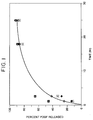
- Figure 1 shows the release rate profile of PDGF from a single layer collagen film (thickness 0.1 mm) made from insoluble collagen fibrils as described in Example 1B.
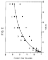
- Figure 2 shows the release rate profile of PDGF from a single layer collagen film (thickness 0.36 mm) made from insoluble collagen fibrils as described in Example 1B.
- Figure 3 shows the release rate profile of PDGF from a single layer collagen film (thickness 0.48 mm) made from insoluble collagen fibrils as described in Example 1B.
- Figure 4 shows the release rate profile of PDGF from a double layer collagen film (thickness 0.01-3.0 mm) made from soluble collagen as described in Example 2.
- Figure 5 shows the release rate profile of PDGF from a four layer collagen film (thicknesses 0.01-3.0 mm) made from soluble collagen as described in Example 3.
- Figure 6 shows the release rate profile of various active ingredients from a single layer collagen film (thicknesses 0.01-3.0 mm) made from soluble collagen as described in Example 1A.
- Figure 7 shows the measurement of protein concentration in samples taken from Costar Transwell Cells by three different methods: ELISA (closed circles); 125I labelled PDGF (open squares); and 3H-Thymidine uptake assay (closed squares).
- Figure 8 shows the maximum height (MH) of granulation tissue at the advancing edges of the wound (light bars; units of mm) and approximate area and volume measurements for new granulation tissue were calculated (dark bars; units of mm3), based on the assumption that the wounds healed concentrically and did not contract.
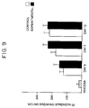
- Figure 9 shows effects of PDGF on the wound breaking strength when compared to untreated animals in the gastric linear wound model.
- the present invention relates to a collagen film comprising one or two rate controlling layers and one or more drug reservoir layers, said layers contacting each other in a stacked conformation such that the rate controlling layer is situated at one or both ends of the stack, with the proviso that each of said rate controlling layer contacts only one other layer, said other layer being a drug reservoir layer.
- a collagen film comprising one or two rate controlling layers and one or more drug reservoir layers, said layers contacting each other in a stacked conformation such that the rate controlling layer is situated at one or both ends of the stack, with the proviso that each of said rate controlling layer contacts only one other layer, said other layer being a drug reservoir layer.
- there is only one rate controlling layer which is situated at one end of the stack.
- Rate controlling layers can be produced from a solution of soluble collagen.
- Soluble collagen is collagen that has an average molecular weight of less than 400,000, preferably having a molecular weight of about 300,000.
- a particularly suitable soluble collagen is Semex S (Semex Medical Co., Malvern, Pennsylvania).
- This particular soluble collagen is also advantageous because it is the atelopeptide form of the collagen.
- Atelopeptide collagen is collagen that is free of telopeptide, which is a peptide located at one end of purified collagen often associated with immunogenicity.
- a solution of the telopeptide form of collagen can be converted to the atelopeptide form of collagen via hydrolysis using organic acid.
- Another preferred characteristic of the soluble collagen is that it possesses a minimal amount of crosslinking, i.e., 0.5% or less.
- the soluble collagen can be dissolved in a suitable solvent such as water to produce a solution that contains from about 0.5 to about 10% of collagen by weight, preferably from about 1 to about 5% by weight, and more preferably about 2% by weight.
- Rate controlling layers can also be produced from a dispersion of collagen fibrils in suspension.
- Collagen fibrils which are commercially available (e.g., Vitaphore Co., Menlo Park, California), can be dispersed into a suitable solvent such as water to produce a suspension that contains from about 0.1 to about 10% of collagen by weight, preferably from about 1 to about 2% of collagen by weight.
- a suitable dilute acid can be present in the solvent.
- a particularly suitable acid is acetic acid at a concentration of about 5%.
- a solution of soluble collagen or a dispersion of collagen fibrils in suspension is prepared as a film using a solvent casting method.
- the collagen solution is poured into a mold and allowed to dry.
- the mold has the property of being nonstick so that the dried collagen film will not adhere to the surface of the mold.
- a particularly suitable mold surface is TeflonTM.
- Suitable conditions include allowing the poured solution to dry at a suitable temperature for a suitable period of time. Generally, the amount of drying time necessary is shorter as the drying temperature is raised.
- a suitable temperature is from about 15°C to 35°C, preferably about room temperature
- a suitable drying time is sufficient time so that the marginal loss of solvent content is essentially zero (e.g., drying time of about an hour to about 10 days, preferably about one to about five days).
- the thickness of the film and the presence/amount of plasticizers in the rate controlling layer it is critical to achieve a suitable thickness of the dried film.
- Particularly suitable thicknesses for such rate controlling layers are from about 0.01 to about 1 mm, preferably from about 0.05 to about 0.5 mm, most preferably from about 0.1 to about 0.2 mm.
- various additives may be optionally included in the collagen solution and film.
- Such desirable characteristics include flexibility, stability, accelerated drying time and a pH compatible with the active ingredient to be utilized.
- a suitable plasticizer can be used.
- Suitable plasticizers include polyethylene glycol and glycerol, preferably glycerol. Such plasticizers can be present in an amount from zero to about 100% of the weight of collagen present, preferably from about 10 to about 30% of the weight of collagen present, most preferably about 20% of the weight of collagen present.
- a suitable stabilizing agent can be used in the film.
- Suitable stabilizing agents include most sugars, preferably mannitol, lactose, and glucose, more preferably mannitol.
- Such stabilizing agents can be present in an amount from zero to about 5% of the weight of collagen present, preferably about 1% of the weight of collagen present.
- a drying enhancer can be used.
- Suitable drying enhancers include alcohols, preferably ethanol, methanol and isopropyl alcohol, more preferably ethanol.
- Such drying enhancers can be present in an amount from zero to about 50% of the weight of the total solution or suspension, preferably from about 10 to about 30% of the weight of the total solution or suspension, more preferably about 20% of the weight of the total solution or suspension.
- a suitable buffer can be used in the film.
- Suitable buffers include most of the commmonly known and utilized biological buffers, preferably acetate, phosphate and citrate, more preferably acetate and phosphate. Such buffers can be present in an amount of from about 0.01% to about 2% of the weight of the collagen.
- a compatible pH is one that maintains the stability of an active ingredient, optimizes its therapeutic effect or protects against its degradation.
- a suitable pH is generally from about 3 to about 8, preferably about 5 to about 8, and most preferably about neutral pH of from about 7.0 to about 7.5.
- the active ingredient is not usually present in the rate controlling layer, the present invention does contemplate such an embodiment. Active ingredient can, therefore, be formulated into the rate controlling layer. However, the presence of any active ingredient in the rate controlling layer is preferably at a concentration that is lower than the concentration of active ingredient in any drug reservoir layer.
- Drug reservoir layers are produced in the same manner as the rate controlling layer with additional active ingredient if there is active ingredient in the rate controlling layer or with the presence of active ingredient if there is no active ingredient in the rate controlling layer.
- Preferred active ingredients are those biological agents which enhance wound healing or regeneration of nerve tissue, particularly recombinant proteins.
- Such preferred active ingredients include platelet derived growth factor (PDGF), epidermal growth factor (EGF), fibroblast growth factor (FGF), platelet derived epidermal growth factor (PDEGF),platelet derived endotheleal cell growth factor (PD-ECGF) keratinocyte growth factor (KGF), insulin-like growth factors 1 and 2 (IGF-1 and IGF-2), tumor necrosis factor (TNF), brain derived neurotrophin factor (BDNF), ciliary neurotrophic factor (CNTF) and neurotrophin-3 (NT-3).
- PDGF platelet derived growth factor
- EGF epidermal growth factor
- FGF fibroblast growth factor
- PEGF platelet derived epidermal growth factor
- PD-ECGF platelet derived endotheleal cell growth factor
- KGF keratinocyte growth factor
- IGF-1 and IGF-2 insulin-like growth factors 1 and 2
- TNF tumor necrosis factor
- BDNF brain derived neurotrophin factor
- CNTF ciliary neurotroph
- the actual amount of the active ingredient will be determined by the attending clinician and will depend on various factors such as the severity of the wound, the condition of the patient, the age of the patient and any collateral injuries or medical ailments possessed by the patient. Generally, the amount of active ingredient will be in the range of about 1 ⁇ g/cm2 to 5 mg/cm2.
- Particularly suitable thicknesses for drug reservoir layers are from about 0.01 to about 1 mm, preferably from about 0.05 to about 0.5 mm, most preferably from about 0.1 to about 0.2 mm.
- the various layers contact each other by any number of methods.
- One such method is to place layers adjacent to each other and apply pressure to the outer sides of the layers to force the layers together.
- Another method is to coat the surface of each of the layers to be contacted with a solvent, such as water, before placing the layers together. In this way, a thin portion of each surface will become soluble thereby producing adhesion upon contact.
- Another method is to use a known adhesive on one or more of the contacting surfaces.
- the adhesive is one that will not interfere with the release of the active ingredient from a layer.
- the preferred method of contacting the layers is with the application of equal pressure on each of the layers to be contacted.
- the number of drug reservoir layers is determined by the desired release characteristics. Generally, more layers produce a more steady and more sustained release of the active ingredient. Preferably, the number of drug reservoir layers is from 1 to 10, more preferably from 1 to 5 and most preferably from 1 to 3.
- the concentration of active ingredient in different layers can be varied and the thickness of the different layers need not be the same.
- the rate controlling layer may be at one or both ends of the stack.
- a stack of layers with a rate controlling layer at only one end is particularly suited to deliver an active ingredient to an epidermal surface.
- a stack of layers with a rate controlling layer at both ends is particularly suited to deliver an active ingredient to an internal wound or to a two surfaced wound, such as a surgical incision.
- the other end of the stack may optionally consist of a backing layer.
- a backing layer can be any of the conventionally known backing layers.
- the backing layer comprises polyurethanes.
- the collagen films of the present invention are useful as a means of delivering the active ingredient to cells or tissue with which it is in contact.
- a collagen film with one rate controlling layer and one backing layer can be placed on the wound to deliver a suitable active ingredient to the traumatized area.
- PDGF is a particularly suitable active ingredient for such uses.
- Collagen films with rate controlling layers at both ends of the stack can be used to accelerate healing of surgical wounds. When used in such a way, the film can be placed in the surgical incision and stitched into the wound as an interface between the two surgical wound surfaces.
- Collagen films can also be used to deliver neurotrophic factors. When used in such a manner, the collagen film can be placed in direct contact with or adjacent to the nerve tissue to be treated with the neurotrophic factor.
- Example 1 Preparation of a Single Layer Collagen Film .
- Collagen films containing various growth factors were prepared by the solvent casting method from a solution of soluble collagen.
- the soluble collagen was purchased from Semex Co. (Frazer, Pennsylvania). This collagen is from bovine origin and it contains 99% type I collagen and 1% type III collagen. The molecular weight of the collagen is 300 K dalton and the density is 0.044 gram/cc. The antigenicity of the collagen is minimal since the telopeptide is removed from the collagen.
- a collagen solution (about 1 to 8%) was prepared by dissolving the soluble collagen in 0-5% acetic acid solution at 18-70°C. After the addition of the plasticizer glycerol (about 20% of the dry weight of the collagen), ethanol was added to the solution to facilitate the solvent evaporation process. The amount of alcohol was about 20% of the amount of the solution. The solution is then centrifuged to remove the undissolved material.
- a growth factor solution (with or without radioactive material) was added to the solution.
- the solution was cast on a TeflonTM surface and dried at room temperature until the weight of the film was constant (for about 1-3 days) to produce collagen films containing various amounts of growth factor.
- Table I shows the thickness of the films prepared from the different concentrations of the collagen solution.
- Figure 6 shows the release profiles of PDGF from the collagen films with the release profiles being obtained by using the methods of Example 4 for various single layer films.
- Wafers made of insoluble collagen fibril were prepared and the release rate of PDGF was measured. 2 grams of insoluble fibril collagen (Vitaphore Co., Menlo Park, California) was dispersed in about 110 ml of 5% acetic acid solution containing 0.2 ml glycerol. To the resulting solution, about 10 ml of PDGF solution (782 ⁇ g/ml) containing a trace amount of 125I-PDGF was added. Five ml of alcohol was added to expedite the solvent evaporation process. Three films were cast from the mixture to obtain three different film thicknesses. Several wafers were cut from each film.
- the average thickness of the films was 0.10, 0.36, and 0.48 mm for Film-A, Film-B, and Film-C, respectively.
- the release rate of PDGF from each wafer was measured with a Franz diffusion cell (Crown Glass Co., Sommerville, New Jersey), which is commonly used for the determination of drug release rate from a transdermal patch. By using a Franz diffusion cell, a reasonable perfect sink condition could be obtained.
- a piece of Durapore membrane (Millipore Co., Bedford, Massachusetts) (pore size 5 ⁇ m) was used to separate the collagen wafer from the receiver solution.
- Figure 1 shows the release profile of PDGF from wafers cut from Film-A (thickness 0.1 mm). Most of the PDGF was released in 24 hours.
- Figure 2 shows the release profile of PDGF from wafers cut from Film-B (thickness 0.36 mm). About 77% of PDGF was released in 96 hours.
- Figure 3 shows the release profile of PDGF from wafers cut from Film-C (thickness 0.48 mm). About 73% of PDGF was released. Data shown in Figures 1, 2, and 3 indicate that the duration of PDGF release may be controlled from one day to more than one week.
- Example 2 Preparation of Double Layer Collagen Film .
- Double layer films of collagens were used to produce wound dressings which can deliver growth factors at a nearly constant rate to the wound sites for long periods of time (12 hours or longer).
- one collagen layer (membrane A) was prepared by a solvent casting method from an aqueous solution containing soluble collagen (4% collagen in 10mM acetate buffer (pH 4) in 0.85% NaCl solution), glycerol (20% w/w of collagen), and ethanol (20% of the solution).
- the second film (membrane B) had almost the same thickness (0.01 - 3mm) and the same composition as the first film except it contained PDGF (20 ⁇ g/cm2 film).
- the two films were combined into one by attaching them together by evenly applying pressure.
- In vitro release rate study conducted according to Example 4 showed that the growth factor was released at constant rate for more than 12 hours ( Figure 4).
- Example 3 Preparation of a Multiple Layer Collagen Film .
- a three layer film or a four layer film was prepared to produce a long term delivery device of growth factors.
- a four layer film was prepared by the following method. Four different castings were performed as in Example 1A and then the films were combined into one by attaching them together by evenly applying pressure. The thickness of each layer was similar (0.01-3mm) however, different thickness layers could be used.
- the first collagen layer which will contact the skin did not contain PDGF.
- the concentrations of PDGF were 0.07%, 0.15%, and 0.30% for the second, the third, and the fourth layers respectively. Subsequent release study showed that a nearly constant release rate of the growth factor was maintained up to 100 hours Figure 5. At that time, almost 90% of the growth factor was released.
- Coster Transwell Cells (Costar Co., Cambridge, Massachusetts) as follows. Collagen films were produced as described in Examples 1, 2 and 3, and wafers (1.6 cm diameter) were cut from the films. Each wafer was transferred to a Coster Transwell Cell and placed on top of the polycarbonate membrane. 2.5 ml of the receiver solution (water and 1% bovine serum albumin, or water and 0.25% human serum albumin) was added to the Cell holder. The Cells were set on the solution and the release study was initiated. At specified times, 20 ⁇ l of the receiver solution was pipetted out and the same amount of fresh solution was replaced in the receiver solution. The sampling procedure was repeated to get another 20 ⁇ l sample.
- the receiver solution water and 1% bovine serum albumin, or water and 0.25% human serum albumin
- the radioactivity of the sample was measured with a gamma counter (Beckman Instruments, Co., Irvine, California).
- the concentration of protein in the receiver solution at any given time was calculated based on the radioactivity and confirmed using other methods such as ELISA and 3H-thymidine uptake bioassays. Results obtained using this assay are shown in Examples 1, 2, and 3 for films of various layers and thicknesses ( Figure 1 to 6).
- Figure 7 shows the agreement of the various methods of measuring protein concentration.
- the diffusion coefficient should be independent of the thickness of the film.
- the release profile of PDGF from the single layer collagen film can be described by the following equation.
- the equation shows that the plot of F versus square root of time should be linear. A plot of F versus square root of time showed a linear relationship between the amount of PDGF released and the square root of time.
- the apparent diffusion coefficients (Da) of PDGF in the collagen films calculated from the data were 2.5, 2.4, 2.5 and 7.6 (all x 10 ⁇ 10 cm2/sec) for films of Groups A, B, C and D, respectively. Ideally, the diffusion coefficient should be the same regardless of the film thickness.
- the resulting data indicate that the current film casting method is very reliable up to the film thickness of at least 0.16 mm.
- the higher value of the diffusion coefficient at 0.29 mm is probably caused by the solvent present in the thick films due to incomplete solvent evaporation, indicating that more drying time or elevated temperature is required for the preparation of thick films.
- Example 5 Measurment of the Tissue Volume Using the Blood Bundle Model .
- This example measures the influence of growth factors in a collagen film on the rate of healing of surgically created 6mm diameter dermal ulcers in the rabbit ear.
- This excisional wound model replicates the healing parameters (i.e., minimal wound contraction, generation of new granulation tissue, reepithelialization) associated with full thickness dermal wounds such as human leg ulcers.
- the full thickness wound model permitted histiologic quantification of both reepithelialization and formation of granulation tissue while excluding wound contracture as a variable.
- cartilage is avascular, and the perichondrium was removed during surgery, new granulation tissue and new epithelium arises solely from the periphery of the wound. PDGF was applied at the time of surgery.
- the inner surface of the ears was wiped with saline-soaked gauze followed by application of a 70% alcohol solution.
- the dermis of the inner surface on one ear of each rabbit was blanched by infiltration of the ear with a 2% xylocaine solution containing 1:1000 epinephrine (this requires 1.5 to 3.0 mls total volume) using a 30 gauge needle.
- the infiltrated area was then scrubbed with 3 cycles of betadine followed by the 70% alcohol solution. Where necessary, the ear plugs were replaced with dry plugs at this point.
- the rabbits were then transferred to a sterile surgical room.
- the blanched ear was immobilized on a plexiglass "ear board" (Washington University Medical Center, Division of Technical Services, St. Louis, Missouri) which utilizes two bar clamps, one at the tip and one at the base of the animal's ear, to stabilize the rabbit ear without compromising its blood supply.
- the animal was draped, and the surgical field (i.e., the inner surface of the blanched ear) sprayed with Betadine and allowed to dry for 3 to 5 minutes.
- the ear Upon completion of one ear, the ear was covered with saline-moistened gauze and then taped shut around the gauze to retain moisture until application of PDGF. The second ear was then blanched, scrubbed, immobilized and wounded in the manner as the first ear. Blood was removed from the biopsy site of each second ear and each completed biopsy covered with a small piece of saline-soaked gauze. Uppon completion of the second ear, it was covered with saline-moistened gauze until application of PDGF. Any rabbit that showed evidence of recovery from anesthesia at any time prior to this point in the procedure was reanesthetized with 25 mg/kg ketamine, administered intramuscularly.
- Collagen wafers (diameter of 0.5 cm) containing 5.9 ⁇ g of PDGF per wafer were prepared from the soluble collagen.
- the rabbits were allowed to recover from anesthesia under the observation of the investigator performing the surgery. Upon recovery, a plastic neck collar (Canine Center, St. Louis, Missouri) extending approximately 15 to 25 cm outward was placed around each rabbit's neck to prevent the rabbit from disrupting the wounds or dressings. The rabbits were returned to an isolation cage where they were maintained until harvest. The wounds of any rabbits which had removed their collars, and any wound on which the Tegaderm® had been disrupted in some way prior to the harvest date, were reevaluated as soon as the problem was noted, and discarded from analysis if the wounds appeared to be damaged.
- the rabbits were anesthesized in the same manner as described for pre-operative preparation. Each rabbit's weight was measured and recorded (each wound being photographed), and a qualitative description of the condition of the wounds was recorded, noting in particular the presence or absence of the Tegaderm® and of any excess fluid under the dressing.
- the rabbits were sacrificed with a 50 ml/kg air embolism administered by intracardiac injection. Both ears were then amputated from the body using a #15 surgical blade mounted on a knife handle.
- Each biopsy with approximately 5 mm of surrounding tissue on any side and the Tegaderm® still intact, was excised from the ear, and the biopsy site measured in order to bisect it accurately at the midline, making reference to notes taken on the day of wounding to avoid bisecting through natural holes or nicks in the cartilage.
- the biopsy was carefully bisected with a single edge razor blade, using a single downward motion to avoid disrupting the wound orientation.
- the bisected biopsies were immediately placed in cassettes labeled with the rabbit identification number, and fixed in 10% buffered formalin for routine histologic processing.
- India ink tatoos were placed at the wound periphery on day zero to assess the degree of contraction during healing. After 7 days, wound diameters were unchanged. Infected wounds (less than 5%) or desicated wounds (less than 5%) were excluded before measurements were made. Cultures of clinically noninfected wounds were repeatedly conducted and showed no growth of pathogens.
- Example 7 Measurement of Wound Healing in the Rabbit Stomach Stretch Model .
- the median gastric linear wound models were performed on an animal using a protocol approved by the institution's Animal Care and Use Committee. New Zealand White rabbits, 2.7 to 3.5 kg (Doe Valley Farms, Bentonville, Arkansas) were preanesthetized by subcutaneous injection of atropine (0.1 mg/kg) and acepromazine (0.75 mg/kg). After a time span of 10 minutes, the animals were anesthetized with ketamine (0.75 mg/kg) and xylazine (5 mg/kg). Their abdomens were shaved with a #40 blade and sterilely prepared for surgery. A 10 cm midline laparotomy was made, the cecum was mobilized to expose the sacculus rotundus and approximately 20 cm of the cecum.
- Sutures were placed through the serosal, muscular, and submuscular layers so as to draw these layers up and over the collagen strip against the muscularis mucosae.
- the loop was trimmed in the control wounds and left intact in the experimental wounds so as to remove any chance of error at time of harvesting.
- the suture was atraumatically removed from each wound and three standardized 8 mm strips were cut across each incision with the use of a punch template (Washington University machine shop). Histological samples were taken from areas between the wound strips. Wound breaking strength was measured in grams/mm2 on a tensometer (Tensometer 10, Monsanto, St. Louis, Missouri) on three strips from each incision (six experimental, six control/rabbit). If evidence of infection, hematoma, or poor coaptation were evident, the sample was disregarded ( ⁇ 2% of all wounds were disregarded from analysis). All wounds were tested on the tesometer at 20 mm/min with the use of electrical claw clamps to insure breaking at the wound site only. Tensometry analysis was divided into three categories: fundal tissue, antral tissue, and cecal tissue and the data reported individually for each.
- Histological analysis was performed on matched samples from each group. Samples were sutured into micro-casettes and stored in formalin and hemotoxlin and eosin staining was performed at a later date. The histological samples were then examined for thickness, amount of granulation tissue, and signs of necrosis, and this data was recorded.
Landscapes
- Health & Medical Sciences (AREA)
- Engineering & Computer Science (AREA)
- Chemical & Material Sciences (AREA)
- Life Sciences & Earth Sciences (AREA)
- Animal Behavior & Ethology (AREA)
- General Health & Medical Sciences (AREA)
- Public Health (AREA)
- Veterinary Medicine (AREA)
- Epidemiology (AREA)
- Bioinformatics & Cheminformatics (AREA)
- Hematology (AREA)
- Materials Engineering (AREA)
- Pharmacology & Pharmacy (AREA)
- Medicinal Chemistry (AREA)
- Chemical Kinetics & Catalysis (AREA)
- General Chemical & Material Sciences (AREA)
- Nuclear Medicine, Radiotherapy & Molecular Imaging (AREA)
- Organic Chemistry (AREA)
- Medicines That Contain Protein Lipid Enzymes And Other Medicines (AREA)
- Medicinal Preparation (AREA)
- Materials For Medical Uses (AREA)
Applications Claiming Priority (4)
| Application Number | Priority Date | Filing Date | Title |
|---|---|---|---|
| US716862 | 1985-03-28 | ||
| US71516591A | 1991-06-14 | 1991-06-14 | |
| US715165 | 1991-06-14 | ||
| US71686291A | 1991-06-18 | 1991-06-18 |
Publications (2)
| Publication Number | Publication Date |
|---|---|
| EP0518697A2 true EP0518697A2 (de) | 1992-12-16 |
| EP0518697A3 EP0518697A3 (en) | 1993-06-23 |
Family
ID=27109285
Family Applications (1)
| Application Number | Title | Priority Date | Filing Date |
|---|---|---|---|
| EP19920305437 Ceased EP0518697A3 (en) | 1991-06-14 | 1992-06-12 | Collagen film for sustained delivery of proteins |
Country Status (6)
| Country | Link |
|---|---|
| US (1) | US5418222A (de) |
| EP (1) | EP0518697A3 (de) |
| JP (1) | JPH06500802A (de) |
| AU (1) | AU654574B2 (de) |
| CA (1) | CA2089487A1 (de) |
| WO (1) | WO1992022304A1 (de) |
Cited By (20)
| Publication number | Priority date | Publication date | Assignee | Title |
|---|---|---|---|---|
| EP0568334A1 (de) * | 1992-05-01 | 1993-11-03 | Amgen Inc. | Kollagen enthaltende Schwämme mit Freisetzung von Proteinen |
| WO1995028964A1 (de) * | 1994-04-27 | 1995-11-02 | Lts Lohmann Therapie-Systeme Gmbh | Kollagenzubereitung zur gesteuerten abgabe von wirkstoffen |
| WO1996005857A1 (en) * | 1994-08-19 | 1996-02-29 | Cambridge Neuroscience, Inc. | Device for delivery of substances and methods of use thereof |
| EP0901792A1 (de) * | 1997-09-05 | 1999-03-17 | Dr. Suwelack Skin & Health Care AG | Mittel zur peroralen Verabreichung, seine Herstellung und Verwendung |
| US6335388B1 (en) | 1997-10-03 | 2002-01-01 | Lavipharm Laboratories Inc. | Prolamine-plant polar lipid composition, its method of preparation and applications thereof |
| EP1196187A4 (de) * | 1999-06-02 | 2003-02-05 | Human Genome Sciences Inc | FORMULIERUNGEN üR DEN KERATINOZYTEN-WACHSTUMS-KAKTOR 2 |
| GB2382775A (en) * | 2001-12-06 | 2003-06-11 | Johnson & Johnson Medical Ltd | Controlled release therapeutic wound dressings |
| US6653284B2 (en) | 1997-12-22 | 2003-11-25 | Human Genome Sciences, Inc. | Keratinocyte growth factor-2 formulations |
| GB2393724A (en) * | 2002-10-01 | 2004-04-07 | Johnson & Johnson Medical Ltd | Controlled release therapeutic wound dressings |
| WO2008070166A1 (en) * | 2006-12-05 | 2008-06-12 | Collengin, Inc. | Collagen materials, films and methods of making same |
| EP2574349A1 (de) | 2011-09-29 | 2013-04-03 | Biorigen S.r.l | Therapeutische Verwendung von Gelatin-Hydrogelen mit Gel-Sol-Übergang bei Körpertemperatur |
| WO2013045689A1 (en) | 2011-09-29 | 2013-04-04 | BIORIGEN Srl | Therapeutic use of gelatin hydrogels with a gel-sol transition at body temperature |
| US8465537B2 (en) | 2003-06-17 | 2013-06-18 | Gel-Del Technologies, Inc. | Encapsulated or coated stent systems |
| US8492332B2 (en) | 2007-04-19 | 2013-07-23 | Fibralign Corporation | Oriented collagen-based materials, films and methods of making same |
| US8513382B2 (en) | 2008-08-11 | 2013-08-20 | Fibralign Corporation | Biocomposites and methods of making the same |
| US8871267B2 (en) | 1998-09-25 | 2014-10-28 | Gel-Del Technologies, Inc. | Protein matrix materials, devices and methods of making and using thereof |
| US9724308B2 (en) | 2010-09-10 | 2017-08-08 | Fibralign Corporation | Biodegradable multilayer constructs |
| US10065046B2 (en) | 2010-07-15 | 2018-09-04 | Fibralign Corporation | Conductive biopolymer implant for enhancing tissue repair and regeneration using electromagnetic fields |
| US10086079B2 (en) | 2008-08-11 | 2018-10-02 | Fibralign Corporation | Biocomposites and methods of making the same |
| US11890371B2 (en) | 2007-12-26 | 2024-02-06 | Petvivo Holdings, Inc. | Biocompatible protein-based particles and methods thereof |
Families Citing this family (72)
| Publication number | Priority date | Publication date | Assignee | Title |
|---|---|---|---|---|
| US5578566A (en) * | 1993-05-04 | 1996-11-26 | The United States Of America As Represented By The Department Of Health And Human Services | KGF receptor-derived antagonists of KGF binding |
| FR2726571B1 (fr) * | 1994-11-03 | 1997-08-08 | Izoret Georges | Colle biologique, procede de preparation et dispositif d'application pour colle biologique, et durcisseurs pour colle biologique |
| ATE244584T1 (de) * | 1995-01-16 | 2003-07-15 | Baxter Int | Selbsttragende flächengebilde aus vernetztem fibrin zur hemmung von postoperativen adhäsionen |
| US6693077B1 (en) | 1995-02-14 | 2004-02-17 | Human Genome Sciences, Inc. | Keratinocyte growth factor-2 |
| US7232667B2 (en) | 1995-02-14 | 2007-06-19 | Human Genome Sciences, Inc. | Keratinocyte growth factor-2 polynucleotides |
| US6077692A (en) | 1995-02-14 | 2000-06-20 | Human Genome Sciences, Inc. | Keratinocyte growth factor-2 |
| AR013829A1 (es) | 1996-07-12 | 2001-01-31 | Baxter Int | Un dispositivo medico para suministrar cantidades volumetricas de un primer y un segundo fluido, bioquimicamente reactivos, y metodo para suministrarfibrina a una superficie con dicho dispositivo |
| US6066325A (en) | 1996-08-27 | 2000-05-23 | Fusion Medical Technologies, Inc. | Fragmented polymeric compositions and methods for their use |
| US7871637B2 (en) | 1996-08-27 | 2011-01-18 | Baxter International Inc. | Dry hemostatic compositions and methods for their preparation |
| US8303981B2 (en) | 1996-08-27 | 2012-11-06 | Baxter International Inc. | Fragmented polymeric compositions and methods for their use |
| US7435425B2 (en) * | 2001-07-17 | 2008-10-14 | Baxter International, Inc. | Dry hemostatic compositions and methods for their preparation |
| US8603511B2 (en) | 1996-08-27 | 2013-12-10 | Baxter International, Inc. | Fragmented polymeric compositions and methods for their use |
| AU778308B2 (en) * | 1996-08-30 | 2004-11-25 | Genzyme Corporation | Method, instruments and kit for autologous transplantation |
| US6743422B1 (en) | 1996-10-15 | 2004-06-01 | Amgen, Inc. | Keratinocyte growth factor-2 products |
| US6593290B1 (en) * | 1996-11-01 | 2003-07-15 | Genentech, Inc. | Treatment of inner ear hair cells |
| TW586934B (en) * | 1997-05-19 | 2004-05-11 | Sumitomo Pharma | Immunopotentiating composition |
| US6869927B1 (en) | 1997-12-22 | 2005-03-22 | Human Genome Sciences, Inc. | Keratinocyte growth factor-2 formulations |
| EP1013280A4 (de) * | 1998-06-10 | 2004-04-28 | Bml Inc | Hautpreparationen zur äusseren anwendung |
| US20030007991A1 (en) * | 1998-09-25 | 2003-01-09 | Masters David B. | Devices including protein matrix materials and methods of making and using thereof |
| BE1012536A3 (fr) | 1998-11-04 | 2000-12-05 | Baxter Int | Element muni d'une couche de fibrine sa preparation et son utilisation. |
| WO2000045858A1 (en) * | 1999-02-08 | 2000-08-10 | Collagenesis, Inc. | Collagen-based delivery of radioactivity for use in brachytherapy |
| AU2001249079B2 (en) * | 2000-02-28 | 2005-11-03 | Gel-Del Technologies, Inc. | Protein matrix materials, devices and methods of making and using thereof |
| US7455854B2 (en) * | 2002-04-18 | 2008-11-25 | University Of Florida Research Foundation, Inc. | Method for producing a mineral fiber |
| US7514249B2 (en) * | 2002-04-18 | 2009-04-07 | The University Of Florida Research Foundation, Inc. | Biomimetic organic/inorganic composites |
| AU2003228587A1 (en) * | 2002-04-18 | 2003-11-03 | University Of Florida | Biomimetic organic/inorganic composites, processes for their production, and methods of use |
| US7261734B2 (en) * | 2002-04-23 | 2007-08-28 | Boston Scientific Scimed, Inc. | Resorption-controllable medical implants |
| CA2483778A1 (en) * | 2002-04-29 | 2003-11-13 | Gel-Del Technologies, Inc. | Biomatrix structural containment and fixation systems and methods of use thereof |
| US7090868B2 (en) * | 2002-09-13 | 2006-08-15 | University Of Florida | Materials and methods for drug delivery and uptake |
| US7135027B2 (en) * | 2002-10-04 | 2006-11-14 | Baxter International, Inc. | Devices and methods for mixing and extruding medically useful compositions |
| WO2005081699A2 (en) * | 2003-04-18 | 2005-09-09 | University Of Florida | Biomimetic organic/inorganic composites, processes for their production, and methods of use |
| US8834864B2 (en) * | 2003-06-05 | 2014-09-16 | Baxter International Inc. | Methods for repairing and regenerating human dura mater |
| TW200505394A (en) * | 2003-06-06 | 2005-02-16 | Asahi Medical Co | Material promoting wound healing |
| US7927626B2 (en) | 2003-08-07 | 2011-04-19 | Ethicon, Inc. | Process of making flowable hemostatic compositions and devices containing such compositions |
| WO2005055945A2 (en) | 2003-12-08 | 2005-06-23 | Gel-Del Technologies, Inc. | Mucoadhesive drug delivery devices and methods of making and using thereof |
| IL159273A0 (en) * | 2003-12-09 | 2004-06-01 | Transpharma Medical Ltd | Transdermal delivery system for sustained release of polypeptides |
| US20050283256A1 (en) * | 2004-02-09 | 2005-12-22 | Codman & Shurtleff, Inc. | Collagen device and method of preparing the same |
| WO2006073514A2 (en) * | 2004-08-25 | 2006-07-13 | Tufts University | Compositions, methods and kits for repressing virulence in gram positive bacteria |
| US7429241B2 (en) * | 2005-09-29 | 2008-09-30 | Codman & Shurtleff, Inc. | Dural graft and method of preparing the same |
| MX2008014847A (es) * | 2006-05-31 | 2009-04-30 | Baxter Int | Metodo para crecimiento interno en la celula dirigido y regeneracion controlada de los tejidos en la cirugia espinal. |
| DE102006043216A1 (de) * | 2006-06-02 | 2007-12-06 | Inoviscoat Gmbh | Verbundmaterial und Verfahren zum Herstellen eines derartigen Verbundmaterials |
| AU2007269712B2 (en) * | 2006-06-30 | 2013-02-07 | Biomimetic Therapeutics, Llc | PDGF-biomatrix compositions and methods for treating rotator cuff injuries |
| WO2008016163A1 (en) * | 2006-08-01 | 2008-02-07 | Nichiban Co., Ltd. | Crosslinked gelatin gel multilayered structure, carrier for bioactive factor, preparation for release of bioactive factor, and their production methods |
| TWI436793B (zh) * | 2006-08-02 | 2014-05-11 | 巴克斯特國際公司 | 快速作用之乾密封膠及其使用和製造方法 |
| ES2840251T3 (es) * | 2007-07-10 | 2021-07-06 | Lifecell Corp | Composiciones acelulares de matriz tisular para reparación de tejidos |
| ES2662647T3 (es) | 2007-10-30 | 2018-04-09 | Baxter International Inc. | Uso de una biomatriz de colágeno biofuncional regenerativa para tratar defectos viscerales o parietales |
| CA2716872C (en) | 2008-02-29 | 2015-02-10 | Ferrosan Medical Devices A/S | Device for promotion of hemostasis and/or wound healing |
| AU2009201541B2 (en) * | 2008-04-23 | 2014-12-04 | Integra Lifesciences Corporation | Flowable collagen material for dural closure |
| BR122020000059B8 (pt) * | 2008-09-09 | 2021-06-22 | Biomimetic Therapeutics Inc | composição que compreende uma matriz biocompatível e um fator de crescimento derivado de plaqueta e kit |
| US20110150976A1 (en) * | 2008-09-10 | 2011-06-23 | Transpharma Medical Ltd. | Transdermal delivery of oligosaccharides |
| US9039783B2 (en) * | 2009-05-18 | 2015-05-26 | Baxter International, Inc. | Method for the improvement of mesh implant biocompatibility |
| HRP20150255T1 (hr) * | 2009-06-16 | 2015-05-08 | Baxter International Inc. | Hemostatiäśna spužva |
| US8771258B2 (en) * | 2009-12-16 | 2014-07-08 | Baxter International Inc. | Hemostatic sponge |
| SA111320355B1 (ar) | 2010-04-07 | 2015-01-08 | Baxter Heathcare S A | إسفنجة لايقاف النزف |
| ES2682302T3 (es) | 2010-06-01 | 2018-09-19 | Baxter International Inc | Proceso para la producción de composiciones hemostáticas secas y estables |
| JP5973997B2 (ja) | 2010-06-01 | 2016-08-23 | バクスター・インターナショナル・インコーポレイテッドBaxter International Incorp0Rated | 乾燥した安定な止血用組成物を作製するためのプロセス |
| AU2011260260B2 (en) | 2010-06-01 | 2015-09-03 | Baxter Healthcare S.A. | Process for making dry and stable hemostatic compositions |
| MX346958B (es) | 2011-10-11 | 2017-04-06 | Baxter Int | Composición hemostatica. |
| WO2013053759A2 (en) | 2011-10-11 | 2013-04-18 | Baxter International Inc. | Hemostatic compositions |
| SG11201401878SA (en) | 2011-10-27 | 2014-09-26 | Baxter Int | Hemostatic compositions |
| CN104159527B (zh) | 2012-03-06 | 2017-04-12 | 弗罗桑医疗设备公司 | 包含止血糊剂的压力容器 |
| RU2636240C2 (ru) | 2012-06-12 | 2017-11-21 | Ферросан Медикал Дивайсиз А/С | Сухая гемостатическая композиция |
| JP6390873B2 (ja) | 2013-06-21 | 2018-09-19 | フェッローサン メディカル ディバイス エー/エス | 減圧膨張させた乾燥組成物およびそれを保持するためのシリンジ |
| BR112016013322B1 (pt) | 2013-12-11 | 2020-07-21 | Ferrosan Medical Devices A/S | métodos para preparação de uma composição seca e para reconstituir uma composição seca, composição seca, uso de uma composição seca, e, kit |
| JP6384749B2 (ja) * | 2014-04-14 | 2018-09-05 | 国立研究開発法人農業・食品産業技術総合研究機構 | 多層構造のガラス化後のハイドロゲル乾燥体の製造方法、多層構造のガラス化後のハイドロゲル乾燥体、多層構造のガラス化後のハイドロゲル、徐放剤乾燥体の製造方法、接着方法、徐放剤乾燥体、及び徐放剤水和体 |
| EP3185922B1 (de) * | 2014-08-27 | 2025-04-02 | Purdue Research Foundation | Abgabesysteme für therapeutikum auf kollagenbasis |
| BR112017007466B1 (pt) | 2014-10-13 | 2021-03-02 | Ferrosan Medical Devices A/S | método para preparar uma composição seca, método para reconstituir a composição seca, pasta, composição seca, recipiente, kit homeostático, e, uso de uma composição seca |
| RU2705905C2 (ru) | 2014-12-24 | 2019-11-12 | Ферросан Медикал Дивайсиз А/С | Шприц для удерживания и смешивания первого и второго веществ |
| CA2986981A1 (en) | 2015-07-03 | 2017-01-12 | Ferrosan Medical Devices A/S | Syringe for mixing two components and for retaining a vacuum in a storage condition |
| US12551509B2 (en) | 2015-11-18 | 2026-02-17 | Lucina Patent Holdco, Llc | Acellular regenerative products and methods of their manufacture |
| EP3386557B1 (de) | 2015-12-11 | 2021-03-31 | LifeCell Corporation | Verfahren und systeme zur versteifung von gewebe zur verbesserten verarbeitung |
| EP4321182A3 (de) | 2018-05-09 | 2024-05-29 | Ferrosan Medical Devices A/S | Verfahren zur herstellung einer hämostatischen zusammensetzung |
| CN113750286B (zh) * | 2021-09-30 | 2022-11-29 | 振德医疗用品股份有限公司 | 一种皮肤创面覆盖膜及其制备方法 |
Family Cites Families (17)
| Publication number | Priority date | Publication date | Assignee | Title |
|---|---|---|---|---|
| US4060081A (en) * | 1975-07-15 | 1977-11-29 | Massachusetts Institute Of Technology | Multilayer membrane useful as synthetic skin |
| US4148664A (en) * | 1976-05-10 | 1979-04-10 | Avicon, Inc. | Preparation of fibrous collagen product having hemostatic and wound sealing properties |
| US4294241A (en) * | 1977-06-09 | 1981-10-13 | Teruo Miyata | Collagen skin dressing |
| DE3214337C2 (de) * | 1982-04-19 | 1984-04-26 | Serapharm - Michael Stroetmann, 4400 Münster | Resorbierbares Flachmaterial zum Abdichten und Heilen von Wunden und Verfahren zu dessen Herstellung |
| EP0227955A3 (de) * | 1982-07-21 | 1987-11-25 | University of Strathclyde | Mehrschichtiger Wundverband |
| US4837285A (en) * | 1984-03-27 | 1989-06-06 | Medimatrix | Collagen matrix beads for soft tissue repair |
| MX163953B (es) * | 1984-03-27 | 1992-07-03 | Univ New Jersey Med | Procedimiento para preparar una matriz biodegradable a base de colageno |
| US4600533A (en) * | 1984-12-24 | 1986-07-15 | Collagen Corporation | Collagen membranes for medical use |
| US4759354A (en) * | 1986-11-26 | 1988-07-26 | The Kendall Company | Wound dressing |
| US4909244B1 (en) * | 1986-11-26 | 1994-07-05 | Kendall & Co | Hydrogel wound dressing |
| US4808402A (en) * | 1987-05-29 | 1989-02-28 | Northwestern University | Method and compositions for modulating neovascularization |
| NZ226171A (en) * | 1987-09-18 | 1990-06-26 | Ethicon Inc | Gel formulation containing polypeptide growth factor |
| US4865846A (en) * | 1988-06-03 | 1989-09-12 | Kaufman Herbert E | Drug delivery system |
| US4950483A (en) * | 1988-06-30 | 1990-08-21 | Collagen Corporation | Collagen wound healing matrices and process for their production |
| US5196185A (en) * | 1989-09-11 | 1993-03-23 | Micro-Collagen Pharmaceutics, Ltd. | Collagen-based wound dressing and method for applying same |
| US5104660A (en) * | 1989-11-21 | 1992-04-14 | Bruce A. Barber | Method of preparing an antimicrobial wound dressing |
| US5030215A (en) * | 1990-01-03 | 1991-07-09 | Cryolife, Inc. | Preparation of fibrinogen/factor XIII precipitate |
-
1992
- 1992-06-11 CA CA002089487A patent/CA2089487A1/en not_active Abandoned
- 1992-06-11 JP JP5501020A patent/JPH06500802A/ja active Pending
- 1992-06-11 WO PCT/US1992/004929 patent/WO1992022304A1/en not_active Ceased
- 1992-06-11 AU AU21926/92A patent/AU654574B2/en not_active Ceased
- 1992-06-12 EP EP19920305437 patent/EP0518697A3/en not_active Ceased
-
1994
- 1994-06-28 US US08/267,647 patent/US5418222A/en not_active Expired - Lifetime
Cited By (29)
| Publication number | Priority date | Publication date | Assignee | Title |
|---|---|---|---|---|
| US5399361A (en) * | 1992-05-01 | 1995-03-21 | Amgen Inc. | Collagen-containing sponges as drug delivery compositions for proteins |
| US5512301A (en) * | 1992-05-01 | 1996-04-30 | Amgen Inc. | Collagen-containing sponges as drug delivery compositions for proteins |
| EP0568334A1 (de) * | 1992-05-01 | 1993-11-03 | Amgen Inc. | Kollagen enthaltende Schwämme mit Freisetzung von Proteinen |
| AU695621B2 (en) * | 1994-04-27 | 1998-08-20 | Lts Lohmann Therapie-Systeme Gmbh | Collagen preparation for the controlled release of active substances |
| WO1995028964A1 (de) * | 1994-04-27 | 1995-11-02 | Lts Lohmann Therapie-Systeme Gmbh | Kollagenzubereitung zur gesteuerten abgabe von wirkstoffen |
| US6761908B1 (en) | 1994-04-27 | 2004-07-13 | Lohmann & Rauscher Gmbh & Co., Kg | Collagen preparation for the controlled release of active substances |
| WO1996005857A1 (en) * | 1994-08-19 | 1996-02-29 | Cambridge Neuroscience, Inc. | Device for delivery of substances and methods of use thereof |
| US5681568A (en) * | 1994-08-19 | 1997-10-28 | Cambridge Neuroscience, Inc. | Device for delivery of substances and methods of use thereof |
| EP0901792A1 (de) * | 1997-09-05 | 1999-03-17 | Dr. Suwelack Skin & Health Care AG | Mittel zur peroralen Verabreichung, seine Herstellung und Verwendung |
| US6335388B1 (en) | 1997-10-03 | 2002-01-01 | Lavipharm Laboratories Inc. | Prolamine-plant polar lipid composition, its method of preparation and applications thereof |
| US6653284B2 (en) | 1997-12-22 | 2003-11-25 | Human Genome Sciences, Inc. | Keratinocyte growth factor-2 formulations |
| US8871267B2 (en) | 1998-09-25 | 2014-10-28 | Gel-Del Technologies, Inc. | Protein matrix materials, devices and methods of making and using thereof |
| EP1196187A4 (de) * | 1999-06-02 | 2003-02-05 | Human Genome Sciences Inc | FORMULIERUNGEN üR DEN KERATINOZYTEN-WACHSTUMS-KAKTOR 2 |
| GB2382775B (en) * | 2001-12-06 | 2005-05-25 | Johnson & Johnson Medical Ltd | Controlled release therapeutic wound dressings |
| US7732655B2 (en) | 2001-12-06 | 2010-06-08 | Systagenix Wound Management (Us), Inc. | Controlled release therapeutic wound dressings |
| GB2382775A (en) * | 2001-12-06 | 2003-06-11 | Johnson & Johnson Medical Ltd | Controlled release therapeutic wound dressings |
| GB2393724A (en) * | 2002-10-01 | 2004-04-07 | Johnson & Johnson Medical Ltd | Controlled release therapeutic wound dressings |
| GB2393724B (en) * | 2002-10-01 | 2006-04-12 | Johnson & Johnson Medical Ltd | Controlled release therapeutic wound dressings |
| US8465537B2 (en) | 2003-06-17 | 2013-06-18 | Gel-Del Technologies, Inc. | Encapsulated or coated stent systems |
| WO2008070166A1 (en) * | 2006-12-05 | 2008-06-12 | Collengin, Inc. | Collagen materials, films and methods of making same |
| US8227574B2 (en) | 2006-12-05 | 2012-07-24 | Fibralign Corporation | Collagen materials, films and methods of making same |
| US8492332B2 (en) | 2007-04-19 | 2013-07-23 | Fibralign Corporation | Oriented collagen-based materials, films and methods of making same |
| US11890371B2 (en) | 2007-12-26 | 2024-02-06 | Petvivo Holdings, Inc. | Biocompatible protein-based particles and methods thereof |
| US8513382B2 (en) | 2008-08-11 | 2013-08-20 | Fibralign Corporation | Biocomposites and methods of making the same |
| US10086079B2 (en) | 2008-08-11 | 2018-10-02 | Fibralign Corporation | Biocomposites and methods of making the same |
| US10065046B2 (en) | 2010-07-15 | 2018-09-04 | Fibralign Corporation | Conductive biopolymer implant for enhancing tissue repair and regeneration using electromagnetic fields |
| US9724308B2 (en) | 2010-09-10 | 2017-08-08 | Fibralign Corporation | Biodegradable multilayer constructs |
| WO2013045689A1 (en) | 2011-09-29 | 2013-04-04 | BIORIGEN Srl | Therapeutic use of gelatin hydrogels with a gel-sol transition at body temperature |
| EP2574349A1 (de) | 2011-09-29 | 2013-04-03 | Biorigen S.r.l | Therapeutische Verwendung von Gelatin-Hydrogelen mit Gel-Sol-Übergang bei Körpertemperatur |
Also Published As
| Publication number | Publication date |
|---|---|
| US5418222A (en) | 1995-05-23 |
| AU654574B2 (en) | 1994-11-10 |
| EP0518697A3 (en) | 1993-06-23 |
| AU2192692A (en) | 1993-01-12 |
| WO1992022304A1 (en) | 1992-12-23 |
| CA2089487A1 (en) | 1992-12-15 |
| JPH06500802A (ja) | 1994-01-27 |
Similar Documents
| Publication | Publication Date | Title |
|---|---|---|
| US5418222A (en) | Multi-layered collagen film compositions for delivery of proteins and methods of using same | |
| US5399361A (en) | Collagen-containing sponges as drug delivery compositions for proteins | |
| US4703108A (en) | Biodegradable matrix and methods for producing same | |
| DE68924069T2 (de) | Kollagen-wundheilmatrizen und verfahren zur herstellung. | |
| US20060159731A1 (en) | Multi-layer collagenic article useful for wounds healing and a method for its production thereof | |
| US20050232979A1 (en) | Multi-layer collagenic article useful for wounds healing | |
| A˚ gren | Four alginate dressings in the treatment of partial thickness wounds: a comparative experimental study | |
| EP1494728B1 (de) | Keratin-netzwerke enthaltende wundverbände für gewebedefekte | |
| CA2031817C (en) | Use of thrombospondin to promote wound healing | |
| US20030095997A1 (en) | Natural polymer-based material for use in human and veterinary medicine and method of manufacturing | |
| Agren | An amorphous hydrogel enhances epithelialisation of wounds. | |
| JP7785127B2 (ja) | クラゲコラーゲンの使用 | |
| US20080118551A1 (en) | Denatured human albumin biomaterial as dressing for burns and wounds of skin | |
| CA2134447A1 (en) | Collagen-containing sponges as drug delivery for proteins | |
| Ghamsari et al. | Evaluation of low level laser therapy on open wound healing of the teat in dairy cattle | |
| US20030124175A1 (en) | Cicatrizant hydrocolloidal patch containing hyaluronic acid and chondroitin sulphate | |
| HK40116416A (en) | Jellyfish collagen use |
Legal Events
| Date | Code | Title | Description |
|---|---|---|---|
| PUAI | Public reference made under article 153(3) epc to a published international application that has entered the european phase |
Free format text: ORIGINAL CODE: 0009012 |
|
| AK | Designated contracting states |
Kind code of ref document: A2 Designated state(s): AT BE CH DE DK ES FR GB GR IT LI LU MC NL PT SE |
|
| PUAL | Search report despatched |
Free format text: ORIGINAL CODE: 0009013 |
|
| AK | Designated contracting states |
Kind code of ref document: A3 Designated state(s): AT BE CH DE DK ES FR GB GR IT LI LU MC NL PT SE |
|
| 17P | Request for examination filed |
Effective date: 19930621 |
|
| 17Q | First examination report despatched |
Effective date: 19950206 |
|
| GRAG | Despatch of communication of intention to grant |
Free format text: ORIGINAL CODE: EPIDOS AGRA |
|
| STAA | Information on the status of an ep patent application or granted ep patent |
Free format text: STATUS: THE APPLICATION HAS BEEN REFUSED |
|
| 18R | Application refused |
Effective date: 19970927 |
|
| RAP1 | Party data changed (applicant data changed or rights of an application transferred) |
Owner name: AMGEN INC., |