EP0520202A2 - Dispositif d'analyse pour la détermination immunologique et quantitative de substances nuisibles - Google Patents
Dispositif d'analyse pour la détermination immunologique et quantitative de substances nuisibles Download PDFInfo
- Publication number
- EP0520202A2 EP0520202A2 EP92109022A EP92109022A EP0520202A2 EP 0520202 A2 EP0520202 A2 EP 0520202A2 EP 92109022 A EP92109022 A EP 92109022A EP 92109022 A EP92109022 A EP 92109022A EP 0520202 A2 EP0520202 A2 EP 0520202A2
- Authority
- EP
- European Patent Office
- Prior art keywords
- tracer
- analyte
- reaction
- layer
- antibody
- Prior art date
- Legal status (The legal status is an assumption and is not a legal conclusion. Google has not performed a legal analysis and makes no representation as to the accuracy of the status listed.)
- Withdrawn
Links
Images


Classifications
-
- G—PHYSICS
- G01—MEASURING; TESTING
- G01N—INVESTIGATING OR ANALYSING MATERIALS BY DETERMINING THEIR CHEMICAL OR PHYSICAL PROPERTIES
- G01N33/00—Investigating or analysing materials by specific methods not covered by groups G01N1/00 - G01N31/00
- G01N33/48—Biological material, e.g. blood, urine; Haemocytometers
- G01N33/50—Chemical analysis of biological material, e.g. blood, urine; Testing involving biospecific ligand binding methods; Immunological testing
- G01N33/53—Immunoassay; Biospecific binding assay; Materials therefor
- G01N33/536—Immunoassay; Biospecific binding assay; Materials therefor with immune complex formed in liquid phase
- G01N33/537—Immunoassay; Biospecific binding assay; Materials therefor with immune complex formed in liquid phase with separation of immune complex from unbound antigen or antibody
- G01N33/538—Immunoassay; Biospecific binding assay; Materials therefor with immune complex formed in liquid phase with separation of immune complex from unbound antigen or antibody by sorbent column, particles or resin strip, i.e. sorbent materials
Definitions
- the invention relates to a device for the quantitative analysis of pollutants or pollutant-containing aerosols with the aid of an immunological reaction, in which the pollutant (analyte) to be detected is brought to an immunological reaction with an antibody in cooperation with a labeled analyte (tracer).
- EP-374 685 describes the use of highly specific enzymes for the detection of formaldehyde in the air.
- the analyte reaches the plaque-shaped dosimeter from the open side.
- the analyte is implemented in the highly specific enzyme reaction.
- the more dye is produced the more analyte can penetrate the badge.
- the evaluation is also carried out here by a color comparison.
- enzymes can only convert relatively small molecules of approximately up to 100 g per mol in a highly specific manner.
- analytes with higher molecular weights such as crop protection agents, explosives and drugs, but also microorganisms such as viruses and bacteria or allergens.
- EP-51 183 describes a multilayer analysis element which consists of at least one carrier layer to which the antibodies necessary for the specificity of the reaction are covalently immobilized and at least one reagent layer which carries the reagents necessary for color development.
- the sample containing the analyte is mixed with a defined amount of one with a tracer (this is to be understood as an analyte or analyte derivative which is covalently bound to one or more labeling elements). The mixture, and not the aqueous sample itself, is then applied to the analysis element.
- a competitive immunoassay takes place in the analysis element. Tracers that do not respond due to the presence of analyte the immobilized antibodies can bind, diffuse into the reagent layer and use their enzyme portion to initiate the enzymatic color reaction used for quantification.
- a fluorescence measuring device Since one does not want to use reagents in the known assay, a fluorescence measuring device has to be used to quantify the amount of displaced tracer. Since exactly one tracer is displaced per analyte, there is a linear relationship between analyte concentration and measurement signal in the displacement immunoassay.
- An immunoassay that works with fluorophores as a labeling element allows a quantitative reaction statement, but requires complex measuring means.
- the present invention is therefore based on the object of a simple, directly displaying and evaluable device of the type mentioned to develop, which makes the very high specificity of the reaction of a specific binding pair usable for the measurement of air, water and soil samples as well as polluted aerosols.
- the object is achieved in that the antibodies are present together with the labeled tracers in a non-transparent part of the support which is exposed to the analyte and permeable to them, through which the free, not bound to an antibody, remains in the non-transparent part after the immunological reaction Diffuse the tracer into a transparent part of the carrier so that the progress of the tracer diffusion on the outer surface of the transparent part of the carrier can be recognized by its discoloration.
- the advantage of the invention is that the antibodies immobilized on the carrier bind part of the present tracer to another part of it, as a result of the immunological reaction (eg competition reaction or displacement reaction).
- This free part of tracers diffuses through the carrier due to the concentration gradient present in relation to the tracer concentration in the carrier until it can be recognized on the surface of the visible part of the carrier.
- Antibodies and tracers are immobilized on the surface of the carrier exposed to the pollutant to be detected.
- An example of a membrane-like support can be mentioned by Pall Corporation, sold under the name Immunodyne membrane. This membrane enables direct covalent binding of antibodies, which means that they are irreversibly immobilized on the membrane. Further membranes are known from EP-51 183.
- these membranes For the passive, diffusion-related measurement of gaseous samples, these membranes must be provided with the moisture required for the reaction. This can be done by installing or applying a moisture storage, or by a wick soaked in water, or by wetting or spraying a water mist. The gas sample then penetrates the carrier by diffusion.
- a rehydration of the antibody and tracer is brought about by using a buffered aqueous or aqueous / organic solution for this.
- the device according to the invention is suitable for use in a simple, directly evaluable quantitative analysis device, which results in direct monitoring the environment, for example at the workplace, without complex wet chemical reactions with different Reagents carried out, or complex detection devices, such as fluorescence measuring device, must be used for the subsequent quantitative evaluation.
- An additional reaction layer in which the displaced tracers enter into a color reaction with a reaction partner can be dispensed with. After the reaction, the displaced tracers can diffuse into the detection layer as unbound, free tracers.
- the device according to the invention is also suitable for allowing a competitive reaction to take place between the tracers and the analytes as the immunological detection reaction.
- moistening e.g.
- the tracer is rehydrated by means of a buffered solution in which the pollutant to be examined is located, as a result of which both the analyte and the tracer compete for a free binding site on the antibody.
- the tracers remaining without a binding site are free and able to diffuse into the detection layer.
- the support has two layers, one of which is exposed as a reaction layer with the tracer immobilized on it to the analyte to be examined, to which the other detection layer permeable for the free tracer not bound to an antibody in the diffusion contact.
- layer is to be understood to mean a single membrane whose different layer areas fulfill the desired function.
- the detection layer can thus be located in the specially structured outer surface of a membrane facing away from the analyte exist, the reaction layer being formed by the membrane body.
- marking elements In order to increase the detection sensitivity of the device, it is expedient to bind several marking elements to one analyte. This is done by first providing the analyte or suitable derivatives (analytes provided with coupling groups) with a polymer which contains a repetitive sequence of suitable coupling groups. Then one marker element per coupling group, ie several elements per analyte or derivative, is bound. Thus, the greater the number of marking elements, the higher the sensitivity of the detection device will be. Expediently, dye molecules which are visible to the naked eye are bound to the analyte as marking elements.
- the two layers it is advantageous to design the two layers in such a way that they have an overall different electrical charge or polarity.
- the reaction layer is, for example, hydrophilic
- the tracers must then be designed in their own polarity so that they have an affinity for the detection layer.
- the tracers are hydrophobic and the detection layer is also hydrophobic, the tracers will diffuse preferentially towards the detection layer due to their hydrophobicity. This reduces cross-diffusions or interfering diffusion interactions between the tracers. In this way, an increased diffusion gradient is generated for the displaced tracers in the direction of the detection layer.
- the two-dimensional layer structure of the detection device with the opaque reaction layer will be exposed to the analyte, and the rear side of the transparent detection layer will be observable in its color change caused by color-marked tracers.
- the layer structure from one of the end faces again the opaque reaction layer remaining unchanged during the measurement process, but the translucent detection layer, viewed from its end face, changes its color can be seen.
- one end face of the detection layer can be irradiated with a light source, and opposite the other end face, the optionally provided with a cylindrical lens or other suitable optical aids, attach a detector which is sensitive to certain wavelengths suitable in connection with the detection, so that the received light signal is a measure of the accumulated tracer concentration and thus of the analyte.
- This evaluation is to be carried out especially if the tracers are not provided with a marking element that can be seen in visible light.
- a useful design of the device as a monitor for monitoring gaseous or liquid pollutants is achieved in that the carrier is accommodated in a housing which has an inlet cover and an outlet cover, each having an inlet and outlet provided for the passage of the sample to be examined have between which the support is clamped, which has a layer structure such that starting from the inlet, first the reaction layer and then the detection layer is in diffusion contact.
- Such a monitor is suitable for carrying out passive or active sampling in a simple manner.
- a pump can be connected at the rear of the housing to the intended outlet, which can be in the form of a nozzle.
- the pump actively draws the gas or liquid through the inlet and through the carrier where the analyte participates in the desired reaction.
- the carrier can be designed as a membrane filter, in which commercially available membranes of suitable pore size are selected by the manufacturers, for example Millipore, Pall or Satorius.
- the antibody specific for the analyte is covalently bound in the reaction layer and marked, for example, with a color support.
- the tracer / antibody complex thus formed enters into a displacement reaction with the analyte, by which the tracer is replaced by the analyte.
- the marked tracer diffuses into the transparent detection layer and accumulates there.
- a reaction layer can expediently precede the reaction layer, in which the analyte is first collected before it starts an immunological reaction with the tracers in the reaction layer.
- the discoloration that corresponds to the correspondingly colored marking area represents a measure of the concentration or dosage of the analyte assigned to the associated marking area.
- a pump with a predetermined output is connected to the outlet and, after a certain period of time, sucks a defined amount of gas or air through the filter device. Pollutants to be examined are retained as analyte on the collecting layer.
- the collection layer is flushed with a certain amount of aqueous / organic-aqueous buffer solution.
- the liquid can be sucked actively through the carrier like the air sample before, or it can passively drip through the carrier.
- the liquid dripping or sucked through is sucked off into a collecting bottle via a suction nozzle at the outlet.
- the buffered solution ensures that the concentrated analytes from the collecting layer pass through this into the reaction layer.
- the reaction layer is soaked and the bound, labeled antibody / antibody-tracer complexes are rehydrated.
- analyte and antibody can connect and lead to a displacement or competitive reaction.
- the housing covers are opened, for example by unscrewing or opening, so that the carrier is visible and the more or less discolored detection layer is evaluated by comparison with the colored marking areas.
- insecticides pyrethroids, triazine
- explosives TNT
- drugs as well as highly toxic substances (dioxins)
- monovalent analytes as analytes, whereby bacteria such as E. coli K-12 are also used to monitor Genetic engineering laboratories, pseudomonas in ventilation from hospitals, legionella in air conditioning systems can be detected.
- the antibodies suitable for this can be produced by known methods, such as those e.g. in Peters, JH and Baumgarten, H. (Editor) "Monoclonal Antibodies, Production and Characterization” (1985), Springer-Verlag, Berlin, Germany, or also in Harlow, E. and Lane, D. (Editor), "Antibodies , a laboratory manual "(1988), Cold Spring Harbor Lab., Cold Spring Harbor, USA.
- the carrier (100) shown in Figure 1 consists of two sections in the form of layers, which are either made in a self-supporting embodiment, or are clamped and received in a frame, not shown.
- the analytes (1) (shown as open rhombuses) in the environment to be examined move according to the directional arrows (2) shown on the surface of an opaque reaction layer (3) in which the tracer / antibody complex (5, 7) is located.
- the antibodies (5) (symbolically represented by a Y-shape) are immobilized with their one branch in the first opaque layer (3), so that the fork-shaped branches of the antibody (5) are exposed to the analyte.
- the analyte (1) marked with a dye (4) is referred to as a tracer (7).
- a light-transmitting detection layer (8) follows the reaction layer (3).
- the layers (3, 8) are connected to one another in diffusion contact.
- the analytes (1) hit the reaction layer (3), diffuse into it (FIG. 2), displace the tracer (7) from its binding site on the antibody (5) and take their place.
- the displaced tracers (7) diffuse through the reaction layer (3) into the detection layer (8), through which they become visible from the back (9) of the detection layer (8) because of their dye (4) coupled to them.
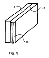
- the layer structure shown in FIG. 3 essentially corresponds to that of FIG. 1.
- the opaque reaction layer (3) and the translucent detection layer (8) are placed against one another in direct diffusion contact.
- the end face of the detection layer (8) facing the observer is designed in the form of a cylindrical lens (10).
- FIG. 4 shows an exploded perspective view of the device, in which the carrier (100) is clamped between an inlet cover (102) and an outlet cover (103).
- the covers (102, 103) have a not-shown latching, as a result of which, in the compressed state, they clamp the carrier (100) between them.
- the carrier (100) is shown in the drawing in three layers, which, however, can be divided objectively both in a three-layer embodiment and in a single membrane by differently designed membrane layer regions.
- a first layer is a collection layer (104) facing the inlet cover (102), which is followed by a middle reaction layer (105) in diffusion contact.
- the layer sequence is completed with a detection layer (106) which has circular marking areas (107) along one of its edges.
- the figure shows five such areas (107), each with the same intensity Coloring is equipped. Depending on the required resolution accuracy, more or fewer such marking areas (107) should be provided.
- the inlet cover is provided with an inlet (108) and the outlet cover (103) with an outlet (109), the inlet providing access to the detection sample (liquid or gaseous) or also a buffer solution required for the detection, and to the Outlet (109) a suction pump, not shown, can be connected.
- a suction nozzle (110) is used to extract excess amounts of liquid.
Landscapes
- Health & Medical Sciences (AREA)
- Immunology (AREA)
- Life Sciences & Earth Sciences (AREA)
- Engineering & Computer Science (AREA)
- Molecular Biology (AREA)
- Biomedical Technology (AREA)
- Chemical & Material Sciences (AREA)
- Hematology (AREA)
- Urology & Nephrology (AREA)
- Biotechnology (AREA)
- Microbiology (AREA)
- Cell Biology (AREA)
- Food Science & Technology (AREA)
- Medicinal Chemistry (AREA)
- Physics & Mathematics (AREA)
- Analytical Chemistry (AREA)
- Biochemistry (AREA)
- General Health & Medical Sciences (AREA)
- General Physics & Mathematics (AREA)
- Pathology (AREA)
- Investigating Or Analysing Materials By The Use Of Chemical Reactions (AREA)
Applications Claiming Priority (2)
| Application Number | Priority Date | Filing Date | Title |
|---|---|---|---|
| DE4121493 | 1991-06-28 | ||
| DE4121493A DE4121493A1 (de) | 1991-06-28 | 1991-06-28 | Analysevorrichtung zur quantitativen, immunologischen bestimmung von schadstoffen |
Publications (2)
| Publication Number | Publication Date |
|---|---|
| EP0520202A2 true EP0520202A2 (fr) | 1992-12-30 |
| EP0520202A3 EP0520202A3 (en) | 1994-06-22 |
Family
ID=6435026
Family Applications (1)
| Application Number | Title | Priority Date | Filing Date |
|---|---|---|---|
| EP19920109022 Withdrawn EP0520202A3 (en) | 1991-06-28 | 1992-05-29 | Analyzing device for the immunological and quantitative determination of harmful substances |
Country Status (2)
| Country | Link |
|---|---|
| EP (1) | EP0520202A3 (fr) |
| DE (1) | DE4121493A1 (fr) |
Cited By (6)
| Publication number | Priority date | Publication date | Assignee | Title |
|---|---|---|---|---|
| EP0650054A3 (fr) * | 1993-10-20 | 1995-12-13 | Boehringer Mannheim Gmbh | Procédé et dispositif d'obtention et de détection de substances réactives immunologiquement d'une phase gazeuse. |
| FR2721112A1 (fr) * | 1994-06-13 | 1995-12-15 | Gks Technologies | Dispositif de diagnostic rapide de toute molécule glyquée et procédé de mise en Óoeuvre. |
| WO1996035121A1 (fr) * | 1995-05-03 | 1996-11-07 | Karl Cammann | Dispositif d'echantillonnage a affichage integre dosimetrique et fonctionnel et procede d'echantillonnage |
| US5712170A (en) * | 1992-12-29 | 1998-01-27 | Oy Medix Biochemica Ab | Test strip, its production and use |
| WO2001073432A3 (fr) * | 2000-03-29 | 2002-04-11 | November Ag Molekulare Medizin | Procede de detection et/ou de quantification de premieres molecules |
| DE102004058093A1 (de) * | 2004-12-01 | 2006-06-08 | Nivoclean Gmbh | Vorrichtung zur Detektion von Mikroorganismen |
Families Citing this family (9)
| Publication number | Priority date | Publication date | Assignee | Title |
|---|---|---|---|---|
| US6605444B1 (en) | 1993-10-20 | 2003-08-12 | Securetec Detektions-Systems Ag | Method and device for obtaining and detecting immunologically active substances from the gas phase |
| DE4343842C1 (de) * | 1993-12-22 | 1995-02-23 | Draegerwerk Ag | Reaktionsgefäß zur immunologischn Analytik von Aerosolen |
| EP0665054A1 (fr) * | 1994-01-27 | 1995-08-02 | SECURETEC GmbH | Matière de sorption pour l'enrichissement et la récupération de substances à l'état de trace dans une phase gazeuze |
| DE19711281C1 (de) * | 1997-03-18 | 1998-04-16 | Inst Chemo Biosensorik | Vorrichtung und Verfahren zur Durchführung von Fluoreszenzimmunotests |
| US6686208B2 (en) | 1997-03-18 | 2004-02-03 | Institut Fur Chemo- Und Biosensorik Munster E.V. | Device and method for carrying out fluoresence immunotests |
| DE19751363B4 (de) * | 1997-11-20 | 2004-07-22 | Dräger Safety AG & Co. KGaA | Verfahren und Vorrichtung zum Nachweis von Substanzemissionen |
| CN100491957C (zh) | 2003-04-16 | 2009-05-27 | 株式会社崛场制作所 | 颗粒状物质捕集用过滤膜、使用该过滤膜的取样器以及颗粒状物质的分析装置 |
| DE10356951A1 (de) * | 2003-12-05 | 2005-06-30 | Human Gesellschaft für Biochemica und Diagnostica mbH | Testvorrichtung |
| DE102005056118A1 (de) * | 2005-11-23 | 2007-05-24 | Bundesdruckerei Gmbh | Sicherheits- und/oder Wertdokument mit Gefahrenstoffsensor |
Family Cites Families (11)
| Publication number | Priority date | Publication date | Assignee | Title |
|---|---|---|---|---|
| US3654090A (en) * | 1968-09-24 | 1972-04-04 | Organon | Method for the determination of antigens and antibodies |
| US4258001A (en) * | 1978-12-27 | 1981-03-24 | Eastman Kodak Company | Element, structure and method for the analysis or transport of liquids |
| US4366241A (en) * | 1980-08-07 | 1982-12-28 | Syva Company | Concentrating zone method in heterogeneous immunoassays |
| JPS5767860A (en) * | 1980-10-15 | 1982-04-24 | Fuji Photo Film Co Ltd | Material for multilayer analysis |
| US4446232A (en) * | 1981-10-13 | 1984-05-01 | Liotta Lance A | Enzyme immunoassay with two-zoned device having bound antigens |
| IT1200382B (it) * | 1985-02-08 | 1989-01-18 | Boehringer Biochemia Srl | Sistema di rilevazione e/o dosaggio di parametri clinici per via immuno enzimatica |
| US4803170A (en) * | 1985-05-09 | 1989-02-07 | Ultra Diagnostics Corporation | Competitive immunoassay method, device and test kit |
| WO1988009798A1 (fr) * | 1987-06-09 | 1988-12-15 | Peck Dana P | Immunoanalyse pour composes contenant un cycle aromatique |
| US5039607A (en) * | 1988-05-17 | 1991-08-13 | Syntex (U.S.A.) Inc. | Method for immunochromatographic analysis |
| DE3826057A1 (de) * | 1988-07-30 | 1990-02-01 | Boehringer Mannheim Gmbh | Testtraeger zur analytischen bestimmung eines bestandteils einer fluessigen probe |
| GB8903627D0 (en) * | 1989-02-17 | 1989-04-05 | Unilever Plc | Assays |
-
1991
- 1991-06-28 DE DE4121493A patent/DE4121493A1/de not_active Withdrawn
-
1992
- 1992-05-29 EP EP19920109022 patent/EP0520202A3/de not_active Withdrawn
Cited By (8)
| Publication number | Priority date | Publication date | Assignee | Title |
|---|---|---|---|---|
| US5712170A (en) * | 1992-12-29 | 1998-01-27 | Oy Medix Biochemica Ab | Test strip, its production and use |
| US5965458A (en) * | 1992-12-29 | 1999-10-12 | Oy Medix Biochemica A.B. | Test strip, its production and use |
| EP0650054A3 (fr) * | 1993-10-20 | 1995-12-13 | Boehringer Mannheim Gmbh | Procédé et dispositif d'obtention et de détection de substances réactives immunologiquement d'une phase gazeuse. |
| FR2721112A1 (fr) * | 1994-06-13 | 1995-12-15 | Gks Technologies | Dispositif de diagnostic rapide de toute molécule glyquée et procédé de mise en Óoeuvre. |
| WO1995034815A1 (fr) * | 1994-06-13 | 1995-12-21 | Gks Technologies | Dispositif de diagnostic rapide de toute molecule glyquee et procede de mise en ×uvre |
| WO1996035121A1 (fr) * | 1995-05-03 | 1996-11-07 | Karl Cammann | Dispositif d'echantillonnage a affichage integre dosimetrique et fonctionnel et procede d'echantillonnage |
| WO2001073432A3 (fr) * | 2000-03-29 | 2002-04-11 | November Ag Molekulare Medizin | Procede de detection et/ou de quantification de premieres molecules |
| DE102004058093A1 (de) * | 2004-12-01 | 2006-06-08 | Nivoclean Gmbh | Vorrichtung zur Detektion von Mikroorganismen |
Also Published As
| Publication number | Publication date |
|---|---|
| DE4121493A1 (de) | 1993-01-07 |
| EP0520202A3 (en) | 1994-06-22 |
Similar Documents
| Publication | Publication Date | Title |
|---|---|---|
| DE60037268T2 (de) | Verfahren zur durchführung magnetischer chromatographischer assays | |
| DE60219207T2 (de) | Diagnostisches testverfahren | |
| DE60018537T2 (de) | Teststreifen zur Bestimmung eines Analyts in eine flüssige Probe | |
| DE69530861T2 (de) | Vorrichtung zur durchführung eines oder mehrerer kompetitiver immunoassays | |
| DE19909891C1 (de) | Wischanalysator für den immunchemischen Stoffnachweis | |
| DE69837544T2 (de) | Testgerät und methode zur entdeckung von analyten in proben | |
| DE3586983T2 (de) | Verfahren und vorrichtung fuer immunotest. | |
| DE69328120T2 (de) | Prüfungsstreifen für immunoassays | |
| DE69033569T2 (de) | Festphasen-analytische Vorrichtung | |
| DE3786388T2 (de) | Analytisches Gerät und Verfahren zum Nachweis von Chlamydia Trachomatis und Neisseria Gonorrhoeae. | |
| DE2518721A1 (de) | Vorrichtung zur analyse von loesungen und koerperfluessigkeiten | |
| DE4121493A1 (de) | Analysevorrichtung zur quantitativen, immunologischen bestimmung von schadstoffen | |
| EP0995992B1 (fr) | Couches de diffusion, agents mouillants pour leurs préparation et leurs utilisation dans les bandes d'essai | |
| DE69330613T2 (de) | Gehäuse für analytisches teststrip | |
| EP2823309B1 (fr) | Procédé et dispositif destinés à la vérification d'analytes | |
| WO2000042434A1 (fr) | Procede et dispositif pour determiner un analyte | |
| DE60115016T2 (de) | Prüfvorrichtung mit zeiteinstellfunktion | |
| DE3226407A1 (de) | Mikro-analyse-kapillar-system | |
| DE60031204T2 (de) | VERFAHREN und KIT ZUM NACHWEIS UND ZUR IDENTIFIZIERUNG VON KRISTALLEN IN BIOLOGISCHEN FLÜSSIGKEITEN | |
| DE4343842C1 (de) | Reaktionsgefäß zur immunologischn Analytik von Aerosolen | |
| DE19808598B4 (de) | Verfahren und Vorrichtung zum Nachweis von staubassoziierten Substanzen | |
| EP3500840B1 (fr) | Dispositif de mesure comprenant un injecteur et une protection contre des giclées | |
| DE10153925A1 (de) | Testgerät zum Nachweis von pharmakologisch wirksamen Verbindungen aus Speichel | |
| EP3361253B1 (fr) | Dispositif de contrôle ciblé et d'affichage de la présence ou de l'absence d'analytes dans un échantillon liquide | |
| DE69817793T2 (de) | Detektion von hydrophoben analyten mit niedrigem gehalt in umweltproben |
Legal Events
| Date | Code | Title | Description |
|---|---|---|---|
| PUAI | Public reference made under article 153(3) epc to a published international application that has entered the european phase |
Free format text: ORIGINAL CODE: 0009012 |
|
| AK | Designated contracting states |
Kind code of ref document: A2 Designated state(s): DE FR GB NL |
|
| PUAL | Search report despatched |
Free format text: ORIGINAL CODE: 0009013 |
|
| AK | Designated contracting states |
Kind code of ref document: A3 Designated state(s): DE FR GB NL |
|
| STAA | Information on the status of an ep patent application or granted ep patent |
Free format text: STATUS: THE APPLICATION IS DEEMED TO BE WITHDRAWN |
|
| 18D | Application deemed to be withdrawn |
Effective date: 19941223 |