EP0529084B1 - Verfahren zur bestimmung der zahl lebender mikroorganismen - Google Patents
Verfahren zur bestimmung der zahl lebender mikroorganismen Download PDFInfo
- Publication number
- EP0529084B1 EP0529084B1 EP92904999A EP92904999A EP0529084B1 EP 0529084 B1 EP0529084 B1 EP 0529084B1 EP 92904999 A EP92904999 A EP 92904999A EP 92904999 A EP92904999 A EP 92904999A EP 0529084 B1 EP0529084 B1 EP 0529084B1
- Authority
- EP
- European Patent Office
- Prior art keywords
- membrane filter
- luminescence
- hydrophilic
- sections
- hydrophobic
- Prior art date
- Legal status (The legal status is an assumption and is not a legal conclusion. Google has not performed a legal analysis and makes no representation as to the accuracy of the status listed.)
- Expired - Lifetime
Links
Images

Classifications
-
- C—CHEMISTRY; METALLURGY
- C12—BIOCHEMISTRY; BEER; SPIRITS; WINE; VINEGAR; MICROBIOLOGY; ENZYMOLOGY; MUTATION OR GENETIC ENGINEERING
- C12Q—MEASURING OR TESTING PROCESSES INVOLVING ENZYMES, NUCLEIC ACIDS OR MICROORGANISMS; COMPOSITIONS OR TEST PAPERS THEREFOR; PROCESSES OF PREPARING SUCH COMPOSITIONS; CONDITION-RESPONSIVE CONTROL IN MICROBIOLOGICAL OR ENZYMOLOGICAL PROCESSES
- C12Q1/00—Measuring or testing processes involving enzymes, nucleic acids or microorganisms; Compositions therefor; Processes of preparing such compositions
- C12Q1/66—Measuring or testing processes involving enzymes, nucleic acids or microorganisms; Compositions therefor; Processes of preparing such compositions involving luciferase
-
- C—CHEMISTRY; METALLURGY
- C12—BIOCHEMISTRY; BEER; SPIRITS; WINE; VINEGAR; MICROBIOLOGY; ENZYMOLOGY; MUTATION OR GENETIC ENGINEERING
- C12Q—MEASURING OR TESTING PROCESSES INVOLVING ENZYMES, NUCLEIC ACIDS OR MICROORGANISMS; COMPOSITIONS OR TEST PAPERS THEREFOR; PROCESSES OF PREPARING SUCH COMPOSITIONS; CONDITION-RESPONSIVE CONTROL IN MICROBIOLOGICAL OR ENZYMOLOGICAL PROCESSES
- C12Q1/00—Measuring or testing processes involving enzymes, nucleic acids or microorganisms; Compositions therefor; Processes of preparing such compositions
- C12Q1/02—Measuring or testing processes involving enzymes, nucleic acids or microorganisms; Compositions therefor; Processes of preparing such compositions involving viable microorganisms
- C12Q1/04—Determining presence or kind of microorganism; Use of selective media for testing antibiotics or bacteriocides; Compositions containing a chemical indicator therefor
- C12Q1/06—Quantitative determination
-
- C—CHEMISTRY; METALLURGY
- C12—BIOCHEMISTRY; BEER; SPIRITS; WINE; VINEGAR; MICROBIOLOGY; ENZYMOLOGY; MUTATION OR GENETIC ENGINEERING
- C12Q—MEASURING OR TESTING PROCESSES INVOLVING ENZYMES, NUCLEIC ACIDS OR MICROORGANISMS; COMPOSITIONS OR TEST PAPERS THEREFOR; PROCESSES OF PREPARING SUCH COMPOSITIONS; CONDITION-RESPONSIVE CONTROL IN MICROBIOLOGICAL OR ENZYMOLOGICAL PROCESSES
- C12Q1/00—Measuring or testing processes involving enzymes, nucleic acids or microorganisms; Compositions therefor; Processes of preparing such compositions
- C12Q1/02—Measuring or testing processes involving enzymes, nucleic acids or microorganisms; Compositions therefor; Processes of preparing such compositions involving viable microorganisms
- C12Q1/24—Methods of sampling, or inoculating or spreading a sample; Methods of physically isolating an intact microorganisms
-
- Y—GENERAL TAGGING OF NEW TECHNOLOGICAL DEVELOPMENTS; GENERAL TAGGING OF CROSS-SECTIONAL TECHNOLOGIES SPANNING OVER SEVERAL SECTIONS OF THE IPC; TECHNICAL SUBJECTS COVERED BY FORMER USPC CROSS-REFERENCE ART COLLECTIONS [XRACs] AND DIGESTS
- Y10—TECHNICAL SUBJECTS COVERED BY FORMER USPC
- Y10S—TECHNICAL SUBJECTS COVERED BY FORMER USPC CROSS-REFERENCE ART COLLECTIONS [XRACs] AND DIGESTS
- Y10S435/00—Chemistry: molecular biology and microbiology
- Y10S435/975—Kit
Definitions
- the present invention relates to a method for counting the number of living microorganisms in a test solution. More paricularly, the invention relates to a rapid, convenient, and highly sensitive method for counting the number of living microorganisms existing in a test solution from, for example, water, raw materials, intermediates, and products used in the industries of foods, pharmacy, cosmetics, electronics, etc.
- a conventional, so called “standard agar plate method” is generally employed according to the food hygiene testing guidance, in which method living microorganisms in test solutions are cultured on an agar plate medium to form the colonies for their detection.
- This process is rather troublesome and, moreover, requires a long time for obtaining the results, causing a substantial delay in judgement on the existence of microorganisms or the number thereof. This delay compels one to spare some "waiting time for judgement", particularly during the production or shipment step, thus causing a considerable loss of time and space.
- the process essentially comprises collecting a small amount of test solution in a small test tube, adding an extracting reagent plus a luminescence-inducing reagent to the solution to determine the amount of adenosine triphosphate (hereinafter referred to as ATP) contained in the microorganisms using a luminometer, and calculating the number of the living microorganisms.
- ATP adenosine triphosphate
- test solution can only be tested directly in the case of the number of microorganisms is sufficiently large, and if it is relatively small, the test solution requires to be filtrated to entrap and concentrate the population of microorganisms in the test solution on a membrane filter, followed by removing the filter, immersing it in a quite small amount of, for example, sterile water to suspend the living microorganisms, and taking a portion of the suspension into a small test tube, before the above mentioned reagents are added.
- test solution contains an extremely small number of living microorganisms (e.g., from 10 3 to 10 4 or less), however, the amount of ATP falls below the lower limit of determination for the luminometer, making this process unfeasible.
- further advanced techniques are required, in which, for example, the membrane filter element with living microorganisms entrapped is placed in a culture medium containing several nutrients suitable for the growth of the microorganisms, and, after the microorganisms are cultured to increase in their number up to the above determination limit, measurement can be carried out. This fact constitutes a major drawback in the method, thus making further improvement inevitable.
- microorganisms are prevented from overlapping and adhering mutually, to provide an easier way to measure a dense population of microorganisms, and at the same time high optical contrast between the microbial colonies and the surface of the membrane filter is produced to facilite an automatic measurement of the colonies.
- the membrane filter according to the above mentioned U. S. patent has not been developed in an initial attempt to detect a single cell of living microorganisms, but in order to measure the colonies formed by entrapping and culturing living microorganisms on a membrane filter.
- Such a method is advantageous in that microorganisms are allowed to form colonies separately within the sections of the non-wetting lattice pattern, so that the mutual overlapping of the colonies is prevented, even if a large number of microorganisms are to be counted, thus enabling an accurate measurement of the colonies.
- the apparatus for counting the number of microorganisms is to count the number of colonies that have been grown, isolated within the lattice pattern, after the culture, for example, at 20°C for 24 hours on said membrane filter.
- the method according to said patent provides a non-wettable lattice on a membrane filter to separate colonies, to maintain their state of relatively high density, uniformity, and optical contrast, and to facilitate an accurate automatic measurement of the number of microorganisms, it is not suitable to prevent any overflow and dispersion of the microorganisms to adjacent sections, as required for detecting a single microbial cell to which the present invention is directed, or to prevent the dilution of microbial ingredients caused by a large area of the individual sections, thus none of such a concept is stated in the patent.
- the present invention aims to provide a membrane filter with sections placed thereon, which will achieve more perfect separation among the sections, prevent the extracted ingredients from dispersing or flowing out, and have small areas. Furthermore, the present invention also aims to provide a rapid, convenient, and highly sensitive method for counting the number of living microorganisms through an improved process of adding the reagents for extraction, luminescence inducing. etc., along with a newly incorporated highly sensitive photometric system.
- partitions which constitute a hydrophobic lattice produced on a membrane filter and slighly protrude therefrom so as to form hydrophilic sections having as small areas as possible within said hydrophobic lattice; thereby preparing membrane filter elements in which the hydrophilic sections are substantially perfectly separated from each other by said partitions; applying a microparticulate spray to the membrane filter in order to add extracting and luminescence-inducing reagents in such an amount that allows the filter membrane within each section to become wet; and thereafter treating the obtained sample with a highly sensitive bioluminescence image analyzing system.
- the present invention provides a method for counting the number of living microorganisms in a test solution, which comprises: filtrating said solution through a membrane filter element having a plurality of small hydrophilic membrane filter sections substantially surrounded by a plurality of hydrophobic partitions, so that said living microorganisms contained in said solution may be entrapped within said sections;
- the membrane filter element according to the present invention is formed by dividing a hydrophilic membrane filter into a plurality of hydrophilic sections surrounded by a plurarity of hydrophobic partitions.
- the hydrophilic membrane filter is a finely made film- or sheet-like product of plastic materials, such as hydrophilic polytetrafluoroethylene, hydrophilic poly(vinylidene difluoride), hydrophilic polysulfone, and hydrophilic polyamide, polycarbonate cellulosic materials, such as acetyl cellulose, nitrocellulose, and mixtures thereof, having almost uniform micropores with a pore size from 0.1 to 1 ⁇ m.
- a plurality of said small sections comprising hydrophilic membrane filter portions are surrounded by thin, hydrophobic partitions (hereinafter referred to as "partitions"), preferably slightly protruding from the surface of said membrane filter sections.
- the shape of the section is square or rectangular lattice, or a ring, though a honeycomblike shape may be employed.
- the shape is preferably such that ensures said sections to have wide filter areas, and makes the required processing as easy as possible to provide the partitions, preferably, slightly protruded from the membrane filter.
- the partition is processed so that it may penetrate and divide the hydrophilic membrane filter into substantially perfect sections.
- the present invention enables a required amount of reagents to be distributed uniformly, movement of the extracted ingredients and reagents to adjacent sections can be substantially prevented.
- desired results of the measurement can successfully be obtained, even if the partition does not penetrate the hydrophilic membrane filter in full thickness from one surface to the other.
- the height of the partition is generally from 0.01 to 0.05 mm above the surface of the membrane filter, and preferably from 0.02 to 0.04 mm, and its width is generally from 0.1 to 2 mm, and preferably from 0.2 to 1 mm.
- the area of each section is less than 2 mm 2 , and preferably less than 1 mm 2 .
- the materials suitable for constructing the partitions include waxes, epoxy resins, silicone waxes, silicone oils, fluorinated resin waxes, polystyrene resins, etc.
- any suitable solvents and/or monomers and oligomers possessing proper compatibility may be added.
- hydrophobic resins permeate into the hydrophilic membrane filter, allowing portions thereof to become hydrophobic and form hydrophobic partitions in a lattice arrangement which substantially penetrate the membrane filter from one surface to the other.
- the membrane filter elements to be used according to the present invention can be produced using a porous hydrophobic membrane; the surface thereof is coated with a monomer capable of crosslinking to form a hydrophilic polymer when irradiated with UV, and thereafter said surface is irradiated with UV to induce said polymerization or copolymerization, with portions which should become said hydrophobic partitions being shaded, or said surface is irradiated with UV through a shading mask during the formation of hydrophilic polymers or copolymers.
- the unshaded portion thus becomes substantially hydrophilic.
- the porous hydrophobic membrane filter may be a membrane of polymers selected in particular from a group consisting of polytetrafluoroethylene, poly(vinylidene difluoride), polycarbonate.
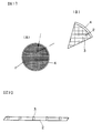
- the membrane filter element used according to the present invention comprises a sheet of hydrophilic membrane filter 1; portions of said hydrophilic membrane filter 1 consisting of a plurarity of small divisions, namely hydrophilic membrane filter sections 2; hydrophobic partitions 3, formed by, for example, printing hydrophobic resins on the membrane filter 1, as described above, to surround said hydrophilic membrane filter sections 2 substantially perfectly not only in planar extent but in vertical extent; and a frame of the membrane filter 1.
- a spray containing extracting and luminescence-inducing reagents is applied to the hydrophilic membrane filter sections 2, immediately after entrapping living organisms.
- the hydrophobic partitions 3 slightly protrude from the surface of said membrane filter sections 2 to prevent the extracted solution, containing the reagents and ATP from the living organisms, from dispersing and flowing out of the particular hydrophilic membrane filter section. Theoretically, this despersion and overflow can be prevented by applying a spray of particles of smaller size, even if the height of the protrusion is nearly zero. In this case, however, extremely fine microparticles usually require a prolonged time period for completion of spraying, and sometimes fail to achieve a preferable extraction and/or luminescence. It is thus recommended that the hydrophobic partitions 3 protrude up to some height. In addition, a membrane filter element having a large number of smaller area and partitions of narrower width is theoretically preferred, but this will cause an additional restriction of greater difficulties in manufacturing thereof.
- the above mentioned membrane filter element is mounted on a cup-shaped filter container, or the like, such as Milliflex Filter Unit - SterifilTM from Nippon Millipore Limited, etc., so that the test solution is filtrated, and living microorganisms are entrapped within a plurarity of sections confined with the above mentioned partitions.
- a cup-shaped filter container such as Milliflex Filter Unit - SterifilTM from Nippon Millipore Limited, etc.
- the membrane filter is removed and dried, and thereafter an extracting reagent is applied thereto by spraying to extract microbial ingredients on the membrane filter.
- the particle size of spray should be varied according to the area of the membrane filter sections, using, for example, an atomizer, or a nebulizer of either pneumatic or ultrasonic type (hereinafter referred to as "sprayers"). It is preferable that sprayers are those which can effect a uniform and fine spraying, and too close spraying should be avoided. In addition, spraying should be carried out uniformly all over the sections lest a too large amount of reagents should be added to a particular section to desperse the microbial ingredients over the partitions. If the above description is not well attended to, a sufficient accuracy in determination may not be achieved due to dilution of potentially luminescent ingredients.
- a luminescence-inducing reagent is sprayed onto the membrane filter sections to induce luminescence.
- a sprayer it is also preferable to carry out the spraying using a sprayer, paying attention to the above descriptions regarding spraying.
- sample The membrane filter element emitting luminescence through the above stated procedures (hereinafter referred to as "sample") is mounted to a sample holder, and, having equipped with a total reflection plate, the sample can be subjected to a bioluminescence image analyzer system (e.g., ARGUS - 50/CLTM of tapered fiber input type from Hamamatsu Photonics Co., Ltd. to count the number of living microorganisms. Measurement is carried out by processing the images after accumulation of the luminescence for 2 minutes, further processing the images to eliminate any luminescence which is considered as a noise, and finally counting the number of remained bright spots.
- a bioluminescence image analyzer system e.g., ARGUS - 50/CLTM of tapered fiber input type from Hamamatsu Photonics Co., Ltd.
- the bioluminesence image analyzing system used herein is a novel and indeed innovative device, in which even such a faint luminescence that could not be detected through conventional instruments can be detected with high sensitivity, and intensified for processing. Also, the processing and analysis of data are carried out rapidly and conveniently. Moreover, said system enables to detect even a single living microbial cell, in cooperation with the effect of spraying using an unusual membrane filter and a reagent sprayer according to the present invention, thereby considerably contributing to the actualization of this unusually excellent counting method.
- This system comprises a sample holder 9 for supporting a membrane filter element (sample) 5 after treatment with the above mentioned extracting and luminescence-inducing reagents; a total reflection plate 6; shading housing 8; a tapered fiber 7, juxtaposed to said membrane filter element as closely as possible to detect luminescence in a two-dimensional extent; an untrahighly sensitive television camera 10 consisting of a photoamplifying component and a camera tube; a camera controller 11; an image processor 12; a data analyzing apparatus 13; and a monitor television 14.
- the system equipped with ARGUS-50/CLTM of tapered fiber input type from Hamamatsu Photonics Co., Ltd., or those having a similar counting performance is particularly preferred.
- a cooled solid state camera device for example, a cooled CCD digital imaging system from Hamamatsu Photonics is available.
- the above procedure can be carried out by inverting both the tapered fiber 7 in the camera tube portion and the ultrahighly sensitive television camera 10, and placing thereon a sample holder containing a sample.
- Sample 5 is preferably placed as closely as possible to tapered fiber 7 therby to significantly enhance the measurement sensitivity.
- sprayer(s) for extracting and luminescence-inducing reagents and sample carrier, etc. may be set in combination in order to carry out an automatic counting.
- sample holder 9 bearing the sample (membrane filter element retaining microorganisms to be examined) after luminescence-inducing treatment, is placed in close contact with surfaces of the tapered fibers 7, then, the luminescence emitted from microbial bodies is introduced through ultrahigh sensitive television camera 10 and camera controller 11 into image processor 12, where photons are accumulated for 30 through 180 seconds, e.g., 120 seconds in a two-dimensional extent to pick up the image.
- Data analyzing apparatus 13 then processes the image to eliminate any weaker noise luminescence, and displays, at monitor television 13, the only remained bright luminescence originated from living microorganisms as a bright spot. By this processing, luminescence from other than microbial bodies are erased, resulting in substantial correspondence of the counted number of bright spots to the number of living microorganisms.
- the microbial ingredient to be extracted is ATP.
- ATP extracting reagent e.g., NRBTM, Lumac Co.
- the reagent is sprayed in the form of very fine particles, using a sprayer (e.g., Ultrasonic AspiratorTM, Matsushita Electric Industries Co., Ltd.), to extract ATP from living organisms entrapped within the membrane filter sections.
- Luminescence-inducing reagent e.g., Lumit-PMTM, Lumac Co.
- the above bioluminescence image analyzing system As stated above, thus obtained sample is then subjected to the above bioluminescence image analyzing system to accumulate the luminescence for 30 through 180 second, and to display the only luminescence brighter than backgroud luminescence as a bright spot in the monitor television.
- the maximum brightness of the background luminescence i.e., background level
- an equal volume of the same test solution as is to be examined is prepared and sterilized. After filtrating this solution on the membrane filter according to the present invention, the membrane filter is treated in the same manner as described above to induce luminescence.
- the maximum brightness of the bright spot displayed in the monitor television when thus obtained sample is subjected to said bioluminescence image analyzing system is the background level (hereinafter referred to as "threshold").
- the bright spots displayed in the monitor television from the rest of the luminescence i.e., luminescence brighter than the threshold
- a membrane filter element consisting of a plurarity of small membrane filter sections and having hydrophobic partitions slightly protruding above the filter surface is employed, so that all the living microorganisms are entrapped within any one of said sections.
- an extracting solution ATP extracting solution
- luminescence-inducing reagent luciferin-lrciferase reagent
- a contrivance is made that the dilution within a section may be retarded so far as possible.
- Luminescent microbial ingredients are thereby retained in higher concentrations, which enables to easily determine even an extremely small amount of microbial ingredients.
- a television camera head composed of a tapered fiber, photoamplifier, and a camera tube has enabled luminescence originated from living microorganisms to be indicated (recognized) as very bright spots, so that any noise luminescence from other materials than living microorganisms can easily be eliminated in comparison with a threshold luminescence, thus enabling to count the number of living microorganisms automatically, rapidly, and conveniently, even when the number is extremely small (e.g., less than a few cells/100 ml of test solution).
- the number is extremely small (e.g., less than a few cells/100 ml of test solution).
- it is rather significant to judge the existence of a single living microorganism in sample solutions as of the case in coliform test for foods or cooling beverages.
- the membrane filter element after filtrating test solutions and entrapping living microorganisms are, more preferably, placed on a pad or nutrient agar plate containing the most suitable nutrients for the growth of said living microorganisms.
- a short-term (e.g., several hours) culture significantly luminous bright spots will be available, thus providing a more accurate judgement means.
- the culturing condition to such extent does not naturally allow a single cell to form any colonies.
- Saccharomyces cerevisiae (IFO 0209) cultured in a glucose peptone medium (Eiken Chemicals Co., Ltd.) at 30°C for overnight was diluted with physiological saline to microbial concentration of about 20 CFU/ml (CFU: colony forming unit), and 1 ml of the solution was collected as a test solution.
- CFU colony forming unit
- a membrane filter made of hydrophilic polycarbonate with pore size of 0.45 ⁇ m and diameter of 25 mm latticed hydrophobic partitions as shown in Figs. 1 and 2 were printed, using an ink of well transmittable and readily curable properties with UV irradiation, and conventional screen printing technique, to obtain a membrane filter element comprising square-shaped hydrophilic sections with side length of 0.5 mm surrounded by the partitions with height above the surface of the membrane of 20 ⁇ m and width of 0.2 mm.
- the membrane filter was then removed form the filter equipment, dried, and mounted on a sample holder.
- An ATP extracting reagent made by Lumac Co. (NRBTM) was sprayed onto the membrane filter for 10 seconds using an atomizer (Koike Chemicals Co.) held above it in an angle of 45° at a distance of about 15 cm, carefully lest large droplets should be splashed.
- luciferin-luciferase luminescence-inducing reagent (Lumit-PMTM, Lumac Co.) was sprayed for 10 seconds using the same atomizer, to allow the membrane filter to emit luminescence.
- the membrane filter was subjected to ARGUS - 50/CLTM bioluminescence image analyzing system (Hamamatsu Photonics Co.), and photon accumulation was carried out for 2 minutes. After displaying the luminescence as bright spots on the television monitor, noise luminescence was eliminated to count the number of living microorganisms. The results obtained are shown in Table 1.
- Saccharomyces cervisiae (IFO 0209) cultured in the same manner as described in Example 1 was diluted with physiological saline to microbial concentration of about 200 CFU/ml, and 1 ml of the solution was collected as a test solution. Following the same procedures as described in Example 1 to count the number of the bright spots, the results shown in Table 2 were obtained.
- Pseudomonas diminuta (IFO 14213) was diluted with physiological saline to microbial concentration of about 50 CFU/ml, and 1 ml was collected as a test solution.
- a membrane filter made of hydrophilic polytetrafluoroethylene with pore size of 0.45 ⁇ m and diameter of 25 mm square-shaped hydrophilic sections with side length of 0.4 mm, and partitions with height of 20 ⁇ m and width of 0.3 mm were produced using an ink permeating into the membrane to some degree, of well transmittable and readily curable properties by UV irradiation, and the conventional screen printing technique.
- the test solution was filtrated as described in Example 1.
- the membrane filter was then removed, dried, extracted, and allowed to emit luminescence as described in Example 1. After it was subjected to image analyzing process to eliminate background luminescence, the number of bright spots were counted.
- Table 3 Exp. No. Standard Agar Plate (CFU/ml test solution) Present Invention (bright spot/element) 1 62 53 2 45 41 3 48 46
- Escherichia coli (IFO 13898) was diluted with physiological saline to microbial concentration of about 100 CFU/ml, and 1 ml of the solution was collected as a test solution.
- the test solution was filtrated. Then, the membrane filter was removed, dried, extracted, and allowed to emit luminescence. After it was subjected to image analyzing process to put out background luminescence, the number of bright spots was counted.
- Table 4 Exp. No. Standard Agar Plate (CFU/ml test solution) Present Invention (bright spot/element) 1 113 108 2 105 112 3 106 98
- a membrane filter element was used, in which latticed hydrophobic partitions with height of 20 ⁇ m and width of 0.3 mm were printed on a membrane filter (25 mm diameter) made of hydrophilic poly(vinylidene difluoride), using an ink permeating into the membrane to some degree, of well transmittable and readily curable properties by UV irradiation, and the conventional screen printing technique, to form square-shaped hydrophilic membrane filter sections with side length of 0.3 mm.
- the membrane filter was washed, and dried, then an extracting reagent (NRBTM, Lumac CO.) was sprayed for 10 seconds through an ultrasonic type sprayer (Matsushita Electric Industries) at a distance of about 10 cm, followed by spraying a luminescence-inducing reagent (Lumit-PMTM, Lumac Co.) for 10 seconds in the same way as above.
- the membrane filter was then subjected to image analyzing process in the same procedures as in Example 1, using a system as described referring to Fig. 3, to eliminate any noise luminescence. The number of bright spots was counted to obtain results as shown in Table 5.
- a membrane filter made of hydrophobic polytetrafluoroethylene with pore size of 0.45 ⁇ m, diameter of 25 mm, and thickness of 50 ⁇ m was wetted down with methanol, and washed with water following the procedure described in Example 34 in U. S. Patent No. 4,618,533 (to Michael J. Steuck).
- Example 9 Using a similar membrane filter as described in Example 8, and following the same procedures as described in Example 5, the test solution was filtrated, and the membrane filter element was washed, dried, extracted, and allowed to emit luminescence. Then, image analyzing process was carried out to put out any noise luminescence, and the number of bright spots was counted to obtain the results as shown in Table 9.
Landscapes
- Chemical & Material Sciences (AREA)
- Life Sciences & Earth Sciences (AREA)
- Organic Chemistry (AREA)
- Health & Medical Sciences (AREA)
- Engineering & Computer Science (AREA)
- Zoology (AREA)
- Wood Science & Technology (AREA)
- Proteomics, Peptides & Aminoacids (AREA)
- Molecular Biology (AREA)
- Bioinformatics & Cheminformatics (AREA)
- Immunology (AREA)
- Microbiology (AREA)
- Biophysics (AREA)
- Analytical Chemistry (AREA)
- Physics & Mathematics (AREA)
- Genetics & Genomics (AREA)
- Biochemistry (AREA)
- Biotechnology (AREA)
- General Engineering & Computer Science (AREA)
- General Health & Medical Sciences (AREA)
- Toxicology (AREA)
- Pathology (AREA)
- Measuring Or Testing Involving Enzymes Or Micro-Organisms (AREA)
- Apparatus Associated With Microorganisms And Enzymes (AREA)
- Investigating Or Analysing Materials By The Use Of Chemical Reactions (AREA)
Claims (15)
- Verfahren zum Zählen der Anzahl an lebenden Mikroorganismen in einer Testlösung durchFiltrieren der betreffenden Lösung durch ein Membranfilterelement mit einer Mehrzahl von kleinen hydrophilen Membranfilterabschnitten, die im wesentlichen von einer Mehrzahl von hydrophoben Trenneinsätzen bzw. -wänden umgeben sind, so daß die in der Lösung enthaltenen lebenden Mikroorganismen in diesen Abschnitten festgehalten werden können;Trocknen des Membranfilterelements;Aufsprühen eines Extraktionsreagens auf das Membranfilterelement zur Extraktion eines möglicherweise lumineszierenden Bestandteils der in den betreffenden Abschnitten eingeschlossenen bzw. festgehaltenen lebenden Mikroorganismen;Aufsprühen eines Lumineszenz induzierenden Reagens auf das Element, damit der lumineszierende Bestandteil innerhalb der betreffenden Abschnitte Lumineszenz emittieren kann;Darstellen der emittierten Lumineszenz als hellen Fleck unter Benutzung eines Biolumineszenzbildanalysesystems undZählen der Zahl der hellen Flecken als Repräsentanten für die Zahl der lebenden Mikroorganismen in der Testlösung.
- Verfahren nach Anspruch 1, wobei zusätzlich das Membranfilterelement nach dem Filtrieren der Probenlösung und vor dem Trocknen des Membranfilterelements auf ein geeignetes Kulturmedium für das Wachstum der lebenden Mikroorganismen gelegt und die lebenden Mikroorganismen kurzzeitig zur Erhöhung ihrer Zahl gezüchtet werden.
- Verfahren nach Anspruch 1 oder 2, wobei die hydrophoben Trenneinsätze bzw. -wände aus der Oberfläche der hydrophilen Membranfilterabschnitte, durch welche die Testlösung hindurchlaufen gelassen wird, herausragen.
- Verfahren nach Anspruch 3, wobei die hydrophoben Trenneinsätze bzw. -wände in einer Höhe von 0,01 bis 0,05 mm über die Oberfläche der hydrophilen Membranfilterabschnitte herausragen und eine Breite von 0,1 bis 2 mm aufweisen.
- Verfahren nach einem der Ansprüche 1 bis 4, wobei der möglicherweise lumineszierende Bestandteil der lebenden Mikroorganismen aus Adenosin-5'-triphosphat (ATP) und das Lumineszenz induzierende Reagens aus LuciferinLuciferase-Reagens bestehen.
- Verfahren nach einem der Ansprüche 1 bis 5, wobei jeder der hydrophilen Membranfilterabschnitte eine Oberfläche zwischen 0,04 mm2 und 2 mm2 aufweist.
- Verfahren nach einem der Ansprüche 1 bis 6, wobei die hydrophoben Trenneinsätze bzw. -wände durch Aufdrucken, Aufstempeln bzw. -prägen oder Aufzeichnen eines Musters aus einem hydrophoben Werkstoff, ausgewählt aus der Gruppe Wachse, Epoxyharze, Siliconharzwachse, Siliconöle, fluorierte Harzwachse und Polystyrolharze, auf ein hydrophiles Membranfilter zur Herstellung der betreffenden Membranfilterabschnitte oder mit Hilfe eines Photosensibilisatorverfahrens unter Verwendung lichtempfindlicher Harze als Rohmaterialien für das Membranfilterelement gebildet werden.
- Verfahren nach einem der Ansprüche 1 bis 4 und 7, wobei die hydrophilen Membramfilterabschnitte aus einem hydrophilen Werkstoff, ausgewählt aus der Gruppe Polytetrafluorethylen, Poly(vinylidenfluorid), Polycarbonat, Polyamid, Polysulfon, Polymerlegierungen hiervon, Acetylcellulose, Nitrocellulose und Mischungen, derselben hergestellt sind.
- Verfahren nach einem der Ansprüche 1 bis 4, wobei das Aufsprühen von Reagenzien durch Aufsprühen eines Nebels einer Teilchengröße zwischen 5 µm bis 20 µm unter Verwendung einer Sprühpistole oder eines Zerstäubers vom entweder pneumatischen Typ oder Ultraschall-Typ erfolgt.
- Verfahren nach einem der Ansprüche 1 bis 4, wobei das Biolumineszenzbildanalysesystem einen Fernsehkamerakopf aus einem konischen Faserlichtleiter, einem Lichtverstärker und einer Kameraröhre oder einem konischen Faserlichtleiter und einer gekühlten CCD-Videokamera enthält.
- Verfahren nach einem der Ansprüche 1 bis 10, wobei ein Probenhalter verwendet wird und dieser das Membranfilterelement mit einer daneben anordneten total reflektierenden Scheibe enthält.
- Verfahren nach einem der Ansprüche 1 bis 4 und 10, wobei das photoelektrische Bildanalysesystem eine Bildverarbeitung zur Eliminierung eines von irgendwelchen anderen Substanzen als den nachzuweisenden Mikroorganismen herrührenden Lumineszenzrauschens und zur Zählung der hellen Punkte oberhalb eines vorgegebenen Niveaus durchführt.
- Verfahren nach einem der Ansprüche 1 bis 4 und 10, wobei das Membranfilterelement mit einer Reihe von hydrophilen Membranfilterabschnitten, die von einer Reihe von hydrophoben Trennwänden umgeben sind, durch Beschichten einer porösen hydrophoben Filtermembran mit einem polymerisierbaren Monomer mit der Fähigkeit, die hydrophobe Membran mit einer hydrophilen Oberfläche zu versehen, Einwirkenlassen einer UV-Strahlung unter Maskierung der (später) die hydrophoben Trennwände bildenden Flächen zur Polymerisation des Monomers an die oder zur Copolymerisation des Monomers mit der poröse(n) hydrophobe(n) Filtermembran oder zur Polymerisation des Monomers als solchem zur Umwandlung der nicht maskierten Flächen zur Hydrophilisierung der nicht maskierten Fläche hergestellt wird.
- Verfahren nach Anspruch 13, wobei die poröse hydrophobe Filtermembran aus der Gruppe Polytetrafluorethylen, Polyvinylidenfluorid und Polycarbonat ausgewählt ist.
- Verfahren nach Anspruch 1, wobei das Aufsprühen in einem derart feinverteilten Zustand erfolgt, daß es während der ATP-Extraktions- und Lumineszenzemissionsstufen nicht zu einer nennenswerten Verdünnung und Diffusion kommen kann.
Applications Claiming Priority (3)
| Application Number | Priority Date | Filing Date | Title |
|---|---|---|---|
| JP4061591 | 1991-02-13 | ||
| JP40615/91 | 1991-02-13 | ||
| PCT/JP1992/000145 WO1992014838A1 (fr) | 1991-02-13 | 1992-02-13 | Procede de determination quantitative de micro-organismes viables |
Publications (3)
| Publication Number | Publication Date |
|---|---|
| EP0529084A1 EP0529084A1 (de) | 1993-03-03 |
| EP0529084A4 EP0529084A4 (en) | 1994-06-01 |
| EP0529084B1 true EP0529084B1 (de) | 1997-05-28 |
Family
ID=12585434
Family Applications (1)
| Application Number | Title | Priority Date | Filing Date |
|---|---|---|---|
| EP92904999A Expired - Lifetime EP0529084B1 (de) | 1991-02-13 | 1992-02-13 | Verfahren zur bestimmung der zahl lebender mikroorganismen |
Country Status (5)
| Country | Link |
|---|---|
| US (2) | US5627042A (de) |
| EP (1) | EP0529084B1 (de) |
| JP (1) | JP3133328B2 (de) |
| DE (1) | DE69219963T2 (de) |
| WO (1) | WO1992014838A1 (de) |
Cited By (3)
| Publication number | Priority date | Publication date | Assignee | Title |
|---|---|---|---|---|
| EP1826276A1 (de) * | 2006-02-24 | 2007-08-29 | Millipore Corporation | Mikrobiologisches Schnellanalyseverfahren |
| EP1826275A1 (de) * | 2006-02-24 | 2007-08-29 | Millipore Corporation | Mikrobiologisches Schnellanalyseverfahren |
| US8642321B2 (en) | 2007-04-26 | 2014-02-04 | Emd Millipore Corporation | Microbiological analysis assembly and method |
Families Citing this family (61)
| Publication number | Priority date | Publication date | Assignee | Title |
|---|---|---|---|---|
| EP0529084B1 (de) * | 1991-02-13 | 1997-05-28 | Nihon Millipore Kogyo Kabushiki Kaisha | Verfahren zur bestimmung der zahl lebender mikroorganismen |
| DE563858T1 (de) * | 1992-04-01 | 1994-03-03 | Nihon Millipore Kogyo K K | Verfahren zur Bestimmung der Anzahl lebender Mikroorganismen. |
| JP3228812B2 (ja) * | 1993-02-10 | 2001-11-12 | 日本マイクロリス株式会社 | 生菌数を測定する方法 |
| FR2719602B1 (fr) * | 1994-05-05 | 1996-07-26 | Biocom Sa | Procédé et installation pour la numérisation de cellules et micro-organismes, notamment des produits alimentaires ou des fluides biologiques. |
| WO1996030542A1 (en) * | 1995-03-28 | 1996-10-03 | Idemitsu Kosan Company Limited | Method for rapidly determining number of bacteria and equipment for determining number of bacteria |
| FR2735255B1 (fr) * | 1995-06-12 | 1998-06-19 | Biocom Sa | Procede de numerisation de particules faiblement luminescentes |
| GB9626932D0 (en) * | 1996-12-24 | 1997-02-12 | Lumitech Limited | Assay methods and kits therefor |
| US6703211B1 (en) * | 1998-03-13 | 2004-03-09 | Promega Corporation | Cellular detection by providing high energy phosphate donor other than ADP to produce ATP |
| EP0974827A3 (de) | 1998-07-09 | 2002-10-02 | Sapporo Breweries Ltd. | Gerät zur Vorbereitung von Proben inklusive Sprühgerät |
| US6197254B1 (en) | 1999-01-11 | 2001-03-06 | International Food Protection | Self-contained assaying apparatus |
| US6328931B1 (en) | 1999-01-11 | 2001-12-11 | International Food Protection, Inc. | Analyte collection and assaying assembly |
| WO2001019504A2 (en) | 1999-09-17 | 2001-03-22 | Millipore Corporation | Three dimensional patterned porous structures |
| JP3405301B2 (ja) | 1999-12-17 | 2003-05-12 | 理化学研究所 | 生体組織スライス標本用実験装置および標本保持具 |
| WO2001059157A2 (en) * | 2000-02-08 | 2001-08-16 | Millipore Corporation | Process for the enumeration and identification of microorganisms |
| US6927851B2 (en) | 2000-03-31 | 2005-08-09 | Neogen Corporation | Methods and apparatus to improve the sensitivity and reproducibility of bioluminescent analytical methods |
| US6653147B2 (en) | 2000-03-31 | 2003-11-25 | Neogen Corporation | Apparatus and method for chemiluminescent assays |
| AU2001219539A1 (en) * | 2000-09-05 | 2002-03-22 | Wesley L. Bradford | Reverse osmosis membrane and process for making same |
| IL139593A (en) * | 2000-11-09 | 2010-12-30 | Biogem Optical Ltd | Method for the detection of viable microorganisms |
| US7588886B2 (en) * | 2001-01-26 | 2009-09-15 | Millipore Corporation | Process for the enumeration and identification of microorganisms |
| US7268229B2 (en) * | 2001-11-02 | 2007-09-11 | Promega Corporation | Compounds to co-localize luminophores with luminescent proteins |
| WO2003100086A1 (fr) * | 2002-05-23 | 2003-12-04 | Fuji Electric Holdings Co., Ltd. | Procede et dispositif de comptage de cellules vivantes |
| WO2003104383A1 (en) * | 2002-06-07 | 2003-12-18 | Millipore Corporation | Kit and process for microbiological for on-site examination of a liquid sample |
| AU2003253840A1 (en) * | 2002-07-11 | 2004-02-02 | Pall Corporation | Uv treated membranes |
| EP1539993A4 (de) * | 2002-08-01 | 2006-11-22 | Merck & Co Inc | Neuartiges verfahren zur anzucht, abbildung und auszahlung von mikrobenkolonien für serologische oder screening-assays |
| JP4429577B2 (ja) * | 2002-09-30 | 2010-03-10 | 株式会社ニコン | 細胞内反応測定装置および細胞内反応測定方法 |
| KR100573621B1 (ko) * | 2003-07-18 | 2006-04-25 | 주식회사 디지탈바이오테크놀러지 | 세포 개체수 계수용 장치 및 그 제조방법 |
| CA2557487C (en) * | 2004-03-01 | 2015-04-21 | Mycometer Aps | Detection of contaminants retained on a filter by a detectable moiety-producing substrate |
| US20050236318A1 (en) * | 2004-04-23 | 2005-10-27 | Millipore Corporation | Low holdup volume multiwell plate |
| US20050236319A1 (en) * | 2004-04-23 | 2005-10-27 | Millipore Corporation | Pendant drop control in a multiwell plate |
| US20050236317A1 (en) * | 2004-04-23 | 2005-10-27 | Millipore Corporation | Pendant drop control in a multiwell plate |
| JP2006223230A (ja) * | 2005-02-18 | 2006-08-31 | Institute Of National Colleges Of Technology Japan | 生菌検出方法および生菌検出用容器 |
| GB0513535D0 (en) * | 2005-07-01 | 2005-08-10 | Iseao Technologies Ltd | Improved methods of detecting microbes |
| FR2897941B1 (fr) * | 2006-02-24 | 2009-01-16 | Millipore Corp | Dispositif et procede pour l'analyse microbiologique rapide. |
| FR2897783B1 (fr) * | 2006-02-24 | 2008-05-30 | Millipore Corp | Dispositif pour le controle microbiologique, ensembles de controle et d'incubation le comportant et procede le mettant en oeuvre |
| US7628823B2 (en) * | 2007-01-22 | 2009-12-08 | Washing Systems, Llc | Method of testing for ATP load in commercial laundry and for data tracking the results |
| US7871444B2 (en) * | 2007-01-22 | 2011-01-18 | Washing Systems, Llc | Method of testing for ATP load in commercial laundry and for data tracking the results |
| US8858681B2 (en) * | 2007-04-23 | 2014-10-14 | W. L. Gore & Associates, Inc. | Patterned porous venting materials |
| US7927405B2 (en) * | 2007-04-23 | 2011-04-19 | Gore Enterprise Holdings, Inc | Porous composite article |
| WO2008128760A1 (en) | 2007-04-23 | 2008-10-30 | W.L Gore & Associates Gmbh | Composite material |
| WO2010058373A1 (en) * | 2008-11-24 | 2010-05-27 | Koninklijke Philips Electronics N.V. | Method and apparatus for rapid filter analysis of fluid samples |
| EP2194368B1 (de) | 2008-12-03 | 2019-07-17 | Grundfos Management A/S | Sensorsystem zum Erfassen und Spezifizieren von einzelnen Partikeln in einem Fluid |
| US20130045496A1 (en) | 2010-02-12 | 2013-02-21 | Gijsbert Johan Jansen | Automatic Fluid Sample Preparation Module, Automatic Analysis System and Method for Use Thereof |
| WO2011099862A1 (en) * | 2010-02-12 | 2011-08-18 | Dutch Water Technologies B.V. | Automatic toxicological fluid sample preparation module, automatic analysis system and method for use thereof |
| US8808848B2 (en) | 2010-09-10 | 2014-08-19 | W. L. Gore & Associates, Inc. | Porous article |
| CN104662144B (zh) | 2012-09-25 | 2018-05-01 | 通用电气公司 | 用于生物负载样品收集和检测的可弃容器 |
| US12350627B2 (en) | 2013-02-28 | 2025-07-08 | Aqua Membranes, Inc. | Permeate flow patterns |
| EP2859933A3 (de) * | 2013-10-11 | 2015-06-03 | Andreas Massold | Filter |
| JP5947476B1 (ja) * | 2015-03-31 | 2016-07-06 | 第一公害プラント株式会社 | バチルス属細菌の計数方法 |
| JP2019529099A (ja) | 2016-09-20 | 2019-10-17 | アクア メンブレインズ エルエルシー | 透過流パターン |
| KR102033982B1 (ko) | 2016-11-19 | 2019-10-18 | 아쿠아 멤브레인스 엘엘씨 | 나선형 권취 요소를 위한 간섭 패턴 |
| JP7112736B2 (ja) * | 2017-01-18 | 2022-08-04 | 国立研究開発法人農業・食品産業技術総合研究機構 | 半透膜及びその使用 |
| WO2018190937A1 (en) | 2017-04-12 | 2018-10-18 | Aqua Membranes Llc | Graded spacers for filtration wound elements |
| CN120094407A (zh) | 2017-04-20 | 2025-06-06 | 阿夸曼布拉尼斯公司 | 用于螺旋卷绕元件的不嵌套、不变形图案 |
| WO2018195367A1 (en) | 2017-04-20 | 2018-10-25 | Aqua Membranes Llc | Mixing-promoting spacer patterns for spiral-wound elements |
| CN111344053A (zh) | 2017-10-13 | 2020-06-26 | 阿夸曼布拉尼斯公司 | 螺旋缠绕元件的桥支撑件和减少的进给间隔件 |
| WO2020154734A1 (en) | 2019-01-27 | 2020-07-30 | Aqua Membranes Inc. | Composite membranes |
| US12303838B2 (en) | 2019-08-06 | 2025-05-20 | Aqua Membranes, Inc. | Preferred flow paths for spiral-wound elements |
| EP3865583A1 (de) * | 2020-02-14 | 2021-08-18 | Testo bioAnalytics GmbH | Verfahren zum nachweis von mikroorganismen und scheibenförmiger probenträger |
| US11633700B2 (en) | 2020-04-07 | 2023-04-25 | Aqua Membranes Inc. | Independent spacers and methods |
| CN118742376B (zh) | 2021-12-28 | 2025-12-05 | 阿夸曼布拉尼斯公司 | 具有保护特征的高截留率螺旋卷绕元件 |
| CN114591823A (zh) * | 2022-04-07 | 2022-06-07 | 河北木美土里科技有限公司 | 一种展示微生物的培养皿及其制作方法 |
Family Cites Families (10)
| Publication number | Priority date | Publication date | Assignee | Title |
|---|---|---|---|---|
| US3616253A (en) * | 1967-05-01 | 1971-10-26 | Du Pont | Method for determining bacterial populations |
| US4587213A (en) * | 1971-09-29 | 1986-05-06 | Malecki George J | Methods and means of determining microorganism population |
| US3929583A (en) * | 1975-08-14 | 1975-12-30 | Canadian Patents Dev | Apparatus for enumerating microorganisms |
| US4618533A (en) * | 1984-11-30 | 1986-10-21 | Millipore Corporation | Porous membrane having hydrophilic surface and process |
| US4908236A (en) * | 1986-12-04 | 1990-03-13 | Millipore Corporation | Transparent porous membrane having hydrophilic surface and process |
| JPH0257197A (ja) * | 1988-02-03 | 1990-02-26 | Japan Organo Co Ltd | 生菌の定量法 |
| JPH02163098A (ja) * | 1988-12-14 | 1990-06-22 | Japan Organo Co Ltd | 生菌の検出法 |
| JPH04257197A (ja) * | 1991-02-08 | 1992-09-11 | Nippon Telegr & Teleph Corp <Ntt> | 遠隔監視装置 |
| EP0529084B1 (de) * | 1991-02-13 | 1997-05-28 | Nihon Millipore Kogyo Kabushiki Kaisha | Verfahren zur bestimmung der zahl lebender mikroorganismen |
| DE563858T1 (de) * | 1992-04-01 | 1994-03-03 | Nihon Millipore Kogyo K K | Verfahren zur Bestimmung der Anzahl lebender Mikroorganismen. |
-
1992
- 1992-02-13 EP EP92904999A patent/EP0529084B1/de not_active Expired - Lifetime
- 1992-02-13 DE DE69219963T patent/DE69219963T2/de not_active Expired - Lifetime
- 1992-02-13 JP JP04505080A patent/JP3133328B2/ja not_active Expired - Fee Related
- 1992-02-13 WO PCT/JP1992/000145 patent/WO1992014838A1/ja not_active Ceased
-
1995
- 1995-06-01 US US08/456,350 patent/US5627042A/en not_active Expired - Lifetime
-
1997
- 1997-03-17 US US08/819,424 patent/US5811251A/en not_active Expired - Lifetime
Cited By (6)
| Publication number | Priority date | Publication date | Assignee | Title |
|---|---|---|---|---|
| EP1826276A1 (de) * | 2006-02-24 | 2007-08-29 | Millipore Corporation | Mikrobiologisches Schnellanalyseverfahren |
| EP1826275A1 (de) * | 2006-02-24 | 2007-08-29 | Millipore Corporation | Mikrobiologisches Schnellanalyseverfahren |
| FR2897873A1 (fr) * | 2006-02-24 | 2007-08-31 | Millipore Corp | Procede d'analyse microbiologique rapide. |
| FR2897872A1 (fr) * | 2006-02-24 | 2007-08-31 | Millipore Corp | Procede d'analyse microbiologique rapide. |
| US8642321B2 (en) | 2007-04-26 | 2014-02-04 | Emd Millipore Corporation | Microbiological analysis assembly and method |
| US9102909B2 (en) | 2007-04-26 | 2015-08-11 | Emd Millipore Corporation | Microbiological analysis assembly and method |
Also Published As
| Publication number | Publication date |
|---|---|
| JP3133328B2 (ja) | 2001-02-05 |
| EP0529084A4 (en) | 1994-06-01 |
| EP0529084A1 (de) | 1993-03-03 |
| US5811251A (en) | 1998-09-22 |
| WO1992014838A1 (fr) | 1992-09-03 |
| US5627042A (en) | 1997-05-06 |
| DE69219963T2 (de) | 1997-10-16 |
| DE69219963D1 (de) | 1997-07-03 |
Similar Documents
| Publication | Publication Date | Title |
|---|---|---|
| EP0529084B1 (de) | Verfahren zur bestimmung der zahl lebender mikroorganismen | |
| JPWO1992014838A1 (ja) | 生菌数測定法 | |
| EP0563858A1 (de) | Verfahren zur Bestimmung der Anzahl lebender Mikroorganismen | |
| JP3228812B2 (ja) | 生菌数を測定する方法 | |
| EP0816513B1 (de) | Druckempfindliches klebendes Blatt zum Nachweis von Mikroorganismen und Methode zum Nachweis von Mikroorganismen | |
| CA1125152A (en) | Selective determination of viable somatic and microbial cells | |
| JP2648376B2 (ja) | 微生物の検出計数装置および方法 | |
| US20050202523A1 (en) | Method for the detection of viable microorganisms | |
| WO2003012397A1 (fr) | Puce collectrice de micro-organismes, trousse collectrice de micro-organismes, methode de quantification de micro-organismes, specimen destine a confirmer l'etat normal d'un appareil de quantification de micro-organismes et ledit appareil de quantification de micro-organismes | |
| Singh et al. | Rapid enumeration of viable bacteria by image analysis | |
| CA1201963A (en) | Method of concentrating and measuring unicellular organisms | |
| EP0333560A1 (de) | Verfahren zur Bestimmung von mikro-organismen | |
| JPH11137293A (ja) | 微生物数の迅速測定法とそれに使用するろ過膜 | |
| GB2152660A (en) | Analysis of biological products | |
| JP2005287337A (ja) | 糸状菌計量方法 | |
| JP4118927B2 (ja) | 微生物計量装置の正常状態確認検査用検体 | |
| FR2556741B1 (fr) | Procede et dispositif de detection de l'activite d'une substance sur un micro-organisme ou sur un melange de micro-organismes | |
| JP2000069994A (ja) | 微生物検出方法および微生物検出システム | |
| JP3678469B2 (ja) | 微生物数の迅速測定法 | |
| CN101054554B (zh) | 用于提高微生物细胞壁透性的组合物和用于在膜上检测所述微生物的方法 | |
| CN114729880A (zh) | 用于分析流体样品的生物活性的方法和系统 | |
| JPH11178596A (ja) | 微生物検出用シート状物、それを含む微生物検出キットおよびそれを用いた微生物検出方法 | |
| JP3208892B2 (ja) | 生菌数の測定方法 | |
| JP4004091B2 (ja) | 微生物試験用粘着性シートを用いた微生物試験方法 | |
| JP3529453B2 (ja) | 大腸菌群の選択的検出方法 |
Legal Events
| Date | Code | Title | Description |
|---|---|---|---|
| PUAI | Public reference made under article 153(3) epc to a published international application that has entered the european phase |
Free format text: ORIGINAL CODE: 0009012 |
|
| AK | Designated contracting states |
Kind code of ref document: A1 Designated state(s): DE FR GB |
|
| 17P | Request for examination filed |
Effective date: 19930302 |
|
| A4 | Supplementary search report drawn up and despatched | ||
| AK | Designated contracting states |
Kind code of ref document: A4 Designated state(s): DE FR GB |
|
| GRAG | Despatch of communication of intention to grant |
Free format text: ORIGINAL CODE: EPIDOS AGRA |
|
| GRAH | Despatch of communication of intention to grant a patent |
Free format text: ORIGINAL CODE: EPIDOS IGRA |
|
| 17Q | First examination report despatched |
Effective date: 19960909 |
|
| GRAH | Despatch of communication of intention to grant a patent |
Free format text: ORIGINAL CODE: EPIDOS IGRA |
|
| GRAA | (expected) grant |
Free format text: ORIGINAL CODE: 0009210 |
|
| AK | Designated contracting states |
Kind code of ref document: B1 Designated state(s): DE FR GB |
|
| REF | Corresponds to: |
Ref document number: 69219963 Country of ref document: DE Date of ref document: 19970703 |
|
| ET | Fr: translation filed | ||
| PLBE | No opposition filed within time limit |
Free format text: ORIGINAL CODE: 0009261 |
|
| STAA | Information on the status of an ep patent application or granted ep patent |
Free format text: STATUS: NO OPPOSITION FILED WITHIN TIME LIMIT |
|
| 26N | No opposition filed | ||
| REG | Reference to a national code |
Ref country code: GB Ref legal event code: IF02 |
|
| RAP2 | Party data changed (patent owner data changed or rights of a patent transferred) |
Owner name: NIHON MILLIPORE KOGYO KABUSHIKI KAISHA |
|
| REG | Reference to a national code |
Ref country code: GB Ref legal event code: 732E |
|
| REG | Reference to a national code |
Ref country code: FR Ref legal event code: TP Ref country code: FR Ref legal event code: CD |
|
| PGFP | Annual fee paid to national office [announced via postgrant information from national office to epo] |
Ref country code: FR Payment date: 20100303 Year of fee payment: 19 |
|
| PGFP | Annual fee paid to national office [announced via postgrant information from national office to epo] |
Ref country code: GB Payment date: 20100224 Year of fee payment: 19 Ref country code: DE Payment date: 20100226 Year of fee payment: 19 |
|
| GBPC | Gb: european patent ceased through non-payment of renewal fee |
Effective date: 20110213 |
|
| REG | Reference to a national code |
Ref country code: FR Ref legal event code: ST Effective date: 20111102 |
|
| REG | Reference to a national code |
Ref country code: DE Ref legal event code: R119 Ref document number: 69219963 Country of ref document: DE Effective date: 20110901 |
|
| PG25 | Lapsed in a contracting state [announced via postgrant information from national office to epo] |
Ref country code: FR Free format text: LAPSE BECAUSE OF NON-PAYMENT OF DUE FEES Effective date: 20110228 |
|
| PG25 | Lapsed in a contracting state [announced via postgrant information from national office to epo] |
Ref country code: GB Free format text: LAPSE BECAUSE OF NON-PAYMENT OF DUE FEES Effective date: 20110213 |
|
| PG25 | Lapsed in a contracting state [announced via postgrant information from national office to epo] |
Ref country code: DE Free format text: LAPSE BECAUSE OF NON-PAYMENT OF DUE FEES Effective date: 20110901 |