EP0533976B1 - Automatische Optimalisierung der gleichmässigen Intensität eines Bildes für ein Ultraschallabbildungsystem - Google Patents
Automatische Optimalisierung der gleichmässigen Intensität eines Bildes für ein Ultraschallabbildungsystem Download PDFInfo
- Publication number
- EP0533976B1 EP0533976B1 EP19910116397 EP91116397A EP0533976B1 EP 0533976 B1 EP0533976 B1 EP 0533976B1 EP 19910116397 EP19910116397 EP 19910116397 EP 91116397 A EP91116397 A EP 91116397A EP 0533976 B1 EP0533976 B1 EP 0533976B1
- Authority
- EP
- European Patent Office
- Prior art keywords
- ultrasound
- average amplitude
- amplitude value
- image
- uis
- Prior art date
- Legal status (The legal status is an assumption and is not a legal conclusion. Google has not performed a legal analysis and makes no representation as to the accuracy of the status listed.)
- Expired - Lifetime
Links
Images
Classifications
-
- G—PHYSICS
- G01—MEASURING; TESTING
- G01S—RADIO DIRECTION-FINDING; RADIO NAVIGATION; DETERMINING DISTANCE OR VELOCITY BY USE OF RADIO WAVES; LOCATING OR PRESENCE-DETECTING BY USE OF THE REFLECTION OR RERADIATION OF RADIO WAVES; ANALOGOUS ARRANGEMENTS USING OTHER WAVES
- G01S7/00—Details of systems according to groups G01S13/00, G01S15/00, G01S17/00
- G01S7/52—Details of systems according to groups G01S13/00, G01S15/00, G01S17/00 of systems according to group G01S15/00
- G01S7/52017—Details of systems according to groups G01S13/00, G01S15/00, G01S17/00 of systems according to group G01S15/00 particularly adapted to short-range imaging
- G01S7/52023—Details of receivers
- G01S7/52033—Gain control of receivers
Definitions
- the invention relates to a method and apparatus for the automatic optimization of image uniformity of an ultrasound imaging system (UIS).
- UIS ultrasound imaging system
- Such ultrasound imaging systems are especially used in the field of medical examination by means of ultrasound echos.
- the image quality of an ultrasound imaging system is determined by three parameters:
- the present invention addresses the image quality of an ultrasound imaging system from the point of view of image uniformity.
- the image uniformity is determined by the ultrasound attenuation in tissues as well as by several factors related to the settings of the UIS, e.g., transmitted ultrasound power, and signal processing algorithms.
- the time-gain compensation can be done automaticly, but other parameters which influence image uniformity, still have to be adjusted manually.
- EP 0 220 631 A1 it is known to amplify echo signals supplied by an ultrasound transducer in an amplifier having controllable gain and to feed the thus amplified echo signals into an image memory.
- a control signal generator is provided to output control signals for the amplifier gain.
- the content of the image memory is transmitted to a histogram buffer in order to have the histogram analyzed by a histogram analyzer to finally generate gain and thereshold values for the control signal generator and a comparator, respectively, which provides breakpoints for tissue identity to the control signal generator.
- a low-pass filter is used during further signal processing in connection with histogram analyzation.
- This known system does, however, not make use of a two dimensional low-pass filtering being applied directly to the content of the image memory and does not take into account other factors of influence, like transmission power, grey scale level and target depth, as does the present invention.
- a feed-back loop is implemented based on which the factors are determined which beside ultrasound attenuation influence the image uniformity of an UIS.
- the variation of these factors with respect to their optimal values is calculated and the results are used in the above mentioned feedback loop in order to drive the system in its optimal state with respect to image uniformity.
- the feed back loop simultaneously provides an improved automatic attenuation compensation.
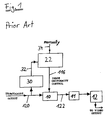
- Fig. 1 illustrates a device prior art for the automatic control of the ultrasound attenuation compensation (time gain compensation) in an UIS according to the US 47 33 668.
- the core of the apparatus is a control signal generator 22.
- One input signal 32 of the said signal generator 22 comes from a threshold circuit 30.
- the threshold circuit 30 is used to drive the control signal generator 22 as a function of ultrasound echo patterns coming from the ultrasound transducer output 120.
- the output signal 116 of the control signal generator 22 drives an amplifier 10 to amplify the output 120 of the ultrasound transducer in order to compensate for the ultrasound attenuation in the examined body.
- the amplified signal 122 is then A/D-converted (A/D-converter 11) and fed into the image memory 12 of the UIS.
- the attenuation compensation is the only means for the automatic control of image uniformity.
- Other influencing factors have to be adjusted manually via a second input 34 of the control signal generator 22.
- time-gain compensation prior art is done by using threshold circuits and by using prior knowledge about characteristic echo patterns.
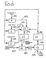
- Fig. 2 illustrates a device for the automatic control of image uniformity according to the present invention.
- the transducer output signal 120 of the UIS is amplified by the amplifier 10, A/D-converted by the A/D-converter 11 and fed to the image memory 12 of the UIS.
- One output 126 of the image memory 12 leads to a video output processor of the UIS.
- Another output 128 of the image memory 12 is connected with a two-dimensional low-pass spatial filter 20, where the content of the ultrasound image memory 12 is filtered in order to obtain an average amplitude value 100 as output signal.
- the output signal 100 of the spatial low-pass filter 20 is weighted by the multiplier 1 with a signal 102 proportional to the transmitted ultrasound power of the UIS.
- the output signal 100 of the low-pass filter 20 is weighted by the multiplier 2 with a signal 104 proportional to the grey scale characteristics of the display unit of the UIS and by the multiplier 3 with a signal 106 proportional to the depth of the displayed target, expressed by the zoom/scroll factor of the UIS.
- the output signals 108,110,112 of the multipliers 1,2,3 are added with the output signal 100 of the spatial low-pass filter 20.
- the not weighted signal 100 takes into account the ultrasound attenuation in the region of interest in the examined body.
- the output signal 114 of the adder 21 drives a control signal generator 22 in accordance with the feed-back information to control the gain of the amplifier 10.
- the output signal 122 of the amplifier 10 represents the optimized ultrasound echo signal with respect to image uniformity.
- the described signal paths represent a feed-back loop: the stored ultrasound information is used in order to automaticly adjust the actual values of the factors, that affect image uniformity.
Landscapes
- Engineering & Computer Science (AREA)
- Computer Networks & Wireless Communication (AREA)
- Physics & Mathematics (AREA)
- General Physics & Mathematics (AREA)
- Radar, Positioning & Navigation (AREA)
- Remote Sensing (AREA)
- Ultra Sonic Daignosis Equipment (AREA)
- Investigating Or Analyzing Materials By The Use Of Ultrasonic Waves (AREA)
Claims (2)
- Verfahren zum automatischen Optimieren der Bildgleichmäßigkeit eines durch eine Bilddarstellungsvorrichtung eines Ultraschall-Bilderzeugungssystems (UBS) darzustellenden Bildes, welches die Schritte des Verstärkens in einem Verstärker (10) der von einem Ultraschallwandler gelieferten Echosignale (120) und des Einführens der so verstärkten Echosignale in einen Ultraschallbild-Speicher (12), das Gebrauchens eines Tiefpaßfilters während der weiteren Signalverarbeitung und schließlich des Bildens eines Regelsignals (116) zum Regeln der Verstärkung des genannten Verstärkers (10) enthält, dadurch gekennzeichnet, daß- auf den Inhalt des Ultraschallbild-Speichers (12) eine zweidimensionale räumliche Tießpaßfilterung angewandt wird, um einen Durchschnitts-Amplitudenwert (100) zu erzeugen, welcher die Ultraschalldämpfung in der interessierenden Region des untersuchten Körpers berücksichtigt,- der genannte Durchschnitts-Amplitudenwert (100) mit einem zur tatsächlich vom UBS ausgesendeten Ultraschalleistung proportionalen Signal (102) gewichtet wird,- die genannte Durchschnitts-Amplitude (100) mit einem zur tatsächlichen Graustufencharakteristik der Bilddarstellungseinheit des UBS proportionalen Signal (104) gewichtet wird, und- der genannte Durchschnitts-Amplitudenwert (100) mit einem zur Tiefe des dargestellten Zieles, ausgedrückt in Zoom/Scroll-Faktoren, proportionalen Signal (106) gewichtet wird,- wodurch gewichtete Signale (108, 110, 112) gebildet werden,- der genannte Durchschnitts-Amplitudenwert (100) den genannten gewichteten Signalen (108, 110, 112) hinzuaddiert wird, um ein Summensignal (114) zu erzeugen, und- das genannte Summensignal (114) gebraucht wird, um das genannte Regelsignal (116) zu erzeugen.
- Vorrichtung für das automatische Optimieren der Bildgleichmäßigkeit eines durch eine Bilddarstellungsvorrichtung eines Ultraschall-Bilderzeugungssystems (UBS) darzustellenden Bildes, mit einem Verstärker (10) zur Verstärkung von durch einen Ultraschallwandler gelieferten Echosignalen (120), einem Bildspeicher (12) zum Empfang der genannten Echosignale nach deren Verstärkung, einem Tiefpaßfilter und einem Regelsignal-Generator (22) zur Regelung der Verstärkung des genannten Verstärkers (10), sowie weiterhin mit- einem zweidimensionalen Tiefpaßfilter (20), welches an einen Ausgang (128) des genannten Bildspeichers (12) angeschlossen ist, zur Erzeugung eines Durchschnitts-Amplitudenwertes (100),- einer Gewichtungsschaltung mit Mitteln (1, 2, 3) zum Gewichten des genannten Durchschnitts-Amplitudenwertes (100) mit Signalen (102, 104, 106), welche vom Niveau der ausgesendeten Ultraschalleistung, der tatsächlichen Graustufencharakteristik und dem tatsächlichen Zoom/Scroll-Faktor des UBS abhängen, wodurch gewichtete Signale (108, 110, 112) gebildet werden,- einem Addierer (21) zum Hinzuaddieren des genannten Durchschnitts-Amplitudenwertes (100) zu den genannten gewichteten Signalen (108, 110, 112) zum Erzeugen eines Summensignales (114), wobei der Ausgang des genannten Addierers (21) mit dem genannten Regelsignal-Generator (22) verbunden ist.
Priority Applications (2)
| Application Number | Priority Date | Filing Date | Title |
|---|---|---|---|
| EP19910116397 EP0533976B1 (de) | 1991-09-26 | 1991-09-26 | Automatische Optimalisierung der gleichmässigen Intensität eines Bildes für ein Ultraschallabbildungsystem |
| DE1991619589 DE69119589T2 (de) | 1991-09-26 | 1991-09-26 | Automatische Optimalisierung der gleichmässigen Intensität eines Bildes für ein Ultraschallabbildungsystem |
Applications Claiming Priority (1)
| Application Number | Priority Date | Filing Date | Title |
|---|---|---|---|
| EP19910116397 EP0533976B1 (de) | 1991-09-26 | 1991-09-26 | Automatische Optimalisierung der gleichmässigen Intensität eines Bildes für ein Ultraschallabbildungsystem |
Publications (2)
| Publication Number | Publication Date |
|---|---|
| EP0533976A1 EP0533976A1 (de) | 1993-03-31 |
| EP0533976B1 true EP0533976B1 (de) | 1996-05-15 |
Family
ID=8207198
Family Applications (1)
| Application Number | Title | Priority Date | Filing Date |
|---|---|---|---|
| EP19910116397 Expired - Lifetime EP0533976B1 (de) | 1991-09-26 | 1991-09-26 | Automatische Optimalisierung der gleichmässigen Intensität eines Bildes für ein Ultraschallabbildungsystem |
Country Status (2)
| Country | Link |
|---|---|
| EP (1) | EP0533976B1 (de) |
| DE (1) | DE69119589T2 (de) |
Cited By (1)
| Publication number | Priority date | Publication date | Assignee | Title |
|---|---|---|---|---|
| US10786226B2 (en) | 2017-02-09 | 2020-09-29 | Clarius Mobile Health Corp. | Ultrasound systems and methods for optimizing multiple imaging parameters using a single user interface control |
Families Citing this family (5)
| Publication number | Priority date | Publication date | Assignee | Title |
|---|---|---|---|---|
| US6468212B1 (en) * | 1997-04-19 | 2002-10-22 | Adalberto Vara | User control interface for an ultrasound processor |
| US6579238B1 (en) * | 2000-04-24 | 2003-06-17 | Acuson Corporation | Medical ultrasonic imaging system with adaptive multi-dimensional back-end mapping |
| US7306561B2 (en) * | 2004-09-02 | 2007-12-11 | Scimed Life Systems, Inc. | Systems and methods for automatic time-gain compensation in an ultrasound imaging system |
| US8235905B2 (en) | 2009-05-26 | 2012-08-07 | General Electric Company | System and method for automatic ultrasound image optimization |
| US8270701B2 (en) | 2010-01-08 | 2012-09-18 | 3M Innovative Properties Company | Optical web-based defect detection using intrasensor uniformity correction |
Family Cites Families (3)
| Publication number | Priority date | Publication date | Assignee | Title |
|---|---|---|---|---|
| DE2806176C3 (de) * | 1978-02-14 | 1980-08-07 | Siemens Ag, 1000 Berlin Und 8000 Muenchen | Verfahren und Vorrichtung zur Gewinnung und Aufzeichnung von Ultraschall-Schnittbildern |
| CA1159945A (en) * | 1979-09-20 | 1984-01-03 | George Kossoff | Signal processing equipment for ultrasonic visualisation |
| US4662380A (en) * | 1985-10-31 | 1987-05-05 | General Electric Company | Adaptive time gain compensation system for ultrasound imaging |
-
1991
- 1991-09-26 EP EP19910116397 patent/EP0533976B1/de not_active Expired - Lifetime
- 1991-09-26 DE DE1991619589 patent/DE69119589T2/de not_active Expired - Fee Related
Cited By (2)
| Publication number | Priority date | Publication date | Assignee | Title |
|---|---|---|---|---|
| US10786226B2 (en) | 2017-02-09 | 2020-09-29 | Clarius Mobile Health Corp. | Ultrasound systems and methods for optimizing multiple imaging parameters using a single user interface control |
| US12336860B2 (en) | 2017-02-09 | 2025-06-24 | Clarius Mobile Health Corp. | Ultrasound systems and methods for optimizing multiple imaging parameters using a single user interface control |
Also Published As
| Publication number | Publication date |
|---|---|
| DE69119589T2 (de) | 1996-12-19 |
| DE69119589D1 (de) | 1996-06-20 |
| EP0533976A1 (de) | 1993-03-31 |
Similar Documents
| Publication | Publication Date | Title |
|---|---|---|
| EP0707221B1 (de) | Diagnostik-Ultraschall-System mit Regelung des Verstärkungsfaktors | |
| US4852576A (en) | Time gain compensation for ultrasonic medical imaging systems | |
| KR100908252B1 (ko) | 영상 처리 시스템 및 방법 | |
| EP0220631B1 (de) | System für Ultraschallabbildung mit adaptiver Verstärkungskompensation gemäss der Zeit | |
| US4057049A (en) | Apparatus for and methods of pulse-echo examination | |
| CA1159945A (en) | Signal processing equipment for ultrasonic visualisation | |
| US11076835B2 (en) | Model-based control of a dynamic range of an ultrasound image | |
| US4763661A (en) | Filtered ultrasonic wave method and apparatus for detecting diseased tissue | |
| US6258033B1 (en) | Ultrasound method employing echoes from a region of interest to enable quantization of backscatter signals | |
| EP0533976A1 (de) | Automatische Optimalisierung der gleichmässigen Intensität eines Bildes für ein Ultraschallabbildungsystem | |
| US6733452B2 (en) | Ultrasound imaging method and apparatus for forming color images by using a variable power threshold level | |
| EP0107172B1 (de) | Ultraschall-Abbildungssystem mit einem durch den Bedienungsmann geregelten Filter zur Dämpfungskompensierung des reflektierten Signals | |
| JP4454293B2 (ja) | 超音波診断装置 | |
| JP3274194B2 (ja) | 超音波診断装置 | |
| JPH0647046A (ja) | グレースケールウインドウ化方法および装置 | |
| JPS59232540A (ja) | 超音波診断装置 | |
| JPH056955B2 (de) | ||
| JP3518910B2 (ja) | 超音波診断装置 | |
| JPS6154416B2 (de) | ||
| US6361498B1 (en) | Contrast agent imaging with suppression of nonlinear tissue response | |
| JPS58165826A (ja) | 超音波診断装置 | |
| JPH1014917A5 (de) | ||
| JP3388954B2 (ja) | 超音波診断装置 | |
| Herrick et al. | Automated gain control for medical diagnostic ultrasound imaging | |
| JP2800241B2 (ja) | 超音波診断装置 |
Legal Events
| Date | Code | Title | Description |
|---|---|---|---|
| PUAI | Public reference made under article 153(3) epc to a published international application that has entered the european phase |
Free format text: ORIGINAL CODE: 0009012 |
|
| AK | Designated contracting states |
Kind code of ref document: A1 Designated state(s): DE ES FR GB IT |
|
| 17P | Request for examination filed |
Effective date: 19930726 |
|
| 17Q | First examination report despatched |
Effective date: 19940131 |
|
| GRAH | Despatch of communication of intention to grant a patent |
Free format text: ORIGINAL CODE: EPIDOS IGRA |
|
| GRAA | (expected) grant |
Free format text: ORIGINAL CODE: 0009210 |
|
| AK | Designated contracting states |
Kind code of ref document: B1 Designated state(s): DE ES FR GB IT |
|
| PG25 | Lapsed in a contracting state [announced via postgrant information from national office to epo] |
Ref country code: ES Free format text: THE PATENT HAS BEEN ANNULLED BY A DECISION OF A NATIONAL AUTHORITY Effective date: 19960515 |
|
| ET | Fr: translation filed | ||
| REF | Corresponds to: |
Ref document number: 69119589 Country of ref document: DE Date of ref document: 19960620 |
|
| ITF | It: translation for a ep patent filed | ||
| PGFP | Annual fee paid to national office [announced via postgrant information from national office to epo] |
Ref country code: GB Payment date: 19960819 Year of fee payment: 6 |
|
| PLBE | No opposition filed within time limit |
Free format text: ORIGINAL CODE: 0009261 |
|
| STAA | Information on the status of an ep patent application or granted ep patent |
Free format text: STATUS: NO OPPOSITION FILED WITHIN TIME LIMIT |
|
| 26N | No opposition filed | ||
| PG25 | Lapsed in a contracting state [announced via postgrant information from national office to epo] |
Ref country code: GB Free format text: LAPSE BECAUSE OF NON-PAYMENT OF DUE FEES Effective date: 19970926 |
|
| GBPC | Gb: european patent ceased through non-payment of renewal fee |
Effective date: 19970926 |
|
| PGFP | Annual fee paid to national office [announced via postgrant information from national office to epo] |
Ref country code: FR Payment date: 20030917 Year of fee payment: 13 |
|
| PGFP | Annual fee paid to national office [announced via postgrant information from national office to epo] |
Ref country code: DE Payment date: 20031030 Year of fee payment: 13 |
|
| PG25 | Lapsed in a contracting state [announced via postgrant information from national office to epo] |
Ref country code: DE Free format text: LAPSE BECAUSE OF NON-PAYMENT OF DUE FEES Effective date: 20050401 |
|
| PG25 | Lapsed in a contracting state [announced via postgrant information from national office to epo] |
Ref country code: FR Free format text: LAPSE BECAUSE OF NON-PAYMENT OF DUE FEES Effective date: 20050531 |
|
| REG | Reference to a national code |
Ref country code: FR Ref legal event code: ST |
|
| PG25 | Lapsed in a contracting state [announced via postgrant information from national office to epo] |
Ref country code: IT Free format text: LAPSE BECAUSE OF NON-PAYMENT OF DUE FEES;WARNING: LAPSES OF ITALIAN PATENTS WITH EFFECTIVE DATE BEFORE 2007 MAY HAVE OCCURRED AT ANY TIME BEFORE 2007. THE CORRECT EFFECTIVE DATE MAY BE DIFFERENT FROM THE ONE RECORDED. Effective date: 20050926 |