EP0538241A2 - Dispositif ultrasonique pour usage thérapeutique - Google Patents
Dispositif ultrasonique pour usage thérapeutique Download PDFInfo
- Publication number
- EP0538241A2 EP0538241A2 EP93100294A EP93100294A EP0538241A2 EP 0538241 A2 EP0538241 A2 EP 0538241A2 EP 93100294 A EP93100294 A EP 93100294A EP 93100294 A EP93100294 A EP 93100294A EP 0538241 A2 EP0538241 A2 EP 0538241A2
- Authority
- EP
- European Patent Office
- Prior art keywords
- drug
- ultrasonic wave
- irradiation
- ultrasonic
- acoustic
- Prior art date
- Legal status (The legal status is an assumption and is not a legal conclusion. Google has not performed a legal analysis and makes no representation as to the accuracy of the status listed.)
- Granted
Links
Images




Classifications
-
- A—HUMAN NECESSITIES
- A61—MEDICAL OR VETERINARY SCIENCE; HYGIENE
- A61M—DEVICES FOR INTRODUCING MEDIA INTO, OR ONTO, THE BODY; DEVICES FOR TRANSDUCING BODY MEDIA OR FOR TAKING MEDIA FROM THE BODY; DEVICES FOR PRODUCING OR ENDING SLEEP OR STUPOR
- A61M37/00—Other apparatus for introducing media into the body; Percutany, i.e. introducing medicines into the body by diffusion through the skin
- A61M37/0092—Other apparatus for introducing media into the body; Percutany, i.e. introducing medicines into the body by diffusion through the skin using ultrasonic, sonic or infrasonic vibrations, e.g. phonophoresis
-
- A—HUMAN NECESSITIES
- A61—MEDICAL OR VETERINARY SCIENCE; HYGIENE
- A61N—ELECTROTHERAPY; MAGNETOTHERAPY; RADIATION THERAPY; ULTRASOUND THERAPY
- A61N7/00—Ultrasound therapy
-
- A—HUMAN NECESSITIES
- A61—MEDICAL OR VETERINARY SCIENCE; HYGIENE
- A61N—ELECTROTHERAPY; MAGNETOTHERAPY; RADIATION THERAPY; ULTRASOUND THERAPY
- A61N7/00—Ultrasound therapy
- A61N7/02—Localised ultrasound hyperthermia
-
- A—HUMAN NECESSITIES
- A61—MEDICAL OR VETERINARY SCIENCE; HYGIENE
- A61B—DIAGNOSIS; SURGERY; IDENTIFICATION
- A61B17/00—Surgical instruments, devices or methods
- A61B17/22—Implements for squeezing-off ulcers or the like on inner organs of the body; Implements for scraping-out cavities of body organs, e.g. bones; for invasive removal or destruction of calculus using mechanical vibrations; for removing obstructions in blood vessels, not otherwise provided for
- A61B17/22004—Implements for squeezing-off ulcers or the like on inner organs of the body; Implements for scraping-out cavities of body organs, e.g. bones; for invasive removal or destruction of calculus using mechanical vibrations; for removing obstructions in blood vessels, not otherwise provided for using mechanical vibrations, e.g. ultrasonic shock waves
- A61B2017/22005—Effects, e.g. on tissue
- A61B2017/22007—Cavitation or pseudocavitation, i.e. creation of gas bubbles generating a secondary shock wave when collapsing
- A61B2017/22008—Cavitation or pseudocavitation, i.e. creation of gas bubbles generating a secondary shock wave when collapsing used or promoted
-
- A—HUMAN NECESSITIES
- A61—MEDICAL OR VETERINARY SCIENCE; HYGIENE
- A61B—DIAGNOSIS; SURGERY; IDENTIFICATION
- A61B17/00—Surgical instruments, devices or methods
- A61B17/22—Implements for squeezing-off ulcers or the like on inner organs of the body; Implements for scraping-out cavities of body organs, e.g. bones; for invasive removal or destruction of calculus using mechanical vibrations; for removing obstructions in blood vessels, not otherwise provided for
- A61B17/22004—Implements for squeezing-off ulcers or the like on inner organs of the body; Implements for scraping-out cavities of body organs, e.g. bones; for invasive removal or destruction of calculus using mechanical vibrations; for removing obstructions in blood vessels, not otherwise provided for using mechanical vibrations, e.g. ultrasonic shock waves
- A61B2017/22027—Features of transducers
- A61B2017/22028—Features of transducers arrays, e.g. phased arrays
-
- A—HUMAN NECESSITIES
- A61—MEDICAL OR VETERINARY SCIENCE; HYGIENE
- A61B—DIAGNOSIS; SURGERY; IDENTIFICATION
- A61B17/00—Surgical instruments, devices or methods
- A61B17/22—Implements for squeezing-off ulcers or the like on inner organs of the body; Implements for scraping-out cavities of body organs, e.g. bones; for invasive removal or destruction of calculus using mechanical vibrations; for removing obstructions in blood vessels, not otherwise provided for
- A61B2017/22082—Implements for squeezing-off ulcers or the like on inner organs of the body; Implements for scraping-out cavities of body organs, e.g. bones; for invasive removal or destruction of calculus using mechanical vibrations; for removing obstructions in blood vessels, not otherwise provided for after introduction of a substance
- A61B2017/22088—Implements for squeezing-off ulcers or the like on inner organs of the body; Implements for scraping-out cavities of body organs, e.g. bones; for invasive removal or destruction of calculus using mechanical vibrations; for removing obstructions in blood vessels, not otherwise provided for after introduction of a substance ultrasound absorbing, drug activated by ultrasound
-
- A—HUMAN NECESSITIES
- A61—MEDICAL OR VETERINARY SCIENCE; HYGIENE
- A61N—ELECTROTHERAPY; MAGNETOTHERAPY; RADIATION THERAPY; ULTRASOUND THERAPY
- A61N7/00—Ultrasound therapy
- A61N2007/0056—Beam shaping elements
- A61N2007/006—Lenses
-
- A—HUMAN NECESSITIES
- A61—MEDICAL OR VETERINARY SCIENCE; HYGIENE
- A61N—ELECTROTHERAPY; MAGNETOTHERAPY; RADIATION THERAPY; ULTRASOUND THERAPY
- A61N7/00—Ultrasound therapy
- A61N2007/0078—Ultrasound therapy with multiple treatment transducers
-
- A—HUMAN NECESSITIES
- A61—MEDICAL OR VETERINARY SCIENCE; HYGIENE
- A61N—ELECTROTHERAPY; MAGNETOTHERAPY; RADIATION THERAPY; ULTRASOUND THERAPY
- A61N7/00—Ultrasound therapy
- A61N2007/0086—Beam steering
- A61N2007/0095—Beam steering by modifying an excitation signal
-
- A—HUMAN NECESSITIES
- A61—MEDICAL OR VETERINARY SCIENCE; HYGIENE
- A61N—ELECTROTHERAPY; MAGNETOTHERAPY; RADIATION THERAPY; ULTRASOUND THERAPY
- A61N7/00—Ultrasound therapy
- A61N7/02—Localised ultrasound hyperthermia
- A61N2007/027—Localised ultrasound hyperthermia with multiple foci created simultaneously
-
- G—PHYSICS
- G01—MEASURING; TESTING
- G01S—RADIO DIRECTION-FINDING; RADIO NAVIGATION; DETERMINING DISTANCE OR VELOCITY BY USE OF RADIO WAVES; LOCATING OR PRESENCE-DETECTING BY USE OF THE REFLECTION OR RERADIATION OF RADIO WAVES; ANALOGOUS ARRANGEMENTS USING OTHER WAVES
- G01S15/00—Systems using the reflection or reradiation of acoustic waves, e.g. sonar systems
- G01S15/88—Sonar systems specially adapted for specific applications
- G01S15/89—Sonar systems specially adapted for specific applications for mapping or imaging
- G01S15/8906—Short-range imaging systems; Acoustic microscope systems using pulse-echo techniques
- G01S15/899—Combination of imaging systems with ancillary equipment
Definitions
- This invention relates to a ultrasonic apparatus for therapeutical use for remedying a malignant tumor or the like, to a ultrasonic apparatus for litholysis by utilizing drug activation by a ultrasonic wave or to a ultrasonic apparatus for promoting a chemical reaction.
- a heretofore known anticancer drug such as adriamycin or daunomycin is used as the anticancer drug and ultrasonic wave irradiation means is the one that can generate a plane wave having an expansion somewhat greater than the diameter of the tumor to be treated.
- the paper reports that the decrease in the tumor diameter and the extension of life after the treatment were significant for the group to which the ultrasonic wave was irradiated after the administration of the anticancer drug in comparison with the group to which only the anticancer drug was dosed and with the group to which only the ultrasonic wave was irradiated.
- the prior art technique described above involves the problem in that it is not effective for the remedy of a deep tumor because it uses the plane wave. Moreover, since the effect of local activation of the drug by the irradiation of the ultrasonic wave is not sufficient, the kind and amount of the drug used might give the side effect on the normal tissue, too.
- Utilization of cavitation is effective for the activation of a drug by a ultrasonic wave and such an activation action can be obtained particularly at the time of rupture of cavitation.
- the ultrasonic apparatus of the present invention causes efficiently the generation and rupture of the cavitation in a selected region of the body by use of a convergent ultrasonic wave.
- the present invention provides a ultrasonic wave irradiation apparatus for causing the generation and rupture of cavitation by irradiating interruptedly a convergent ultrasonic wave with a suitable time interval or by irradiating a ultrasonic wave while changing over within a short period a plurality of kinds of convergent acoustic fields having mutually overlapping focal zones, though their focal points or acoustic wave distribution shapes are different.
- the accurate position at which cavitation is more likely to be generated among the positions near the focal point is in agreement with the maximum acoustic pressure position so long as its size is very small but when cavitation grows to the size such as the one that can be detected by a ultrasonic echo of a frequency substantially equal to the irradiated ultrasonic wave, the accurate position moves close to the minimum acoustic pressure position.
- efficiency of the formation and rupture of cavitation can be improved further by irradiating sequentially or alternately a plurality of kinds of convergent ultrasonic waves which, though their focal zones overlap with one another, have mutually different acoustic pressure distribution shapes or more definitely, have mutually different maximum acoustic pressure positions, while changing them over within a short period.
- the present invention proposes a ultrasonic therapeutical effect promoter consisting, as a principal agent, of a compound which does not have the anticancer effect by itself or has a very low effect, but exhibits a remarkable anticancer effect when activated by the irradiation of a ultrasonic wave, and its derivatives, such as chelation formation compounds represented by porphyrin compounds and alkylating agents, and ascorbic salts.
- Such drugs do not at all have any side effect on the normal tissue not irradiated with the ultrasonic wave or their side effect, and even when it exists, they can be used so that the side effect can be neglected substantially. Accordingly, they are extremely suitable for the calcinomatic therapy by the application of the ultrasonic wave irradiation as the object of the present invention.
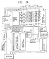
- a ultrasonic apparatus for therapeutical use in accordance with one embodiment of the present invention which has the function of supporting the cooperation between the dose of a drug and the irradiation of a ultrasonic wave, is shown in Fig. 1(A)
- Fig. 1(B) an example of the display of this apparatus is shown in Fig. 1(B)
- the structure of a ultrasonic wave applicator is shown in Figs. 2 and 3
- the structure of a drug dose support portion is shown in Fig. 4.
- Fig. 1(A) the information on the ultrasound irradiation therapeutical schedule is inputted from a keyboard 19 to a main controller 20 and an irradiation focus code signal which determines the focal zone position and sound pressure distribution shape of the irradiating field on the basis of the information is given from the main controller 20 to a driving phase controller 21.
- the driving phase controller 21 designates the phase of the driving signals generated from driving signal generators 6-1 ⁇ 6-N (where N is the total number of transducer independent elements) in accordance with the code signal given thereto.
- Each driving signal thus generated is power-amplified by each driver 2-1 ⁇ 2-N and is applied to each transducer element 1-1 ⁇ 1-N so that a ultrasonic wave converged to a desired portion is irradiated.
- a signal given directly from the main controller 20 controls the generation and stop of the driving signal of each driver 2-1 ⁇ 2-N so that the duration time of the irradiated ultrasonic wave can be controlled and the intensity can be changed without changing the irradiation focus and emergency stop of the ultrasound irradiation can be made when any abnormality occurs.
- Each transducer for irradiation operates also as a reception transducer for detecting cavitation occurring in the object of the irradiation.
- the component of an irradiation signal band is removed from the signal received by each transducer element 1-1 ⁇ 1-N by each band-pass filter 3-1 ⁇ 3-N, the signal is led to each receiving amplifier 5-1 ⁇ 5-N and amplified there. Thereafter, the signal is applied to a receiving beam former 22.
- a series inductance resonating with the sum of an element capacity and a cable capacity at a driving frequency f o is inserted into the output portion of each driver 2-1 ⁇ 2-N.
- Each receiving amplifier 5-1 ⁇ 5-N has a variable gain and this gain is controlled by the signal given directly from the main controller 20. This gain is lowered to avoid saturation of the amplifier in the time zone where great quantities of unnecessary signal components other than the center frequency of the irradiated ultrasonic wave occurs, such as at the time of change-over of the irradiation focus.
- a receiving beam former 22 has a plurality of focus circuits converging to a plurality of focuses arranged with gaps therebetween that correspond to the spatial resolution of a reception system inside the irradiation focal zone, detects the occurrence and occurrence positions of subharmonic components such as f o /2 and f o /3 that are emitted by the cavitation and higher harmonic components and provides the detection signal to the display 18.
- Fig. 1(B) shows the display surface in the display and the cavitation occurrence position detected as described above is displayed as represented by reference numeral 27-6 on the display surface 27.
- the information on the position and size of the cavitation is applied to the irradiation main controller 20 and when the cavitation is judged to be greater than a predetermined standard, the irradiation focus is switched and the maximum of the sound pressure is moved to that position.
- the production cost of the receiving beam former 22 can be reduced by use of a smaller number of parallel processing focus circuits so as to scan their respective focuses in the irradiation focal zone.
- reference numeral 4 represents an array type ultrasonic probe for imaging and reference numeral 23 is a motor which rotates the probe around the axis perpendicular to the probe surface, and obtains a plurality of ultrasonic echo tomograms 27-1 and 27-2 necessary for locating the irradiation target.
- Each element of the probe 4 is connected to the transmission controller 24 and to the receiving beam former 26 through the transmitting and receiving amplifier 25.
- the echo tomograms thus obtained are displayed as represented by 27-1 and 27-2 in Fig. 1(B).
- an irradiation focal zone mark 27-4, intersection 27-5 of sectional planes of a plurality of tomograms and a cavitation detection position mark 27-6 are superposed and displayed in mutually distinguishable colors on the display surface 27 under the control of the display 18.
- the ultrasonic wave frequency band of the probe 4 is at least 2 f o .
- the higher harmonic components such as 2 f o , 3 f o , etc., that are emitted by the cavitation may be detected by the probe 4.
- the irradiation focus is controlled to move in such a manner as to follow up the motion of the object portion on the basis of the signal that is applied from the receiving beam former 26 to the irradiation main controller 20. If the motion of the object portion is so great that it exceeds the range where the irradiation focusing is possible or tracking becomes difficult, the ultrasonic wave irradiation timing is synchronized with the aspiration on the basis of the signal given from a breath detector 29 to the main controller 20 so that the ultrasonic irradiation can be made within a certain predetermined range of the aspiration time phase.
- Reference numerals 15 to 17 in Fig. 1(A) represents a mechanism for supporting the cooperation between the drug dose and the ultrasonic wave irradiation. This portion is shown more definitely in Fig. 4.
- a dip drug 10 whose anti-tumor effect is activated by the ultrasonic wave irradiation is placed into a transparent container 11 and is communicated with a needle 14 through a flexible tube.
- the output from an optical drop counter 12 is inputted to a flow rate analyzer 15 and the flow rate per unit time is inputted to a pharmacokinetic analyzer 17.
- the information such as the name of the drip drug, the dose method, the name, weight, length, age and blood protein concentration of a patient, the name and portion of the remedial object, the kind of tumor, and the like, are inputted from the keyboard 19 into the pharmacokinetic analyzer 17.
- the dose time of the drug, that has been dosed before the start of instillation is also inputted from the keyboard 19 into the pharmacokinetic analyzer 17.
- the pharmacokinetic analyzer 17 estimates the past, present and future drug concentrations of the object region and those of the portions nearby through numeric calculation on the basis of the pharmacokinetical aspect and provides the estimation values to the display 18, which displays the result on the display surface 27 such as 27-3 of Fig.
- a present time marker 27-7 is displayed, too, on the graph displayed.
- a "pharmacokinetical ultrasonic dose” is calculated from both the internal drug concentration and the ultrasonic wave dose at each portion in the body and is also displayed on the display surface 27. If the difference between the estimated value of the interior drug concentration and the scheduled value for the therapeutical plan is automatically judged to be greater than a preset value, the pressure-feed quantity of a drug pump 13 is controlled through a pump controller 16 to bring it closer to the scheduled value.
- the keyboard 19 is equipped with recording means such as a magnetic or optical disc or an IC card, records the pharmacokinetical ultrasonic dose in addition to the various information on the patient and the drug dose method and uses them as the recorded information for the future diagnosis and remedy of the patient.
- recording means such as a magnetic or optical disc or an IC card
- the focuses of the transmission controller 24 and the receiving beam former 26 are brought into conformity with the blood vessels and the output of the receiving beam former 26 is processed by a Doppler signal processor 28 in order to determine the blood flow velocity and the estimated value of the blood flow rate per unit time of the object organ from the diameter of the blood vessel as well as its change with time.
- These data are given to the pharmacokinetic analyzer 17. Using these data, the pharmacokinetic analyzer 17 corrects the drug concentration estimated value in the object organ. If it is difficult to measure the blood flow rate of the object organ itself, the change with time of the blood flow rate of the more whole body such as that of the main artery of the abdominal region and the result is given to the pharmacokinetic analyzer 17 to correct the estimated drug concentration.
- Hematoporphyrin which is likely to cause the photochemical reaction is often used as the drip drug 10 shown in Fig. 4. Therefore, the light source used for the optical drop counter 12 must have the minimum necessary intensity.
- a shield cover is put to the transparent container 11, whenever necessary, to prevent the change with time of the drug due to light.
- Fig. 2 shows an example of 16-sector 2-track array type applicator having a geometric focus F by way of example, Fig. 2(A) is its plane view and Fig. 2(B) is its sectional view.
- the ultrasonic irradiation transducer elements represented by reference numerals 1-1, 1-2 and the like are arranged in a double circle.
- a piezoelectric ceramic of a lead titanium type is used as the electro-optical conversion material of this element.
- each element is bonded to an acoustic matching layer 5 made of a light metal consisting principally of magnesium by use of an epoxy type specific adhesive having a small coefficient of thermal expansion as the polymer.
- an epoxy type specific adhesive having a small coefficient of thermal expansion as the polymer.
- the matching layer 5 describes a concave forming a part of the spherical plane with the geometric focus F on the ultrasonic wave irradiation plane side being its center, and its pack is a polished polyhedron for bonding thereto the piezoelectric ceramic element.
- This light metal acoustic matching layer 5 has good thermal conductivity and is therefore effective for cooling the piezoelectric ceramic element at the time of irradiation of the ultrasonic wave.
- the matching layer 5 forms a part of the applicator housing.
- a coolant fluid passage 6 is disposed so as to absorb the heat generated at the time of irradiation of the ultrasonic wave and a water bag 7 storing therein deaerated water is fitted so as to facilitate acoustic coupling with the body surface.
- the array type ultrasonic probe for imaging 4 is stored in the round hole at the array center.
- the probe structure is the same as the one that is used for a ultrasonic diagnostical apparatus and the ultrasonic wave frequency used in this embodiment is from 2 to 4 MHz.
- the probe 4 is rotated round the center axis of the applicator with respect to the applicator housing 5 so that a plurality of tomograms can be obtained by a single unidirectional array probe, and this rotation is caused by the motor 23.
- the geometric focal length of the applicator is 12 cm
- the outer diameter 2r2 is 12 cm
- its inner diameter 2r o is 4 cm
- the diameter 2r1 of the circle dividing these two tracks is 8 cm.
- the acoustic field generation method of the present invention will be described in detail hereafter by setting the polar coordinates (r, ⁇ ) on the array plane, the angular coordinates at the center of the n th element to ⁇ n and the driving signal of the n th element of the i th track to A i ( ⁇ n ).
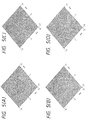
- a cavitation is formed and grows near the sound pressure minimum point encompassed by the sound pressure maximum portion and gets stable.
- the irradiation of the ultrasonic wave is switched and made within a short period to the acoustic field having the acoustic pressure maximum point at the position of the acoustic pressure minimum point such as shown in Fig. 5B, the stable cavitation is broken and insteads, a large number of fine cavitations repeat for a while the formation and rupture which are useful for the activation of the drug near the acoustic pressure maximum point.
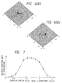
- the acoustic field in which the ultrasonic wave power distribution is approximated by the same M- th order Bessel function is two fold: one having a positive M and the other, a negative M.
- the rotating directions of their wave planes are mutually different as shown in Figs. 6(A) and 6(B).
- This acoustic field will be hereinafter referred to as a "conjugate acoustic field".
- This conjugate acoustic field has different positions of points at which the cavitation gets stable because the inclinations of the wave planes are different, even though the ultrasound power distribution is the same, in a heterogeneous medium such as in the living body. Therefore, the formation and rupture can be conducted extremely efficiently by the irradiation of the ultrasonic wave using alternately the conjugate acoustic fields.
- Another important factor for conducting efficiently the acoustic chemical reaction by utilizing the formation and rupture of the cavitation is a time interval in which a plurality of acoustic fields are switched.
- the reaction rate is determined from the change of absorbancy by utilizing the color change of an aqueous solution to violet due to iodine precipitated by the chemical reaction caused by the irradiation of the ultrasonic wave in the system prepared by adding starch and carbon tetrachloride to the aqueous potassium iodide solution.
- the acoustic field of the simple focus shown in Fig. 5(B) is used interruptedly for the sake of simplicity.
- Fig. 7 shows the experimental result when the experiment is conducted by setting the duration time and rest time in the interrupted irradiation to the 1:1 basis.
- the abscissa represents the logarithmic scale representation of the burst wave duration time.
- the duration time of the burst wave must be from 0.01 msec to 10 msec and preferably, from 0.05 msec to 2 msec.
- the transducer is divided into a plurality of segments and the ultrasonic wave is irradiated from the different portions of the transducer so as to form convergent acoustic fields whose focal points and acoustic pressure distributions in the focal zone are substantially the same. Then, the duration time of the irradiation of the ultrasonic wave from one segment of the transducer is switched so that it is shorter than the value T o described above so that it is sufficiently greater then T o at the focus but is shorter than T o at the other positions. In this manner the factors of the spatial selectivity of the reaction can be obtained besides the difference in the density of the ultrasonic wave power.
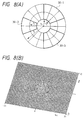
- the array transducer is divided into segments 30-1, 30-2, 30-3 encompassed by thick lines in the drawing (A) and the rest of portions and when only the segments encompassed by the thick lines are driven and the ultrasonic wave is irradiated, a convergent acoustic field whose concentration distribution is shown in (B) is formed on the focal plane.
- the segments 30-1, 30-2, 30-3 are selected so that they are complementary with the partial transducer which is point-symmetric with respect to the center of the transducer. Accordingly, the collapse of the beam of the focus due to the partial transducer transmittion can be restricted sufficiently.
- the partial transducers 30-1, 30-2, 30-3 used for the transmission are rotated round the center of the transducer within the time interval below about T o described above, or they are used alternately with the complementary partial transducer so that the irradiation duration time becomes shorter than T o immediately below the transducer.
- the beam of the focus at the center of the drawing (B) does not change substantially, and the chemical reaction is allowed to proceed only at this portion.
- the position of the steady cavitation occurring due to the irradiation of the ultrasonic wave of such an acoustic field is not always acurately at the position of the acoustic pressure minimum point but in some cases, at a deviated position.
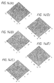
- the focus can be moved as shown in Fig. 5C and 5D without forming an unnecessary acoustic field such as a grating lobe of about ⁇ 8 mm round the geometric focal plane F. Therefore, even if the stable cavitation somewhat deviates from the center in the drawing, the acoustic pressure maximum point can be formed at the cavitation position itself if that position can be detected by the means described already.
- Figs. 5B, C and D can be regarded as a spatial response function when the transducer for the irradiation is used as a receiving transducer for detecting the cavitation.
- the ultrasonic wave frequency to be handled is 0.5/n Mhz
- the scale of the distance is read n times and when it is 0.5 x n MHz, the scale must be read l/n times. Therefore, when the cavitation is detected by the fractional harmonic components, high location accuracy cannot be expected but scanning can be made over a broad range. On the other hand, when detection is made by a higher harmonic component, high location accuracy can be obtained, though the scannable range is not much broad.
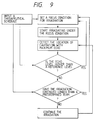
- Fig. 9 shows the flowchart of an example of the algorithm for a series of operations such as setting of the irradiation focus, detection of the stable cavitation and change of the irradiation focus.
- the gas elution quantity near the acoustic pressure maximum and minimum points is judged insufficient and the irradiation focus condition is changed automatically to move the acoustic pressure maximum and minimum positions.
- the acoustic fields A and B are formed by use of the same ultrasonic wave frequency but the frequency of A may be reduced to be relatively lower than that of B. This is directed to generate the cavitation by the ultrasonic wave having a lower frequency which is more effective for the generation of the cavitation and to rupture the cavitation by the ultrasonic wave having a higher frequency which is more likely to concentrate the spatial energy.
- each element is driven by the driving signal obtained by approximating the cosine function and sine function in the formulas by the rectangular function.
- the acoustic pressure maximum point of the acoustic field B is situated at the position of the acoustic pressure minimum point interposed by the acoustic pressure maximum points of the acoustic field A, while the acoustic pressure maximum point of the acoustic field A is situated at the position of the acoustic pressure minimum point interposed by the acoustic pressure maximum points of the acoustic field B, on the contrary.
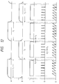
- Fig. 12 shows the time chart of the ultrasonic wave irradiation and the cavitation detection in this embodiment.
- Symbols A and B represent the time chart of the amplitude of the driving signals for generating the two irradiation acoustic fields having the relation such as described above. Irradiation is effected continuously for the periods T1 and T2, respectively, and their switching is made rapidly by electronic control.
- Symbol C represents the time chart of the gain of each receiving amplifier 5-1 ⁇ 5-N for detecting the cavitation. The gain is lowered to avoid the saturation of the amplifier during the period from before the switching of the irradiation focus T3 to the time T4 after the switching.
- Symbols D and E represent the amplitude of the transmission driving signal of the probe 4 for imaging and the gain of each receiving amplifier, respectively.
- the time interval T5 of the transmission pulse is set to be longer than the time necessary for the ultrasonic wave to reciprocate from the probe to the object region and the gain of the receiving amplifier is controlled as the function of time in accordance with the distance of the echo source.
- the gain control is conducted in the same way as C.
- the drug activation can be carried out efficiently by selecting suitably the switching time intervals T1 and T2 of the irradiation acoustic fields from within the range of 0.01 msec to 10 msec, as described already.
- an electrocardiograph represented by reference numeral 29 in Fig. 1 is used for a patient having the problem of a cardiac function and switching of the acoustic fields from the first irradiation means to the second irradiation means is conducted in such a manner as to avoid the timing immediately before the contraction period. If the patient has other cardiac problems, the irradiation of the therapeutical ultrasonic wave is stopped for the short period from immediately before till immediately after the contraction period.
- Fig. 3 shows the array applicator for irradiating the ultrasonic wave using the rectangular array in accordance with still another embodiment of the present invention.
- like reference numerals are used to identify those components or portions which have the same functions and the same names as in Figs. 1 and 2.
- the rectangular array having major sides of 16 cm and the minor sides of 4 cm is divided into 3 x N1 elements and the elements at both ends of each of the three divided groups are connected electrically with each other.
- Both irradiation sides of the acoustic matching layer 5 made of an alluminum alloy form a part of a cylindrical surface and an acoustic packing material 8 of a polymer material with a sound speed equal to, or lower than, that of water is packed into the recess.
- the surface is shaped as a plane or convex so that the applicator forms a geometric focus converting to a line F' -F'' as a whole.
- the major side is divided by N1 while the minor side is divided by 3, too. Therefore, the movement of focus in the direction of depth becomes possible even for the focus on the minor side.
- Fig. 13B shows the intensity distribution of the acoustic field formed by an ordinary converging method which drives each element by a signal having a phase difference corresponding to the sound wave propagation time from each element to the focus on the focal plane.
- the acoustic pressure maximum point of the acoustic pressure B is situated at the acoustic pressure minimum point encompassed by the acoustic pressure maximum points of the acoustic field A and the combination of the acoustic fields is suitable so that the stable cavitation generated near the acoustic pressure minimum point generated by the ultrasonic wave irradiation by use of the acoustic field A is ruptured by the acoustic field B.
- the movement of spot focus such as the acoustic field B in the direction of the major side becomes easy by effecting convergence in the direction of the major side or by moving the operture.
- Fig. 14 shows an example when the acoustic field generated by the same converging method as the acoustic field A in Fig. 13A is moved in the direction of the major side.
- a and B, between B and C, between C and D and between D and E in Fig. 14 such that the acoustic pressure maximum point of one of them is situated at the acoustic pressure minimum point of the other while the acoustic pressure minimum point of one of them is situated at the acoustic pressure maximum point of the other, on the contrary, in the same way as the relation between the acoustic fields A and B of Figs. 10 and 11.
- the ultrasonic wave is irradiated by switching the acoustic fields with suitable time interval such as A ⁇ B ⁇ C ⁇ D ⁇ E ⁇ D ⁇ C ⁇ B ⁇ A, ... , the drug activation can be attained in a broader range.
- the drug can be activated by irradiating the ultrasonic wave while switching a plurality of kinds of convergent acoustic fields with the suitable time intervals described above having the superposed focal zones, such as the combination of the acoustic fields (B) and (D) in the drawing, though the acoustic pressure maximum point of one of the fields is not always situated at the zero point of one of them.
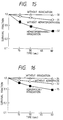
- Fig. 15 shows the result of examination of the cell killing effect for tumor cells in a suspension placed into a polyethylene testing tube placed in degassed water by the irradiation of the ultrasonic wave described above.
- the cells used are sarcoma 180 and the number of surviving cells is determined by counting the number in a predetermined quantity of sample through a microscope.
- the survival fractions of the tumor cells of a group to which hematoporphyrin is added up to a 5 wt% concentration, a group for which only the ultrasonic wave irradiation is made and a group for which the ultrasonic wave irradiation is made after the addition of hematoporphyrin are represented by numerals 31, 32 and 33 as the function of time, respectively.
- Hematoporphyrin alone does not at all exhibit the cell killing function and the effect is not great even by the ultrasonic wave irradiation alone, either. When they are combined, however, the number of surviving cells drops to about 1/3 within 60 seconds and a great cell killing effect can be observed. The appearance of the similar cell killing effect by the irradiation of the ultrasonic wave is also observed in the case of a protoporphyrin. The similar effect can be observed in methylene blue which is similarly a chelating agent, though it is not a prophyrin type compound.
- adriamycin which is an alkylating agent having the similar chelate forming function.
- This compound provides a remarkable effect of the cell killing effect of the drug by the irradiation of the ultrasonic wave, though it is somewhat lower than that of hematoporphyrin.
- Adriamycin is known as a compound having by itself the tumor cell killing action but such an action cannot be detected within a short period of 1 minute in this experiment. It can therefore be concluded that the appearance of the cell killing effect of adriamycin by the irradiation of the ultrasonic wave is independent of the tumor cell killing action of the drug alone. This is in common to other alkylating agents such as daunomycin.
- the calculation method of the drug concentration in the body in the system for supporting the cooperation between the drug dose and the ultrasonic wave irradiation in the present invention will be explained about the case where the drug is in advance dosed at a predetermined time before the ultrasonic wave irradiation as described above.
- the drug migration constant from the dosed portion into the blood is k A
- the elimination rate constant of the drug from the blood is k E
- the drug dose is D
- the drug and the drug distribution volume is V.
- the blood concentration B(t) is given as follows:
- the drug distribution volume V is substantially proportional to the volume of the patient but is affected by the protein combinability of the drug, too.
- the drug distribution volume V becomes relatively smaller if a patient is greater in length than another, though they have the same volume, because the proportion occupied by the skeletal structure in the weight and volume is generally greater in the former than the latter.
- the drug distribution volume V is determined by correcting the volume value on the basis of the information on the patient and the drug name, and the calculation of the formula (4) is made.
- the values k A and k E are corrected by use of the measured value to make the calculation of the formula (4).
- the drug concentration in the tumor C(t) is given by the following formula when the free fractions in the blood and in the tumor are F B and F T , respectively, and the tumor blood flow rate per unit time and per unit volume is k1:
- the drug concentration in the tumor C(0) at the time of dose is set to 0.
- the quantities of the free fractions F B and F T are primarily determined by the drug used but they are also affected by the protein concentration in the blood of the patient, the organ and portion of the organ in which the tumor exists, and whether the tumor is primary or metastatic. Therefore, the values are corrected and used on the basis of the information inputted from the keyboard.
- k1 can be determined more accurately.
- the change of the flow rate of the more whole body can be measured in place of the flow rate flowing into and out from the tumor, such a processing is effective as the information for correcting k1.
- the formula (5) is derived, the effect that the drug concentration drops as the dosed drug changes to other substances due to the irradiation of the ultrasonic wave and the change of the blood vessel systems of the tumor due to the effects of the irradiation of the ultrasonic wave and the dosed drug are neglected.
- the drug concentration in the tissues other than in the tumor can be calculated by use of a formula similar to the formula (5). Therefore, the drug concentration is calculated and displayed as the function of time for the portions around the tumor subjected to the ultrasonic wave irradiation in order to assist the therapy having higher safety.
- the "pharmacokinetical ultrasonic dose" Du(t) which is defined by the following formula by use of the ultrasonic wave irradiation intensity I(t) and the drug concentration in the tumor C(t) determined from the formula (5), is calculated and displayed every moment, and its final value is recorded for the future diagnosis and therapy of the patient:
- symbols G and H are those functions which convert the ultrasonic wave irradiation intensity and the drug concentration in the tumor to the effective intensity and to the effective concentration, respectively.
- the effects of both the ultrasonic wave irradiation intensity and the drug concentration in the tumor appear when they exceed certain threshold values, respectively, and their effects get into saturation when they exceed certain greater threshold values, respectively. Therefore, a quantitative scale can be obtained by correcting and converting these values.
- the drug of the present invention is dosed to the patient and the acoustic field for generating and rupturing selectively and efficiently the cavitation at the object region by use of the ultrasound irradiation apparatus of the invention in good cooperation by utilizing the therapeutical schedule support system of the invention, the anticancer activation of the drug can be attained locally at the object region and the cancer therapy can be accomplished with a reduced side effect.
- the embodiments using the two kinds of array type ultrasonic wave applicators have been described particularly in detail but the present invention is not limited thereto.
- the application range of the present invention includes the case where independent electro-acoustic transducers are used for generating a plurality of kinds of convergent acoustic fields and the case where the focuses of the convergent acoustic fields are scanned mechanically.
- the application range of the present invention is not limited thereto but includes also the litholytical therapy by the application of the drug activation by the ultrasonic wave irradiation and the synthetic chemical industry and biochemical industry.
Landscapes
- Health & Medical Sciences (AREA)
- Engineering & Computer Science (AREA)
- Veterinary Medicine (AREA)
- General Health & Medical Sciences (AREA)
- Biomedical Technology (AREA)
- Public Health (AREA)
- Life Sciences & Earth Sciences (AREA)
- Animal Behavior & Ethology (AREA)
- Radiology & Medical Imaging (AREA)
- Nuclear Medicine, Radiotherapy & Molecular Imaging (AREA)
- Heart & Thoracic Surgery (AREA)
- Hematology (AREA)
- Dermatology (AREA)
- Medical Informatics (AREA)
- Anesthesiology (AREA)
- Surgical Instruments (AREA)
- Thermotherapy And Cooling Therapy Devices (AREA)
- Percussion Or Vibration Massage (AREA)
Applications Claiming Priority (3)
| Application Number | Priority Date | Filing Date | Title |
|---|---|---|---|
| JP162512/88 | 1988-07-01 | ||
| JP16251288 | 1988-07-01 | ||
| EP89112008A EP0351610B1 (fr) | 1988-07-01 | 1989-06-30 | Dispositif ultrasonique pour usage thérapeutique |
Related Parent Applications (2)
| Application Number | Title | Priority Date | Filing Date |
|---|---|---|---|
| EP89112008.1 Division | 1989-06-30 | ||
| EP89112008A Division EP0351610B1 (fr) | 1988-07-01 | 1989-06-30 | Dispositif ultrasonique pour usage thérapeutique |
Publications (3)
| Publication Number | Publication Date |
|---|---|
| EP0538241A2 true EP0538241A2 (fr) | 1993-04-21 |
| EP0538241A3 EP0538241A3 (en) | 1993-06-16 |
| EP0538241B1 EP0538241B1 (fr) | 1998-02-04 |
Family
ID=15756032
Family Applications (2)
| Application Number | Title | Priority Date | Filing Date |
|---|---|---|---|
| EP93100294A Expired - Lifetime EP0538241B1 (fr) | 1988-07-01 | 1989-06-30 | Dispositif ultrasonique pour usage thérapeutique |
| EP89112008A Expired - Lifetime EP0351610B1 (fr) | 1988-07-01 | 1989-06-30 | Dispositif ultrasonique pour usage thérapeutique |
Family Applications After (1)
| Application Number | Title | Priority Date | Filing Date |
|---|---|---|---|
| EP89112008A Expired - Lifetime EP0351610B1 (fr) | 1988-07-01 | 1989-06-30 | Dispositif ultrasonique pour usage thérapeutique |
Country Status (4)
| Country | Link |
|---|---|
| US (1) | US5158071A (fr) |
| EP (2) | EP0538241B1 (fr) |
| JP (1) | JP2741907B2 (fr) |
| DE (2) | DE68928578T2 (fr) |
Cited By (17)
| Publication number | Priority date | Publication date | Assignee | Title |
|---|---|---|---|---|
| EP0679371A1 (fr) * | 1994-04-30 | 1995-11-02 | Orthosonics Limited | Système thérapeutique aux ultrasons |
| US6385474B1 (en) | 1999-03-19 | 2002-05-07 | Barbara Ann Karmanos Cancer Institute | Method and apparatus for high-resolution detection and characterization of medical pathologies |
| EP1393775A1 (fr) * | 2002-08-28 | 2004-03-03 | Hitachi, Ltd. | Appareil à ultrasons pour utilisation thérapeutique |
| US6837854B2 (en) | 2002-12-18 | 2005-01-04 | Barbara Ann Karmanos Cancer Institute | Methods and systems for using reference images in acoustic image processing |
| US6926672B2 (en) | 2002-12-18 | 2005-08-09 | Barbara Ann Karmanos Cancer Institute | Electret acoustic transducer array for computerized ultrasound risk evaluation system |
| US6984210B2 (en) | 2002-12-18 | 2006-01-10 | Barbara Ann Karmanos Cancer Institute | Diagnostic analysis of ultrasound data |
| EP1255488A4 (fr) * | 2000-02-09 | 2006-05-10 | Spencer Technologies Inc | Procede et appareil associant le diagnostic par ultrason a la therapie par ultrason de fa on a ameliorer les thrombolyses |
| US8376946B2 (en) | 2002-05-16 | 2013-02-19 | Barbara Ann Karamanos Cancer Institute | Method and apparatus for combined diagnostic and therapeutic ultrasound system incorporating noninvasive thermometry, ablation control and automation |
| EP2349483B1 (fr) * | 2008-11-05 | 2017-05-03 | Oxford University Innovation Limited | Cartographie et caractérisation d'activité de cavitation |
| US9763641B2 (en) | 2012-08-30 | 2017-09-19 | Delphinus Medical Technologies, Inc. | Method and system for imaging a volume of tissue with tissue boundary detection |
| US9814441B2 (en) | 2010-02-12 | 2017-11-14 | Delphinus Medical Technologies, Inc. | Method of characterizing tissue of a patient |
| US10123770B2 (en) | 2013-03-13 | 2018-11-13 | Delphinus Medical Technologies, Inc. | Patient support system |
| US10143443B2 (en) | 2014-05-05 | 2018-12-04 | Delphinus Medical Technologies, Inc. | Method for representing tissue stiffness |
| US10201324B2 (en) | 2007-05-04 | 2019-02-12 | Delphinus Medical Technologies, Inc. | Patient interface system |
| US10278672B2 (en) | 2010-02-12 | 2019-05-07 | Delphinus Medical Technologies, Inc. | Method of characterizing the pathological response of tissue to a treatment plan |
| US10285667B2 (en) | 2014-08-05 | 2019-05-14 | Delphinus Medical Technologies, Inc. | Method for generating an enhanced image of a volume of tissue |
| US10743837B2 (en) | 2014-08-04 | 2020-08-18 | Delphinus Medical Technologies, Inc. | Ultrasound waveform tomography method and system |
Families Citing this family (159)
| Publication number | Priority date | Publication date | Assignee | Title |
|---|---|---|---|---|
| US5149319A (en) * | 1990-09-11 | 1992-09-22 | Unger Evan C | Methods for providing localized therapeutic heat to biological tissues and fluids |
| US5399158A (en) * | 1990-05-31 | 1995-03-21 | The United States Of America As Represented By The Secretary Of The Army | Method of lysing thrombi |
| DE4205030C2 (de) * | 1991-02-28 | 1997-04-03 | Hitachi Ltd | Therapeutische Ultraschallvorrichtung |
| EP0732106A3 (fr) | 1991-03-22 | 2003-04-09 | Katsuro Tachibana | Amplificateur pour la thérapie de maladies à ultrason contenant des microbulles |
| JP3181071B2 (ja) * | 1991-06-28 | 2001-07-03 | 俊郎 立花 | 血液処理装置 |
| DE4227800C2 (de) * | 1991-08-21 | 1996-12-19 | Toshiba Kawasaki Kk | Thrombuslösende Behandlungsvorrichtung |
| SE469778B (sv) * | 1992-02-17 | 1993-09-13 | Bertil Olsson Enheten Foer Kar | Apparat för arteriell reperfusion genom noninvasiv ultraljudspåverkan |
| DE69331692T2 (de) * | 1992-09-16 | 2002-10-24 | Hitachi, Ltd. | Ultraschallbestrahlungsgeraet |
| DE4446429C1 (de) * | 1994-12-23 | 1996-08-22 | Siemens Ag | Vorrichtung zur Behandlung eines Objektes mit fokussierten Ultraschallwellen |
| US6176842B1 (en) | 1995-03-08 | 2001-01-23 | Ekos Corporation | Ultrasound assembly for use with light activated drugs |
| DE69630285T2 (de) * | 1995-03-08 | 2004-07-15 | Ekos Corp., Bothell | Ultraschalltherapievorrichtung |
| AU736153B2 (en) * | 1995-06-06 | 2001-07-26 | Imarx Therapeutics, Inc. | Methods and apparatus for performing diagnostic and therapeutic ultrasound simultaneously |
| US5558092A (en) * | 1995-06-06 | 1996-09-24 | Imarx Pharmaceutical Corp. | Methods and apparatus for performing diagnostic and therapeutic ultrasound simultaneously |
| US5833613A (en) | 1996-09-27 | 1998-11-10 | Advanced Technology Laboratories, Inc. | Ultrasonic diagnostic imaging with contrast agents |
| WO1997017018A1 (fr) * | 1995-11-09 | 1997-05-15 | Brigham & Women's Hospital | Groupement aperiodique d'elements a ultra-sons commandes en phase |
| EP0869828A1 (fr) * | 1995-11-24 | 1998-10-14 | Ekos Corporation | Matiere hemostatique et appareil d'hemostase |
| AU1983397A (en) * | 1996-02-29 | 1997-09-16 | Acuson Corporation | Multiple ultrasound image registration system, method and transducer |
| US5957851A (en) * | 1996-06-10 | 1999-09-28 | Acuson Corporation | Extended bandwidth ultrasonic transducer |
| WO1998007373A1 (fr) * | 1996-08-21 | 1998-02-26 | Brigham & Women's Hospital | Methodes et dispositifs pour administrer un traitement par ultrasons non invasif au cerveau a travers une boite cranienne intacte |
| US5879303A (en) * | 1996-09-27 | 1999-03-09 | Atl Ultrasound | Ultrasonic diagnostic imaging of response frequency differing from transmit frequency |
| US6283919B1 (en) | 1996-11-26 | 2001-09-04 | Atl Ultrasound | Ultrasonic diagnostic imaging with blended tissue harmonic signals |
| US7104956B1 (en) * | 1996-11-08 | 2006-09-12 | Research Corporation Technologies, Inc. | Finite amplitude distortion-based inhomogeneous pulse echo ultrasonic imaging |
| US6458083B1 (en) | 1996-11-26 | 2002-10-01 | Koninklijke Philips Electronics N.V. | Ultrasonic harmonic imaging with adaptive image formation |
| US6045508A (en) * | 1997-02-27 | 2000-04-04 | Acuson Corporation | Ultrasonic probe, system and method for two-dimensional imaging or three-dimensional reconstruction |
| US6723063B1 (en) | 1998-06-29 | 2004-04-20 | Ekos Corporation | Sheath for use with an ultrasound element |
| US6676626B1 (en) | 1998-05-01 | 2004-01-13 | Ekos Corporation | Ultrasound assembly with increased efficacy |
| US6582392B1 (en) * | 1998-05-01 | 2003-06-24 | Ekos Corporation | Ultrasound assembly for use with a catheter |
| US6050944A (en) * | 1997-06-17 | 2000-04-18 | Acuson Corporation | Method and apparatus for frequency control of an ultrasound system |
| US6132374A (en) * | 1997-08-01 | 2000-10-17 | Acuson Corporation | Ultrasonic imaging method and system |
| US6128958A (en) * | 1997-09-11 | 2000-10-10 | The Regents Of The University Of Michigan | Phased array system architecture |
| US5897500A (en) * | 1997-12-18 | 1999-04-27 | Acuson Corporation | Ultrasonic imaging system and method for displaying composite fundamental and harmonic images |
| US6416478B1 (en) | 1998-05-05 | 2002-07-09 | Acuson Corporation | Extended bandwidth ultrasonic transducer and method |
| FR2778573B1 (fr) | 1998-05-13 | 2000-09-22 | Technomed Medical Systems | Reglage de frequence dans un appareil de traitement par ultrasons focalises de haute intensite |
| US6048316A (en) * | 1998-10-16 | 2000-04-11 | Acuson Corporation | Medical diagnostic ultrasonic imaging system and method for displaying composite fundamental and harmonic images |
| US7089054B2 (en) * | 2002-10-02 | 2006-08-08 | Standen Ltd. | Apparatus and method for treating a tumor or the like |
| US7146210B2 (en) * | 2000-02-17 | 2006-12-05 | Standen Ltd. | Apparatus and method for optimizing tumor treatment efficiency by electric fields |
| US8447395B2 (en) * | 2000-02-17 | 2013-05-21 | Novocure Ltd | Treating bacteria with electric fields |
| US7136699B2 (en) * | 2002-10-02 | 2006-11-14 | Standen, Ltd. | Apparatus for destroying dividing cells |
| US6868289B2 (en) * | 2002-10-02 | 2005-03-15 | Standen Ltd. | Apparatus for treating a tumor or the like and articles incorporating the apparatus for treatment of the tumor |
| CN1416466A (zh) | 2000-02-17 | 2003-05-07 | 约朗姆·帕尔蒂 | 破坏正在分裂的细胞的方法和装置 |
| US7805201B2 (en) * | 2000-02-17 | 2010-09-28 | Standen Ltd. | Treating a tumor or the like with an electric field |
| US8175698B2 (en) | 2000-02-17 | 2012-05-08 | Novocure Ltd. | Treating bacteria with electric fields |
| US7599746B2 (en) | 2000-02-17 | 2009-10-06 | Standen Ltd | Apparatus and method for preventing the spread of cancerous metastases and for elimination of metastases |
| US7016725B2 (en) * | 2001-11-06 | 2006-03-21 | Standen Ltd. | Method and apparatus for destroying dividing cells |
| US6654638B1 (en) | 2000-04-06 | 2003-11-25 | Cardiac Pacemakers, Inc. | Ultrasonically activated electrodes |
| US6589174B1 (en) | 2000-10-20 | 2003-07-08 | Sunnybrook & Women's College Health Sciences Centre | Technique and apparatus for ultrasound therapy |
| US6475148B1 (en) | 2000-10-25 | 2002-11-05 | Acuson Corporation | Medical diagnostic ultrasound-aided drug delivery system and method |
| US7846096B2 (en) * | 2001-05-29 | 2010-12-07 | Ethicon Endo-Surgery, Inc. | Method for monitoring of medical treatment using pulse-echo ultrasound |
| US7211044B2 (en) | 2001-05-29 | 2007-05-01 | Ethicon Endo-Surgery, Inc. | Method for mapping temperature rise using pulse-echo ultrasound |
| US20030013972A1 (en) | 2001-05-29 | 2003-01-16 | Makin Inder Raj. S. | Treatment of lung lesions using ultrasound |
| CN1157239C (zh) * | 2001-11-05 | 2004-07-14 | 北京源德生物医学工程股份有限公司 | 用于体外高能聚焦超声波治疗机波源的传导介质容纳结构 |
| US20040019318A1 (en) * | 2001-11-07 | 2004-01-29 | Wilson Richard R. | Ultrasound assembly for use with a catheter |
| EP1450900B1 (fr) | 2001-12-03 | 2006-07-26 | Ekos Corporation | Catheter a ultrasons pour petits vaisseaux |
| US7220239B2 (en) | 2001-12-03 | 2007-05-22 | Ekos Corporation | Catheter with multiple ultrasound radiating members |
| US7141044B2 (en) | 2001-12-11 | 2006-11-28 | Ekos Corporation | Alternate site gene therapy |
| JP4167178B2 (ja) | 2001-12-14 | 2008-10-15 | イコス コーポレイション | 血液の流れの再開の判定 |
| WO2003057275A2 (fr) * | 2001-12-28 | 2003-07-17 | Ekos Corporation | Catheter ultrasonore a resonances multiples |
| JP3816809B2 (ja) | 2002-01-30 | 2006-08-30 | 株式会社日立製作所 | 薬剤、薬剤キャリア、薬剤の製造方法及び腫瘍の治療方法 |
| AU2003212481A1 (en) * | 2002-02-28 | 2003-09-09 | Ekos Corporation | Ultrasound assembly for use with a catheter |
| US8226629B1 (en) | 2002-04-01 | 2012-07-24 | Ekos Corporation | Ultrasonic catheter power control |
| US6921371B2 (en) * | 2002-10-14 | 2005-07-26 | Ekos Corporation | Ultrasound radiating members for catheter |
| WO2004060448A2 (fr) | 2003-01-03 | 2004-07-22 | Ekos Corporation | Catheter a ultrasons comprenant un champ d'energie axial |
| WO2004093656A2 (fr) | 2003-04-22 | 2004-11-04 | Ekos Corporation | Catheter veineux central ultrasonore perfectionne |
| US20050070961A1 (en) * | 2003-07-15 | 2005-03-31 | Terumo Kabushiki Kaisha | Energy treatment apparatus |
| JP4481600B2 (ja) * | 2003-07-31 | 2010-06-16 | テルモ株式会社 | エネルギー治療装置 |
| US20050137520A1 (en) * | 2003-10-29 | 2005-06-23 | Rule Peter R. | Catheter with ultrasound-controllable porous membrane |
| US7201737B2 (en) * | 2004-01-29 | 2007-04-10 | Ekos Corporation | Treatment of vascular occlusions using elevated temperatures |
| WO2005072391A2 (fr) * | 2004-01-29 | 2005-08-11 | Ekos Corporation | Catheter ultrasonore pour petits vaisseaux |
| CA2553165A1 (fr) | 2004-01-29 | 2005-08-11 | Ekos Corporation | Methode et appareil de detection d'etats vasculaires a l'aide d'un catheter |
| US7341569B2 (en) * | 2004-01-30 | 2008-03-11 | Ekos Corporation | Treatment of vascular occlusions using ultrasonic energy and microbubbles |
| WO2005094701A1 (fr) * | 2004-03-31 | 2005-10-13 | Toudai Tlo, Ltd. | Procede et dispositif d'exposition a des ondes ultrasonores |
| US7494467B2 (en) | 2004-04-16 | 2009-02-24 | Ethicon Endo-Surgery, Inc. | Medical system having multiple ultrasound transducers or an ultrasound transducer and an RF electrode |
| US7883468B2 (en) * | 2004-05-18 | 2011-02-08 | Ethicon Endo-Surgery, Inc. | Medical system having an ultrasound source and an acoustic coupling medium |
| US7951095B2 (en) * | 2004-05-20 | 2011-05-31 | Ethicon Endo-Surgery, Inc. | Ultrasound medical system |
| US7473250B2 (en) | 2004-05-21 | 2009-01-06 | Ethicon Endo-Surgery, Inc. | Ultrasound medical system and method |
| US7806839B2 (en) | 2004-06-14 | 2010-10-05 | Ethicon Endo-Surgery, Inc. | System and method for ultrasound therapy using grating lobes |
| US8750983B2 (en) | 2004-09-20 | 2014-06-10 | P Tech, Llc | Therapeutic system |
| US7452357B2 (en) | 2004-10-22 | 2008-11-18 | Ethicon Endo-Surgery, Inc. | System and method for planning treatment of tissue |
| US7833221B2 (en) | 2004-10-22 | 2010-11-16 | Ethicon Endo-Surgery, Inc. | System and method for treatment of tissue using the tissue as a fiducial |
| US20060184070A1 (en) * | 2004-11-12 | 2006-08-17 | Hansmann Douglas R | External ultrasonic therapy |
| DE602005020504D1 (de) * | 2004-12-07 | 2010-05-20 | Standen Ltd | Vivo über einen längeren zeitraum |
| US20060173387A1 (en) * | 2004-12-10 | 2006-08-03 | Douglas Hansmann | Externally enhanced ultrasonic therapy |
| CA2594231C (fr) * | 2004-12-27 | 2016-04-19 | Standen Ltd. | Traitement d'une tumeur ou autre trouble semblable a l'aide de champs electriques sur differentes orientations |
| US20090221902A1 (en) * | 2005-06-02 | 2009-09-03 | Cancercure Technology As | Ultrasound Treatment Center |
| US20070016111A1 (en) * | 2005-06-30 | 2007-01-18 | Ken Hara | Ultrasonic treatment apparatus and method of ultrasonic treatment |
| US10548659B2 (en) | 2006-01-17 | 2020-02-04 | Ulthera, Inc. | High pressure pre-burst for improved fluid delivery |
| US8518069B2 (en) | 2005-09-07 | 2013-08-27 | Cabochon Aesthetics, Inc. | Dissection handpiece and method for reducing the appearance of cellulite |
| US9011473B2 (en) | 2005-09-07 | 2015-04-21 | Ulthera, Inc. | Dissection handpiece and method for reducing the appearance of cellulite |
| US7967763B2 (en) * | 2005-09-07 | 2011-06-28 | Cabochon Aesthetics, Inc. | Method for treating subcutaneous tissues |
| US9486274B2 (en) | 2005-09-07 | 2016-11-08 | Ulthera, Inc. | Dissection handpiece and method for reducing the appearance of cellulite |
| US9358033B2 (en) | 2005-09-07 | 2016-06-07 | Ulthera, Inc. | Fluid-jet dissection system and method for reducing the appearance of cellulite |
| EP1928540A4 (fr) * | 2005-09-07 | 2010-03-10 | The Foundry Inc | Appareil et procede permettant de desintegrer des structures sous-cutanees |
| US10219815B2 (en) | 2005-09-22 | 2019-03-05 | The Regents Of The University Of Michigan | Histotripsy for thrombolysis |
| US8057408B2 (en) | 2005-09-22 | 2011-11-15 | The Regents Of The University Of Michigan | Pulsed cavitational ultrasound therapy |
| CN101321555B (zh) * | 2005-10-03 | 2020-12-08 | 诺沃库勒有限责任公司 | 优化电场特征以增加电场在增殖细胞上的效果 |
| US20080195036A1 (en) * | 2005-12-02 | 2008-08-14 | Cabochon Aesthetics, Inc. | Devices and methods for selectively lysing cells |
| US20080014627A1 (en) * | 2005-12-02 | 2008-01-17 | Cabochon Aesthetics, Inc. | Devices and methods for selectively lysing cells |
| US7885793B2 (en) | 2007-05-22 | 2011-02-08 | International Business Machines Corporation | Method and system for developing a conceptual model to facilitate generating a business-aligned information technology solution |
| US9248317B2 (en) * | 2005-12-02 | 2016-02-02 | Ulthera, Inc. | Devices and methods for selectively lysing cells |
| US20080200864A1 (en) * | 2005-12-02 | 2008-08-21 | Cabochon Aesthetics, Inc. | Devices and methods for selectively lysing cells |
| US20080200863A1 (en) * | 2005-12-02 | 2008-08-21 | Cabochon Aesthetics, Inc. | Devices and methods for selectively lysing cells |
| US20080197517A1 (en) * | 2005-12-02 | 2008-08-21 | Cabochon Aesthetics, Inc. | Devices and methods for selectively lysing cells |
| US8019414B2 (en) * | 2006-04-05 | 2011-09-13 | Novocure Ltd. | Treating cancer using electromagnetic fields in combination with other treatment regimens |
| EP2015846A2 (fr) | 2006-04-24 | 2009-01-21 | Ekos Corporation | Systeme de therapie par ultrasons |
| WO2008018019A2 (fr) * | 2006-08-09 | 2008-02-14 | Koninklijke Philips Electronics N. V. | Dispositif et procédé d'activation d'une substance physiologiquement efficace par des ondes ultrasonores, et capsule |
| US8192363B2 (en) | 2006-10-27 | 2012-06-05 | Ekos Corporation | Catheter with multiple ultrasound radiating members |
| US20100056924A1 (en) * | 2006-11-20 | 2010-03-04 | Koninklijke Philips Electronics N.V. | Control and display of ultrasonic microbubble cavitation |
| US10182833B2 (en) | 2007-01-08 | 2019-01-22 | Ekos Corporation | Power parameters for ultrasonic catheter |
| US10188410B2 (en) | 2007-01-08 | 2019-01-29 | Ekos Corporation | Power parameters for ultrasonic catheter |
| DK2167194T3 (en) | 2007-03-06 | 2017-06-19 | Novocure Ltd | TREATMENT OF CANCER USING ELECTROMAGNETIC FIELDS IN COMBINATION WITH PHOTODYNAMIC THERAPY |
| US8870771B2 (en) * | 2007-05-04 | 2014-10-28 | Barbara Ann Karmanos Cancer Institute | Method and apparatus for categorizing breast density and assessing cancer risk utilizing acoustic parameters |
| ES2471118T3 (es) | 2007-06-22 | 2014-06-25 | Ekos Corporation | Método y aparato para el tratamiento de hemorragias intracraneales |
| EP2183024B1 (fr) * | 2007-08-14 | 2019-05-15 | Novocure Ltd. | Traitement de parasites par des champs électriques |
| US8439940B2 (en) | 2010-12-22 | 2013-05-14 | Cabochon Aesthetics, Inc. | Dissection handpiece with aspiration means for reducing the appearance of cellulite |
| EP2231230A4 (fr) | 2007-12-21 | 2013-01-23 | Carticept Medical Inc | Système d'injection articulaire |
| US8545440B2 (en) | 2007-12-21 | 2013-10-01 | Carticept Medical, Inc. | Injection system for delivering multiple fluids within the anatomy |
| US9044542B2 (en) | 2007-12-21 | 2015-06-02 | Carticept Medical, Inc. | Imaging-guided anesthesia injection systems and methods |
| US20090247911A1 (en) * | 2008-03-25 | 2009-10-01 | Petr Novak | Multiple-angle switched high intensity focused ultrasound |
| ES2885054T3 (es) * | 2009-03-20 | 2021-12-13 | Univ Cincinnati | Inducción, detección y mejora mediada por ultrasonido de la cavitación estable |
| WO2010127495A1 (fr) * | 2009-05-07 | 2010-11-11 | Li Peng | Appareil de traitement par ultrasons pulsés |
| WO2011003031A1 (fr) | 2009-07-03 | 2011-01-06 | Ekos Corporation | Paramètres d'énergie pour cathéter ultrasonore |
| US9358064B2 (en) | 2009-08-07 | 2016-06-07 | Ulthera, Inc. | Handpiece and methods for performing subcutaneous surgery |
| US11096708B2 (en) | 2009-08-07 | 2021-08-24 | Ulthera, Inc. | Devices and methods for performing subcutaneous surgery |
| WO2011022411A2 (fr) | 2009-08-17 | 2011-02-24 | Histosonics, Inc. | Récipient de milieu de couplage acoustique jetable |
| WO2011021412A1 (fr) * | 2009-08-19 | 2011-02-24 | オリンパスメディカルシステムズ株式会社 | Système médical et méthode de commande médicale |
| US9943708B2 (en) | 2009-08-26 | 2018-04-17 | Histosonics, Inc. | Automated control of micromanipulator arm for histotripsy prostate therapy while imaging via ultrasound transducers in real time |
| US9901753B2 (en) | 2009-08-26 | 2018-02-27 | The Regents Of The University Of Michigan | Ultrasound lithotripsy and histotripsy for using controlled bubble cloud cavitation in fractionating urinary stones |
| US8539813B2 (en) | 2009-09-22 | 2013-09-24 | The Regents Of The University Of Michigan | Gel phantoms for testing cavitational ultrasound (histotripsy) transducers |
| GB0916634D0 (en) * | 2009-09-22 | 2009-11-04 | Isis Innovation | Ultrasound systems |
| GB0916635D0 (en) | 2009-09-22 | 2009-11-04 | Isis Innovation | Ultrasound systems |
| US8740835B2 (en) | 2010-02-17 | 2014-06-03 | Ekos Corporation | Treatment of vascular occlusions using ultrasonic energy and microbubbles |
| WO2012028898A1 (fr) * | 2010-08-31 | 2012-03-08 | Inserm (Institut National De La Sante Et De La Recherche Medicale) | Dispositif de distribution d'une substance à l'intérieur d'un milieu, et procédé de distribution de la substance |
| US11458290B2 (en) | 2011-05-11 | 2022-10-04 | Ekos Corporation | Ultrasound system |
| JP5775751B2 (ja) * | 2011-06-15 | 2015-09-09 | オリンパス株式会社 | 超音波照射装置 |
| US9144694B2 (en) | 2011-08-10 | 2015-09-29 | The Regents Of The University Of Michigan | Lesion generation through bone using histotripsy therapy without aberration correction |
| US9049783B2 (en) | 2012-04-13 | 2015-06-02 | Histosonics, Inc. | Systems and methods for obtaining large creepage isolation on printed circuit boards |
| US9636133B2 (en) | 2012-04-30 | 2017-05-02 | The Regents Of The University Of Michigan | Method of manufacturing an ultrasound system |
| EP2903688A4 (fr) | 2012-10-05 | 2016-06-15 | Univ Michigan | Rétroaction par doppler couleur induite par des bulles lors d'une histotripsie |
| EP4166194A1 (fr) | 2013-07-03 | 2023-04-19 | Histosonics, Inc. | Séquences d'excitation d'histotripsie optimisées pour la formation de nuage de bulles à l'aide de la diffusion de choc |
| US11432900B2 (en) | 2013-07-03 | 2022-09-06 | Histosonics, Inc. | Articulating arm limiter for cavitational ultrasound therapy system |
| JP6389521B2 (ja) * | 2013-07-24 | 2018-09-12 | コーニンクレッカ フィリップス エヌ ヴェKoninklijke Philips N.V. | 頸動脈狭窄の自動スクリーニングのための非撮像型2次元アレイプローブ及びシステム |
| US10780298B2 (en) | 2013-08-22 | 2020-09-22 | The Regents Of The University Of Michigan | Histotripsy using very short monopolar ultrasound pulses |
| US10092742B2 (en) | 2014-09-22 | 2018-10-09 | Ekos Corporation | Catheter system |
| CN107427695B (zh) | 2015-03-09 | 2019-08-16 | 纽约州立大学研究基金会 | 用于组织维护、修复和再生的促进细胞活性的系统和方法 |
| EP3307388B1 (fr) | 2015-06-10 | 2022-06-22 | Ekos Corporation | Cathéter à ultrasons |
| EP4230262A3 (fr) | 2015-06-24 | 2023-11-22 | The Regents Of The University Of Michigan | Systèmes de thérapie par histotripsie pour le traitement de tissu cérébral |
| EP3245988B1 (fr) * | 2016-05-18 | 2023-12-27 | Sonikure Holdings Limited | Système pour l'administration transsclérale de médicaments par ultrasons |
| CN113286552B (zh) | 2018-11-28 | 2025-05-02 | 希斯托索尼克斯公司 | 组织摧毁术系统及方法 |
| WO2021155026A1 (fr) | 2020-01-28 | 2021-08-05 | The Regents Of The University Of Michigan | Systèmes et procédés d'immunosensibilisation par histotripsie |
| JP2023530477A (ja) | 2020-06-18 | 2023-07-18 | ヒストソニックス,インコーポレーテッド | 組織破砕音響/患者結合システムおよび方法 |
| CA3185803A1 (fr) * | 2020-08-07 | 2022-02-10 | Vijay Agarwal | Reseaux ultrasonores pour une therapie sonodynamique amelioree pour le traitement du cancer |
| EP4204084A4 (fr) | 2020-08-27 | 2024-10-09 | The Regents Of The University Of Michigan | Transducteur ultrasonore à capacité d'émission-réception pour l'histotripsie |
| WO2022221649A1 (fr) | 2021-04-15 | 2022-10-20 | The Regents Of The University Of Michigan | Conception et fabrication d'un transducteur ultrasonore thérapeutique ayant des éléments modulaires amovibles de forme arbitraire et à garniture dense |
| IL308943A (en) | 2021-06-07 | 2024-01-01 | Univ Michigan Regents | All-inclusive ultrasound systems and methods that include histotripsy |
| EP4351718A4 (fr) | 2021-06-07 | 2025-03-26 | The Regents of The University of Michigan | Méthodes et systèmes d'histotripsie minimalement invasifs |
| JP7022861B1 (ja) * | 2021-06-25 | 2022-02-18 | 株式会社日立パワーソリューションズ | アレイ型超音波送受信装置 |
| JP7170359B1 (ja) * | 2022-01-04 | 2022-11-14 | ソニア・セラピューティクス株式会社 | 超音波画像処理装置 |
| AU2023366591A1 (en) | 2022-10-28 | 2025-04-24 | Histosonics, Inc. | Histotripsy systems and methods |
| WO2024221001A2 (fr) | 2023-04-20 | 2024-10-24 | Histosonics, Inc. | Systèmes d'histotripsie et procédés associés comprenant des interfaces utilisateur et des flux de travail pour la planification et la thérapie de traitement |
Family Cites Families (16)
| Publication number | Priority date | Publication date | Assignee | Title |
|---|---|---|---|---|
| US4315514A (en) * | 1980-05-08 | 1982-02-16 | William Drewes | Method and apparatus for selective cell destruction |
| DE3119295A1 (de) * | 1981-05-14 | 1982-12-16 | Siemens AG, 1000 Berlin und 8000 München | Einrichtung zum zerstoeren von konkrementen in koerperhoehlen |
| EP0068961A3 (fr) * | 1981-06-26 | 1983-02-02 | Thomson-Csf | Dispositif d'échauffement localisé de tissus biologiques |
| US4622972A (en) * | 1981-10-05 | 1986-11-18 | Varian Associates, Inc. | Ultrasound hyperthermia applicator with variable coherence by multi-spiral focusing |
| US4441486A (en) * | 1981-10-27 | 1984-04-10 | Board Of Trustees Of Leland Stanford Jr. University | Hyperthermia system |
| EP0111386B1 (fr) * | 1982-10-26 | 1987-11-19 | University Of Aberdeen | Dispositif ultrasonique d'hyperthermie |
| US4620546A (en) * | 1984-06-30 | 1986-11-04 | Kabushiki Kaisha Toshiba | Ultrasound hyperthermia apparatus |
| JPS61209643A (ja) * | 1985-03-15 | 1986-09-17 | 株式会社東芝 | 超音波診断治療装置 |
| US4913156A (en) * | 1985-05-24 | 1990-04-03 | Elscint Ltd. | Lithotripsy system having an ultrasonic calculi locator |
| DE3614657A1 (de) * | 1986-04-30 | 1987-11-05 | Dornier Medizintechnik | Pharmaka enthaltende lipidvesikel, verfahren zu ihrer herstellung und einbringung in den koerper eines lebewesens und freisetzung der in den lipidvesikeln enthaltende pharmaka |
| US4875487A (en) * | 1986-05-02 | 1989-10-24 | Varian Associates, Inc. | Compressional wave hyperthermia treating method and apparatus |
| FR2601544A1 (fr) * | 1986-07-08 | 1988-01-15 | Thomson Csf | Diaphragme pour faisceau de radiations electromagnetiques et son utilisation dans un dispositif de collimation de ce faisceau |
| JPS63111886A (ja) * | 1986-10-29 | 1988-05-17 | 呉羽化学工業株式会社 | 光ダイオ−ドを用いた癌治療装置 |
| US5040537A (en) * | 1987-11-24 | 1991-08-20 | Hitachi, Ltd. | Method and apparatus for the measurement and medical treatment using an ultrasonic wave |
| JPH0629196B2 (ja) * | 1987-12-01 | 1994-04-20 | 甲子郎 梅村 | 超音波による腫瘍治療用生理作用増強剤 |
| US4893624A (en) * | 1988-06-21 | 1990-01-16 | Massachusetts Institute Of Technology | Diffuse focus ultrasound hyperthermia system |
-
1989
- 1989-06-27 US US07/373,264 patent/US5158071A/en not_active Expired - Lifetime
- 1989-06-30 DE DE68928578T patent/DE68928578T2/de not_active Expired - Fee Related
- 1989-06-30 EP EP93100294A patent/EP0538241B1/fr not_active Expired - Lifetime
- 1989-06-30 JP JP1166653A patent/JP2741907B2/ja not_active Expired - Fee Related
- 1989-06-30 DE DE68929178T patent/DE68929178T2/de not_active Expired - Fee Related
- 1989-06-30 EP EP89112008A patent/EP0351610B1/fr not_active Expired - Lifetime
Cited By (28)
| Publication number | Priority date | Publication date | Assignee | Title |
|---|---|---|---|---|
| US5656015A (en) * | 1994-04-30 | 1997-08-12 | Orthosonics, Ltd. | Ultrasonic therapeutic system |
| EP0679371A1 (fr) * | 1994-04-30 | 1995-11-02 | Orthosonics Limited | Système thérapeutique aux ultrasons |
| US6728567B2 (en) | 1998-03-20 | 2004-04-27 | Barbara Ann Karmanos Cancer Institute | Method and apparatus for high-resolution detection and characterization of medical pathologies |
| US6385474B1 (en) | 1999-03-19 | 2002-05-07 | Barbara Ann Karmanos Cancer Institute | Method and apparatus for high-resolution detection and characterization of medical pathologies |
| US7425198B2 (en) | 2000-02-09 | 2008-09-16 | Spentech, Inc. | Method and apparatus for automatic location of blood flow with Doppler ultrasound |
| EP1255488A4 (fr) * | 2000-02-09 | 2006-05-10 | Spencer Technologies Inc | Procede et appareil associant le diagnostic par ultrason a la therapie par ultrason de fa on a ameliorer les thrombolyses |
| US8376946B2 (en) | 2002-05-16 | 2013-02-19 | Barbara Ann Karamanos Cancer Institute | Method and apparatus for combined diagnostic and therapeutic ultrasound system incorporating noninvasive thermometry, ablation control and automation |
| US7125387B2 (en) | 2002-08-28 | 2006-10-24 | Hitachi, Ltd. | Ultrasonic apparatus for therapeutical use |
| EP1393775A1 (fr) * | 2002-08-28 | 2004-03-03 | Hitachi, Ltd. | Appareil à ultrasons pour utilisation thérapeutique |
| US7285092B2 (en) | 2002-12-18 | 2007-10-23 | Barbara Ann Karmanos Cancer Institute | Computerized ultrasound risk evaluation system |
| US6984210B2 (en) | 2002-12-18 | 2006-01-10 | Barbara Ann Karmanos Cancer Institute | Diagnostic analysis of ultrasound data |
| US6926672B2 (en) | 2002-12-18 | 2005-08-09 | Barbara Ann Karmanos Cancer Institute | Electret acoustic transducer array for computerized ultrasound risk evaluation system |
| US6837854B2 (en) | 2002-12-18 | 2005-01-04 | Barbara Ann Karmanos Cancer Institute | Methods and systems for using reference images in acoustic image processing |
| US12161505B2 (en) | 2007-05-04 | 2024-12-10 | Delphinus Medical Technologies, Inc. | Patient interface system |
| US10201324B2 (en) | 2007-05-04 | 2019-02-12 | Delphinus Medical Technologies, Inc. | Patient interface system |
| EP2349483B1 (fr) * | 2008-11-05 | 2017-05-03 | Oxford University Innovation Limited | Cartographie et caractérisation d'activité de cavitation |
| US10278672B2 (en) | 2010-02-12 | 2019-05-07 | Delphinus Medical Technologies, Inc. | Method of characterizing the pathological response of tissue to a treatment plan |
| US9814441B2 (en) | 2010-02-12 | 2017-11-14 | Delphinus Medical Technologies, Inc. | Method of characterizing tissue of a patient |
| US11399798B2 (en) | 2010-02-12 | 2022-08-02 | Delphinus Medical Technologies, Inc. | Method of characterizing tissue of a patient |
| US10231696B2 (en) | 2010-02-12 | 2019-03-19 | Delphinus Medical Technologies, Inc. | Method of characterizing tissue of a patient |
| US9763641B2 (en) | 2012-08-30 | 2017-09-19 | Delphinus Medical Technologies, Inc. | Method and system for imaging a volume of tissue with tissue boundary detection |
| US11064974B2 (en) | 2013-03-13 | 2021-07-20 | Delphinus Medical Technologies, Inc. | Patient interface system |
| US10123770B2 (en) | 2013-03-13 | 2018-11-13 | Delphinus Medical Technologies, Inc. | Patient support system |
| US11147537B2 (en) | 2014-05-05 | 2021-10-19 | Delphinus Medical Technologies, Inc. | Method for representing tissue stiffness |
| US10143443B2 (en) | 2014-05-05 | 2018-12-04 | Delphinus Medical Technologies, Inc. | Method for representing tissue stiffness |
| US10743837B2 (en) | 2014-08-04 | 2020-08-18 | Delphinus Medical Technologies, Inc. | Ultrasound waveform tomography method and system |
| US10285667B2 (en) | 2014-08-05 | 2019-05-14 | Delphinus Medical Technologies, Inc. | Method for generating an enhanced image of a volume of tissue |
| US11298111B2 (en) | 2014-08-05 | 2022-04-12 | Delphinus Medical Technologies, Inc. | Method for generating an enhanced image of a volume of tissue |
Also Published As
| Publication number | Publication date |
|---|---|
| EP0351610A3 (fr) | 1990-05-23 |
| EP0351610B1 (fr) | 2000-03-15 |
| EP0351610A2 (fr) | 1990-01-24 |
| DE68928578T2 (de) | 1998-10-01 |
| JPH02126848A (ja) | 1990-05-15 |
| EP0538241A3 (en) | 1993-06-16 |
| DE68929178T2 (de) | 2000-12-14 |
| DE68929178D1 (de) | 2000-04-20 |
| EP0538241B1 (fr) | 1998-02-04 |
| JP2741907B2 (ja) | 1998-04-22 |
| US5158071A (en) | 1992-10-27 |
| DE68928578D1 (de) | 1998-03-12 |
Similar Documents
| Publication | Publication Date | Title |
|---|---|---|
| EP0538241B1 (fr) | Dispositif ultrasonique pour usage thérapeutique | |
| US6623430B1 (en) | Method and apparatus for safety delivering medicants to a region of tissue using imaging, therapy and temperature monitoring ultrasonic system | |
| Chang et al. | Second harmonic imaging and harmonic Doppler measurements with Albunex | |
| US6846290B2 (en) | Ultrasound method and system | |
| EP0214782B1 (fr) | Dispositif d'irradiation ultrasonore | |
| EP0619104B1 (fr) | Dispositif d'irradiation ultrasonore | |
| Barber et al. | Ultrasonic duplex echo-Doppler scanner | |
| JP6779864B2 (ja) | 超音波診断治療装置 | |
| Goss et al. | Sparse random ultrasound phased array for focal surgery | |
| US5833615A (en) | Excitation enhanced ultrasound system | |
| CA2605531C (fr) | Diagnostic ultrasons et appareil de traitement | |
| US20190290939A1 (en) | Ultrasound therapeutic and scanning apparatus | |
| EP1909908B1 (fr) | Systeme de traitement a ultrasons | |
| US5720286A (en) | Use of A-mode echography for monitoring the position of a patient during ultrasound therapy | |
| US4059010A (en) | Ultrasonic inspection and diagnosis system | |
| JPWO1994006380A1 (ja) | 超音波照射装置及びそれによる処理装置 | |
| WO2004066856A1 (fr) | Sonde ultrasonore et dispositif ultrasonore | |
| US20110208095A1 (en) | Method for modifying glomerular permeability and function with focused ultrasound | |
| JP2022517968A (ja) | 血管内超音波装置 | |
| Umemura et al. | Sonodynamic approach to tumor treatment | |
| Born | New concepts in echocardiography | |
| Popp et al. | Ultrasonic diagnostic instruments | |
| RU2806736C1 (ru) | Способ предотвращения повреждения акустического блока устройства для комплексной диагностики и терапии отраженным излучением HIFU | |
| Zhu et al. | Passive microlesion detection and mapping for treatment of hypertrophic cardiomyopathy | |
| Compact | Doppler ultrasound systems |
Legal Events
| Date | Code | Title | Description |
|---|---|---|---|
| PUAI | Public reference made under article 153(3) epc to a published international application that has entered the european phase |
Free format text: ORIGINAL CODE: 0009012 |
|
| 17P | Request for examination filed |
Effective date: 19930111 |
|
| AC | Divisional application: reference to earlier application |
Ref document number: 351610 Country of ref document: EP |
|
| AK | Designated contracting states |
Kind code of ref document: A2 Designated state(s): DE FR GB |
|
| PUAL | Search report despatched |
Free format text: ORIGINAL CODE: 0009013 |
|
| AK | Designated contracting states |
Kind code of ref document: A3 Designated state(s): DE FR GB |
|
| 17Q | First examination report despatched |
Effective date: 19950313 |
|
| GRAG | Despatch of communication of intention to grant |
Free format text: ORIGINAL CODE: EPIDOS AGRA |
|
| GRAG | Despatch of communication of intention to grant |
Free format text: ORIGINAL CODE: EPIDOS AGRA |
|
| GRAH | Despatch of communication of intention to grant a patent |
Free format text: ORIGINAL CODE: EPIDOS IGRA |
|
| GRAH | Despatch of communication of intention to grant a patent |
Free format text: ORIGINAL CODE: EPIDOS IGRA |
|
| GRAA | (expected) grant |
Free format text: ORIGINAL CODE: 0009210 |
|
| AC | Divisional application: reference to earlier application |
Ref document number: 351610 Country of ref document: EP |
|
| AK | Designated contracting states |
Kind code of ref document: B1 Designated state(s): DE FR GB |
|
| REF | Corresponds to: |
Ref document number: 68928578 Country of ref document: DE Date of ref document: 19980312 |
|
| ET | Fr: translation filed | ||
| PLBE | No opposition filed within time limit |
Free format text: ORIGINAL CODE: 0009261 |
|
| STAA | Information on the status of an ep patent application or granted ep patent |
Free format text: STATUS: NO OPPOSITION FILED WITHIN TIME LIMIT |
|
| 26N | No opposition filed | ||
| REG | Reference to a national code |
Ref country code: GB Ref legal event code: IF02 |
|
| PGFP | Annual fee paid to national office [announced via postgrant information from national office to epo] |
Ref country code: FR Payment date: 20060602 Year of fee payment: 18 Ref country code: DE Payment date: 20060602 Year of fee payment: 18 |
|
| PGFP | Annual fee paid to national office [announced via postgrant information from national office to epo] |
Ref country code: GB Payment date: 20060613 Year of fee payment: 18 |
|
| GBPC | Gb: european patent ceased through non-payment of renewal fee |
Effective date: 20070630 |
|
| REG | Reference to a national code |
Ref country code: FR Ref legal event code: ST Effective date: 20080229 |
|
| PG25 | Lapsed in a contracting state [announced via postgrant information from national office to epo] |
Ref country code: DE Free format text: LAPSE BECAUSE OF NON-PAYMENT OF DUE FEES Effective date: 20080101 |
|
| PG25 | Lapsed in a contracting state [announced via postgrant information from national office to epo] |
Ref country code: GB Free format text: LAPSE BECAUSE OF NON-PAYMENT OF DUE FEES Effective date: 20070630 |
|
| PG25 | Lapsed in a contracting state [announced via postgrant information from national office to epo] |
Ref country code: FR Free format text: LAPSE BECAUSE OF NON-PAYMENT OF DUE FEES Effective date: 20070702 |