EP0539497B1 - Procede de detection de lipopolysaccharides bacteriens gram-negatifs dans des liquides biologiques - Google Patents
Procede de detection de lipopolysaccharides bacteriens gram-negatifs dans des liquides biologiques Download PDFInfo
- Publication number
- EP0539497B1 EP0539497B1 EP91913737A EP91913737A EP0539497B1 EP 0539497 B1 EP0539497 B1 EP 0539497B1 EP 91913737 A EP91913737 A EP 91913737A EP 91913737 A EP91913737 A EP 91913737A EP 0539497 B1 EP0539497 B1 EP 0539497B1
- Authority
- EP
- European Patent Office
- Prior art keywords
- antibody
- endotoxin
- los
- lysate
- container
- Prior art date
- Legal status (The legal status is an assumption and is not a legal conclusion. Google has not performed a legal analysis and makes no representation as to the accuracy of the status listed.)
- Expired - Lifetime
Links
Images
Classifications
-
- G—PHYSICS
- G01—MEASURING; TESTING
- G01N—INVESTIGATING OR ANALYSING MATERIALS BY DETERMINING THEIR CHEMICAL OR PHYSICAL PROPERTIES
- G01N33/00—Investigating or analysing materials by specific methods not covered by groups G01N1/00 - G01N31/00
- G01N33/48—Biological material, e.g. blood, urine; Haemocytometers
- G01N33/50—Chemical analysis of biological material, e.g. blood, urine; Testing involving biospecific ligand binding methods; Immunological testing
- G01N33/579—Chemical analysis of biological material, e.g. blood, urine; Testing involving biospecific ligand binding methods; Immunological testing involving limulus lysate
-
- G—PHYSICS
- G01—MEASURING; TESTING
- G01N—INVESTIGATING OR ANALYSING MATERIALS BY DETERMINING THEIR CHEMICAL OR PHYSICAL PROPERTIES
- G01N33/00—Investigating or analysing materials by specific methods not covered by groups G01N1/00 - G01N31/00
- G01N33/48—Biological material, e.g. blood, urine; Haemocytometers
- G01N33/50—Chemical analysis of biological material, e.g. blood, urine; Testing involving biospecific ligand binding methods; Immunological testing
- G01N33/53—Immunoassay; Biospecific binding assay; Materials therefor
- G01N33/569—Immunoassay; Biospecific binding assay; Materials therefor for microorganisms, e.g. protozoa, bacteria, viruses
- G01N33/56911—Bacteria
- G01N33/56916—Enterobacteria, e.g. shigella, salmonella, klebsiella, serratia
-
- G—PHYSICS
- G01—MEASURING; TESTING
- G01N—INVESTIGATING OR ANALYSING MATERIALS BY DETERMINING THEIR CHEMICAL OR PHYSICAL PROPERTIES
- G01N33/00—Investigating or analysing materials by specific methods not covered by groups G01N1/00 - G01N31/00
- G01N33/48—Biological material, e.g. blood, urine; Haemocytometers
- G01N33/50—Chemical analysis of biological material, e.g. blood, urine; Testing involving biospecific ligand binding methods; Immunological testing
- G01N33/53—Immunoassay; Biospecific binding assay; Materials therefor
- G01N33/577—Immunoassay; Biospecific binding assay; Materials therefor involving monoclonal antibodies binding reaction mechanisms characterised by the use of monoclonal antibodies
-
- Y—GENERAL TAGGING OF NEW TECHNOLOGICAL DEVELOPMENTS; GENERAL TAGGING OF CROSS-SECTIONAL TECHNOLOGIES SPANNING OVER SEVERAL SECTIONS OF THE IPC; TECHNICAL SUBJECTS COVERED BY FORMER USPC CROSS-REFERENCE ART COLLECTIONS [XRACs] AND DIGESTS
- Y10—TECHNICAL SUBJECTS COVERED BY FORMER USPC
- Y10S—TECHNICAL SUBJECTS COVERED BY FORMER USPC CROSS-REFERENCE ART COLLECTIONS [XRACs] AND DIGESTS
- Y10S435/00—Chemistry: molecular biology and microbiology
- Y10S435/81—Packaged device or kit
-
- Y—GENERAL TAGGING OF NEW TECHNOLOGICAL DEVELOPMENTS; GENERAL TAGGING OF CROSS-SECTIONAL TECHNOLOGIES SPANNING OVER SEVERAL SECTIONS OF THE IPC; TECHNICAL SUBJECTS COVERED BY FORMER USPC CROSS-REFERENCE ART COLLECTIONS [XRACs] AND DIGESTS
- Y10—TECHNICAL SUBJECTS COVERED BY FORMER USPC
- Y10S—TECHNICAL SUBJECTS COVERED BY FORMER USPC CROSS-REFERENCE ART COLLECTIONS [XRACs] AND DIGESTS
- Y10S435/00—Chemistry: molecular biology and microbiology
- Y10S435/8215—Microorganisms
- Y10S435/822—Microorganisms using bacteria or actinomycetales
- Y10S435/879—Salmonella
-
- Y—GENERAL TAGGING OF NEW TECHNOLOGICAL DEVELOPMENTS; GENERAL TAGGING OF CROSS-SECTIONAL TECHNOLOGIES SPANNING OVER SEVERAL SECTIONS OF THE IPC; TECHNICAL SUBJECTS COVERED BY FORMER USPC CROSS-REFERENCE ART COLLECTIONS [XRACs] AND DIGESTS
- Y10—TECHNICAL SUBJECTS COVERED BY FORMER USPC
- Y10S—TECHNICAL SUBJECTS COVERED BY FORMER USPC CROSS-REFERENCE ART COLLECTIONS [XRACs] AND DIGESTS
- Y10S436/00—Chemistry: analytical and immunological testing
- Y10S436/815—Test for named compound or class of compounds
-
- Y—GENERAL TAGGING OF NEW TECHNOLOGICAL DEVELOPMENTS; GENERAL TAGGING OF CROSS-SECTIONAL TECHNOLOGIES SPANNING OVER SEVERAL SECTIONS OF THE IPC; TECHNICAL SUBJECTS COVERED BY FORMER USPC CROSS-REFERENCE ART COLLECTIONS [XRACs] AND DIGESTS
- Y10—TECHNICAL SUBJECTS COVERED BY FORMER USPC
- Y10S—TECHNICAL SUBJECTS COVERED BY FORMER USPC CROSS-REFERENCE ART COLLECTIONS [XRACs] AND DIGESTS
- Y10S436/00—Chemistry: analytical and immunological testing
- Y10S436/821—Chemistry: analytical and immunological testing involving complement factors or complement systems
Definitions
- the present invention relates to a method for the detection of bacterial endotoxin in a biological fluid or in a fluid intended for clinical or pharmaceutical use, using antibodies cross-reactive with a broad range of gram-negative bacteria to act as lipopolysaccharide capture agents.
- the method provides a sensitive assay that can be tailored to detect the endotoxin of selected gram-negative bacteria.
- Septicemia is a potentially fatal clinical condition which is currently increasing in importance, possibly because of the longer survival of immunocompromised patients and greater use of invasive techniques in medicine (1,2). It has been estimated that the incidence of this disease has increased ten-fold during the last 20 years and that the number of cases annually is from 100,000 to 300,000 in the United States alone (3). From 20% to 40% of the patients with gram-negative bacterial septicemia have shock and, of these, approximately 75% will die (1). In children, Haemophilus influenzae type b (Hib) is responsible for about 40% of cases of septic shock (4). Pseudomonas aeruginosa bacteremia in neutropenic patients is a particularly virulent form of septicemia.
- Endotoxin is considered to be a key element in the initiation of the inflammatory cascade during gram-negative bacterial infections (5). Therefore, quantitation of these molecules in blood samples of septic patients has been considered to be important.
- An easy and relatively sensitive way to detect endotoxin involves the Limulus amoebocyte lysate (LAL) assay (6).
- the LAL assay has several problems which have limited its usefulness in the diagnosis of septicemia. This assay is sensitive to trace amounts of LPS contamination in laboratory fluids and reagents, which then cause false-positive reactions.
- plasma of patients has several nonspecific activators and inhibitors of the enzymes involved in the LAL reaction.
- the color and turbidity of normal plasma impedes the high sensitivity of a recent refinement of the LAL method known as the CLAL assay.
- the latter method measures color generated by the action of activated lysate enzymes on a synthetic chromogenic substrate. Because of these problems, the sensitivity and specificity of the LAL and CLAL assays are thought to be suboptimal for reliable clinical diagnosis.
- LPS lipopolysaccharide
- the basic structure of lipopolysaccharide involves three relatively well defined regions and is similar in all gram-negative bacteria. These regions are an O-specific side chain, the core oligosaccharide, and lipid A.
- the O-specific region is composed of repeating oligosaccharide units each having 2-6 saccharides.
- the core lies between the O-specific side chains and lipid A and is a branching oligosaccharide having representative sugars such as glucose, N-acetylglucosamine and galactose. In the core region proximal to Lipid A, heptose and keto-deoxyoctonate are commonly found.
- LOS lipooligosaccharide
- Some gram-negative bacteria including Haemophilus influenzae , Neisseria meningitidis , N. gonorrhoeae and Bordetella pertussis , synthesize a different type of LPS molecule that has been designated as lipooligosaccharide (LOS).
- LOS is very similar to the LPS molecule except that LOS does not have an O-antigen but consists of lipid A and core oligosaccharide.
- LOS is, like LPS, an endotoxin.
- XMMEN-OE5 produces a monoclonal antibody that binds epitopes on LPS associated with the endotoxin core glycolipid of gram-negative bacteria (11).
- the disclosed antibodies have broad cross reactivity with gram-negative bacteria of different genera and effectively neutralize endotoxin.
- Potential assays using these monoclonal antibodies have been suggested, including quantification of endotoxin using standard ELISA techniques well known to those skilled in the art.
- standard methods in immunodiagnosis lack the high sensitivity of the Limulus assay.
- amoebocyte lysate used in the CLAL assay to react with gram-negative endotoxin has been used to develop an assay for detecting lipopoly-saccharides.
- Endotoxin is bound to a capture agent prepared from an amoebocyte lysate (12). The bound endotoxin is then detected, for example, by antigenic analysis.
- the limit of detection for Escherichia coli K235 LPS by this method was 10 ng.
- the method of this patent is said to be selective for gram-negative endotoxin, but it is far less sensitive than the CLAL assay which can detect picogram quantities.
- the general utility of the claimed method is therefore limited due to its lack of sensitivity, especially in clinical applications where it is important to detect even very small amounts of LPS or very small numbers of bacteria. Furthermore, very low limits of detection are critical in analysis of sterile solutions for in vivo use.
- the present invention relates generally to a sensitive and selective method for the detection of bacterial endotoxins.
- the method combines the use of monoclonal or polyclonal antibodies as capture agents and the known sensitivity of the chromogenic Limulus amoebocyte lysate detection system.
- detection of bacterial endotoxin in accordance with the invention includes the steps of contacting a sample suspected of containing a bacterial endotoxin with at least one antibody capable of binding such bacterial endotoxin, wherein the sample is contacted with the antibody under conditions effective to bind the antibody to endotoxin that may be present in the sample. Then, the antibody-bound endotoxin is washed to remove contaminants, and the endotoxin then detected through the application of an amoebocyte lysate.
- the method is performed by attaching antibodies of the desired specificity to a solid surface, incubating the surface with a sample suspected of containing endotoxin, washing the matrix-bound endotoxin, adding and incubating the bound endotoxin with Limulus amoebocyte lysate and finally adding a substrate of the lysate to form a product that can be detected.
- the amount of endotoxin may be measured, in that the amount of product will generally be proportional to the amount of bound endotoxin.
- Attachment of antibodies to a solid surface is commonly used for immobilization and is most often achieved by simply coating a hard surface with the antibodies.
- the antibodies can be attached to any solid surface to which they will adhere.
- antibodies are adsorbed to the plastic surface of microtiter plate wells, but adsorption could be to any suitable surface. It is important to block uncoated sites to prevent nonspecific binding of interfering substances from the sample. This is normally done with proteins, for example, albumin, but other surface blocking agents that do not interfere with the assay could be used.
- wash solution is usually pyrogen-free phosphate buffered saline (pf-PBS) containing Tween 20. It is likely that the presence of detergent increases washing effectiveness. It should be noted that where amoebocyte lysate was used as the capture agent, washing the surface with surfactants such as polyoxyethylene sorbitan monolaurate or deoxycholate caused high levels of nonspecific binding to the immobilized lysate (12); however, that system differs from that of the present invention in that the immobilized lysate is used for capture, not detection. It is possible that detergents may affect antibody capture binding, but in view of the high sensitivity of the method, detrimental effects appear unlikely.
- pf-PBS pyrogen-free phosphate buffered saline
- the nature of the antibody determines the broadness or narrowness of detection of specific endotoxins.
- any cross-reactive antibodies can be used as capture agents for bacterial endotoxin, it is preferable to use a limited number of monoclonal antibodies directed toward the highly conserved-region of endotoxin core glycolipid in detecting lipopolysaccharide (LPS) and lipooligosaccharide (LOS) found in a wide range of gram-negative bacteria. This includes intact bacteria as well as vesicles or blebs shed or exposed on bacteria and to which the capture antibodies can bind.
- LPS lipopolysaccharide
- LOS lipooligosaccharide
- Examples, not intended to be limiting, of endotoxin from bacteria which could be detected include Escherichia , Bordetella , Branhamella , Salmonella , Haemophilus , Klebsiella , Proteus , Enterobacter , Pseudomonas , Pasteurella , Acinetobacter , Chlamydia and Neisseria and in general any bacteria whose LPS is capable of binding to the selected antibodies.
- a preferred antibody is directed toward the O-antigen of Salmonella typhi.
- antibodies act as capture agents in selectively binding endotoxin, a toxic component of the gram-negative bacterial membrane.
- a surface could be coated with antibodies directed specifically to the core oligosaccharide distal to lipid A in any species of gram-negative bacteria. Capture specificity would be then directed toward one or a limited group of bacteria, depending on cross reactivity.
- antibodies directed toward epitopes proximal to lipid A or to lipid A itself would be expected to display broad cross reactivity toward virtually all classes of gram-negative bacteria.
- Monoclonal antibodies to be used as capture agents is an aspect of this invention that shows its versatility. For example, to detect a range of bacterial endotoxin from several species, one could select antibodies to lipid A or the lipid A/KDO region of LPS since this core region is highly conserved among gram-negative bacteria. Some experimentation may be necessary to obtain an optimal panel of MAb, but there are several references with detailed procedures for producing antibodies of good cross reactivity (7,13,14,16). At least two clones found to secrete monoclonal antibody stably are available as hybridomas XMMEN-OE5 and XMMEN-LY1 and are on deposit with the American Type Culture Collection with respective ATCC Accession Nos. HB9081 and HB9082 (11). The monoclonal antibodies produced by these cell lines show broad cross reactivity against gram-negative bacteria and could be used as capture agents for detecting bacterial endotoxin.
- Antibodies could also be developed using experimental techniques known to those skilled in the art.
- suitable antibodies may be selected from hybridomas obtained from immunizations and hybridoma fusions involving several species of rough mutants of gram-negative bacteria.
- Rough mutants can be selected from wild-type colonies on the basis of their appearance which contrasts with the smooth appearance of wild-type colonies. The rough appearance is the result of deletion of O-antigen. As a result of the deletions, portions of the core become more exposed, enabling formation of a wider selection of antibodies directed to epitopes in the exposed regions.
- Most preferred monoclonal antibodies would be specific for lipid A or perhaps alternatively for a smaller fragment of lipid A.
- a panel of 3-4 monoclonal antibodies would be selected, preferably those directed toward heptose/KDO regions of the core. This region is adjacent to lipid A and comprises several unusual 2-keto sugars, particularly 3-deoxy-D-manno-octulosonic acid (KDO).
- Selected antibodies could include MAbs 4-7B5 (7), 8A1 (9), 7G (16), A6(H4C5) (15), 8-2C1 (7), or HA-1A (21).
- the broadly cross-reactive monoclonal antibody produced by hybridoma cell line XMMEN-OE5 (ATCC Accession No. HB9081) would also serve as a capture agent, either in combination with the other antibodies or, depending on the degree of cross reactivity required, by itself (11).
- the sample in which bacterial LOS or LPS is to be detected is usually a body fluid such as plasma, serum, cerebrospinal fluid, urine, saliva, urethral secretions, sputum or the like, all being fluids that either are normally sterile or do not contain the organism of interest.
- the method is, however, of general application and could be used to detect endotoxin contamination in sterile preparations, and in fluids intended for clinical or pharmaceutical use, or use in food products.
- Samples to be tested for endotoxin may need to be diluted, usually with pyrogen-free diluent, preferably phosphate-buffered saline. Dilution would depend on the type of sample and on the amount of endotoxin present.
- pyrogen-free diluent preferably phosphate-buffered saline.
- Dilution would depend on the type of sample and on the amount of endotoxin present.
- the sample for testing prior to incubation with the immobilized capture antibody, the sample for testing is subjected to heat treatment, preferably at 75°C for about 12 min, in order to inactivate materials that would later interfere with the Limulus chromogenic assay. Heating may also facilitate exposure of binding sites to the capture agent.
- the sample is then incubated with the capture antibody or antibodies.
- the capture antibody or antibodies When microtiter plate wells are used, this is simply a matter of adding a measured amount of sample to the wells and incubating for a time sufficient for binding to occur, usually about an hour incubation at 37°C. Other incubation times may be employed as required depending on the species of endotoxin detected. Incubation could also be performed at room temperature, although longer times to effect binding may be necessary.
- measurement of endotoxin is accomplished by adding an agent capable of detecting the protease, whose release is stimulated by the presence of endotoxin.
- Amoebocyte lysate contains factors that, in the presence of endotoxin, initiate a cascade that releases, among others, serine proteases.
- a most preferred source of lysate is the blood of Limulus polyphemus , but other organisms may be used, for example Tachypleus tridentatus , Tachypleus gigas or Carcinoscorpius rotundicauda.
- protease substrate usually a chromogenic compound
- Adding a protease substrate, usually a chromogenic compound, to the lysate sample containing endotoxin allows cleavage of the substrate and release of a chromophore which can be detected spectrophotometrically.
- the amount of protease activated is proportional to the amount of endotoxin bound by the capture monoclonal antibody and thus the rate of color formation will be proportional to the amount of endotoxin present in the sample.
- the chromogenic substrate is N-benzoyl-val-arg-p-nitroanilide in which release of the C-terminal chromogenic moiety is measured at 410 nm.
- Other chromogens could be used, for example tetramethylbenzidine, or different p-nitroanilide substrates.
- Quantitation could also be effected using other labels and other detection means, including fluorescent and isotopic labels or initiation of secondary reactions so long as the reaction is proportional to the protease activated by the endotoxin.
- a standard curve can be generated using a purified endotoxin or, preferably, the U.S. standard endotoxin as the standard.
- the capture monoclonal antibody is bound to a solid surface typically plastic microtiter wells, which preferably is then blocked with a protein, usually bovine serum albumin, fetal calf serum, etc . Any Hib LOS endotoxin present in the sample will be bound to the capture antibody and not to unexposed surface.
- the capture agent comprises two monoclonal antibodies, one directed against an epitope in the oligosaccharide region of a first endotoxin and the other directed against an epitope in the oligosaccharide region of a second endotoxin.
- a preferred embodiment employs a first antibody that recognizes endotoxin from a first strain, such as-Hib LOS DL26, and a second antibody that recognizes endotoxin from a second strain, such as Hib LOS DL42.
- Preferred examples include monoclonal antibodies 12D9 and 4C4, respectively (ATCC accession numbers HB10462 and HB10461, respectively). Both are IgG3 type immunoglobulins.
- a single capture agent could be used but not all strains of Haemophilus influenza type b would be detected.
- bound LOS is washed to remove nonbinding substances present, preferably with pf-PBS containing a detergent such as Tween 20.
- the surface-bound LOS is incubated with an amoebocyte lysate, preferably Limulus amoebocyte lysate. The incubation is usually performed at 37°, allowing activation of a protease system from the lysate by bound endotoxin.
- a chromogenic substrate is added and the optical density of the sample is read after standing at room temperature, preferably for 30 minutes. Optical density may be read on a spectrophotometer, such as the ELISA reader. Standards may be included with the test sample and from them concentrations of endotoxin in the samples determined.
- the present invention may be applied to provide a rapid, definitive diagnosis of chancroid, the causative agent of which is Haemophilus ducreyi .
- the causative agent of which is Haemophilus ducreyi .
- a monoclonal antibody specific for LOS of H. ducreyi in the special Limulus amoebocyte lysate assay of the present invention as few as 50 cfu of H. cuteryi grown in vitro and resuspended in saline could be detected.
- as few as 500 cfu in aspirated material from a rabbit model of chancroid could be detected.
- the method of the present invention is simple and rapid. Tests can generally be completed within three hours after sample preparation. Typically, the sample is incubated with the capture antibody for an hour at 37°C, washed, incubated about 20 min at room temperature with amoebocyte lysate and further incubated at room temperature after addition of chromogenic substrate. Thus, measurements may be made within three hours of incubating the sample in microtiter wells coated with capture antibody.
- the inventors have demonstrated the specificity of the invention with Hib LOS detection; however, it will be appreciated that other pathogens of clinical importance may be readily detected using appropriate antibodies.
- the method is readily adapted to specific detection of Pseudomonas aeruginosa and Pseudomonas maltophilia by employing the monoclonal antibody XMMPS-605 produced by the hybridoma cell line having ATCC Accession No. HB8909 (11).
- the development of other specific diagnoses is limited only by the availability of the capture antibody or antibodies required and not by the general applicability of the method.
- kits will be useful for the practice of the method of the present invention.
- Such kits would contain separate containers comprising monoclonal antibodies suitable for the detection of all or a limited selection of bacterial endotoxins.
- containers comprising an amoebocyte lysate and a chromogenic substrate to detect release of lysate protease would be provided, all preferably in lyophilized form.
- the monoclonal antibody from hybridoma cell line XMMEN-OE5 alone or in combination with one or more of the monoclonal antibodies 4-7B5, 8A1, A6 and 8-2C1 would be provided.
- For the detection of Haemophilus influenzae type b MAb 4C4 and MAb 12D9 would be provided.
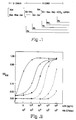
- Figure 1 shows the structure of S. typhimurium lipopolysaccharide which is similar to the lipopolysaccharide of other gram-negative bacteria.
- R a , R b , R c , R d1 and R e designate the structures of LPS molecules synthesized by different mutants of Samonella .
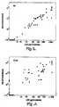
- Figure 2 indicates the detection of Hib DL42 LOS in PBS ( ⁇ ) and in plasma-PBS (diluted 1:3) ( ⁇ ) and Hib DL42 bacteria in plasma-PBS (diluted 1:3) ( ⁇ ) by immunolimulus assay. Open symbols indicate nonspecific background. Results are means from duplicate wells.
- the invention is a sensitive and rapid assay that can be designed to detect gram-negative bacterial endotoxin, or, modified to detect specific species of bacterial endotoxin.
- the method is useful for detection of very low levels of endotoxin.
- the inventors have used antibodies as the basis of selectivity of their method and combined the previously known reaction with amoebocyte lysate to provide an assay that is highly sensitive.
- the antibodies are preferably monoclonal antibodies directed to designated regions of the endotoxin core of LPS/LOS.
- Hib LOS Haemophilus influenzae type b endotoxin
- An inclusive method to detect gram-negative bacterial endotoxins, for example, would require up to several monoclonal antibodies directed to regions at or very close to the endotoxin lipid A core.
- a method to detect Pseudomonas aeruginosa could employ monoclonal antibody XMMPS-605 (ATCC Accession No. HB8909 (11)).
- Hib strain DL42 has been characterized extensively (17). Another Hib strain (DL301) used in this study was a recent isolate from a child with Hib meningitis in Dallas. Both of these strains belong to Hib LOS antigenic group 2, as determined by their reactivity with MAb 4C4 in the colony blot-radioimmunoassay stem (17). Encapsulated Escherichia coli K1 (77-436) and Hib strain DL26, from Hib LOS antigenic group 1 (17) were used for control experiments. These strains do not react with MAb 4C4.
- Hib strains were grown in brain-heart infusion medium (Difco Laboratories, Detroit, MI) supplemented with Levinthal's base (BHIs) as previously described (18).
- Escherichia coli K1 was grown in brain-heart infusion medium.without the supplement.
- LOS from Hib strain DL42 was purified by means of the modified hot phenol-water method (19). Purified LOS was diluted in pyrogen-free saline (Abbott Laboratories, North Chicago, IL) and stored in 1 »g/ml concentrations at -70°C until used as a standard in the assays.
- LPS purified from Escherichia coli 0111:B4 (Sigma No. L-33012) was purchased from Sigma Chemical Company, St. Louis, MO, as were LPS from Escherichia coli 0127:B8 (Sigma No. L-3137), Klebsiella pneumoniae (Sigma No. L-1770) and Pseudomonas aeruginosa (Sigma No. L-8643).
- MAbs Monoclonal Antibodies
- MAb 4C4 ATCC Accession No. HB10461
- This MAb reacts with the LOS from Hib strains DL42 and DL301 and, additionally, was able to recognize 86% of the Hib clinical isolates recently tested (17).
- Sterile polystyrene ELISA plates were purchased from Corning Laboratory Sciences, Houston, TX. All plates were determined to be pyrogen-free prior to use. Each vial of LAL (Pyrotell, Associates of Cape Code, Woods Hole, MA specified to be suitable for use in the CLAL) was reconstituted with 10 ml of pf-water, and stored in multiple portions at -20°C for less than 3 months. For the IML assay, the LAL was diluted with 20 ml of pf-water. N-benzoyl-val-gly-arg-p-nitroanilide hydrochloride (Sigma No. B-4758) was diluted to a concentration of 0.7 mg/ml in pf-water and stored at 4°C until used in the assays.
- Pearson's correlation coefficient was used to assess the strength of the relationship between the results from the CLAL and IML assays and the magnitude of bacteremia in the animals.
- Microtiter plates were coated overnight at room temperature with purified MAb 4C4 diluted 1:500 in 0.1 M sodium carbonate buffer, pH 9.6 (20). The final amount of antibody incubated in the well was 800 ng.
- the microtiter wells were washed three times with pf-PBS containing 0.05% (v/v) Tween® 20 (pf PBS-Tw) and then blocked with 1% (vol/vol) fetal calf serum (20) in carbonate buffer for one hour at 37°C. The plates were again washed three times with pf-PBS-Tw.
- Plasma for dilution of purified LOS samples was obtained by collecting blood by cardiac puncture from 50 normal infant rats, centrifuging and pooling the plasma derived by the sodium citrate method. Plasma aliquots were stored at -70°C and prior to use as diluent were diluted 1:3 in pyrogen-free phosphate buffered saline (pf-PBS, pH 7.4).
- Purified Hib DL42 LOS was diluted in pf-PBS and in plasma:PBS (diluted 1:3) and Hib bacteria were diluted in plasma:PBS (diluted 1:3).
- Purified Hib LOS standards were prepared in 0-1,000 pg/ml concentrations in the appropriate dilution fluid. After heat-inactivation at 75°C for 12 min, 50 »l of the test samples and the standards were incubated in the microtiter wells, for one hour at 37°C. The wells were washed six times with pf-PBS-Tw and then filled with 50 »l LAL extract in pf-water.
- the limit of sensitivity (the concentration of LOS yielding an optical density reading exceeding the mean + 2SD of the background) was 2 pg/ml in pf-PBS and 10 pg/ml in diluted plasma ( Figure 2).
- the sensitivity of the IML method in detecting LOS in diluted plasma containing various numbers of Hib organisms corresponded to a concentration of 300 CFU/ml, respectively.
- the specificity of the IML assay is based on its ability to detect only those LOS molecules bound to the solid phase by the MAbs. As shown in Figure 2, the nonspecific background (obtained with wells without MAb) remained consistently low during testing of plasma samples when the concentration of LOS was less than 1,000 pg/ml or when the concentration of bacteria was less than 105 CFU/ml. With higher concentrations of either LOS or Hib the background increased and the IML assay lost its antibody-dependent specificity although it showed the presence of endotoxin with the CLAL-type reaction. The specific antibody binding could be demonstrated in these cases with the further dilution of the test samples.
- the immunolimulus assay was applied to the detection of H. ducreyi , the causative agent of chancroid.
- H. ducreyi the causative agent of chancroid.
- a study was conducted wherein a monoclonal antibody specific for H. ducreyi LOS was employed in an assay essentially as set forth in Example I.
- the monoclonal antibody to H. ducreyi LOS employed in these studies was prepared as follows:
- H. ducreyi strain 35000 were used to immunize female BALB/c mice. A 50-100»g quantity of cell envelope protein suspended in 0.1 ml of Freund's complete adjuvant was injected intraperitoneally into several mice. Approximately four weeks later, two of these mice were given an intravenous injection with 50 »g of these cell envelopes and then the spleens were removed for use in the hybridoma fusion protocol.
- the hybridoma cell line 3E6 secreting an IgG monoclonal antibody reactive with H. ducreyi LOS was identified by screening hybridoma culture supernatants in Western blot analysis against cell envelopes and then against proteinase K-treated cell envelopes.
- This Mab (3E6) was found to be reactive with every H. ducreyi strain tested to date (at least 20 strains) and does not react with H. influenzae strains, with Treponema pallidum , or with two Neisseria gonorrhoeae strains in colony blot-radioimmunoassay analysis.
- the immunolimulus assay incorporating H. ducreyi LOS-specific antibody 3E6 was applied first to the detection of H. ducreyi that had been grown in vitro and resuspended in pyrogen-free PBS.
- This sample was prepared by growing H. ducreyi strain 35000 on chocolate agar plates overnight at 33°C in a candle extinction jar. The resultant colonies were scraped off the surface of the agar plates and suspended in pyrogen-free PBS to a concentration of 108 cfu. After mixing by vigorous vortexing, the cell suspension was diluted to approximately 107 cfu per ml in pyrogen-free PBS. This suspension was then heated at 75°C for 12 minutes.
- H. ducreyi strain 35000 used for inoculation into animals were cultivated in candle extinction jars at 33°C for 14 to 16 hrs.
- the bacteria were harvested into sterile phosphate-buffered saline (PBS), pH 7.2, using sterile cotton swabs and the cells were washed twice with PBS by repeated centrifugation and resuspension, and cell suspensions containing varying numbers of cfu of H. ducreyi were prepared in pyrogen-free saline.
- the H. ducreyi cell suspensions were injected intradermally into the shaved backs of the rabbits at duplicate sites, using an injection volume of 0.1 ml.
- Rabbits were housed in a temperature-controlled room at 15-17°C to lower their skin temperature. Animals housed at 15-17°C had an average surface skin temperature 2°C lower than that of animals housed at 23-25°C. After about 2-4 days, lesions were found to develop. Two to five days after intradermal injection, material was aspirated from the resultant lesions and the content of viable H. ducreyi organisms in each sample determined. Bacteria were recovered from skin lesions by injecting 0.1 ml of sterile, pyrogen-free PBS into the lesion site with a 25-gauge needle and then aspirating the injected material back into a syringe.
- the material aspirated from each lesion was brought to a final volume of 1 ml using pyrogen-free PBS. Then, 50 ul aliquots were plated to determine the content of viable H. ducreyi in the aspirated material. A 1:5 dilution of this material in pyrogen-free PBS was prepared and heated at 75°C for 12 minutes. Then, 50 ul samples of both this suspension and further dilutions of this suspension were used in the immunolimulus assay. A positive reaction in this assay was obtained with samples containing as few as 200 cfu of viable H. Ducreyi per ml.
Landscapes
- Health & Medical Sciences (AREA)
- Life Sciences & Earth Sciences (AREA)
- Engineering & Computer Science (AREA)
- Immunology (AREA)
- Chemical & Material Sciences (AREA)
- Urology & Nephrology (AREA)
- Hematology (AREA)
- Biomedical Technology (AREA)
- Molecular Biology (AREA)
- Physics & Mathematics (AREA)
- Biochemistry (AREA)
- Cell Biology (AREA)
- Pathology (AREA)
- General Physics & Mathematics (AREA)
- Food Science & Technology (AREA)
- Medicinal Chemistry (AREA)
- Biotechnology (AREA)
- Analytical Chemistry (AREA)
- Microbiology (AREA)
- General Health & Medical Sciences (AREA)
- Tropical Medicine & Parasitology (AREA)
- Virology (AREA)
- Chemical Kinetics & Catalysis (AREA)
- Measuring Or Testing Involving Enzymes Or Micro-Organisms (AREA)
- Peptides Or Proteins (AREA)
- Preparation Of Compounds By Using Micro-Organisms (AREA)
- Investigating Or Analysing Biological Materials (AREA)
Claims (27)
- Procédé de détection d'une endotoxine bactérienne dans un échantillon, comportant les étapes consistant à :(a) mettre en contact un échantillon suspecté contenir une endotoxine bactérienne avec au moins un anticorps capable de se lier à une telle endotoxine bactérienne, l'échantillon étant mis en contact avec l'anticorps dans des conditions efficaces pour lier l'anticorps à l'endotoxine qui peut être présente dans l'échantillon;(b) laver l'endotoxine liée à l'anticorps pour éliminer les contaminants; et(c) détecter l'endotoxine avec un lysat d'amoebocyte.
- Procédé selon la revendication 1, dans lequel l'endotoxine est détectée :(a) en incubant l'endotoxine liée à l'anticorps lavé avec un lysat d'amoebocyte afin d'activer un système de protéase du lysat;(b) en ajoutant au lysat un substrat du système de proétase du lysat; et(c) en mesurant les quantités d'un produit formé à partir de l'action dudit système de protéase sur le substrat ajouté, ledit produit étant proportionnel à un niveau d'endotoxine bactérienne dans l'échantillon.
- Procédé selon la revendication 1, dans lequel l'anticorps est lié à une surface solide.
- Procédé selon la revendication 1, dans lequel l'anticorps a une affinité de liaison pour un anticorps choisi à partir : d'un glycolipide du coeur d'une endotoxine bactérienne, d'un épitope comportant une région proximale au lipide A de l'endotoxine bactérienne, d'un épitope comportant le lipide A et d'un épitope comportant un heptose et une céto-déoxyoctonate.
- Procédé selon la revendication 1, dans lequel l'anticorps est un MAb obtenu à partir d'une lignée cellulaire d'hybridomes XMMEN-OE5 ayant le numéro de référence ATCC HB9081 ou à partir d'une lignée cellulaire d'hybridomes XMMEN-LY1 ayant le numéro de référence ATCC HB9082.
- Procédé selon la revendication 1, dans lequel l'anticorps comporte un fragment de liaison de celui-ci.
- Procédé selon la revendication 1, dans lequel l'endotoxine bactérienne est une lipooligosaccharide ou une lipo-polysaccharide d'une bactérie de gram-négatif, de préférence dans lequel l'endotoxine bactérienne est issue d'une bactérie de gram-négatif du genre Escherichia, Bordetella, Branhamella, Salmonella, Haemophilus, Klebsiella, Proteus, Enterobacter, Pseudomonas, Pasteurella, Acinetobacter ou Neisseria.
- Procédé selon la revendication 1, dans lequel l'anticorps a une affinité de liaison pour un antigène-O, de préférence l'antigène-O de Salmonella typhi.
- Procédé selon la revendication 1, dans lequel le lysat d'amoebocyte est obtenu à partir de sang de Tachypleus tridentatus, Tachypleus gigas, Carcinoscorpius rotundicauda ou Limulus polyphemus.
- Procédé selon la revendication 2, dans lequel le substrat ajouté à la réaction est constitué d'un substrat chromogénique.
- Procédé selon la revendication 1, dans lequel l'échantillon est constitué de plasma, de sérum, de fluide cérébro-spinal ou d'urine.
- Procédé selon la revendication 1, dans lequel l'échantillon est chauffé avant la mise en contact avec l'anticorps, de préférence à une température aux environs de 75°C.
- Procédé de détection du lipooligosaccharide d'Haemophilus influenzae de type b (Hib LOS) dans un échantillon suspecté contenir un tel LOS, comportant les étapes consistant à :(a) mettre en contact l'échantillon avec au moins deux anticorps dirigés contre Hib LOS pour faciliter la formation d'un complexe immunologique entre les anticorps et le Hib LOS;(b) laver le complexe immunologique;(c) incuber le Hib LOS lavé avec un lysat d'amoebocyte de Limulus pour faciliter l'activation d'un système de protéase dans le lysat de Limulus; et(d) détecter la présence de LOS en détectant un produit formé à partir de l'action du système de protéase sur un substrat.
- Procédé selon la revendication 13, dans lequel l'anticorps est dirigé contre l'oligosaccharide du coeur externe en position distale par rapport au lipide A.
- Procédé selon la revendication 13, dans lequel l'anticorps est un anticorps monoclonal IgG3 dirigé contre un épitope dans la région oligosaccharidique de Hib LOS DL26 ou d'Hib LOS DL42.
- Procédé de détection du LOS d'Haemophilus ducreyi dans un échantillon supposé contenir un tel LOS, le procédé comportant les étapes consistant à :(a) mettre en contact l'échantillon avec au moins un anticorps dirigé contre LOS d'Haemophilus ducreyi pour faciliter la formation d'un complexe immunologique entre l'anticorps et LOS qui peut être présent dans l'échantillon;(b) laver le complexe immunologique ;(c) incuber H. ducreyi lavé avec un lysat d'amoebocyte de Limulus pour faciliter l'activation d'un système de protéase dans le lysat de Limulus; et(d) détecter la présence de LOS en détectant un produit formé à partir de l'action du système de protéase sur un substrat.
- Kit comportant :(a) un support, ayant des compartiments pour recevoir-au moins deux conteneurs en confinement;(b) un premier conteneur positionné dans un des compartiments, le premier conteneur comportant au moins un anticorps ayant une affinité de liaison pour une endotoxine bactérienne; et(c) un second conteneur positionné dans un autre compartiment, le second conteneur comportant un lysat d'amoebocyte ou un substrat chromogénique afin de détecter une libération d'un système de protéase par une endotoxine bactérienne pour une utilisation dans un procédé selon l'une quelconque des revendications 1 à 12.
- Kit selon la revendication 17, défini de plus comme incluant à la fois un conteneur comportant un lysat d'amoebocyte et un conteneur comportant un substrat chromogénique.
- Kit selon la revendication 17, dans lequel l'anticorps comporte un anticorps monoclonal.
- Kit selon la revendication 17, dans lequel l'anticorps comporte un anticorps qui reconnaît un épitope d'un oligosaccharide situé en position proximale par rapport au lipide A ou au lipide A situé dans une région interne du coeur d'une bactérie de gram-négatif.
- Kit comportant :(a) un support, ayant des compartiments pour recevoir au moins deux conteneurs en confinement;(b) un premier conteneur positionné dans un des compartiments, le premier conteneur comportant deux anticorps se liant de manière spécifique aux épitopes de l'endotoxine d'Haemophilus influenzae de type b; et(c) un second conteneur positionné dans un compartiment, le second conteneur comportant un lysat d'amoebocyte ou un substrat chromogénique afin de détecter une libération de protéase à partir d'un tel lysat pour une utilisation dans un procédé selon l'une quelconque des revendications 13 à 15.
- Kit selon la revendication 21, défini de plus comme incluant à la fois un conteneur comportant un lysat d'amoebocyte et un conteneur comportant un substrat chromogénique.
- Kit selon la revendication 21, dans lequel le premier conteneur comporte les MAbs 4C4 et DL42.
- Kit selon la revendication 21, dans lequel un ou plusieurs des composants sont apportés sous une forme lyophilisée.
- Kit comportant :(a) un support, ayant des compartiments pour recevoir au moins deux conteneurs en confinement;(b) un premier conteneur positionné dans un compartiment, le premier conteneur comportant au moins un anticorps se liant de manière spécifique à un épitope de LOS d'Haemophilus ducreyi; et(c) un second conteneur positionné dans un compartiment, le second conteneur comportant un lysat d'amoebocyte ou un substrat chromogénique afin de détecter une libération de protéase à partir d'un tel lysat pour une utilisation dans un procédé selon la revendication 16.
- Kit selon la revendication 25, défini de plus comme incluant à la fois un conteneur comportant un lysat d'amoebocyte et un conteneur comportant un substrat chromogénique.
- Kit selon la revendication 25, dans lequel un ou plusieurs composants sont fournis sous une forme lyophilisée.
Applications Claiming Priority (3)
| Application Number | Priority Date | Filing Date | Title |
|---|---|---|---|
| US07/553,072 US5198339A (en) | 1990-07-13 | 1990-07-13 | Method for detection of gram-negative bacterial lipopolysaccharides in biological fluids |
| US553072 | 1990-07-13 | ||
| PCT/US1991/004864 WO1992001228A1 (fr) | 1990-07-13 | 1991-07-10 | Procede de detection de lipopolysaccharides bacteriens gram-negatifs dans des liquides biologiques |
Publications (2)
| Publication Number | Publication Date |
|---|---|
| EP0539497A1 EP0539497A1 (fr) | 1993-05-05 |
| EP0539497B1 true EP0539497B1 (fr) | 1995-06-07 |
Family
ID=24208011
Family Applications (1)
| Application Number | Title | Priority Date | Filing Date |
|---|---|---|---|
| EP91913737A Expired - Lifetime EP0539497B1 (fr) | 1990-07-13 | 1991-07-10 | Procede de detection de lipopolysaccharides bacteriens gram-negatifs dans des liquides biologiques |
Country Status (8)
| Country | Link |
|---|---|
| US (2) | US5198339A (fr) |
| EP (1) | EP0539497B1 (fr) |
| JP (1) | JPH05508714A (fr) |
| AT (1) | ATE123576T1 (fr) |
| AU (1) | AU8297191A (fr) |
| CA (1) | CA2084083A1 (fr) |
| DE (1) | DE69110287T2 (fr) |
| WO (1) | WO1992001228A1 (fr) |
Cited By (1)
| Publication number | Priority date | Publication date | Assignee | Title |
|---|---|---|---|---|
| US6790661B1 (en) | 1999-07-16 | 2004-09-14 | Verax Biomedical, Inc. | System for detecting bacteria in blood, blood products, and fluids of tissues |
Families Citing this family (24)
| Publication number | Priority date | Publication date | Assignee | Title |
|---|---|---|---|---|
| US7118757B1 (en) * | 1988-12-19 | 2006-10-10 | Wyeth Holdings Corporation | Meningococcal class 1 outer-membrane protein vaccine |
| US5198339A (en) * | 1990-07-13 | 1993-03-30 | Board Of Regents, The University Of Texas System | Method for detection of gram-negative bacterial lipopolysaccharides in biological fluids |
| JP3322700B2 (ja) * | 1992-09-14 | 2002-09-09 | 生化学工業株式会社 | 固相系反応用エンドトキシン特異的測定剤 |
| CA2112776C (fr) * | 1993-01-21 | 2002-11-12 | Masakazu Tsuchiya | Methode pour inhiber l'activite d'une endotoxine |
| US5707817A (en) * | 1993-02-05 | 1998-01-13 | Colorado State University Research Foundation | Carbohydrate-based vaccine and diagnostic reagent for trichinosis |
| US5496700A (en) * | 1993-08-06 | 1996-03-05 | The United States Of America As Represented By The Secretary Of The Navy | Optical immunoassay for microbial analytes using non-specific dyes |
| GB9807814D0 (en) * | 1998-04-09 | 1998-06-10 | Allied Therapeutics Limited | Solid phase test for endoxin |
| US6824997B1 (en) | 1998-09-18 | 2004-11-30 | Binax, Inc. | Process and materials for the rapid detection of streptococcus pneumoniae employing purified antigen-specific antibodies |
| US9134303B1 (en) | 1998-08-25 | 2015-09-15 | Alere Scarborough, Inc. | ICT immunoassay for Legionella pneumophila serogroup 1 antigen employing affinity purified antibodies thereto |
| JP2002181824A (ja) * | 2000-12-15 | 2002-06-26 | Kita Kyushu Biophysics Kenkyusho:Kk | 糖抗原認識性エンドトキシン測定法 |
| US6720160B2 (en) | 2001-10-11 | 2004-04-13 | Helica Biosystems, Inc. | Method for simultaneous detection of multiple microbial antigens in biological specimens from mastitic animals |
| US6696254B2 (en) * | 2001-11-21 | 2004-02-24 | Kimberly-Clark Worldwide, Inc. | Detection and identification of enteric bacteria |
| US7828741B2 (en) * | 2002-12-20 | 2010-11-09 | The Charlotte-Mecklenburg Hospital Authority | Utilizing lipopolysaccharide in exhaled breath condensate to diagnose gram negative pneumonia |
| DE60326988D1 (de) * | 2002-12-20 | 2009-05-14 | Charlotte Mecklenburg Hospital | Wegwerf-handvorrichtung zum auffangen von ausgeatmetem atemkondensat |
| WO2004073497A2 (fr) * | 2003-02-14 | 2004-09-02 | The Charlotte-Mecklenburg Hospital Authority | Dispositif et procede de collecte de condensat d'exhalation alveolaire |
| MX306461B (es) * | 2006-06-05 | 2013-01-07 | Ocean Nutrition Canada Ltd | Microcapsulas con cubiertas mejoradas |
| EP2388593A3 (fr) * | 2006-10-18 | 2012-01-18 | bioMérieux | Procédé de diagnostic in vitro des Staphylococcus aureus producteurs de PVL |
| FR2912219A1 (fr) * | 2007-02-06 | 2008-08-08 | Biomerieux Sa | Procede de diagnostic in vitro des staphylococcus aureus producteurs de pvl, anticorps monoclonaux et kits de diagnostic |
| KR100888788B1 (ko) | 2007-07-02 | 2009-03-16 | 다이아텍코리아 주식회사 | 엔도톡신의 정량적 측정 방법 |
| JP5564193B2 (ja) * | 2009-04-07 | 2014-07-30 | 株式会社ペプタイドドア | リポ多糖又はリピッドaの分析方法 |
| US20130084586A1 (en) * | 2011-09-30 | 2013-04-04 | The Penn State Research Foundation | Rapid, specific and sensitive immunoassays for the detection of highly variable gram negative bacterial antigens |
| GB201312635D0 (en) * | 2013-07-15 | 2013-08-28 | Univ Plymouth | Water testing |
| CN111077307B (zh) * | 2018-10-21 | 2022-08-23 | 厦门鲎试剂生物科技股份有限公司 | 一种用革兰氏阴性细菌感染快速检测脓毒病的新方法 |
| GB202009143D0 (en) * | 2020-06-16 | 2020-07-29 | Molendotech Ltd | Testing method and apparatus |
Family Cites Families (5)
| Publication number | Priority date | Publication date | Assignee | Title |
|---|---|---|---|---|
| US4455296A (en) * | 1982-02-02 | 1984-06-19 | Board Of Regents, The University Of Texas System | Hybrid cell lines producing monoclonal antibodies directed against Haemophilus influenzae |
| US4918163A (en) * | 1985-09-27 | 1990-04-17 | Pfizer Inc. | Monoclonal antibodies specific for lipid-A determinants of gram negative bacteria |
| US4906567A (en) * | 1987-01-21 | 1990-03-06 | E. I. Dupont De Nemours And Company | Non-immunochemical binding of lipopolysaccharides and sandwich assays therefor |
| DK255887D0 (da) * | 1987-05-20 | 1987-05-20 | Claus Koch | Immunoassay |
| US5198339A (en) * | 1990-07-13 | 1993-03-30 | Board Of Regents, The University Of Texas System | Method for detection of gram-negative bacterial lipopolysaccharides in biological fluids |
-
1990
- 1990-07-13 US US07/553,072 patent/US5198339A/en not_active Expired - Fee Related
-
1991
- 1991-07-10 US US07/972,498 patent/US5356778A/en not_active Expired - Fee Related
- 1991-07-10 EP EP91913737A patent/EP0539497B1/fr not_active Expired - Lifetime
- 1991-07-10 WO PCT/US1991/004864 patent/WO1992001228A1/fr not_active Ceased
- 1991-07-10 CA CA002084083A patent/CA2084083A1/fr not_active Abandoned
- 1991-07-10 AT AT91913737T patent/ATE123576T1/de not_active IP Right Cessation
- 1991-07-10 JP JP91513070A patent/JPH05508714A/ja active Pending
- 1991-07-10 DE DE69110287T patent/DE69110287T2/de not_active Expired - Fee Related
- 1991-07-10 AU AU82971/91A patent/AU8297191A/en not_active Abandoned
Non-Patent Citations (1)
| Title |
|---|
| JOURNAL OF IMMUNOLOGICAL METHODS, vol. 122, 01 September 1989, New York, NY (US); J. MERTSOLA et al., pp. 219-226# * |
Cited By (1)
| Publication number | Priority date | Publication date | Assignee | Title |
|---|---|---|---|---|
| US6790661B1 (en) | 1999-07-16 | 2004-09-14 | Verax Biomedical, Inc. | System for detecting bacteria in blood, blood products, and fluids of tissues |
Also Published As
| Publication number | Publication date |
|---|---|
| DE69110287T2 (de) | 1996-02-08 |
| JPH05508714A (ja) | 1993-12-02 |
| EP0539497A1 (fr) | 1993-05-05 |
| WO1992001228A1 (fr) | 1992-01-23 |
| AU8297191A (en) | 1992-02-04 |
| US5198339A (en) | 1993-03-30 |
| DE69110287D1 (de) | 1995-07-13 |
| CA2084083A1 (fr) | 1992-01-14 |
| US5356778A (en) | 1994-10-18 |
| ATE123576T1 (de) | 1995-06-15 |
Similar Documents
| Publication | Publication Date | Title |
|---|---|---|
| EP0539497B1 (fr) | Procede de detection de lipopolysaccharides bacteriens gram-negatifs dans des liquides biologiques | |
| Musher et al. | Does naturally acquired IgG antibody to cell wall polysaccharide protect human subjects against pneumococcal infection? | |
| JP2667695B2 (ja) | 試料中のエンドトキシンの存在を決定する方法 | |
| US4683196A (en) | Method and materials for the identification of lipopolysaccharide producing microorganisms | |
| Olling | Sensitivity of gram-negative bacilli to the serum bactericidal activity: a marker of the host-parasite relationship in acute and persisting infections | |
| US4361647A (en) | Sandwich immunoassay and compositions for use therein | |
| Carlsson et al. | Quantitation of Salmonella O-antibodies in human sera by enzyme-linked immunosorbent assay (ELISA) | |
| Barclay et al. | Serological relationships between Escherichia coli and Salmonella smooth-and rough-mutant lipopolysaccharides as revealed by enzyme-linked immunosorbent assay for human immunoglobulin G antiendotoxin antibodies | |
| US20100273177A1 (en) | Methods and Devices for Testing Saliva | |
| AU5693399A (en) | Method for detection of (legionella) bacteria employing purified antigen-specific antibodies | |
| AU616384B2 (en) | Method for the evaluation of oral microbes | |
| Tsang et al. | A murine monoclonal antibody specific for the outer core oligosaccharide of Salmonella lipopolysaccharide | |
| EP0168422A1 (fr) | Procede et substances permettant l'identification de micro-organismes produisant du lipopolysaccharide | |
| Néron et al. | Type 2 T-cell-independent murine immune response to the human ABO blood group antigens | |
| Scott et al. | IgG antibodies to Gram-negative endotoxin in human sera. I. Lipopolysaccharide (LPS) cross-reactivity due to antibodies to LPS core | |
| Mertsola et al. | Detection of experimental Haemophilus influenzae type b bacteremia and endotoxemia by means of an immunolimulus assay | |
| LIM | Diagnostic uses of monoclonal antibodies to Salmonella | |
| Mertsola et al. | Specific detection of Haemophilus influenzae type b lipooligosaccharide by immunoassay | |
| Colling et al. | Association of bacterial carbohydrate-specific cold agglutinin antibody production with immunization by group C, group B type III, and Streptococcus pneumoniae type XIV streptococcal vaccines | |
| Eskenazy et al. | Detection by immunofluorescence of common antigenic determinants in unrelated gram-negative bacteria and their lipopolysaccharides | |
| Carlsson et al. | IMMUNOCHEMISTRY OF SALMONELLA O‐ANTIGENS: Characterization of the Antibody Response to Tyvelose 13 Mannose 1 Bovine Serum Albumin Representative of Salmonella O‐Antigen 9 | |
| Yang et al. | Development of human ABO blood group A antigen on Escherichia coli Y1089 and Y1090 | |
| Nolan et al. | Immunoradiometric assay of lipid A: a test for detecting and quantitating endotoxins of various origins | |
| KÄYHTY | The unspecific antibody response to N. meningitidis group A capsular polysaccharide often seen in bacteraemic diseases | |
| Pongsunk et al. | Production of monoclonal antibodies to Vi polysaccharide antigen of Salmonella typhi |
Legal Events
| Date | Code | Title | Description |
|---|---|---|---|
| PUAI | Public reference made under article 153(3) epc to a published international application that has entered the european phase |
Free format text: ORIGINAL CODE: 0009012 |
|
| AK | Designated contracting states |
Kind code of ref document: A1 Designated state(s): AT BE CH DE DK ES FR GB GR IT LI LU NL |
|
| 17P | Request for examination filed |
Effective date: 19930211 |
|
| RBV | Designated contracting states (corrected) |
Designated state(s): AT BE CH DE DK ES FR GB GR IT LI LU NL SE |
|
| 17Q | First examination report despatched |
Effective date: 19931207 |
|
| GRAA | (expected) grant |
Free format text: ORIGINAL CODE: 0009210 |
|
| AK | Designated contracting states |
Kind code of ref document: B1 Designated state(s): AT BE CH DE DK ES FR GB GR IT LI LU NL SE |
|
| PG25 | Lapsed in a contracting state [announced via postgrant information from national office to epo] |
Ref country code: IT Free format text: LAPSE BECAUSE OF FAILURE TO SUBMIT A TRANSLATION OF THE DESCRIPTION OR TO PAY THE FEE WITHIN THE PRE;WARNING: LAPSES OF ITALIAN PATENTS WITH EFFECTIVE DATE BEFORE 2007 MAY HAVE OCCURRED AT ANY TIME BEFORE 2007. THE CORRECT EFFECTIVE DATE MAY BE DIFFERENT FROM THE ONE RECORDED.SCRIBED TIME-LIMIT Effective date: 19950607 Ref country code: DK Effective date: 19950607 Ref country code: ES Free format text: THE PATENT HAS BEEN ANNULLED BY A DECISION OF A NATIONAL AUTHORITY Effective date: 19950607 Ref country code: LI Effective date: 19950607 Ref country code: BE Effective date: 19950607 Ref country code: CH Effective date: 19950607 Ref country code: GR Free format text: LAPSE BECAUSE OF FAILURE TO SUBMIT A TRANSLATION OF THE DESCRIPTION OR TO PAY THE FEE WITHIN THE PRESCRIBED TIME-LIMIT Effective date: 19950607 Ref country code: FR Effective date: 19950607 Ref country code: AT Effective date: 19950607 Ref country code: NL Effective date: 19950607 |
|
| REF | Corresponds to: |
Ref document number: 123576 Country of ref document: AT Date of ref document: 19950615 Kind code of ref document: T |
|
| REF | Corresponds to: |
Ref document number: 69110287 Country of ref document: DE Date of ref document: 19950713 |
|
| PGFP | Annual fee paid to national office [announced via postgrant information from national office to epo] |
Ref country code: DE Payment date: 19950725 Year of fee payment: 5 |
|
| PG25 | Lapsed in a contracting state [announced via postgrant information from national office to epo] |
Ref country code: LU Free format text: LAPSE BECAUSE OF NON-PAYMENT OF DUE FEES Effective date: 19950731 |
|
| NLT2 | Nl: modifications (of names), taken from the european patent patent bulletin |
Owner name: BOARD OF REGENTS THE UNIVERSITY OF TEXAS SYSTEM |
|
| PG25 | Lapsed in a contracting state [announced via postgrant information from national office to epo] |
Ref country code: GB Effective date: 19950907 Ref country code: SE Effective date: 19950907 |
|
| PGFP | Annual fee paid to national office [announced via postgrant information from national office to epo] |
Ref country code: BE Payment date: 19950911 Year of fee payment: 5 |
|
| REG | Reference to a national code |
Ref country code: CH Ref legal event code: PL |
|
| NLV1 | Nl: lapsed or annulled due to failure to fulfill the requirements of art. 29p and 29m of the patents act | ||
| EN | Fr: translation not filed | ||
| PLBE | No opposition filed within time limit |
Free format text: ORIGINAL CODE: 0009261 |
|
| STAA | Information on the status of an ep patent application or granted ep patent |
Free format text: STATUS: NO OPPOSITION FILED WITHIN TIME LIMIT |
|
| GBPC | Gb: european patent ceased through non-payment of renewal fee |
Effective date: 19950907 |
|
| 26N | No opposition filed | ||
| PG25 | Lapsed in a contracting state [announced via postgrant information from national office to epo] |
Ref country code: DE Effective date: 19970402 |