EP0561003A1 - Dispositif pour détecter des marqueurs de maladies - Google Patents
Dispositif pour détecter des marqueurs de maladies Download PDFInfo
- Publication number
- EP0561003A1 EP0561003A1 EP91310654A EP91310654A EP0561003A1 EP 0561003 A1 EP0561003 A1 EP 0561003A1 EP 91310654 A EP91310654 A EP 91310654A EP 91310654 A EP91310654 A EP 91310654A EP 0561003 A1 EP0561003 A1 EP 0561003A1
- Authority
- EP
- European Patent Office
- Prior art keywords
- membrane
- piston
- housing
- fluid
- assembly
- Prior art date
- Legal status (The legal status is an assumption and is not a legal conclusion. Google has not performed a legal analysis and makes no representation as to the accuracy of the status listed.)
- Withdrawn
Links
Images


Classifications
-
- B—PERFORMING OPERATIONS; TRANSPORTING
- B01—PHYSICAL OR CHEMICAL PROCESSES OR APPARATUS IN GENERAL
- B01L—CHEMICAL OR PHYSICAL LABORATORY APPARATUS FOR GENERAL USE
- B01L3/00—Containers or dishes for laboratory use, e.g. laboratory glassware; Droppers
- B01L3/50—Containers for the purpose of retaining a material to be analysed, e.g. test tubes
- B01L3/508—Rigid containers without fluid transport within
- B01L3/5082—Test tubes per se
-
- A—HUMAN NECESSITIES
- A61—MEDICAL OR VETERINARY SCIENCE; HYGIENE
- A61B—DIAGNOSIS; SURGERY; IDENTIFICATION
- A61B10/00—Instruments for taking body samples for diagnostic purposes; Other methods or instruments for diagnosis, e.g. for vaccination diagnosis, sex determination or ovulation-period determination; Throat striking implements
- A61B10/0045—Devices for taking samples of body liquids
-
- B—PERFORMING OPERATIONS; TRANSPORTING
- B01—PHYSICAL OR CHEMICAL PROCESSES OR APPARATUS IN GENERAL
- B01L—CHEMICAL OR PHYSICAL LABORATORY APPARATUS FOR GENERAL USE
- B01L3/00—Containers or dishes for laboratory use, e.g. laboratory glassware; Droppers
- B01L3/50—Containers for the purpose of retaining a material to be analysed, e.g. test tubes
- B01L3/508—Rigid containers without fluid transport within
-
- G—PHYSICS
- G01—MEASURING; TESTING
- G01N—INVESTIGATING OR ANALYSING MATERIALS BY DETERMINING THEIR CHEMICAL OR PHYSICAL PROPERTIES
- G01N33/00—Investigating or analysing materials by specific methods not covered by groups G01N1/00 - G01N31/00
- G01N33/48—Biological material, e.g. blood, urine; Haemocytometers
- G01N33/50—Chemical analysis of biological material, e.g. blood, urine; Testing involving biospecific ligand binding methods; Immunological testing
- G01N33/53—Immunoassay; Biospecific binding assay; Materials therefor
- G01N33/543—Immunoassay; Biospecific binding assay; Materials therefor with an insoluble carrier for immobilising immunochemicals
- G01N33/54366—Apparatus specially adapted for solid-phase testing
-
- G—PHYSICS
- G01—MEASURING; TESTING
- G01N—INVESTIGATING OR ANALYSING MATERIALS BY DETERMINING THEIR CHEMICAL OR PHYSICAL PROPERTIES
- G01N33/00—Investigating or analysing materials by specific methods not covered by groups G01N1/00 - G01N31/00
- G01N33/48—Biological material, e.g. blood, urine; Haemocytometers
- G01N33/50—Chemical analysis of biological material, e.g. blood, urine; Testing involving biospecific ligand binding methods; Immunological testing
- G01N33/80—Chemical analysis of biological material, e.g. blood, urine; Testing involving biospecific ligand binding methods; Immunological testing involving blood groups or blood types or red blood cells
Definitions
- the present invention is directed to medical and laboratory specimen collecting and testing equipment, and more specifically to an apparatus for detecting disease markers both for screening as well as for a reference laboratory setting.
- a typical specimen collecting apparatus is shown by U.S. Patent 4,741,346.
- This apparatus includes a base stand which supports the specimen vial in an upright position.
- a funnel is inserted in the open end of the specimen vial and surrounds and encloses the upper portion of the vial.
- the base stand has an upwardly extending tubular wall which at least partially surrounds the vial which in connection with the cap allows the user to remove the vial without touching the surface or coming in contact with the specimen.
- Examples of various types of liquid containers for collecting and transporting urine are shown by U.S. Patents 3,777,739; 3,881,465; 4,042,337; 4,084,937; 4,244,920; 4,492,258 and 4,700,714.
- U.S. Patent 4,040,791 discloses a collection receptacle having a nipple upon which is mounted a specimen container which receives a predetermined amount of the specimen in a sealed condition.
- the specimen container is provided with an integrally formed cap which is placed over the opening in which the collector nipple is inserted.
- U.S. Patent 4,557,274 discloses a midstream urine collector having a funnel which transmits urine into a cup member which is covered by a membrane cover.
- U.S. Patent 4,473,530 A combined strip testing device and collection apparatus is shown by U.S. Patent 4,473,530 and is directed to an apparatus which integrates testing and collection by having chemical reagent test strips present within the tube together with specific gravity reading means allowing immediate testing of the urine.
- U. S. Patent 4,573,983 is directed towards a liquid collection system having an antiseptic member on the discharge section which uses a filter of air and bacteria impervious material to filter the urine.
- cytology cups and membranes The use of cytology cups and membranes is known in the art.
- the Nuclepore Schisto-Kit TM is designed for rapid and accurate quantification of Schistosome eggs in urine by the membrane filtration technique.
- a simple syringe filtration permits collection of virtually all eggs onto the smooth flat surface of a transparent Nuclepore polycarbonate membrane filter. Quantitative egg counts without staining are easily made with a low power magnifier.
- Other cytology cups are marketed under the trademark SWIN-LOK and Swinnex Disc Filter Holder.
- Nuclepore polycarbonate membranes are used for diagnostic cytology. The surface allows collection of atypical cells from all types of body fluids.
- a body fluid collection and testing device is in the form of a tubular device having a removable cytology cup which contains a prefiltration/beads housing and cytology membrane for quantitative analysis and a transportable syringe in the housing with a coloration membrane for qualitative test analysis.
- a capture antibody is immobilized on the membrane surface of the syringe head which is in contact with the body fluid.
- the prefilteration/beads housing becomes part of the cytology cup leaving the syringe head with membrane exposed.
- the cytology cup is then detached from the syringe body and the syringe body is inverted upside down to add the coloring reagents to the membrane.
- the bead housing and the cytology cup will be sent to the reference labratory for further analysis (quantitative).
- the preferred embodiment and best mode of the invention is seen in Figures 1 through 11.
- the invention shown therein comprises a modular separable body fluid testing device. While the invention can be used for any body fluid such as sputum, blood, peritoneal cavity fluid, pleural cavity fluid or urine, it is primarily designed for use in collecting urine/blood samples for use in testing for the presence of various kinds of disease markers, such as cancer in the body.
- a sample testing apparatus 20 is constructed of polystyrene and comprises a tubular collection unit or syringe barrel 22, a cytology cup 30 and a piston 50 with associated piston head test assembly 70.
- the tubular collection unit or syringe barrel 22 is constructed with a tubular open ended cylindrical body 24 defining a chamber 23 with an open flared end portion 26 and circular locking rib 27 formed on one end and on the other end a circular locking rib 28.
- the flared end portion 26 has a wide mouth to more easily receive body fluid such as urine or blood which is loaded into the chamber. It should be noted that a prelabelled antibody is added to the body fluid sample along with the buffer reagents.
- a cytology cup 30 is removably secured to the body 24 by virtue of a snap on fit of the cups locking mechanism over the rib 28.
- the cytology cup 30 comprises a cylindrical cup shaped body 32 with a locking lip mechanism comprising a stepped portion 33 and a flexible lip member 34 ending in rib 35.
- the lip rib 35 has an inner lesser diameter than the outer diameter of rib 28, allowing rib 28 to be snap fit into the locking lip mechanism.
- a downwardly extending circular flange member 36 extends inwardly toward the chamber of the cup to hold a bead housing assembly 100 in place in the cytology cup 30.
- the cytology cup is also provided with a cytology membrane housing comprised of a cylindrical barrel body 38 and an end member 39.
- the cytology membrane housing is removably mounted or secured in an aperture formed in the bottom surface of the body 32 with the barrel 38 extending upward into the chamber and the end member seated adjacent the bottom surface of the cytology cup 30.
- the barrel 38 holds a cytology membrane 40 which is seated on end member 39 at the bottom of the cup where cells can be captured at the end of the assay.
- the preferred membrane 40 which is used is manufactured by Nuclepore and can be cut in discs ranging from 13 mm to 293 mm in diameter with a pore size of 2.0 ⁇ m or less and exhibits a tensile strength of over 3000 psi.
- the prefered material composition is polycarbonate although polyester can be used.
- the membrane is flexible and will not crack and is resistent to splitting or breakage.
- membrane filters have a pore size, pore density and pore structure which are geometrically defined and photomicrographs of the same reveal individual pore openings on the surface with diameters closely equal to the rated pore size of the membrane.
- the advantages of a defined surface pore size are the complete surface capture of all particulate larger than the rated pore size, excellent particulate visibility and internal reference scale for particulate sizing.
- the smooth flat surface of the membrane offers an ideal substrate for particulate analysis using either optical or electron microscopy.
- Those membranes with pore size larger than 1.0 ⁇ m are sufficiently transparent to permit transmitted light allowing viewing of objects on the membrane surface without cleaning the membrane.
- the membrane can be coated with a hydrophilic surface that yields nearly instantanous flash-wetting with aqueous solutions.
- Such membranes when coated are coated with polyvinylphrrolidone (PVP) to render them hydrophilic.
- PVP polyvinylphrrolidone
- the membrane can be PVP free if so desired.
- the cytology diagnostic membrane 40 has a smooth flat surface which is ideal for the collection of atypical cells from all types of body fluids.
- Polycarbonate membranes are semi-transparent permitting direct microscopy or may easily be dissolved to remove all pore image artifacts.
- the advantages which occur in the use of a polycarbonate membrane are minimum clogging by red blood cells and protein, well preserved cellular morphology with high recovery rate, rapid filtration with low pressure, and excellent surface capture due to the pore structure and porosity.
- the smooth flat surface permits high cell visibility, improved morphologic resolution and surface capture.
- the membrane thickness allows easy mounting and immediate microscopic examination.
- the membranes low absorption and adsoption provides improved contrast, greater cell isolation and easy mounting while its non-staining characteristics allow improved contrast and simpler microscopic analysis. Furthermore the chemical resistance of the membrane is unaffected by conventioned cytologic fixatives and stains.
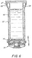
- the barrel also serves as a holder for the bead housing assembly as shown in Figure 6.
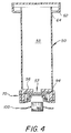
- a piston 50 as shown in Figure 2 is designed to fit within cylindrical body of the syringe barrel 24 and slideably move along the interior wall surface 25 holding a test assembly 70 for deposit within the cytology cup.
- the piston 50 is constructed of a transparent plastic and comprises a hollow cylindrical piston body 52 provided with a thumb cover assembly 54 and a cross sectional U-shaped bottom end member 56 of thicker construction than the piston body 52.
- the thumb cover assembly 54 includes a thumb support member 58 with a downwardly projecting flexible skirt or flange 60 ending in locking rib 62.
- the locking rib 62 is adapted to lock onto syringe rib 27 as the rib 62 cams the flange 60 so that it springs outward allowing rib 62 to ride over rib 27 and then snap back into place thereby securing the piston 50 on the syringe barrel 22.
- An air release aperture 64 is formed in the piston body so that there is communication between the interior chamber 53 of the piston body into the outside atmosphere.
- the bottom endwall 56 is provided with a throughgoing aperture 57 which allows communication with the chamber 53.
- the test assembly 70 as shown in exploded parts in Figure 2 is constructed with a cylindrical base cap shaped member having an interior diameter equal to or slightly less than the exterior diameter of endwall 56 so that it can be friction fit on same and a funnel shaped endwall 81 which funnels into a cylindrical section 83 which serves as a chamber for housing body 102 and a support for seat 84.
- the endwall by virtue of section 83 is provided with a pass through port or opening 82.
- the port 82 is further defined by a circular ring shaped membrane disk seat 84 which sits over the port.
- the seat 84 is provided with a flat upper surface to hold membrane 92 and a flat lower surface 85 forming a stop for the body 102 of the bead housing assembly.
- a membrane clip assembly 86 with a plastic cup body 87 and curved spring skirt 88 is mounted over the disc seat 84 and curls back under the disc seat 84 toward cylindrical section 83 forming a circular channel 89 and then extends outward along the inner surface 81 of the base member body 80 to provide a tight fit for attachment of the membrane clip assembly to the rim of the disc seat 84.
- An elastomeric "O" ring 90 abuts the surface of the skirt 88 in channel 89 to hold the membrane clip assembly tightly on the disc seat and the membrane 92 positioned over port 82.
- the plastic membrane member 92 is provided with immobilized ligands preferably in the form of antibodies and is seated on disc seat 84 over port 82.
- a porous support disc member 94 provides support for membrane 92 against the fluid flow comming through port 82 and sits in the endwall cavity 58 over port 57 so that the fluid pressure will not rupture the membrane 92.
- the disc support member 94 sits in the cavity 58 of bottom end member 56 as is shown in Figures 5 and 6.
- a prefilteration bead housing assembly 100 is seated in cavity 82 formed by cylindrical section 83 against the back of disc seat 84 which as noted operates as a stop. All of the parts of the membrane clip assembly are preferably integrally molded in one piece.
- the bead housing assembly 100 is constructed with a barrel shaped cylindrical body 102 open at both ends and threaded to allow the mounting of circular top cover 104 and bottom cover 106 which are threadably mounted on the inside of the cylindrical body. These endwalls are provided with throughgoing perforations or apertures or are formed with porous septums to allow easy flowthrough of fluids.
- a saucer shaped housing support member 108 with a flat rim 110 is contoured to fit around the outer surface of cylindrical body 102 and keeps the bead housing from contacting cytology membrane 40 while locking the bead housing in the chamber of the cytology cup under rib 36.
- bead housing body 102 may be filled with resin/sample consisting of beads of all forms and sizes which can be specifically manufactured for ion exchange (e.g., fast flow Q-sepharose anion exchange, and Fast Flow S-sepharose cation exchange from Pharmacia), high affinity chromatography or hydrophobicity (e.g., phenylsepharose beads).
- the module holds high affinity resin with specific antibodies immobilized onto the solid phase resin (e.g., protein A, etc.) so that anitgens in the sample can bind to their specific antibodies while passing through the resin module and become immobilized as well.
- the air contained in chamber 53 is pushed out by the fluid entering through port 57 into chamber 53, through air release aperture 64 into a chamber formed by the concentric outer surface of the piston body 52 and the inner surface 25 of syringe barrel 22.
- the test assembly 70 is pushed down by the piston body until it enters into the body cavity 33 of the cytology cup 30 past the spring rib 36.
- the lower surface of the rim 110 of bead housing support 108 engages and deflects the spring rib 36 thus seating the bead housing 100 in a predetermined position held in the cytology cup.
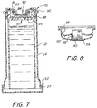
- the body fluid entering the body chamber 53 through port 57 will be trapped inside it even after removal of the cytology cup 30 as shown in Figure 7.
- the membrane surface 92 is provided with immobilized antibodies which having had flow contact with the bodily fluid captures the specific component of the fluid which is to be tested; in this example, antigens caused by cancer cells.
- the cytology cup 30 is then pulled off of the syringe barrel 22 with the bead housing 100 contained therein.
- the piston 50 remains with the syringe barrel 22, and test assembly 70 so when the same is inverted membrane 92 can be tested with a color developing solution as seen in Figures 10(a)-(c).
- the body fluid which will be placed in compartments 23 and 33 contains lyophilized primary labelled antibodies having a binding site contoured to the epitope structure and chemistry of an antigen.
- This antigen has been previously determined as being a marker for a specific type of disease, preferably cancer.
- the antibodies are labelled with HRP (horseradish peroxidase), an enzyme that detoxifies hydrogen peroxide, H2O2, by converting it to water. HRP initiates this transformation when it gives hydrogen peroxide a pair of electrons. The enzyme subsequently collects these electrons from suitable donors.
- HRP horseradish peroxidase
- Membrane 92 contains antibodies immobilized (covalently bound) thereto in area 93 for reception of the complexed antibodies and is provided with a second area 95 which acts as a control.
- the antigen has epitopes which have a high affinity for the binding sites of the primary labelled antibody and immobilized antibody.
- the principle of affinity chromatography requires that a successful separation of a biospecific ligand is available and that it can be chemically immobilized to a chromatographic bed material, the matrix. Numbers of methods well known in the art have been used to couple or immobilize the antibodies to a variety of matrixes.
- Examples of immobilization techniques which exhibit variable linkage are those formed by the reaction of the reactive groups on the support with amino, thiol, hydroxyl, and carboxyl groups on the protein ligand.
- the selection of the ligand is influenced by two factors. First, the ligand should exhibit specific and reversible binding affinity for the substance to be purified and secondly it should have chemically modifiable groups which allow it to be attached to the matrix without destroying its binding activity. (Examples of such are Protein G Sepharose manufactured by Pharmacia, Hydrazide AvidGel Ax manufactured by BioProbe International, and Actigel-ALD manufactured by Sterogene Bioseparation Inc.)
- Actigel-ALD does not cross link proteins therefore allowing proteins to retain high bioactivity after their immobilization.
- Actigel-ALO SUPER FLOW also available from Sterogene Bioseparation Inc., permits a linear flow rate of up to 3000 cm/h which would fit nicely with the flow rates in the apparatus.
- the membrane 92 is preferably soaked with ABTS solution 120 to determine the presence of the disease marker.
- a hydrogen peroxide ( H2O2 ) solution may be alternately placed on the membrane when OPD or TMB or other dual substrate systems are used.
- the color solution 120 used on the membrane 92 is preferably a substrate manufactured by Kirkegaard & Perry Labs under one of several acronyms namely: ABTS (2,2'-azino-di-[3-ethylbenzthiazoline sulfonate (6)]; OPD (orthophenylene diamine); or TMB (tetramethylkbenzidine).
- ABTS 2,2'-azino-di-[3-ethylbenzthiazoline sulfonate (6)]
- OPD orthophenylene diamine
- TMB tetramethylkbenzidine
- the preferred color solution 120 as shown in Figure 9 of the present invention is ABTS.
- the preferred ABTS substrate is a one-component substrate.
- the HRP label on the primary antibody is turned by the ABTS to a blue-green color and there is no change in color or absorbance when the reaction is stopped with SDS (sodium dodecyl sulfate). If the assay optimization indicates the sensitivity of the immunoassay is limited by the color generated by the HRP substrate, then the more sensitive TMB substrate would give more color development without a corresponding increase in the background.
- Another advantage of the TMB substrate is that it often lowers the amount of antibody and antigen reagents required for the immunoassay.
- TMB substrate is a two component liquid substrate and requires hydrogen peroxide.
- HRP converts TMB to a blue product. When the reaction is stopped by acidification, the TMB product becomes yellow.
- ODP is generally provided as a tablet that is dissolved in buffer at the time of use. HRP converts OPD to a yellow product which continues to oxidize into a brown precipitate. Upon acidification the OPD product becomes orange.
- the membrane material 92 with matrix and immobilized ligand (in this case immobilized antibody) having had flow contact with the fluid, captures or immobilizes the antibody through antigen-antibody reaction or immune reaction the specific ligand component carried by the fluid, namely, the complexed primary labelled antibody and antigen which was formerly contained by the body fluid in chambers 23 and 33.
- This antibody as previously noted was provided prelabelled with coloring enzyme HRP.
- the specific antigen is present in the testing sample which is added to the container, the antigen reacts with the antibody to form antigen-antibody complexes.
- This labelling enzyme of the antibody reacts with the ABTS poured on the membrane surface 92 turning the surface of the membrane 93 into a blue green color.
- the positive control area 93 reflects the current state of the coloring reagents as well as the prelabelled antibody at the time the test is performed.
Landscapes
- Health & Medical Sciences (AREA)
- Life Sciences & Earth Sciences (AREA)
- Chemical & Material Sciences (AREA)
- Hematology (AREA)
- Engineering & Computer Science (AREA)
- Immunology (AREA)
- Molecular Biology (AREA)
- General Health & Medical Sciences (AREA)
- Biomedical Technology (AREA)
- Analytical Chemistry (AREA)
- Urology & Nephrology (AREA)
- Pathology (AREA)
- Microbiology (AREA)
- Physics & Mathematics (AREA)
- General Physics & Mathematics (AREA)
- Biochemistry (AREA)
- Medicinal Chemistry (AREA)
- Food Science & Technology (AREA)
- Biotechnology (AREA)
- Cell Biology (AREA)
- Clinical Laboratory Science (AREA)
- Chemical Kinetics & Catalysis (AREA)
- Heart & Thoracic Surgery (AREA)
- Veterinary Medicine (AREA)
- Public Health (AREA)
- Medical Informatics (AREA)
- Animal Behavior & Ethology (AREA)
- Surgery (AREA)
- Investigating Or Analysing Biological Materials (AREA)
Priority Applications (1)
| Application Number | Priority Date | Filing Date | Title |
|---|---|---|---|
| EP91310654A EP0561003A1 (fr) | 1991-11-19 | 1991-11-19 | Dispositif pour détecter des marqueurs de maladies |
Applications Claiming Priority (1)
| Application Number | Priority Date | Filing Date | Title |
|---|---|---|---|
| EP91310654A EP0561003A1 (fr) | 1991-11-19 | 1991-11-19 | Dispositif pour détecter des marqueurs de maladies |
Publications (1)
| Publication Number | Publication Date |
|---|---|
| EP0561003A1 true EP0561003A1 (fr) | 1993-09-22 |
Family
ID=8208475
Family Applications (1)
| Application Number | Title | Priority Date | Filing Date |
|---|---|---|---|
| EP91310654A Withdrawn EP0561003A1 (fr) | 1991-11-19 | 1991-11-19 | Dispositif pour détecter des marqueurs de maladies |
Country Status (1)
| Country | Link |
|---|---|
| EP (1) | EP0561003A1 (fr) |
Cited By (8)
| Publication number | Priority date | Publication date | Assignee | Title |
|---|---|---|---|---|
| DE29505652U1 (de) * | 1995-04-01 | 1996-04-25 | Boehringer Mannheim Gmbh, 68305 Mannheim | Gefäß zur kontaminationsreduzierten Behandlung von Flüssigkeiten |
| WO2000062061A1 (fr) * | 1999-04-12 | 2000-10-19 | Institut für Chemo- und Biosensorik Münster E.V. | Dispositif et procede a utiliser lors de la realisation de tests relatifs a des systemes recepteur-ligand et de tests d'affinite |
| EP1693670A4 (fr) * | 2003-12-04 | 2008-06-11 | Olympus Corp | Recipient de reaction et dispositif de reaction le contenant, dispositif de detection et procede pour produire ledit recipient de reaction |
| DE102010011560A1 (de) * | 2010-03-16 | 2011-09-22 | Gilupi Gmbh | Biodetektor |
| CN102883699A (zh) * | 2010-05-10 | 2013-01-16 | 德国梅尔松根B·布劳恩股份有限公司 | 端口装置 |
| EP2451576A4 (fr) * | 2009-07-09 | 2013-04-10 | Alere Switzerland Gmbh | Dispositif et procédé d analyse d une substance à analyser dans un échantillon liquide |
| US10039882B2 (en) | 2016-09-01 | 2018-08-07 | Arthrex, Inc. | Binding syringe |
| CN119901915A (zh) * | 2025-03-31 | 2025-04-29 | 浙江万泰福生物科技有限公司 | 一种血液检测装置及其使用方法 |
Citations (5)
| Publication number | Priority date | Publication date | Assignee | Title |
|---|---|---|---|---|
| EP0378353A2 (fr) * | 1989-01-10 | 1990-07-18 | La Mina Ltd. | Dispositif pour recueillir un fluide biologique |
| EP0414513A2 (fr) * | 1989-08-22 | 1991-02-27 | La Mina Ltd. | Dispositif modulaire de préparation d'échantillons liquides multiples |
| US5016644A (en) * | 1989-01-10 | 1991-05-21 | La Mina Ltd. | Urine testing module and method of collecting urine antigen |
| US5024238A (en) * | 1989-01-10 | 1991-06-18 | Cancer Diagnostics, Inc. | Blood withdrawing apparatus and antigen testing method |
| US5038793A (en) * | 1989-01-10 | 1991-08-13 | La Mina Ltd. | Urine testing membrane module and method of conducting same |
-
1991
- 1991-11-19 EP EP91310654A patent/EP0561003A1/fr not_active Withdrawn
Patent Citations (6)
| Publication number | Priority date | Publication date | Assignee | Title |
|---|---|---|---|---|
| EP0378353A2 (fr) * | 1989-01-10 | 1990-07-18 | La Mina Ltd. | Dispositif pour recueillir un fluide biologique |
| EP0404527A2 (fr) * | 1989-01-10 | 1990-12-27 | La Mina Ltd. | Dispositif modulaire de préparation d'un échantillon fluide |
| US5016644A (en) * | 1989-01-10 | 1991-05-21 | La Mina Ltd. | Urine testing module and method of collecting urine antigen |
| US5024238A (en) * | 1989-01-10 | 1991-06-18 | Cancer Diagnostics, Inc. | Blood withdrawing apparatus and antigen testing method |
| US5038793A (en) * | 1989-01-10 | 1991-08-13 | La Mina Ltd. | Urine testing membrane module and method of conducting same |
| EP0414513A2 (fr) * | 1989-08-22 | 1991-02-27 | La Mina Ltd. | Dispositif modulaire de préparation d'échantillons liquides multiples |
Cited By (11)
| Publication number | Priority date | Publication date | Assignee | Title |
|---|---|---|---|---|
| DE29505652U1 (de) * | 1995-04-01 | 1996-04-25 | Boehringer Mannheim Gmbh, 68305 Mannheim | Gefäß zur kontaminationsreduzierten Behandlung von Flüssigkeiten |
| US5855852A (en) * | 1995-04-01 | 1999-01-05 | Boehringer Mannheim Gmbh | Vessel for reducing contamination in the treatment of liquids |
| WO2000062061A1 (fr) * | 1999-04-12 | 2000-10-19 | Institut für Chemo- und Biosensorik Münster E.V. | Dispositif et procede a utiliser lors de la realisation de tests relatifs a des systemes recepteur-ligand et de tests d'affinite |
| EP1693670A4 (fr) * | 2003-12-04 | 2008-06-11 | Olympus Corp | Recipient de reaction et dispositif de reaction le contenant, dispositif de detection et procede pour produire ledit recipient de reaction |
| EP2451576A4 (fr) * | 2009-07-09 | 2013-04-10 | Alere Switzerland Gmbh | Dispositif et procédé d analyse d une substance à analyser dans un échantillon liquide |
| DE102010011560A1 (de) * | 2010-03-16 | 2011-09-22 | Gilupi Gmbh | Biodetektor |
| DE102010011560B4 (de) | 2010-03-16 | 2021-09-16 | Gilupi Gmbh | Biodetektor |
| CN102883699A (zh) * | 2010-05-10 | 2013-01-16 | 德国梅尔松根B·布劳恩股份有限公司 | 端口装置 |
| CN102883699B (zh) * | 2010-05-10 | 2014-09-24 | 德国梅尔松根B·布劳恩股份有限公司 | 端口装置 |
| US10039882B2 (en) | 2016-09-01 | 2018-08-07 | Arthrex, Inc. | Binding syringe |
| CN119901915A (zh) * | 2025-03-31 | 2025-04-29 | 浙江万泰福生物科技有限公司 | 一种血液检测装置及其使用方法 |
Similar Documents
| Publication | Publication Date | Title |
|---|---|---|
| US5077012A (en) | Device for detecting disease markers | |
| US5024237A (en) | Modular fluid sample preparation assembly | |
| US5042502A (en) | Urine testing module with cytology cup | |
| US5022411A (en) | Modular fluid testing device | |
| US5038793A (en) | Urine testing membrane module and method of conducting same | |
| US4953561A (en) | Urine testing module and method of collecting urine antigen | |
| US6210909B1 (en) | Liquid specimen container and attachable testing modules | |
| US5003988A (en) | Modular multiple fluid sample preparation assembly | |
| US5998214A (en) | Environmental sample collection and membrane testing device | |
| EP0471570A1 (fr) | Appareil pour l'examen de l'urine avec sédiment urinaire et dispositif | |
| US5024238A (en) | Blood withdrawing apparatus and antigen testing method | |
| US5016644A (en) | Urine testing module and method of collecting urine antigen | |
| HU203287B (en) | Method and device for detecting anolite in fluids | |
| EP0561003A1 (fr) | Dispositif pour détecter des marqueurs de maladies | |
| US5133363A (en) | Modular multiple fluid sample preparation assembly | |
| EP0508010A1 (fr) | Module membraneux pour tester des liquides biologiques et méthode pour leur application | |
| JPH06347385A (ja) | 生物流体試験装置 | |
| CA2055707A1 (fr) | Appareil pour detecter les marqueurs de maladies | |
| EP0509158A1 (fr) | Procédé pour tester des antigènes d'urine | |
| CA2040087A1 (fr) | Module muni d'une membrane pour des tests d'urine et methode d'utilisation |
Legal Events
| Date | Code | Title | Description |
|---|---|---|---|
| PUAI | Public reference made under article 153(3) epc to a published international application that has entered the european phase |
Free format text: ORIGINAL CODE: 0009012 |
|
| AK | Designated contracting states |
Kind code of ref document: A1 Designated state(s): AT BE CH DE ES FR GB GR IT LI LU NL SE |
|
| 17P | Request for examination filed |
Effective date: 19940321 |
|
| 17Q | First examination report despatched |
Effective date: 19941213 |
|
| STAA | Information on the status of an ep patent application or granted ep patent |
Free format text: STATUS: THE APPLICATION IS DEEMED TO BE WITHDRAWN |
|
| 18D | Application deemed to be withdrawn |
Effective date: 19950625 |