EP0567232A2 - Nachweis von Bakterien in Kulturfläschchen durch Messung zeitgelöster Lichtstreuung und -absorption - Google Patents
Nachweis von Bakterien in Kulturfläschchen durch Messung zeitgelöster Lichtstreuung und -absorption Download PDFInfo
- Publication number
- EP0567232A2 EP0567232A2 EP93302293A EP93302293A EP0567232A2 EP 0567232 A2 EP0567232 A2 EP 0567232A2 EP 93302293 A EP93302293 A EP 93302293A EP 93302293 A EP93302293 A EP 93302293A EP 0567232 A2 EP0567232 A2 EP 0567232A2
- Authority
- EP
- European Patent Office
- Prior art keywords
- electromagnetic radiation
- bacteria
- sample
- time
- time characteristics
- Prior art date
- Legal status (The legal status is an assumption and is not a legal conclusion. Google has not performed a legal analysis and makes no representation as to the accuracy of the status listed.)
- Withdrawn
Links
- 238000010521 absorption reaction Methods 0.000 title abstract description 9
- 238000005259 measurement Methods 0.000 title abstract description 9
- 238000000149 argon plasma sintering Methods 0.000 title abstract 2
- 238000009640 blood culture Methods 0.000 title description 11
- 230000014670 detection of bacterium Effects 0.000 title 1
- 230000005670 electromagnetic radiation Effects 0.000 claims abstract description 62
- 241000894006 Bacteria Species 0.000 claims abstract description 42
- 230000003287 optical effect Effects 0.000 claims abstract description 31
- 238000000034 method Methods 0.000 claims abstract description 30
- 230000005284 excitation Effects 0.000 claims abstract description 13
- 239000000523 sample Substances 0.000 claims description 56
- 239000000835 fiber Substances 0.000 claims description 14
- 239000013068 control sample Substances 0.000 claims description 5
- 238000012545 processing Methods 0.000 claims description 3
- 230000008859 change Effects 0.000 abstract description 11
- 230000005855 radiation Effects 0.000 description 25
- 230000001580 bacterial effect Effects 0.000 description 11
- CURLTUGMZLYLDI-UHFFFAOYSA-N Carbon dioxide Chemical compound O=C=O CURLTUGMZLYLDI-UHFFFAOYSA-N 0.000 description 10
- 239000008280 blood Substances 0.000 description 8
- 210000004369 blood Anatomy 0.000 description 8
- 238000001514 detection method Methods 0.000 description 8
- 238000012544 monitoring process Methods 0.000 description 7
- 229910002092 carbon dioxide Inorganic materials 0.000 description 5
- 239000001569 carbon dioxide Substances 0.000 description 5
- 239000001963 growth medium Substances 0.000 description 5
- 230000000737 periodic effect Effects 0.000 description 5
- 239000002609 medium Substances 0.000 description 4
- 230000032683 aging Effects 0.000 description 3
- 230000004071 biological effect Effects 0.000 description 3
- 239000003990 capacitor Substances 0.000 description 3
- 238000010168 coupling process Methods 0.000 description 3
- 238000005859 coupling reaction Methods 0.000 description 3
- 238000002347 injection Methods 0.000 description 3
- 239000007924 injection Substances 0.000 description 3
- 238000001307 laser spectroscopy Methods 0.000 description 3
- 230000005693 optoelectronics Effects 0.000 description 3
- 230000004044 response Effects 0.000 description 3
- 239000000126 substance Substances 0.000 description 3
- 241000588724 Escherichia coli Species 0.000 description 2
- 102000001554 Hemoglobins Human genes 0.000 description 2
- 108010054147 Hemoglobins Proteins 0.000 description 2
- 239000000654 additive Substances 0.000 description 2
- 238000004458 analytical method Methods 0.000 description 2
- QVGXLLKOCUKJST-UHFFFAOYSA-N atomic oxygen Chemical compound [O] QVGXLLKOCUKJST-UHFFFAOYSA-N 0.000 description 2
- 230000008878 coupling Effects 0.000 description 2
- 238000013461 design Methods 0.000 description 2
- 238000011534 incubation Methods 0.000 description 2
- 230000005012 migration Effects 0.000 description 2
- 238000013508 migration Methods 0.000 description 2
- 229910052760 oxygen Inorganic materials 0.000 description 2
- 239000001301 oxygen Substances 0.000 description 2
- 238000006213 oxygenation reaction Methods 0.000 description 2
- 230000010363 phase shift Effects 0.000 description 2
- 230000035945 sensitivity Effects 0.000 description 2
- 230000003595 spectral effect Effects 0.000 description 2
- 239000000725 suspension Substances 0.000 description 2
- XUIMIQQOPSSXEZ-UHFFFAOYSA-N Silicon Chemical compound [Si] XUIMIQQOPSSXEZ-UHFFFAOYSA-N 0.000 description 1
- 239000013543 active substance Substances 0.000 description 1
- 230000006978 adaptation Effects 0.000 description 1
- 230000003679 aging effect Effects 0.000 description 1
- 230000004075 alteration Effects 0.000 description 1
- 230000000721 bacterilogical effect Effects 0.000 description 1
- 230000006399 behavior Effects 0.000 description 1
- 210000001124 body fluid Anatomy 0.000 description 1
- 239000010839 body fluid Substances 0.000 description 1
- 239000006227 byproduct Substances 0.000 description 1
- 238000004891 communication Methods 0.000 description 1
- 238000010276 construction Methods 0.000 description 1
- 238000011109 contamination Methods 0.000 description 1
- 238000012864 cross contamination Methods 0.000 description 1
- 230000023077 detection of light stimulus Effects 0.000 description 1
- 238000006073 displacement reaction Methods 0.000 description 1
- 230000000694 effects Effects 0.000 description 1
- 230000006870 function Effects 0.000 description 1
- 239000007788 liquid Substances 0.000 description 1
- 239000006194 liquid suspension Substances 0.000 description 1
- 238000000691 measurement method Methods 0.000 description 1
- 230000007246 mechanism Effects 0.000 description 1
- 230000002503 metabolic effect Effects 0.000 description 1
- 230000004060 metabolic process Effects 0.000 description 1
- 244000005700 microbiome Species 0.000 description 1
- 238000012986 modification Methods 0.000 description 1
- 230000004048 modification Effects 0.000 description 1
- 239000013307 optical fiber Substances 0.000 description 1
- 230000008569 process Effects 0.000 description 1
- 239000004065 semiconductor Substances 0.000 description 1
- 229910052710 silicon Inorganic materials 0.000 description 1
- 239000010703 silicon Substances 0.000 description 1
- 241000894007 species Species 0.000 description 1
- 238000004611 spectroscopical analysis Methods 0.000 description 1
- 238000012360 testing method Methods 0.000 description 1
- 238000011179 visual inspection Methods 0.000 description 1
Images



Classifications
-
- G—PHYSICS
- G01—MEASURING; TESTING
- G01N—INVESTIGATING OR ANALYSING MATERIALS BY DETERMINING THEIR CHEMICAL OR PHYSICAL PROPERTIES
- G01N21/00—Investigating or analysing materials by the use of optical means, i.e. using sub-millimetre waves, infrared, visible or ultraviolet light
- G01N21/17—Systems in which incident light is modified in accordance with the properties of the material investigated
- G01N21/47—Scattering, i.e. diffuse reflection
- G01N21/49—Scattering, i.e. diffuse reflection within a body or fluid
- G01N21/51—Scattering, i.e. diffuse reflection within a body or fluid inside a container, e.g. in an ampoule
-
- C—CHEMISTRY; METALLURGY
- C12—BIOCHEMISTRY; BEER; SPIRITS; WINE; VINEGAR; MICROBIOLOGY; ENZYMOLOGY; MUTATION OR GENETIC ENGINEERING
- C12M—APPARATUS FOR ENZYMOLOGY OR MICROBIOLOGY; APPARATUS FOR CULTURING MICROORGANISMS FOR PRODUCING BIOMASS, FOR GROWING CELLS OR FOR OBTAINING FERMENTATION OR METABOLIC PRODUCTS, i.e. BIOREACTORS OR FERMENTERS
- C12M41/00—Means for regulation, monitoring, measurement or control, e.g. flow regulation
- C12M41/30—Means for regulation, monitoring, measurement or control, e.g. flow regulation of concentration
- C12M41/36—Means for regulation, monitoring, measurement or control, e.g. flow regulation of concentration of biomass, e.g. colony counters or by turbidity measurements
-
- G—PHYSICS
- G01—MEASURING; TESTING
- G01N—INVESTIGATING OR ANALYSING MATERIALS BY DETERMINING THEIR CHEMICAL OR PHYSICAL PROPERTIES
- G01N21/00—Investigating or analysing materials by the use of optical means, i.e. using sub-millimetre waves, infrared, visible or ultraviolet light
- G01N21/17—Systems in which incident light is modified in accordance with the properties of the material investigated
- G01N2021/1789—Time resolved
-
- Y—GENERAL TAGGING OF NEW TECHNOLOGICAL DEVELOPMENTS; GENERAL TAGGING OF CROSS-SECTIONAL TECHNOLOGIES SPANNING OVER SEVERAL SECTIONS OF THE IPC; TECHNICAL SUBJECTS COVERED BY FORMER USPC CROSS-REFERENCE ART COLLECTIONS [XRACs] AND DIGESTS
- Y10—TECHNICAL SUBJECTS COVERED BY FORMER USPC
- Y10S—TECHNICAL SUBJECTS COVERED BY FORMER USPC CROSS-REFERENCE ART COLLECTIONS [XRACs] AND DIGESTS
- Y10S435/00—Chemistry: molecular biology and microbiology
- Y10S435/808—Optical sensing apparatus
Definitions
- the present invention relates to non-invasive methods and apparatus for detecting biological activities in a specimen such as blood by measuring the absorption and scattering of light, and in particular to systems wherein a specimen and culture medium are introduced into a sealable container and exposed to conditions enabling metabolic processes to take place and thereby permit the detection of the presence of microorganisms in the sample.
- the presence of biologically active agents in a patient's body fluid, and especially in blood is determined using blood culture vials.
- a small quantity of blood is typically injected through an enclosing rubber septum into a sterile vial containing a culture medium.
- the vial is typically incubated at 37°C and monitored for bacterial growth.
- Common visual inspection for bacterial growth involves monitoring the turbidity of the liquid suspension.
- Known instrumented methods detect changes in the carbon dioxide content of the culture bottles, which is a metabolic by-product of the bacterial growth.
- Monitoring the carbon dioxide content can be accomplished by methods well-established in the art, such as radiochemical (e.g., BACTEC®, Becton-Dickinson, Franklin Lakes, NJ, USA), infrared absorption at a carbon dioxide spectral line (e.g., NR-BACTEC@, Becton-Dickinson, Franklin Lakes, NJ, USA), or pressure/vacuum measurement techniques such as those disclosed in U.S. Patent No. 4,152,213 -- Ahnell.
- radiochemical e.g., BACTEC®, Becton-Dickinson, Franklin Lakes, NJ, USA
- infrared absorption at a carbon dioxide spectral line e.g., NR-BACTEC@, Becton-Dickinson, Franklin Lake
- invasive describes a procedure wherein the sample container is opened, pierced or otherwise placed in communication with an external environment during the point at which the presence or absence of bacteria is determined.
- An example of such a device would be a 5-mW green HeNe laser (543.5 nm) externally modulated by means of an acousto-optic light modulator.
- the las- er/modulator combination is rather expensive, requiring that the individual samples be moved to the laser instead of having one light source at each sample.
- Such an instrument would necessarily have a complicated mechanism for effecting the transportation of the individual samples, and the time interval between successive measurements for each sample would be relatively long. Since for the time being it appears unlikely that high-brightness, short-wavelength semiconductor diode lasers will be developed to permit a commercially feasible embodiment of this type of system, such an improved system would suffer serious practical shortcomings.
- the present invention provides optical methods and apparatus for detecting biological activities in a sample that are non-invasive, not intensity-based, do not require chemical sensors or any other additives within the sample, do not require high-brightness, short-wavelength light sources, have no moving parts and allow an almost continuous monitoring of each sample vial.
- the present invention provides methods for detecting the presence of bacteria in a sample by monitoring the time characteristics of modulated electromagnetic radiation that has migrated through the sample.
- the methods of the present invention preferably include the steps of introducing modulated electromagnetic radiation into the sample at a first point and receiving the collected radiation at a second point. The time-averaged intensity of the collected radiation over time is then determined, as well as the amplitude and phase of the collected radiation relative to the injected radiation.
- bacteria are present if the time characteristics of the collected radiation show changes over time.
- a computer is provided with data indicative of the intensity of the collected radiation over time and the amplitude and phase of the collected radiation relative to the injected radiation.
- the computer calculates the modulation of the collected radiation, which is obtained by dividing the amplitude signal by the intensity signal. Since the measured amplitude is directly proportional to the intensity, the calculated modulation is independent of the intensity. Because the phase is also independent of the intensity, an instrument according to the present invention exhibits an excellent long-time stability.
- the present invention determines the presence of bacteria in a plurality of samples by sequentially directing modulated electromagnetic radiation to each sample, sequentially introducing the modulated electromagnetic radiation into each sample at a first point and sequentially detecting the signal that emerges from the sample and thereby determining the presence of bacteria in each of the plurality of samples.
- the step of sequentially directing the modulated electromagnetic radiation may either be carried out using a multiplexer to sequentially excite a laser diode disposed adjacent each of the samples, or may be carried out by transmitting a modulated electrical signal to a laser thereby creating modulated electromagnetic radiation that is transmitted to each of the samples using an optical switch.
- the determination of the time characteristics of the collected radiation overtime either comprises the steps of transmitting the collected radiation to a multichannel plate photomultiplierto create a signal and measuring the amplitude of the signal or, alternatively, the collected radiation may be used to excite an avalanche photodiode that is used as a detector to create a photodiode output signal and the time characteristics of the photodiode output signal are measured.
- the present invention also discloses apparatus for detecting the presence of bacteria in a sample comprising a light source, preferably a laser, for introducing modulated electromagnetic radiation into a sample at a first point and a detector for receiving collected electromagnetic radiation at a second point.
- a processor is provided for determining the intensity of the collected electromagnetic radiation over time, and a vector voltmeter is used to determine the amplitude and phase of the modulated radiation relative to the injected radiation.
- a computer preferably determines the modulation and phase of the collected radiation to detect the presence of bacteria. If bacteria are present, the time characteristics of the collected radiation will change over time.
- the present invention also provides an improved optical switch for use in certain embodiments of the apparatus of the present invention to sequentially introduce modulated electromagnetic radiation into each of a plurality of samples.
- the optical switch comprises a laser disposed to impinge a laser beam on a deflector, wherein the deflector focuses the laser beam upon one of a plurality of excitation fibers.
- the deflector is precisely moved such that the laser beam is sequentially focused upon each of the plurality of excitation fibers.
- the deflector is moved by a stepper motor to rotate the deflector about an axis.
- a preferred form of modulated electromagnetic radiation is given by a sequence of periodic pulses with a wavelength within the range 600 nm to 900 nm, or more specifically, between 650 nm and 800 nm.
- This sequence of periodic pulses is equivalent to electromagnetic radiation that is modulated at the pulse repetition frequency and at harmonics thereof.
- a time-averaged intensity is defined, as well as an amplitude and phase at each of these frequency components.
- the detection electronics are preferably tuned to one particular frequency component. For large sample volumes, low-frequency components are appropriate. For small sample volumes, higher harmonic frequencies can be selected in order to obtain maximum bacterial detection sensitivity.
- the time characteristics for the collected radiation in the time domain may also be monitored.
- data measured in the time domain and data measured in the frequency domain i.e., the phase and modulation of the light
- frequency-domain instruments can be produced at lower cost.
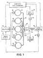
- a signal source 10 delivers periodic electrical sub-nanosecond electrical pulses to a multiplexer 20.
- the computer-controlled multiplexer 20 directs these periodic electrical pulses in a serial mode of operation to a plurality of diode lasers 30 for emitting light of a specified wavelength, each mounted closely to a sample container 32.
- Light reemerges from each sample container 32 and is collected at a small area approximately opposite the light injection area by a light collection means that is most preferably comprised of a fiber-optic bundle 34.
- the reemerging light is directed to a large-area multichan nel-plate photomultiplier tube 40 which is powered by a high voltage source 42.
- the time-averaged output current of the photomultiplier tube 40 is measured by a low-pass filter 50 and a digital voltmeter 52.
- the voltage data are then stored within the memory of a computer 70.
- Two band-pass filters 60 and a high-frequency vector voltmeter 62 allow for determination of the high-frequency photocurrent signal amplitude and signal phase relative to the modulation phase of the diode lasers 30.
- a first band pass filter 60 receives a voltage signal from the output of the photomultiplier tube 40, while a second filter 60 is connected to the signal source 10. By combining these signals in the vector voltmeter 62, the amplitude and phase of the output are determined.
- These data are also stored in the memory of the data acquisition computer 70.
- each diode laser 30 introduces ultrashort periodic electromagnetic radiation pulses into the sample container 32, which preferably contains a liquid blood culture suspension or other sample.
- the electromagnetic radiation is comprised of photons that migrate through the sample and experience multiple scattering and absorption events. Due to these multiple events, the photon path length within the medium is not a discrete one, but can instead be described as a distribution of path lengths. In other words, the reemerging light pulse is much longer than the pulse that was introduced.
- the pulse shape and the pulse duration will depend on the absorption coefficient, f..lA, and on the scattering coefficient, w s , of the blood culture or other sample. If bacteria are present and are consuming oxygen, the hemoglobin oxygenation of the blood will be reduced.
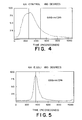
- FIG. 4 shows the measured light output waveform for a BACTEC@ 6A blood culture medium (Becton-Dickinson, Franklin Lakes, NJ, USA) and blood in a typical BACTEC@ vial having a 43 mm outer diameter after 24 hours of incubation. It will be observed that the electromagnetic radiation decay time in this case is about 196 ps.
- FIG. 5 illustrates the results obtained from a similar vial under identical conditions that was inoculated with about 100 cfu per ml of E. coli bacteria. As can be seen, this vial shows a significantly shorter waveform with a decay time of only about 71 ps.
- time characteristics are detected by using either time-domain or frequency-domain principles. In terms of cost, the latter are more advantageous.
- an apparatus made in accordance with the present invention can be constructed using a multichannel plate photomultiplier tube 40. Unfortunately, these detectors are still relatively very expensive.
- time characteristics covers many of the possible properties of a pulse of light.
- a light pulse When a light pulse is introduced into a scattering and absorbing medium, there are numerous properties related to the time course of the light that traverses the medium and reemerges. In general, the intensity increases to a maximum value and decays exponentially. In such a system numerous properties that reflect the information contained in this behavior can be measured. For example, the rise time of the pulse, the full pulse width and the delay time are all time characteristics that might contain useful information.
- the methods and apparatus described herein use phase and modulation data, operable embodiments of the present invention that utilize other time characteristics can be devised.
- FIG. 2 One possible alternative embodiment to the apparatus of the present invention that uses a multichannel plate photomultiplier tube 40 as illustrated in FIG. 1 is shown in FIG. 2, where silicon avalanche photodiodes 80 are used as detectors. Except as noted herein, the apparatus of the embodiment of the present invention shown in FIG. 2 is similar to that described with reference to FIG. 1. Thus, like reference numerals will designate like components.
- the avalanche photodiodes 80 are operated as optoelectronic cross-correlators with a DC output signal. See Berndt, K. "Opto-electronic high-frequency cross-correlation using avalanche photodiodes," Measurement, Vol. 5, No. 4, Oct.-Dec.
- part of the output of the signal source 10 is directed to a band-pass filter 60 where a particular harmonic of the repetition frequency is selected.
- a frequency translator 14 such as a synthesizer or single sideband modulator generates a frequency-shifted signal, preferably at an offset of about 50 to 100 Hz.
- This frequency-shifted modulation signal is divided into a plurality of modulation signals M using a signal splitter 16 and then added to the bias voltage of each of the avalanche photodiodes 80 at each junction M shown on each photodiode 80.
- the connection between each junction M and the signal splitter is not shown for purposes of clarity.
- each signal M transmitted from the signal splitter 16 is equal to the others and is connected to one of the junctions M in a one-to-one correspondence.
- a DC power supply 18 similarly provides an appropriate voltage V to each junction V shown on each photodiode 80. These connections are also not shown for purposes of clarity, however, the same voltage V is provided to each avalanche photodiode 80. Details of the circuitry of the avalanche photodiode 80 are shown in FIG. 3, which is explained below. Adding the modulation signal M to the DC bias voltage V results in a gain modulation.
- the avalanche photodiodes 80 act as opto-electronic mixers with an additional DC signal proportional to the received average optical input power.
- the intermediate frequency (IF) output of the photodiodes 80 has a frequency equal to the offset frequency generated by the frequency translator 14 and is fed to a low-frequency demultiplexer 85. Due to the relatively low frequency IF output, the equivalent noise bandwidth is very low when compared with direct detection high-speed photodetectors. Therefore, excellent detectivity is obtained.
- the IF signal carries all the high-frequency information of the optical signal received by the avalanche photodiodes 80. By separating the DC and the IF component using appropriate low pass filters 50 and measuring both with appropriate voltmeters 52,54, the modulation degree of the optical signal can be calculated and stored within the data acquisition computer 70.
- FIG. 3 Further details of the circuitry of the avalanche photodiodes 80 are illustrated in FIG. 3.
- the voltage input V from the DC power supply 18 is connected to a portion of the circuit comprising R1, R2, C2 and C3.
- the resistors R1 and R2 have values of about 10,000 and 50 Ohms respectively, while capacitors C2 and C3 have values of about 100 and 1 x 10- 9 farads respectively.
- the modulation signal M from the splitter 16 shown in FIG. 2 is connected to junction M shown in FIG. 3.
- a capacitor C1 having a value of about 1 x 10- 9 farads is disposed between the junction M and the photodiode 80.
- the output of the photodiode is connected to a capacitor C4 that has a value of about 10 x 10- 9 farads and a resistor R3 of about 1 x 10 6 Ohms, and the resulting output signal is connected to the demultiplexer 85 shown in FIG. 2.
- These values represent preferred values for each of the noted components. Those of ordinary skill will realize that one or more of these values may be altered, but that such alteration will necessarily result in different requirements for other components if the same output signal is to be obtained.
- Apparatus made in accordance with the present invention does not require a particular diode laser pulse shape. After-pulses or any kind of pulse ringing have no negative impact on the decay time measurement process.
- the present invention requires only a stable light waveform at a constant repetition frequency.
- the band-pass filters 60 shown in FIGS. 1-2 select only one particular harmonic of the repetition frequency of the diode lasers 30. The modulation degree and/or the phase shift are derived using this frequency.
- This feature of the invention allows use of low-cost diode lasers. As explained above, a wavelength within the range of about 600 nm to about 900 nm would be appropriate for detecting bacteria in a sample of blood and a wavelength between about 650 nm to 800 nm is preferred.
- an important aspect of the present invention is that it has now been discovered that bacterial growth can be monitored by introducing modulated electromagnetic radiation into a sample such as a culture medium/blood suspension and performing a time-resolved analysis of the reemerging light at a small area a known distance away from the injection area.
- the use of time-resolved spectroscopy by the present invention is a step towards an absolute detection system.
- the present invention therefore provides optical methods and apparatus for detecting biological activities in blood culture bottles, that is non-invasive, not intensity-based, does not require chemical sensors or any other additives within the blood culture vial, does not require high-brightness short-wavelength light sources, has no moving parts and allows an almost continuous monitoring for each vial.
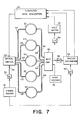
- FIG. 7 Another alternate embodiment of the apparatus of the present invention is shown in FIG. 7.
- like numerals designate like elements described above with reference to FIGS. 1 and 2.
- the optical output of a diode laser 30 is coupled via an optical fiber 34 to the input of an optical switch 100.
- This switch 100 contains a large number of optical output channels. Each output channel of the optical switch 100 is fed via an excitation fiber 113 to a sample container 32 and introduces modulated electromagnetic radiation into the sample.
- the sample containers 32 contain blood culture medium and a blood sample.
- the optical switch 100 is preferably controlled by a computer 70 and in operation, the optical switch 100 directs the modulated electromagnetic radiation serially through the excitation fibers 113 to the sample containers 32.
- the detection of light reemerging from the sample containers 32 is preferably performed in the same way as shown and as described above with reference to FIG. 1 using a plurality of fiber optic bundles 34.
- the fiber-optic coupling 34 between the diode laser 30 and the optical switch 100 is one possible embodiment. It is, however, a preferred embodiment because diode lasers 30 that include fiber-optic pigtail connectors are currently commercially available.
- FIG. 7 Another embodiment of the apparatus disclosed in FIG. 7 provides a system wherein the photomultiplier tube 40 and its associated processing equipment are replaced by the avalanche photodiodes 80, demultiplexer 85 and associated processing equipment as discussed above with reference to FIG. 2.
- FIG. 8 an embodiment of the optical switch 100 is illustrated in FIG. 8 wherein a laser 300 is directly mounted to the optical switch 100.
- This direct-coupling provides compactness and a simple design.
- a laser 300 is fixed to a mounting tube 101 which is in turn mounted to a plate 102, that is attached to a cylindrical housing 104 by several screws 103.
- Aflexible portion 105 is preferably disposed between the plate 102 and the second cylinder 104 to allow for fine angular adjustment of the laser beam 110 with respect to a cylindrical housing 104 by the degree to which the flexible portion 105 is compressed.
- the second cylinder 104 is fixed to a base plate 106, upon which an electric stepper motor 107 is mounted coaxially with respect to the cylindrical housing 104. Fine angular adjustment of the stepper motor 107 is accomplished in a similar manner to that described above by providing several screws 108 and an additional flexible insert 109 disposed between the base plate 106 and the stepper motor 107. As shown, the laser beam 110 impinges upon a 90-degree beam deflector 112 mounted to the stepper motor shaft.
- a movable lens 111 slidably mounted inside the mounting tube 101 allows for exact focussing of the laser beam 110 on the inputs of the plurality of excitation fibers 113 that are mounted in equidistant axial bore-holes along the perimeter of the cylindrical housing 104 after it strikes the deflector 112.
- the computer-controlled stepper motor directs the full optical laser power serially toward each of the inputs of the excitation fibers 113.
- An optical switch of this design can easily accommodate 360 or more excitation fibers 113 by employing standard stepper motors.
- the optical switch 100 illustrated in FIG. 8 also allows for random access operation. It will be realized, however, that it is advantageous to use at least one optical output to establish a reference address.
- the mechanical optical switch 100 described above with reference to FIG. 8 represents one embodiment of such a device that would be useful in certain embodiments of the apparatus of the present invention such as that illustrated in FIG. 7.
- a single laser beam 110 may be selectively time-shared with a plurality of excitation fibers 113.
- a carriage containing the laser 300 could be advanced by a power screw or stepper motor to travel precisely along a linear array of excitation fibers 113 and achieve the same effect.
- the present invention provides methods for detecting the presence of bacteria in a sample by monitoring the time characteristics of modulated electromagnetic radiation that has migrated through the sample.
- the detection principle of the preserit invention is based on the change in the time characteristics, modulation (i.e., amplitude/time-averaged intensity) and phase of the collected modulated radiation during bacterial growth. Since the time characteristics for a control (a sample without bacteria) do not change, a control sample is not required in all embodiments.
- a response such as that shown in FIG. 4 is typical of the response of a certain vial a short time after incubation, while FIG. 5 illustrates a response in the same vial after a time sufficient to permit bacteriological activity.
- the methods of the present invention include the steps of introducing modulated electromagnetic radiation into a sample at a first point and receiving collected electromagnetic radiation at a second point.
- the second point be located across from the first point, however,'it will be understood that these two points need not necessarily be disposed directly opposite each other.
- the intensity of the collected radiation over time is then determined along with the amplitude and phase of the modulated radiation relative to the injected radiation. As noted above and pointed out with reference to FIGS. 4-5 bacteria are present if the time characteristics change over time.
- the methods of the present invention include the steps of transmitting data indicative of the intensity of the collected radiation over time and the amplitude and phase of the radiation relative to the injected radiation to a computer and the computer carries out the steps of determining the modulation and phase of the collected radiation.
Landscapes
- Chemical & Material Sciences (AREA)
- Health & Medical Sciences (AREA)
- Life Sciences & Earth Sciences (AREA)
- Bioinformatics & Cheminformatics (AREA)
- Analytical Chemistry (AREA)
- Biochemistry (AREA)
- General Health & Medical Sciences (AREA)
- Organic Chemistry (AREA)
- Zoology (AREA)
- Wood Science & Technology (AREA)
- Engineering & Computer Science (AREA)
- Biomedical Technology (AREA)
- General Engineering & Computer Science (AREA)
- Biotechnology (AREA)
- Microbiology (AREA)
- Sustainable Development (AREA)
- Pathology (AREA)
- Immunology (AREA)
- Physics & Mathematics (AREA)
- Genetics & Genomics (AREA)
- General Physics & Mathematics (AREA)
- Investigating Or Analysing Materials By Optical Means (AREA)
- Measuring Or Testing Involving Enzymes Or Micro-Organisms (AREA)
- Apparatus Associated With Microorganisms And Enzymes (AREA)
- Investigating, Analyzing Materials By Fluorescence Or Luminescence (AREA)
- Investigating Or Analysing Biological Materials (AREA)
Applications Claiming Priority (2)
| Application Number | Priority Date | Filing Date | Title |
|---|---|---|---|
| US874252 | 1992-04-24 | ||
| US07/874,252 US5293210A (en) | 1992-04-24 | 1992-04-24 | Detection of bacteria in blood culture bottles by time-resolved light scattering and absorption measurement |
Publications (2)
| Publication Number | Publication Date |
|---|---|
| EP0567232A2 true EP0567232A2 (de) | 1993-10-27 |
| EP0567232A3 EP0567232A3 (de) | 1995-08-09 |
Family
ID=25363327
Family Applications (1)
| Application Number | Title | Priority Date | Filing Date |
|---|---|---|---|
| EP93302293A Withdrawn EP0567232A3 (de) | 1992-04-24 | 1993-03-25 | Nachweis von Bakterien in Kulturfläschchen durch Messung zeitgelöster Lichtstreuung und -absorption. |
Country Status (6)
| Country | Link |
|---|---|
| US (1) | US5293210A (de) |
| EP (1) | EP0567232A3 (de) |
| JP (1) | JPH0823528B2 (de) |
| AU (1) | AU667884B2 (de) |
| BR (1) | BR9301615A (de) |
| CA (1) | CA2092985C (de) |
Cited By (5)
| Publication number | Priority date | Publication date | Assignee | Title |
|---|---|---|---|---|
| EP0622455A1 (de) * | 1993-04-26 | 1994-11-02 | Becton, Dickinson and Company | Nachweis biologischer Aktivität in Kulturfläschchen |
| WO1997010494A1 (en) * | 1995-09-11 | 1997-03-20 | Becton Dickinson And Company | Fluorescence detector |
| EP0697460A3 (de) * | 1994-08-15 | 1997-07-02 | Becton Dickinson Co | Optischer Blutkultursensor |
| ITUD20080190A1 (it) * | 2008-08-22 | 2010-02-23 | Alifax S P A | Procedimento per l'indagine batteriologica su plasma |
| KR20180033546A (ko) * | 2015-07-21 | 2018-04-03 | 플루이드센스 인터내셔널 주식회사 | 액체 또는 공기 중의 입자를 탐지하기 위한 시스템 및 방법 |
Families Citing this family (26)
| Publication number | Priority date | Publication date | Assignee | Title |
|---|---|---|---|---|
| CA2092373A1 (en) * | 1992-04-24 | 1993-10-25 | Klaus W. Berndt | Methods and apparatus for detecting biological activities in a specimen |
| JP3107914B2 (ja) * | 1992-07-20 | 2000-11-13 | 浜松ホトニクス株式会社 | 散乱吸収体内部の吸収情報計測装置及び方法 |
| US5746210A (en) * | 1993-02-26 | 1998-05-05 | David A. Benaron | Device and method for detection, localization, and characterization of inhomogeneities in turbid media |
| US5987346A (en) | 1993-02-26 | 1999-11-16 | Benaron; David A. | Device and method for classification of tissue |
| US5422720A (en) * | 1993-07-21 | 1995-06-06 | Becton Dickinson And Company | Blood culture sensor station utilizing two distinct light sources |
| US5593854A (en) * | 1994-02-16 | 1997-01-14 | Becton Dickinson And Company | Data analysis method for use with fluorescent bacterial sensors |
| IES940182A2 (en) * | 1994-03-01 | 1995-11-29 | Teagasc Agric Food Dev Authori | "Rapid detection of bacteria in liquid cultures" |
| CA2179364C (en) * | 1995-06-27 | 1999-09-28 | Klaus W. Berndt | Method and apparatus for detecting microorganisms |
| US6043871A (en) * | 1997-03-03 | 2000-03-28 | Brigham Young University | System and method for measuring blood platelet function |
| AU8026100A (en) | 1999-10-15 | 2001-04-30 | The Administrators Of The Tulane Eductional Fund | Device for and method of simultaneously measuring light scatter from multiple liquid samples |
| US6426505B1 (en) | 2000-01-19 | 2002-07-30 | University Of Maryland Biotechnology Institute | Phase-modulation fluorometer and method for measuring nanosecond lifetimes using a lock-in amplifier |
| US8697029B2 (en) * | 2002-04-18 | 2014-04-15 | The Regents Of The University Of Michigan | Modulated physical and chemical sensors |
| JP2005524445A (ja) * | 2002-05-03 | 2005-08-18 | ガンブロ インコーポレーテッド | 血液製剤中のバクテリアを検出する装置並びに方法 |
| US6940594B2 (en) * | 2002-06-18 | 2005-09-06 | Agilent Technologies, Inc. | Measurement of polarization-resolved optical scattering parameters |
| US7067323B2 (en) * | 2003-10-15 | 2006-06-27 | Lighthouse Instruments, Llc | System and method for automated headspace analysis |
| US20060074348A1 (en) * | 2004-09-30 | 2006-04-06 | Gambro, Inc. | Biologic Fluid Sampling Apparatus |
| US7708944B1 (en) * | 2006-06-13 | 2010-05-04 | Research Foundation Of State University Of New York | Ultra-sensitive, portable capillary sensor |
| US9068977B2 (en) * | 2007-03-09 | 2015-06-30 | The Regents Of The University Of Michigan | Non-linear rotation rates of remotely driven particles and uses thereof |
| WO2009111615A2 (en) * | 2008-03-05 | 2009-09-11 | Thrombovision, Inc. | Systems for measuring properties of a physiological fluid suspension |
| JP5600166B2 (ja) * | 2009-08-04 | 2014-10-01 | エーエスエムエル ネザーランズ ビー.ブイ. | 対象検査システムおよび方法 |
| WO2011133540A2 (en) * | 2010-04-19 | 2011-10-27 | Research Foundation Of State University Of New York | Capillary biosensor system and its method of use |
| WO2012027747A2 (en) | 2010-08-27 | 2012-03-01 | The Regents Of The University Of Michigan | Asynchronous magnetic bead rotation sensing systems and methods |
| WO2012142179A2 (en) | 2011-04-11 | 2012-10-18 | The Regents Of The University Of Michigan | Magnetically induced microspinning for super-detection and super-characterization of biomarkers and live cells |
| US9797817B2 (en) | 2012-05-03 | 2017-10-24 | The Regents Of The University Of Michigan | Multi-mode separation for target detection |
| US9983110B2 (en) | 2013-11-04 | 2018-05-29 | The Regents Of The University Of Michigan | Asynchronous magnetic bead rotation (AMBR) microviscometer for analysis of analytes |
| CN112300914B (zh) * | 2020-11-23 | 2023-03-24 | 济南国科医工科技发展有限公司 | 一种用于细菌培养检测的双路激光检测装置 |
Family Cites Families (8)
| Publication number | Priority date | Publication date | Assignee | Title |
|---|---|---|---|---|
| US4152213A (en) * | 1977-03-10 | 1979-05-01 | Johnston Laboratories, Inc. | Vacuum detection of bacteria |
| US4516858A (en) * | 1982-02-09 | 1985-05-14 | The United States Of America As Represented By The Secretary Of The Air Force | Multiple site laser excited pollution monitoring system |
| FI65332C (fi) * | 1982-06-29 | 1984-04-10 | Labsystems Oy | Foerfarande foer maetning i tvao prov av skillnaden av optiskaegenskaper beroende av ljusets riktning |
| JPS60222753A (ja) * | 1984-04-20 | 1985-11-07 | Hitachi Ltd | 濁り測定装置 |
| DE3675400D1 (de) * | 1985-02-08 | 1990-12-13 | Univ California | Verfahren und vorrichtung zum identifizieren von viren. |
| US5119815A (en) * | 1988-12-21 | 1992-06-09 | Nim, Incorporated | Apparatus for determining the concentration of a tissue pigment of known absorbance, in vivo, using the decay characteristics of scintered electromagnetic radiation |
| AU645557B2 (en) * | 1990-05-07 | 1994-01-20 | University Of Sydney, The | Gas detection by infrared absorption |
| JPH0454439A (ja) * | 1990-06-22 | 1992-02-21 | Hitachi Ltd | 生体計測方法および装置 |
-
1992
- 1992-04-24 US US07/874,252 patent/US5293210A/en not_active Expired - Fee Related
-
1993
- 1993-03-25 EP EP93302293A patent/EP0567232A3/de not_active Withdrawn
- 1993-03-30 CA CA002092985A patent/CA2092985C/en not_active Expired - Fee Related
- 1993-04-16 AU AU36987/93A patent/AU667884B2/en not_active Ceased
- 1993-04-20 BR BR9301615A patent/BR9301615A/pt not_active IP Right Cessation
- 1993-04-23 JP JP5098031A patent/JPH0823528B2/ja not_active Expired - Lifetime
Cited By (9)
| Publication number | Priority date | Publication date | Assignee | Title |
|---|---|---|---|---|
| EP0622455A1 (de) * | 1993-04-26 | 1994-11-02 | Becton, Dickinson and Company | Nachweis biologischer Aktivität in Kulturfläschchen |
| EP0697460A3 (de) * | 1994-08-15 | 1997-07-02 | Becton Dickinson Co | Optischer Blutkultursensor |
| WO1997010494A1 (en) * | 1995-09-11 | 1997-03-20 | Becton Dickinson And Company | Fluorescence detector |
| US5686300A (en) * | 1995-09-11 | 1997-11-11 | Becton Dickinson And Company | Fluorescence detector |
| AU702995B2 (en) * | 1995-09-11 | 1999-03-11 | Becton Dickinson & Company | Fluorescence detector |
| ITUD20080190A1 (it) * | 2008-08-22 | 2010-02-23 | Alifax S P A | Procedimento per l'indagine batteriologica su plasma |
| WO2010020863A3 (en) * | 2008-08-22 | 2010-08-05 | Alifax Holding Spa | Device and method for bacteriological testing on plasma |
| CN102131914A (zh) * | 2008-08-22 | 2011-07-20 | 亚历法克斯控股有限公司 | 用于对血浆进行细菌学测试的装置和方法 |
| KR20180033546A (ko) * | 2015-07-21 | 2018-04-03 | 플루이드센스 인터내셔널 주식회사 | 액체 또는 공기 중의 입자를 탐지하기 위한 시스템 및 방법 |
Also Published As
| Publication number | Publication date |
|---|---|
| JPH0643096A (ja) | 1994-02-18 |
| AU3698793A (en) | 1993-10-28 |
| JPH0823528B2 (ja) | 1996-03-06 |
| CA2092985C (en) | 1999-12-21 |
| BR9301615A (pt) | 1993-10-26 |
| CA2092985A1 (en) | 1993-10-25 |
| AU667884B2 (en) | 1996-04-18 |
| EP0567232A3 (de) | 1995-08-09 |
| US5293210A (en) | 1994-03-08 |
| MX9302224A (es) | 1998-03-31 |
Similar Documents
| Publication | Publication Date | Title |
|---|---|---|
| US5293210A (en) | Detection of bacteria in blood culture bottles by time-resolved light scattering and absorption measurement | |
| JP2738500B2 (ja) | 培養容器内生物学的活性を測定する装置 | |
| US5427920A (en) | Methods and apparatus for detecting biological activities in a specimen | |
| US5473437A (en) | Methods and apparatus for detecting bacterial growth by spectrophotometric sampling of a fiber-optic array | |
| US6426505B1 (en) | Phase-modulation fluorometer and method for measuring nanosecond lifetimes using a lock-in amplifier | |
| Schreiber et al. | Portable, solid‐state fluorometer for the measurement of chlorophyll fluorescence induction in plants | |
| JPH0634788B2 (ja) | 生物医学用光ファイバープローブ | |
| US20130084649A1 (en) | Fluorescence measurement | |
| EP0697460B1 (de) | Optischer Blutkultursensor | |
| JP2008224680A (ja) | マルチチャネル蛍光センサ | |
| JPH11513243A (ja) | 蛍光検出器 | |
| CA2092372C (en) | Methods and apparatus for detecting microorganisms in blood culture vials | |
| US7368694B2 (en) | Device for measuring light absorption characteristics of a biological tissue sample | |
| WO2023109625A1 (zh) | 一种在体双色双光子显微成像系统 | |
| US20070172392A1 (en) | Apparatus, system and method for tissue oximetry | |
| CA2346728C (en) | Characterization of luminescence in a scattering medium | |
| MXPA93002224A (en) | Detection of bacteria in bottles of cultureanguineo by measuring the dispersion and the absorption of the light with resolution tempo | |
| Lo et al. | A micro-light guide system for measuring oxygen by phosphorescence quenching | |
| UA143555U (uk) | Вимірювач потужності люмінесцентного випромінення | |
| RU5912U1 (ru) | Универсальный лазерный фотоплетизмограф | |
| JPS62182653A (ja) | レ−ザ−光線による癌細胞診断自動化装置 |
Legal Events
| Date | Code | Title | Description |
|---|---|---|---|
| PUAI | Public reference made under article 153(3) epc to a published international application that has entered the european phase |
Free format text: ORIGINAL CODE: 0009012 |
|
| AK | Designated contracting states |
Kind code of ref document: A2 Designated state(s): DE ES FR GB IT SE |
|
| PUAL | Search report despatched |
Free format text: ORIGINAL CODE: 0009013 |
|
| AK | Designated contracting states |
Kind code of ref document: A3 Designated state(s): DE ES FR GB IT SE |
|
| 17P | Request for examination filed |
Effective date: 19960129 |
|
| 17Q | First examination report despatched |
Effective date: 19980724 |
|
| STAA | Information on the status of an ep patent application or granted ep patent |
Free format text: STATUS: THE APPLICATION HAS BEEN WITHDRAWN |
|
| 18W | Application withdrawn |
Withdrawal date: 20010822 |