EP0573264A2 - Gerät und Verfahren zum Gebrauch einer für die durch Photosensibilator verbesserte Fluoreszenzbiopsie - Google Patents
Gerät und Verfahren zum Gebrauch einer für die durch Photosensibilator verbesserte Fluoreszenzbiopsie Download PDFInfo
- Publication number
- EP0573264A2 EP0573264A2 EP93304252A EP93304252A EP0573264A2 EP 0573264 A2 EP0573264 A2 EP 0573264A2 EP 93304252 A EP93304252 A EP 93304252A EP 93304252 A EP93304252 A EP 93304252A EP 0573264 A2 EP0573264 A2 EP 0573264A2
- Authority
- EP
- European Patent Office
- Prior art keywords
- tissue
- fluorescence
- biopsy
- light
- blade
- Prior art date
- Legal status (The legal status is an assumption and is not a legal conclusion. Google has not performed a legal analysis and makes no representation as to the accuracy of the status listed.)
- Withdrawn
Links
Images




Classifications
-
- A—HUMAN NECESSITIES
- A61—MEDICAL OR VETERINARY SCIENCE; HYGIENE
- A61B—DIAGNOSIS; SURGERY; IDENTIFICATION
- A61B10/00—Instruments for taking body samples for diagnostic purposes; Other methods or instruments for diagnosis, e.g. for vaccination diagnosis, sex determination or ovulation-period determination; Throat striking implements
- A61B10/02—Instruments for taking cell samples or for biopsy
- A61B10/0233—Pointed or sharp biopsy instruments
-
- G—PHYSICS
- G01—MEASURING; TESTING
- G01N—INVESTIGATING OR ANALYSING MATERIALS BY DETERMINING THEIR CHEMICAL OR PHYSICAL PROPERTIES
- G01N21/00—Investigating or analysing materials by the use of optical means, i.e. using sub-millimetre waves, infrared, visible or ultraviolet light
- G01N21/62—Systems in which the material investigated is excited whereby it emits light or causes a change in wavelength of the incident light
- G01N21/63—Systems in which the material investigated is excited whereby it emits light or causes a change in wavelength of the incident light optically excited
- G01N21/64—Fluorescence; Phosphorescence
- G01N21/6428—Measuring fluorescence of fluorescent products of reactions or of fluorochrome labelled reactive substances, e.g. measuring quenching effects, using measuring "optrodes"
-
- G—PHYSICS
- G01—MEASURING; TESTING
- G01N—INVESTIGATING OR ANALYSING MATERIALS BY DETERMINING THEIR CHEMICAL OR PHYSICAL PROPERTIES
- G01N21/00—Investigating or analysing materials by the use of optical means, i.e. using sub-millimetre waves, infrared, visible or ultraviolet light
- G01N21/84—Systems specially adapted for particular applications
- G01N21/85—Investigating moving fluids or granular solids
- G01N21/8507—Probe photometers, i.e. with optical measuring part dipped into fluid sample
-
- A—HUMAN NECESSITIES
- A61—MEDICAL OR VETERINARY SCIENCE; HYGIENE
- A61B—DIAGNOSIS; SURGERY; IDENTIFICATION
- A61B17/00—Surgical instruments, devices or methods
- A61B2017/00017—Electrical control of surgical instruments
- A61B2017/00022—Sensing or detecting at the treatment site
- A61B2017/00057—Light
- A61B2017/00061—Light spectrum
-
- A—HUMAN NECESSITIES
- A61—MEDICAL OR VETERINARY SCIENCE; HYGIENE
- A61B—DIAGNOSIS; SURGERY; IDENTIFICATION
- A61B90/00—Instruments, implements or accessories specially adapted for surgery or diagnosis and not covered by any of the groups A61B1/00 - A61B50/00, e.g. for luxation treatment or for protecting wound edges
- A61B90/30—Devices for illuminating a surgical field, the devices having an interrelation with other surgical devices or with a surgical procedure
- A61B2090/306—Devices for illuminating a surgical field, the devices having an interrelation with other surgical devices or with a surgical procedure using optical fibres
-
- A—HUMAN NECESSITIES
- A61—MEDICAL OR VETERINARY SCIENCE; HYGIENE
- A61B—DIAGNOSIS; SURGERY; IDENTIFICATION
- A61B90/00—Instruments, implements or accessories specially adapted for surgery or diagnosis and not covered by any of the groups A61B1/00 - A61B50/00, e.g. for luxation treatment or for protecting wound edges
- A61B90/39—Markers, e.g. radio-opaque or breast lesions markers
- A61B2090/3937—Visible markers
- A61B2090/3941—Photoluminescent markers
-
- G—PHYSICS
- G01—MEASURING; TESTING
- G01N—INVESTIGATING OR ANALYSING MATERIALS BY DETERMINING THEIR CHEMICAL OR PHYSICAL PROPERTIES
- G01N21/00—Investigating or analysing materials by the use of optical means, i.e. using sub-millimetre waves, infrared, visible or ultraviolet light
- G01N21/62—Systems in which the material investigated is excited whereby it emits light or causes a change in wavelength of the incident light
- G01N21/63—Systems in which the material investigated is excited whereby it emits light or causes a change in wavelength of the incident light optically excited
- G01N21/64—Fluorescence; Phosphorescence
- G01N2021/6417—Spectrofluorimetric devices
-
- G—PHYSICS
- G01—MEASURING; TESTING
- G01N—INVESTIGATING OR ANALYSING MATERIALS BY DETERMINING THEIR CHEMICAL OR PHYSICAL PROPERTIES
- G01N21/00—Investigating or analysing materials by the use of optical means, i.e. using sub-millimetre waves, infrared, visible or ultraviolet light
- G01N21/62—Systems in which the material investigated is excited whereby it emits light or causes a change in wavelength of the incident light
- G01N21/63—Systems in which the material investigated is excited whereby it emits light or causes a change in wavelength of the incident light optically excited
- G01N21/64—Fluorescence; Phosphorescence
- G01N21/645—Specially adapted constructive features of fluorimeters
Definitions
- the present invention relates generally to a method and apparatus for the instant intraoperative detection and biopsy of metastatic cancer using fluorescence spectroscopy.
- metastatic colonies are harder to detect and eliminate because they often are not visible to the unaided eye, being microscopic in size or hidden within tissues or organs. This difficulty in detecting and eradicating metastasis enhances cancer's image as a fleeting and unpredictable disease.
- biopsy is a crucial technique in the management of cancer. Since different types of neoplasms have their own responses to the various modalities of therapy, a histological diagnosis is imperative in planning the appropriate management of malignant disease. Moreover, biopsy provides the pathologist with adequate samples of tumor. In addition to the biopsy, an immediate frozen section during intraoperative procedures is often necessary for final diagnosis of malignancy. However, frozen section histology is both time consuming and expensive. Performed during the operation, this procedure requires the surgeon to halt the surgery midstream until the pathology of the suspected cancerous tissue is determined. Once determined, the operation can then be resumed. Consequently, the availability of a device that provides an instant indication of metastasis, as well as a simultaneous biopsy sample of the suspected tissue during the operative procedure would satisfy the need for a definitive biopsy and eliminate the disadvantages of frozen tissue histology.
- porphyrins are selectively retained by neoplastic tissue.
- These same porphyrins also emit a characteristic dual-peaked red fluorescence after being exposed to light containing the appropriate wavelength to excite fluorescence.
- any fluorescent agent selectively retained by cancerous tissue can be used, Lipson in 1961, used these two properties of porphyrins to develop a primary tumor detection system.
- HPD hematoporphyrin derivative
- HPD Hematoporphyrin Derivative Photodynamic Therapy
- HPD When injected intravenously, HPD localizes at higher levels in malignant tumor tissues than in normal tissues. The HPD is then activated by light to catalyze the production of singlet oxygen from available triplet oxygen. Although the exact mechanism of necrosis is unclear, it has been suggested that the reactive singlet oxygen oxidizes unsaturated carbon-carbon bonds in amino acids and fatty acids. The ensuing loss of the structural integrity of cellular macromolecules results in cytocidal effects and tumor necrosis. Li, et al., Application of HPD and Laser-Induced Photo Dynamical Reaction in the Treatment of Lung Cancer , Laser in Surgery and Medicine, 4:31-7 (1984).
- HPD photodynamic cancer treatment
- a complete cure for cancer is impossible without specific detection and ablation of those cancer cells that have disseminated throughout the organism via the lymphatic or circulatory system.
- the present invention deals with this problem of metastasis and facilitates a potential cancer cure by utilizing HPD's tumor-seeking properties and fluorescence to instantly detect and simultaneously biopsy cancerous tissue undetected by conventional methods.
- the present invention is capable of detecting and biopsying metastatic sites, as well as determining tumor depth and size.
- the present invention is based in part on the discovery that metastatic sites which generally are undetected by conventional diagnostic methods produce a fluorescence spectra different from that of primary tumor and normal tissue.
- the photosensitizer enhanced fluorescence spectra of metastatic sites are consistently and significantly higher than the fluorescence spectra of both the primary tumor and normal tissue.
- HPD has one main disadvantage, that being its tendency to accumulate in the skin and thereby cause prolonged photosensitivity of the skin.
- Photofrin II® contains Dihematoporphyrin ether (DHE), the agent primarily responsible for the tumor localizing and photosensitizing activity of the drug. After irradiation by blue light, Photofrin II® emits an intense dual peaked red fluorescence that can be detected and analyzed.
- DHE Dihematoporphyrin ether
- BDP Benzoporphyrin derivative
- Profio, et al. described a fluorescence bronchoscopy system for localizing small lung tumors and carcinomas in situ by HPD fluorescence.
- E. Profio Fluorescence Bronchoscopy for Localization of Carcinoma In Situ , Med. Phys. 10 (1), pp. 35-39 (Jan./Feb. 1983).
- Ankerst, Montan and Svanberg each studied HPD laser-induced fluorescence in normal and tumor rat tissue to determine optimal HPD surface fluorescence for tumor detection.
- J. Ankerst, et al. Laser-Induced Fluorescence Studies of Hematoporphyrin Derivative (HPD) in Normal and Tumor Tissue of Rat , Applied Spectroscopy, Vol.
- HPD Hematoporphyrin Derivative
- HPD tumor seeking properties to detect cancer
- HPD's tumor seeking properties to detect cancer
- This failure was apparently due to an intrinsic abundance of free porphyrins in cancerous tissue and HPD's uptake in normal tissue. It therefore was concluded that HPD did not provide a good in vivo technique for detection.
- the present invention overcomes these failures, and is capable of detecting cancer in situ as well as difficult to find metastasis. Moreover, once detected, the present invention also allows for the immediate biopsy of metastatic sites.
- the present invention for the instant intraoperative detection and biopsy of metastatic cancer involves, according to one embodiment of the invention, administering prior to surgery, a photosensitizing agent selectively retained by cancerous tissue.
- the photosensitizing agent may be a porphyrin or derivative thereof such as Photofrin II® or BPD, or any of the other agents discussed above.
- the region to be examined is then illuminated with a beam of monochromatic light and/or incoherent light filtered to a specific wavelength from the fiber optic portion of the fiber optic biopsy probe, and the emitted fluorescence is recorded by a spectrograph and plotted as a spectral curve.
- the intensity ratio (S1/S2) of the photosensitizer induced fluorescence (S1) and autofluorescence (S2) for the examined tissue is compared with the intensity ratio at the same wavelengths for primary tumor and normal tissue. All tissue that displays a fluorescence pattern different from normal can immediately be examined for tumor depth, excised using the biopsy portion of the fiber optic biopsy probe and then subjected to histological examination.
- the apparatus for the present invention includes a light source, a spectrograph, a video camera, a digitizer, a computer, a display means for measuring and comparing the intensity of the emitted light over a plurality of wavelengths, and a biopsy device.
- the apparatus of the present invention includes a light source, optical filters, a photo detector, a display means for measuring the emitted light at different wavelengths and a biopsy device. It is to be understood however, that other embodiments may be utilized and that structural changes may be made without departing from the scope of the invention. The following detailed description is therefore, not to be taken in a limiting sense, and the scope of the present invention is best defined by the appended claims.
- FIG. 1 comprising Fig. 1A and Fig. 1B, is a schematic diagram of an experimental optical biopsy probe.
- FIG. 2 is a fluorescence spectra of rat malignant tissue containing Photofrin II® and the autofluorescence spectra of rat tissue over time.
- FIG. 3 is a graphic distribution of the fluorescence intensity ratio of Photofrin II® to autofluorescence for various Photofrin II® doses in different rat tissues.
- FIG. 4 is a graphic distribution of the fluorescence intensity ratio of Photofrin II® to autofluorescence for the accumulation of Photofrin II® in specific rat tissues over time.
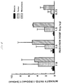
- FIG. 5 is a graphic distribution of the fluorescence intensity ratio of Photofrin II® to autofluorescence for different doses of Photofrin II® in specific rat tissues forty-eight hours after administration.
- FIG. 6 is a fluorescence spectra obtained from tissue stained with BPD.
- FIG. 7 is a graphic distribution of the fluorescence intensity ratio of BPD to autofluorescence for various BPD doses in different rat tissues.
- FIG. 8 is a graphic distribution of the fluorescence intensity ratio of BPD to autofluorescence for the accumulation of BPD in specific rat tissues over time.
- the present invention is directed to a method and apparatus for the instant intraoperative detection and biopsy of metastatic cancer using fluorescence spectroscopy.
- the experimental arrangement used to measure the fluorescence spectra during abdominal exploration included a Helium-cadmium laser operating at 442 nm as an excitation source.
- a hand held 400 micron core diameter fiber was directed at the tissue producing a low power (17 mw) illumination.
- reflected and emitted light was returned to the input of a SPEX 500 spectrometer.
- the spectrometer output was directed to a Silicon Diode Array (Model A 1420 B, EG&G) coupled into an optical multi-channel analyzer (OMA III EG&G). Signals from the OMA were then displayed on a screen for immediate examination.
- a series of normal spectra were recorded from the left medial thigh muscles for baseline.
- the fluorescence intensities at 540 nm (auto- fluorescence) and 630 nm (Photofrin II®) or 690 nm (BPD) were simultaneously monitored. By using the ratio of these intensities (I 630 nm /I 540 nm ) or (I 690 nm /I 540 nm ), a relative value could be assigned to each area studied. An increased value of the ratio of intensities (I 630 nm /I 540 nm ) or (I 690 nm /I 540 nm ) signaled a possible metastatic site. Tissues displaying such an increased ratio of intensities were removed using a biopsy device integrated with the fiberoptic probe and subjected to histological examination.
- the ratios of two probe signals S1 and S2 are first determined for a known non-cancerous region.
- S1 represents the fluorescence intensity at 630 nm (Photofrin II®) or 690 nm (BPD); and
- S2 represents the fluorescence intensity at 540 nm (autofluorescence).
- An increased value of the S1/S2 ratio signals a possible metastatic site.
- the light source may comprise any device emitting substantially monochromatic light and/or incoherent light filtered to a specific wavelength.
- the fiber optic probe is composed of either multiple or single fiber arrays, the core diameter of which is preferably about 400 microns.
- a biopsy needle (Baxter, Tri-Cut® or any conventional biopsy needle) for instant determination of tumor depth and immediate tissue removal.
- the biopsy needle can be calibrated (inches, millimeters) to visually determine the depth of tumor involvement in the affected tissue.
- FIG. 1 A schematic diagram of an experimental optical biopsy probe is illustrated in FIG. 1.
- the optical probe 1 can be integrated in any manner to the top surface of the biopsy blade 2, and then housed in a probe casing 3. However, it is understood that other embodiments may be utilized and structural changes made to the optical biopsy probe without departing from the scope of the invention.
- the fluorescence emitted from cancerous and normal tissues of twenty male Lobund-Wistar rats was investigated. All tumors were subcutaneously implanted by inoculating 105 viable cells of Pollard rat prostatic adenocarcinoma (PA-III) into the right flank of each animal. This tumor model was selected because it is known to metastasize uniformly and spontaneously from extravascular sites only through ipsilateral lymphatic channels. Because of this tumor's unique, predictable spread, the contralateral side of the animal could be used as a control. In addition, cancer detection is facilitated with this model because rats with PA-III cells survive beyond forty days after implantation without evidence of physical impairment.
- PA-III Pollard rat prostatic adenocarcinoma
- Photofrin II® QLT Phototherapeutics, Inc., Vancouver, Canada
- Photofrin II® QLT Phototherapeutics, Inc., Vancouver, Canada
- Metastatic detection was performed during abdominal exploration of the renal, para-aortic, and iliac lymph nodes after laparotomy.
- the rats were anesthetized with ketamine 40 mg/kg and Xylazine 5.0 mg/kg intraperitoneally, and the abdomens opened through a midline incision.
- Advancing caudally to cranially, ipsilateral inguinal, iliac, para-aortal and renal lymph nodes were scanned.
- the areas were scanned in a contact mode with a hand held fiber along the iliac artery to medial and along the aorta to the para-aortic and renal lymph nodes (50 acquisition points/second, 3-4 detection sites/minute, going back and forth. Otherwise, the number of sites could increase.)
- FIG. 2 illustrates the time-related spectral curves for cancerous and normal rat tissue after Photofrin II® administration.
- the Y-axis represents fluorescence intensity
- the X-axis represents wavelength
- the Z-axis represents time.
- the fluorescence spectrum was recorded at every 20th millisecond using a OMA III system while puncturing the targeted rat tissue with the optical biopsy needle. Both the autofluorescence of the tissue and the fluorescence signal of the malignant tissue containing Photofrin II® were recorded.
- the autofluorescence peak is located at 540 nm, and a more prominent peak representing metastasis is located at 630 nm. Note that the intensity of the prominent peak at 630 nm increases as the optical biopsy needle is advanced into the metastasis.
- FIG. 3 illustrates the intensity ratio of Photofrin II® to autofluorescence for the Photofrin II® distribution in rat muscle, primary tumor (right flank) and abdominal metastasis.
- the Y-axis represents the Intensity Ratio (I 630 nm /I 540 nm ), the X-axis represents the particular sample number tested, and the bars represent the three different areas scanned: the circle illustrating metastasis, the shaded bar illustrating tumor, and the solid bar illustrating normal muscle mass.
- the intensity ratio of Photofrin II® fluorescence at 630 nm and tissue autofluorescence at 540 nm was calculated for each of 20 rats to eliminate interference from background fluorescence due to intrinsic porphyrins in cancerous tissue and limited porphyrin uptake in normal tissue. Rats No. 1 and No. 18 were used as controls.
- the intensity ratio was calculated from fluorescence produced by four different doses of Photofrin II® (0.75 mg/kg, 1.5 mg/kg, 2.5 mg/kg and 7.5 mg/kg) and two post-injection times of 24 and 48 hours.
- the prominent peaks for metastasis demonstrates a consistent and significantly higher uptake of Photofrin II® by metastasis than by primary tumor for all post injection times.
- the graph in FIG. 4 uses the same intensity ratio of Photofrin II® fluorescence to autofluorescence to demonstrate the optimum time for Photofrin II® accumulation in muscle, primary tumor, and metastasis after injection.
- the Y-axis represents the Intensity Ratio
- the X-axis represents the two post-injection times of 24 hours for the 3 bars left of center, and 48 hours for the three bars right of center.
- the three bars represent the three different areas of the body where the intensity ratio was monitored: the diagonal shading representing metastasis, the dotted shading representing tumor and the solid bar representing normal muscle mass.
- Signals at the site of the primary tumor and lymph nodes with metastasis show a higher ratio at 48 hours than at 24 hours after Photofrin II® administration. In contrast, the ratio at the thigh muscle site was higher at 24 hours than at 48 hours. (See also Tables II and III below).
- FIG. 5 demonstrates the optimum Photofrin II® dosage range for metastasis detection using the same fluorescence intensity ratio of Photofrin II® fluorescence to autofluorescence for different doses 48 hours after administration.
- the Y-axis represents the Intensity Ratio while the X-axis represents the four different Photofrin II® injection doses tested.
- the first three bars from the left side of the graph illustrate the Intensity Ratio at a dose of 7.5 mg/kg
- the second three bars from the left illustrate the Intensity Ratio at a dose of 2.5 mg/kg
- the third three bars illustrate the Intensity Ratio at a dose of 1.5 mg/kg
- the last three bars illustrate the Intensity Ratio at a dose of 0.75 mg/kg.
- the bars with diagonal shading represent metastasis
- the bars with dotted shading represent tumor and the solid bars represent normal muscle tissue.
- the bar graph illustrates no significant difference in the metastasis intensity ratio for Photofrin II® doses of 1.25 mg/kg, 2.5 mg/kg and 7.5 mg/kg. Although the metastasis intensity ratio at 0.75 mg/kg was somewhat lower than at the other three dosages, it was still useful to distinguish metastatic tissue from primary tumor.
- FIGURES 4 and 5 also demonstrate that the intensity ratio of Photofrin II® in all organs was higher in P-III tumor bearing rats than in healthy rats.
- the ratio of intensities served to assign a comparative value to each organ in relation to the tumor The data for all tissues are provided in Table IV below. Analysis of this data reveals that the fluorescence ratio increases with time in all organs, the fastest increase occurring in metastatic lymph nodes and then in primary tumor.
- a He-Cd laser (442 nm) was used for excitation via a 400 ⁇ m core diameter fiber.
- OMA III optical multichannel analyzer
- Spectra of the liver (L), spleen (S), stomach (St), kidney (K), small bowel (SB), large bowel (LB), mesocolon lymph node (Mc), foot-pad skin (F), primary tumor (T), metastatic lymph node and non-metastatic lymph node were acquired and analyzed.
- the intensity ratio of the characteristic fluorescence peak of BPD ( ⁇ 693 nm) to the autofluorescence intensity was used as an index for drug presence.
- FIGURE 8 illustrates the fluorescence intensity ratio of BPD to autofluorescence for the accumulation of BPD in specific rat tissues over time.
- the fluorescence intensity ratio was higher in liver, small bowel, tumor and metastatic tissue compared to other tissues.
- liver and small bowel tissue also have higher fluorescence intensity ratios than primary tumor and metastasis. As time passes, however, their ratios decrease rapidly whereas the metastasis ratio slightly increases. Large bowel tissue also gives a higher intensity ratio with time while the remaining tissues demonstrate smaller intensity ratios with time. See also Table VII below.
- BPD may be a better alternative photosensitizer than HPD because of its minimal retention in the skin. This advantage of minimal skin retention may reduce the phototoxicity experienced by patients undergoing photodynamic therapy, which is the major clinical limitation of HPD. Based on this knowledge, the detection and subsequent biopsy of disseminated metastatic cancer can be made for cancer diagnosis and treatment.
Landscapes
- Health & Medical Sciences (AREA)
- Life Sciences & Earth Sciences (AREA)
- Physics & Mathematics (AREA)
- Chemical & Material Sciences (AREA)
- General Health & Medical Sciences (AREA)
- Immunology (AREA)
- Pathology (AREA)
- Analytical Chemistry (AREA)
- Biochemistry (AREA)
- General Physics & Mathematics (AREA)
- Chemical Kinetics & Catalysis (AREA)
- Optics & Photonics (AREA)
- Nuclear Medicine, Radiotherapy & Molecular Imaging (AREA)
- Engineering & Computer Science (AREA)
- Biomedical Technology (AREA)
- Heart & Thoracic Surgery (AREA)
- Medical Informatics (AREA)
- Molecular Biology (AREA)
- Surgery (AREA)
- Animal Behavior & Ethology (AREA)
- Public Health (AREA)
- Veterinary Medicine (AREA)
- Investigating, Analyzing Materials By Fluorescence Or Luminescence (AREA)
Applications Claiming Priority (2)
| Application Number | Priority Date | Filing Date | Title |
|---|---|---|---|
| US89158692A | 1992-06-01 | 1992-06-01 | |
| US891586 | 1992-06-01 |
Publications (2)
| Publication Number | Publication Date |
|---|---|
| EP0573264A2 true EP0573264A2 (de) | 1993-12-08 |
| EP0573264A3 EP0573264A3 (en) | 1996-02-07 |
Family
ID=25398468
Family Applications (1)
| Application Number | Title | Priority Date | Filing Date |
|---|---|---|---|
| EP93304252A Withdrawn EP0573264A3 (en) | 1992-06-01 | 1993-06-01 | Apparatus and method of use for a photosensitizer enhanced fluorescence based biopsy needle |
Country Status (3)
| Country | Link |
|---|---|
| EP (1) | EP0573264A3 (de) |
| JP (1) | JPH0638967A (de) |
| CA (1) | CA2097280A1 (de) |
Cited By (7)
| Publication number | Priority date | Publication date | Assignee | Title |
|---|---|---|---|---|
| WO1998001074A1 (en) * | 1996-07-08 | 1998-01-15 | Boston Scientific Corporation | Diagnosing and performing interventional procedures on tissue in vivo |
| DE19854291A1 (de) * | 1998-11-19 | 2000-08-10 | Werner Schramm | Anordnung zur mikroinvasiven Untersuchung von biologischem Gewebe |
| WO2001074251A3 (en) * | 2000-03-31 | 2002-01-31 | Rita Medical Systems Inc | Tissue biopsy and treatment apparatus and method |
| CN102253019A (zh) * | 2011-04-29 | 2011-11-23 | 中国科学院化学研究所 | 一种简便快速检测面粉中过氧化苯甲酰含量的方法 |
| US10143450B2 (en) | 2012-11-02 | 2018-12-04 | Koninklijke Philips N.V. | System with photonic biopsy device for obtaining pathological information |
| CN110710953A (zh) * | 2019-09-10 | 2020-01-21 | 中国科学院上海技术物理研究所 | 一种基于荧光成像的皮肤肿瘤早期筛查装置及使用方法 |
| CN112082942A (zh) * | 2020-09-08 | 2020-12-15 | 宁波市加合灯具有限公司 | 一种节能灯灯头与塑件接合牢固度检测装置 |
Families Citing this family (2)
| Publication number | Priority date | Publication date | Assignee | Title |
|---|---|---|---|---|
| JP2005195379A (ja) * | 2003-12-31 | 2005-07-21 | Yamaguchi Univ | 腫瘍画像検出方法とその装置 |
| EP3401677A4 (de) * | 2016-01-05 | 2019-06-12 | Hamamatsu Photonics K.K. | Lebendkörperbeobachtungsverfahren lebendkörperbeobachtungsvorrichtung |
Family Cites Families (3)
| Publication number | Priority date | Publication date | Assignee | Title |
|---|---|---|---|---|
| JPS5940830A (ja) * | 1982-08-31 | 1984-03-06 | 浜松ホトニクス株式会社 | レ−ザ光パルスを用いた癌の診断装置 |
| US4920143A (en) * | 1987-04-23 | 1990-04-24 | University Of British Columbia | Hydro-monobenzoporphyrin wavelength-specific cytotoxic agents |
| US5318023A (en) * | 1991-04-03 | 1994-06-07 | Cedars-Sinai Medical Center | Apparatus and method of use for a photosensitizer enhanced fluorescence based biopsy needle |
-
1993
- 1993-05-28 CA CA002097280A patent/CA2097280A1/en not_active Abandoned
- 1993-06-01 EP EP93304252A patent/EP0573264A3/en not_active Withdrawn
- 1993-06-01 JP JP5130394A patent/JPH0638967A/ja active Pending
Cited By (11)
| Publication number | Priority date | Publication date | Assignee | Title |
|---|---|---|---|---|
| WO1998001074A1 (en) * | 1996-07-08 | 1998-01-15 | Boston Scientific Corporation | Diagnosing and performing interventional procedures on tissue in vivo |
| US6296608B1 (en) | 1996-07-08 | 2001-10-02 | Boston Scientific Corporation | Diagnosing and performing interventional procedures on tissue in vivo |
| US8135454B2 (en) | 1996-07-08 | 2012-03-13 | Boston Scientific Corporation | Diagnosing and performing interventional procedures on tissue in vivo |
| DE19854291A1 (de) * | 1998-11-19 | 2000-08-10 | Werner Schramm | Anordnung zur mikroinvasiven Untersuchung von biologischem Gewebe |
| DE19854291C2 (de) * | 1998-11-19 | 2001-04-26 | Werner Schramm | Endoskopische Anordnung zum Untersuchen von biologischem Gewebe |
| WO2001074251A3 (en) * | 2000-03-31 | 2002-01-31 | Rita Medical Systems Inc | Tissue biopsy and treatment apparatus and method |
| WO2001074252A3 (en) * | 2000-03-31 | 2002-05-23 | Rita Medical Systems Inc | Tissue biopsy and treatment apparatus and method |
| CN102253019A (zh) * | 2011-04-29 | 2011-11-23 | 中国科学院化学研究所 | 一种简便快速检测面粉中过氧化苯甲酰含量的方法 |
| US10143450B2 (en) | 2012-11-02 | 2018-12-04 | Koninklijke Philips N.V. | System with photonic biopsy device for obtaining pathological information |
| CN110710953A (zh) * | 2019-09-10 | 2020-01-21 | 中国科学院上海技术物理研究所 | 一种基于荧光成像的皮肤肿瘤早期筛查装置及使用方法 |
| CN112082942A (zh) * | 2020-09-08 | 2020-12-15 | 宁波市加合灯具有限公司 | 一种节能灯灯头与塑件接合牢固度检测装置 |
Also Published As
| Publication number | Publication date |
|---|---|
| CA2097280A1 (en) | 1993-12-02 |
| EP0573264A3 (en) | 1996-02-07 |
| JPH0638967A (ja) | 1994-02-15 |
Similar Documents
| Publication | Publication Date | Title |
|---|---|---|
| AU654914B2 (en) | Apparatus and method of use for a photosensitiser enhanced fluorescence based biopsy needle | |
| EP0513986B1 (de) | Nadel mittels eines Photosensibilisators mit verbesserter Fluoreszenzbiopsie | |
| Yoshihiro et al. | Fiberoptic bronchoscopic laser photoradiation for tumor localization in lung cancer | |
| US6846311B2 (en) | Method and apparatus for in VIVO treatment of mammary ducts by light induced fluorescence | |
| Borisova et al. | Endogenous and exogenous fluorescence skin cancer diagnostics for clinical applications | |
| US8858914B2 (en) | Folate targeted enhanced tumor and folate receptor positive tissue optical imaging technology | |
| Poon et al. | Laser-induced fluorescence: experimental intraoperative delineation of tumor resection margins | |
| Kato et al. | Clinical measurement of tumor fluorescence using a new diagnostic system with hematoporphyrin derivative, laser photoradiation, and a spectroscope | |
| Andersson-Engels et al. | Laser-induced fluorescence in medical diagnostics | |
| Andersson-Engels et al. | Fluorescence characteristics of atherosclerotic plaque and malignant tumors | |
| CA2097280A1 (en) | Apparatus and method of use for a photosensitizer enhanced fluorescence based bipsy needle | |
| Zharkova et al. | Fluorescence observations of patients in the course of photodynamic therapy of cancer with the photosensitizer PHOTOSENS | |
| Panjehpour et al. | Quantification of phthalocyanine concentration in rat tissue using laser–induced fluorescence spectroscopy | |
| Andersson-Engels et al. | Fluorescence diagnosis and photochemical treatment of diseased tissue using lasers: Part I | |
| WO1993013403A1 (en) | FLUORESCENCE DIAGNOSTICS OF CANCER USING δ-AMINO LEVULINIC ACID | |
| Gossner et al. | Photodynamic therapy of gastric cancer | |
| Sokolov et al. | Clinical fluorescence diagnostics in the course of photodynamic therapy of cancer with the photosensitizer PHOTOGEM | |
| Tsai et al. | Fluorospectral study of the rat brain and glioma in vivo | |
| Vari et al. | Intraoperative metastases detection by laser-induced fluorescence spectroscopy | |
| Sokolov et al. | Endoscopic fluorescent diagnostics and PDT of early malignancies of lung and esophagus | |
| Chaudhury et al. | Oncologic applications of biophotonics: prospects and problems | |
| Stratonnikov et al. | Spectral fluorescent properties of tissues in vivo with excitation in the red wavelength range | |
| Wang et al. | In-vitro laser-induced fluorescence studies of breast tumors following low-dose injection of Photofrin | |
| Ebert et al. | Characteristic porphyrin-like autofluorescence in primary colon tumors and lymph nodes | |
| Braichotte et al. | Clinical comparison of the pharmacokinetics of m-THPC and Photofrin II as observed by light-induced fluorescence |
Legal Events
| Date | Code | Title | Description |
|---|---|---|---|
| PUAI | Public reference made under article 153(3) epc to a published international application that has entered the european phase |
Free format text: ORIGINAL CODE: 0009012 |
|
| AK | Designated contracting states |
Kind code of ref document: A2 Designated state(s): AT BE CH DE DK ES FR GB GR IE IT LI LU MC NL PT SE |
|
| PUAL | Search report despatched |
Free format text: ORIGINAL CODE: 0009013 |
|
| AK | Designated contracting states |
Kind code of ref document: A3 Designated state(s): AT BE CH DE DK ES FR GB GR IE IT LI LU MC NL PT SE |
|
| STAA | Information on the status of an ep patent application or granted ep patent |
Free format text: STATUS: THE APPLICATION IS DEEMED TO BE WITHDRAWN |
|
| 18D | Application deemed to be withdrawn |
Effective date: 19960808 |