EP0575497B1 - Sequences d'adn codant pour les caracteristiques de virulence de -$(streptococcus suis) et de parties de celui-ci, polypeptides et anticorps derives de celles-ci et leur utilisation pour le diagnostic et la prophylaxie de l'infection par -$(s. suis) chez les mammiferes, y compris l'homme - Google Patents
Sequences d'adn codant pour les caracteristiques de virulence de -$(streptococcus suis) et de parties de celui-ci, polypeptides et anticorps derives de celles-ci et leur utilisation pour le diagnostic et la prophylaxie de l'infection par -$(s. suis) chez les mammiferes, y compris l'homme Download PDFInfo
- Publication number
- EP0575497B1 EP0575497B1 EP92908242A EP92908242A EP0575497B1 EP 0575497 B1 EP0575497 B1 EP 0575497B1 EP 92908242 A EP92908242 A EP 92908242A EP 92908242 A EP92908242 A EP 92908242A EP 0575497 B1 EP0575497 B1 EP 0575497B1
- Authority
- EP
- European Patent Office
- Prior art keywords
- mrp
- strains
- suis
- sequence
- polypeptide
- Prior art date
- Legal status (The legal status is an assumption and is not a legal conclusion. Google has not performed a legal analysis and makes no representation as to the accuracy of the status listed.)
- Expired - Lifetime
Links
Images

















Classifications
-
- C—CHEMISTRY; METALLURGY
- C07—ORGANIC CHEMISTRY
- C07K—PEPTIDES
- C07K14/00—Peptides having more than 20 amino acids; Gastrins; Somatostatins; Melanotropins; Derivatives thereof
- C07K14/195—Peptides having more than 20 amino acids; Gastrins; Somatostatins; Melanotropins; Derivatives thereof from bacteria
- C07K14/315—Peptides having more than 20 amino acids; Gastrins; Somatostatins; Melanotropins; Derivatives thereof from bacteria from Streptococcus (G), e.g. Enterococci
-
- A—HUMAN NECESSITIES
- A61—MEDICAL OR VETERINARY SCIENCE; HYGIENE
- A61P—SPECIFIC THERAPEUTIC ACTIVITY OF CHEMICAL COMPOUNDS OR MEDICINAL PREPARATIONS
- A61P31/00—Antiinfectives, i.e. antibiotics, antiseptics, chemotherapeutics
- A61P31/04—Antibacterial agents
-
- C—CHEMISTRY; METALLURGY
- C12—BIOCHEMISTRY; BEER; SPIRITS; WINE; VINEGAR; MICROBIOLOGY; ENZYMOLOGY; MUTATION OR GENETIC ENGINEERING
- C12Q—MEASURING OR TESTING PROCESSES INVOLVING ENZYMES, NUCLEIC ACIDS OR MICROORGANISMS; COMPOSITIONS OR TEST PAPERS THEREFOR; PROCESSES OF PREPARING SUCH COMPOSITIONS; CONDITION-RESPONSIVE CONTROL IN MICROBIOLOGICAL OR ENZYMOLOGICAL PROCESSES
- C12Q1/00—Measuring or testing processes involving enzymes, nucleic acids or microorganisms; Compositions therefor; Processes of preparing such compositions
- C12Q1/68—Measuring or testing processes involving enzymes, nucleic acids or microorganisms; Compositions therefor; Processes of preparing such compositions involving nucleic acids
- C12Q1/6876—Nucleic acid products used in the analysis of nucleic acids, e.g. primers or probes
- C12Q1/6888—Nucleic acid products used in the analysis of nucleic acids, e.g. primers or probes for detection or identification of organisms
- C12Q1/689—Nucleic acid products used in the analysis of nucleic acids, e.g. primers or probes for detection or identification of organisms for bacteria
-
- A—HUMAN NECESSITIES
- A61—MEDICAL OR VETERINARY SCIENCE; HYGIENE
- A61K—PREPARATIONS FOR MEDICAL, DENTAL OR TOILETRY PURPOSES
- A61K39/00—Medicinal preparations containing antigens or antibodies
-
- Y—GENERAL TAGGING OF NEW TECHNOLOGICAL DEVELOPMENTS; GENERAL TAGGING OF CROSS-SECTIONAL TECHNOLOGIES SPANNING OVER SEVERAL SECTIONS OF THE IPC; TECHNICAL SUBJECTS COVERED BY FORMER USPC CROSS-REFERENCE ART COLLECTIONS [XRACs] AND DIGESTS
- Y10—TECHNICAL SUBJECTS COVERED BY FORMER USPC
- Y10S—TECHNICAL SUBJECTS COVERED BY FORMER USPC CROSS-REFERENCE ART COLLECTIONS [XRACs] AND DIGESTS
- Y10S435/00—Chemistry: molecular biology and microbiology
- Y10S435/8215—Microorganisms
- Y10S435/822—Microorganisms using bacteria or actinomycetales
- Y10S435/885—Streptococcus
-
- Y—GENERAL TAGGING OF NEW TECHNOLOGICAL DEVELOPMENTS; GENERAL TAGGING OF CROSS-SECTIONAL TECHNOLOGIES SPANNING OVER SEVERAL SECTIONS OF THE IPC; TECHNICAL SUBJECTS COVERED BY FORMER USPC CROSS-REFERENCE ART COLLECTIONS [XRACs] AND DIGESTS
- Y10—TECHNICAL SUBJECTS COVERED BY FORMER USPC
- Y10S—TECHNICAL SUBJECTS COVERED BY FORMER USPC CROSS-REFERENCE ART COLLECTIONS [XRACs] AND DIGESTS
- Y10S435/00—Chemistry: molecular biology and microbiology
- Y10S435/975—Kit
Definitions
- the invention is in the field of veterinary and human preventive medicine, in particular that of the diagnosis of and protection against infection by pathogenic strains of the bacterium Streptococcus suis.
- the pig industry in the Netherlands and many other countries has a pyramid structure, with a small number of breeding herds at the top, from where animals are distributed to multiplication herds. These supply a large number of fattening herds, from where the (animal) product finally goes to the slaughterhouses.
- a control program based on diagnosis should primarily aim at creating herds which are free of S. suis type 2 high in this pyramid.
- a vaccine would primarily be useful in affecting herds lower in the pyramid.
- means and methods for diagnosing infections by Streptococcus suis in human medicine can be of value.
- the object of the invention is to provide methods and means which make it possible, in a more effective manner than hitherto, to detect infections by Streptococcus suis on the one hand and to prevent such infections by elimination of infected and carrier pigs on the other hand.
- a virulence characteristic is defined as a polypeptide whose presence is associated with the virulence of an organism, in this case the bacterium S. suis, in particular serotype 2.
- MRP and EF are high molecular weight proteins.
- MRP 136 kD
- EF 110 kD
- EF* higher molecular weight counterparts which are denoted herein as EF*.
- the invention provides new diagnostic methods which are able to differentiate between virulent and avirulent strains. These methods are based on the genes encoding MRP, EF and EF* and their expression products. On the basis of the expression of one or both proteins by said genes, three different phenotypes of S. suis type 2 have been found to date: i.e. the MRP+ EF+ phenotype. the XRP+ EF- phenotype and the MRP- EF- phenotype.
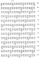
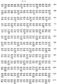
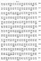
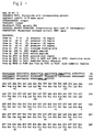
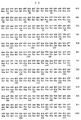
- the invention therefore relates to the DNA sequence of the gene which, apart from coding for specific high molecular weight polypeptides. codes for the 90-120 kDa polypeptide which is a characteristic of S. suis virulence, which gene, hereinafter designated the ef gene has the nucleotide sequence according to Fig. 1A for S. suis serotype 2, strain D-282, and to equivalent sequences and to parts of said sequences. The nucleotide sequence of the entire region coding for EF and the flanking sequences have been determined. Analysis of the sequence of the ef gene (Fig. 1A) provides an open reading frame of 2529 nucleotides which codes for a polypeptide of 843 amino acids (calculated molecular weight 90,014).
- the 110 kDa EF and the high molecular weight proteins are related, which implies that at least part of the ef gene, from strains with a MRP+ EF- phenotype, is identical to the ef gene of strains with the MRP+ EF+ phenotype.
- the higher molecular weight counterpart of the protein EF is designated herein as EF*, and the gene encoding it as the ef* gene.
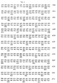
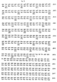
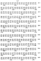
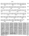
- the corresponding nucleotide and amino acid sequences are represented in Fig. 1B.
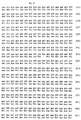
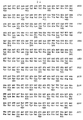
- the invention also relates to the DNA sequence of the gene which codes for the 135-136 kDa polypeptide which is also a virulence characteristic of S. suis, which gene, hereinafter designated the mrp gene, has the nucleotide sequence according to Figure 2 for S. suis serotype 2 strain D-282, and to equivalent sequences and to parts of said sequences.
- the nucleotide sequence of the entire region coding for MRP and the flanking sequences have been determined.
- Analysis of the sequence of the mrp gene (Fig. 2) shows an open reading frame of 3768 nucleotides which codes for a polypeptide of 1256 amino acids (calculated molecular weight 135.794).
- an equivalent sequence comprises a sequence which is essentially the same as the sequence shown but can display slight differences, such as point mutations. or other modifications which may be caused by substitution, deletion, insertion or addition; similarly, an equivalent sequence also comprises a sequence which, despite any differences in nucleotide sequence, hybridises with the sequence shown or with its complement, and also a related sequence which means that it codes for the same amino acid sequence despite differences in nucleotide sequence.
- the invention also relates to a recombinant polynucleotide which contains an ef/ef * gene and/or mrp gene sequence as described above, in the presence of a regulating sequence.
- a recombinant of this type such as a virus vector, a plasmid or a bacterium, can be used for expression of the gene or of relevant parts thereof in a desired environment, for example for the production of immunogenic peptides intended for the diagnosis of an infection, or for controlling infections with virulent strains of S. suis by vaccination.
- Polynucleotide probes which contain a sequence as described above, derived from a gene which codes for a virulence characteristic of S. suis, also form part of the invention.
- a probe of this type in particular corresponds with part of the nucleotide sequence of one of the two said genes.
- the probe can be used for direct detection of the presence of sequences of virulent strains of S. suis.
- the probe can also be used as a basis for a primer for the multiplication of polynucleotides (for example in a polymerase chain reaction) as part of a diagnostic method or a protection method.
- a suitable polynucleotide probe was found to be a partial sequence containing at least 10 nucleotides, preferably at least 15 nucleotides, up to 835 nucleotides from the sequence 1100-1934 of the mrp gene.
- Another suitable polynucleotide probe was found to be a partial sequence containing 10-417, in particular 15-417 nucleotides from the sequence 2890-3306 of the ef * gene. These probes differentiate effectively between pathogenic and non-pathogenic strains of S. suis.
- a combination of such an mrp based probe and an ef* based probe is an especially powerful diagnostic tool.
- the invention also relates to polypeptides which are derived from a polynucleotide sequence described above.
- a polypeptide of this type is either coded by said sequence or obtained by expression of said sequence and essentially corresponds to a S. suis protein characteristic of virulence, or to a part thereof.
- a polypeptide of this type can, for example, be used as an antigen in an immunoassay, as an immunogen in the immunisation of mammals or as an immunogen for the production of antibodies for diagnostic purposes.
- the antibodies generated in this way also form part of the invention.
- Such antibodies can be polyclonal or monoclonal and can be conjugated with a marker (enzyme, isotope, luminescent substance or complex-forming agent); the antibody can also be bound to solid carriers.
- the invention also relates to methods for the detection of an infection by a pathogenic strain or by a non-pathogenic strain of S. suis , in which one or more polynucleotide probes, polypeptides and/or antibodies as described above are used.
- "Infection” signifies here the presence of the pathogenic organism, both in the case where there are clinical signs of disease (infection in a narrow sense) and in the case where there are no clinical signs of disease (infection in a broad sense, or contamination).
- immunoassays such as a determination of the presence of antigens of and/or antibodies against S.
- a polypeptide (110 kDa) which is encoded by the ef/ef * gene or a part thereof, and/or an antibody which has been generated against such a polypeptide it is possible, for example, to use on a microtiter plate a polypeptide (110 kDa) which is encoded by the ef/ef * gene or a part thereof, and/or an antibody which has been generated against such a polypeptide.
- the diagnostic methods can be carried out using procedures known per se. Examples are Enzyme-Linked Immunosorbent Assays (ELISA) and Double Antibody Sandwich (DAS)-ELISA.
- a diagnostic kit according to the invention contains, respectively, at least one polynucleotide or a polypeptide which corresponds to or is derived from a sequence of the ef/ef* gene or mrp gene or a part thereof or contains an antibody which has been generated against the polypeptide derived from one of the said ef/ef* and mrp sequences. It is also possible to use combinations of probes and the like, in particular of ef * diagnostic agents and mrp diagnostic agents. or combinations of primers, for example for carrying out PCR.
- the kits can also contain the components required for carrying out diagnoses, such as reagents (labelling substances, dyes and the like), supports (filters, plates and the like), media and calibrating agents as well as a manual for carrying out the diagnosis.
- the invention also relates to a method for protecting mammals against infection with Streptococcus suis, in which method a polynucleotide, a polypeptide or an antibody as described above is used.
- the method is a passive immunisation, that is to say there is direct provision of antibodies against the pathogenic organism; since antibodies which are derived from EF, EF * and MRP are directed against the most virulent forms of S. suis , a procedure of this type can be an effective method for protecting against, or controlling, infection, especially if the animal to be protected is not itself able to produce sufficient antibodies. for example if infection has already taken place or in the case of young animals.
- Another form of passive immunisation in the case of pigs is the administration of antibodies to the piglets via the colostrum from the sow.
- the dam is actively immunised with one or both polypeptides during pregnancy, that is to say before the birth of the piglets.
- a polypeptide or a polynucleotide (optionally in the form of a recombinant organism) is used, the procedure is an active immunisation, the animal to be protected being stimulated, by means of the immunogenic polypeptide which is administered directly or in the form of a gene for expression, to produce antibodies.
- Another suitable method of immunisation is the administration of a polypeptide from which the activity responsible for virulence has been neutralised.
- a polypeptide should then no longer be pathogenic, while immunogenic characteristics are retained. It can be obtained. for example, by expression of a gene which has been modified with respect to the original ef/ef* or mrp gene. such as by means of deletion.
- Vaccines for protecting mammals against an infection by S. suis. which vaccines contain a polynucleotide, a polypeptide or an antibody as described above, also form part of the invention.
- a particular vaccine according to the invention is a vaccine which contains a S. suis material which does not or does not completely bring to expression at least one of the polypeptides corresponding to EF and MRP.
- This material can originate from or can be formed by a possible live strain which is not virulent or is less virulent.
- the role of virulence factors which are involved in the pathogenesis of S. suis type 2 has been studied in vivo by means of gnotobiotic/germ-free piglets with S. suis type 2 strains defined in respect of virulence factors (MRP and EF). The animal experiments were monitored by means of haematological, bacteriological and (histo)-pathological analytical techniques.
- the presumed ribosome binding site, the -35 and -10 regions of the presumed promoters, and the regions with complementary symmetry are marked.
- the possible cleaving site for signal peptidase is between nucleotides 498-499.
- the putative ribosome binding site, the -35 and -10 regions of the putative promoter sequences, the repetitive regions R1 - R11, and the putative termination signals are indicated.
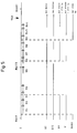
- the region between the nucleotides 2859 and 5228 is absent in the gene encoding the 110 kDa EF protein.
- the region between the nucleotides 3423 and 4456 is absent in the genes encoding the class IV and class V EF* proteins.
- the probable ribosome binding site, the -35 and -10 regions of the presumed promoter sequences, the region of complementary symmetry beyond the mrp gene, the putative cleaving site for signal peptidase, the proline-rich region, the repeating amino acid sequences and the envelope anchor region are indicated.
- ORF1 open reading frame 1
- ORF2 and ORF3 are separated by a TAA stop codon. Restriction sites of interest are indicated.
- the uppermost and middle sequences represent regions flanking the left and right ends of the lacking fragments.
- the bottom sequences show the junctions as found in the class IV and V ef * genes (A) and in the ef gene (B). Directly repeated sequences are shown in boxes.
- the bold nucleotides indicate the first bases of the translational triplets.
- A Restriction maps of the DNA inserts of putative MRP-positive recombinant bacteriophages. The thick line indicates the DNA region which is present in all of these clones. Restriction sites: E: Eco RI; H: Hin dIII; X: Xba I; K: Kpn I; S: Sac I. B. Parts of the DNA inserts subcloned in the plasmid vector pKUN19 ( 24 ) .
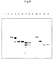
- Lane 1 negative control; proteins extracted from the cell wall of a MRP-negative strain of S. suis.
- Lane 2 crude MRP preparation which contains proteins extracted from the cell wall of strain D282.
- Lane 3 pMR7-1.
- Lane 4 pMR7-2.
- Lane 5 pMR9-1.
- Lane 6 pMR9-2.
- Lane 7 pMR10-1.
- Lane 8 PMR10-2.
- Lane 9 lambda GEM11 with control insert.
- Lane 10 lambda clone 7.
- Lane 11 lambda clone 9.
- Lane 12 lambda clone 10.
- Lane 13 lambda clone 11.
- the amino acid sequence of S. suis MRP was compared with M6 protein of Streptococcus pyogenes (20), protein A of Staphylococcus aureus (16), protein G of group G streptococci (10), AP4 of S. pyogenes (13) , LP of Lactococcus lactis (46), WAP4 of S. mutans (11), T6 of S. pyogenes (38) , and Fn-BP of S. aureus (39).
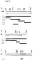
- Fragments of the mrp and ef genes that were used as a probe On top of each figure is the localisation of restriction sites that were used to create the probes. The fragments which were used as probes are indicated with solid bars. Left of the solid bar is the abbreviation of the probe. The arrow indicates the open reading frame (ORF) of each gene.
- Fig. 14a Probes of the mrp gene. The Sac I and Hin dIII sites are not authentic but are generated by subcloning fragments of the mrp gene.
- Fig. 14b Probes of the ef gene.
- Fig. 14c Probe of the ef* gene. The open bar indicates the insert sequence of ef* that is not part of the ef gene.
- Lanes 1 to 4 contained amplified DNA of MRP + EF + strains (D282, 3, 10, and 22), lanes 5, 6, 7, and 9 of MRP+EF* strains (17, 24, 26, 28), lanes 10 to 14 of MRP - EF - strains (T15, 12, 16, 18, and 25), and lane 15 contained the negative control; all ingredients except DNA.
- Lanes 8 and 16 contained 300 ng size marker Lambda DNA digested with Hin dIII and Eco RI .
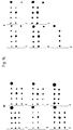
- row A contains 1 ⁇ g/spot DNA of four MRP+EF+ strains; D282, 3, 10 and 22, and one positive control.
- Row B contains four MRP+EF* strains: strain 17, 24, 26 and 28; and row C five MRP-EF- strains; T15, 12, 16, 18 and 25.
- Bacterial strains and growth conditions Bacterial strains and growth conditions .
- E. coli strains JM101 (29) and LE392 (33) were used as hosts for recombinant plasmids and bacteriophages.
- the pathogenic MRP+EF+ strain D282 of S. suis type 2 (43) was used for the isolation of chromosomal DNA.
- E. coli strains were grown in Luria broth (30) . Ampicillin was added as needed to a final concentration of 50 ⁇ g/ml.
- S. suis strains were grown in Todd-Hewitt broth (Oxoid, Ltd., London, England).
- a DNA library of S. suis type 2 strain D282 was constructed in LambdaGEM-11 as recommended by the manufacturer of the cloning vector (Promega, Madison, USA). Recombinant bacteriophages were plated on E. coli strain LE392 and incubated for 16 h at 37°C.
- Nitrocellulose filters (Schleicher and Schuell, Inc., Dassel, Germany) were placed on the plaques, and the plates were further incubated for 2 h at 37°C. Recombinants that produced EF were visualized with monoclonal antibodies (Mabs) directed against EF (Example 4). Bound antibodies were detected with anti-mouse serum conjugated with alkaline phosphatase (Zymed Laboratories, Inc., San Francisco, USA) as described by Maniatis et al. (28). Selected EF positive clones were purified by several rounds of single plaque isolation and immunological screening.
- SDS Sodium dodecyl sulfate
- PAGE polyacrylamide gel electroDhoresis
- Western blot analysis Proteins were separated by SDS gel electrophoresis in which 4% stacking and 6% separating gels were used (26) . The separated proteins were transferred to nitrocellulose in a Semi-Dry transfer cell (Bio-Rad Laboratories, Richmond, USA). Specific proteins were visualized by use of polyclonal antibodies (Pabs, Example 4) or Mabs directed against EF and anti-rabbit or anti-mouse sera conjugated with alkaline phosphatase (Zymed Laboratories).
- DNA manipulations and nucleotide sequence analysis Selected restriction fragments were (sub)-cloned in the plasmid vector pKUN19 (24) by standard molecular biological techniques ( 28 ). Progressive unidirectional deletions were made with the Erase-a-Base system from Promega (Madison, USA). DNA sequences were determined by the dideoxy chain termination method (37) . DNA and protein sequences were analysed by the software packages PCGENE (Intelli-genetics Corp., Mountain View, CA) and Wisconsin GCG (University of Wisconsin).
- a DNA library was constructed by isolating chromosomal DNA from strain D282 of S. suis type 2. This DNA was partially digested with the restriction enzyme Sau 3A and cloned into the bacteriophage LambdaGEM11 replacement vector. The library contained approximately 5 x 10 5 recombinants per ⁇ g of DNA. Two thousand plaques of recombinant phages were tested for the presence of antigenic determinants of EF by use of a Mab directed against EF. Two plaques were positive.
- the plasmid containing the 6.8 kb K pn I- Sal I fragment encoded a protein with a molecular weight identical to EF, that was recognized by Mabs directed against EF. Plasmids containing the 5.8 kb Eco RV- Sal I or the 5.3 kb Bgl II- Sal I fragment, however, did not express EF. These data indicate that the Eco RV and the Bgl II sites are within regions required for EF expression.
- Nucleotide sequence of the ef gene The nucleotide sequence of the fragment comprising the EF encoding region was determined. The sequence (Fig. 1A) showed the presence of 3 major open reading frames (ORFs).
- ORF1 (from nucleotide 361 to 2890), ORF2 (from nucleotide 2856 to 3459) and ORF3 (from nucleotide 3462 to 4053) encoded polypeptides of 843 amino acids, of 201 amino acids and of 197 amino acids respectively.
- ORF1 contained a putative ATG start codon that is preceded by a sequence that is similar to ribosome binding sites of several types of gram-positive bacteria (17) .
- EF is exclusively found in the supernatant of S. suis cultures, and thus the protein is expected to be preceded by a signal peptide.
- the first 46 amino acids of the deduced amino acid sequence of EF are characteristic of a typical signal peptide.
- An N-terminal part that contained six positively charged amino acids was followed by a hydro-phobic core of 21 amino acids and a putative signal peptidase cleavage site (45) .
- the hydropathy pattern (25) of the deduced amino acid sequence showed that, apart from the signal peptide, the EF protein was very hydrophilic and did not contain extended hydrophobic regions (cf. MRP, Example 3). No significant similarities were found between the deduced amino acid sequence of EF and the protein sequences in the EMBL Data Library.
- ORF2 and ORF3 Although appropriate translation initiation signals upstream of ORF2 and ORF3 could not be found, the deduced amino acid sequences of ORF2 and ORF3 showed some properties which raised doubt to the idea that those frames are not expressed.
- the N-terminus of the putative ORF2 protein showed two highly repetative units of 57 amino acids (identity 82%).
- the C-terminus of the putative ORF3 protein is functionally similar to C-terminal regions of several cell-envelope located proteins of gram-positive bacteria ( 10, 12, 13, 16, 41 ) . A hydrophobic region was preceded by the conserved sequence Leu-Pro-X-Thr-Gly-Glu and followed by a highly hydrophilic region. This similarity suggests that the putative ORF3 protein is associated with the cell-envelope.
- Escherichia coli strain JM101 (29) was used as host for recombinant plasmids. Seventeen MRP + EF* strains of S. suis type 2 were isolated from human patients, five strains from tonsils of slaughthered pigs, seven strains from organs of diseased pigs and from two strain the origin was unknown (Example 4). The E. coli strain was grown in Luria broth (30). Ampicillin was added as needed to a final concentration of 50 ⁇ g/ml. Streptococcus suis strains were grown in Todd-Hewitt broth (Oxoid, Ltd., London, England).
- Genomic DNA and oligonucleotides Genomic DNA and oligonucleotides .
- Genomic DNA was isolated by lysis in proteinase K/SDS solution, extraction with phenol/chloroform and precipitation with ethanol (28).
- the sequences of the oligonucleotides used in the polymerase chain reaction (PCR) were: 5'-ATGTAATT GAATTC TCTTTTTAAGT-3' and 5'-AAACGTCCGCAGACT TCTAGA TTAAAAGC-3'. These oligonucleotides correspond to the positions 35 to 59 and 4308 to 4279 in the S. suis type 2 ef gene.
- the underlined sequences indicate the recognition sites for the restriction enzymes Eco RI and Xba I .
- the membranes were washed twice with a solution of 2 x SSC (1 x SSC is 0.15M NaCl plus 0.015 M trisodium citrate, pH 7.0) for 5 min at room temperature and twice with a solution of 0.1 x SSC plus 0.5% SDS for 30 min at 65°C.
- PCR Polymerase Chain Reaction
- ef* genes Cloning of ef* genes .
- the genes encoding the different EF* proteins were obtained using PCR to amplify the ef * containing DNA fragments.
- Genomic DNA of 5 different MRP + EF* strains of S. suis type 2 (one representative of each class) was used as a template.
- the amplified fragments were digested with restriction enzymes Eco RI and Xba I and cloned into E. coli .
- Ef* gene of class I The nucleotide sequence of a 6.8 kb Eco RI -Xba I fragment containing the entire ef* gene of class I and the regions flanking it was determined. Analysis of the sequence revealed two open-reading frames (ORFs, Fig. 1B ). The first ORF (from nucleotide 361 to 5827) and the second ORF (from nucleotide 5830 to 6421) encoded polypeptides of 1822 amino acids and 197 amino acids respectively. Based on its size the first ORF is expected to encode the EF* protein (195 kDa). The ORFs were separated by a single TAA stop codon.
- the first ORF contained a putative ATG start codon that was preceded by a sequence similar to bacterial ribosome-binding sites (17).
- the second ORF was not preceded by an appropriate start codon, nor by a putative ribosome-binding site.
- the first 46 amino acids of the deduced amino acid sequence of the EF* protein had the characteristics of a typical signal peptide (45) .
- the C terminus of the mature part of the protein contained a number of imperfect repeats of 76 amino acids.
- the first four repeats were contiguous as were the last six and a half repeats.
- the fourth and the fifth repeated unit were separated by 113 amino acids and the fifth and the six unit by 22 amino acids (Fig. 5 ).
- the amino acid sequences of the last five and a half unit were highly conserved, whereas the sequences of the first five units were more variable.
- One particular amino acid sequence, Asn-Pro-Asn-Leu was conserved in all repeated units. No significant homology was found between the EF* sequence of class I and any protein sequence in the EMBL Data Library.
- Ef* genes of class II, III, IV and V are Ef* genes of class II, III, IV and V . Because the genes encoding the various EF* proteins differed mainly at their 3' ends, the nucleotide sequences of the small Pst I fragments from the genes of class II, III, IV and V were determined. Comparison of the nucleotide sequences showed that the various ef * genes were highly homologous in this region. The ef * genes differed, however, in the number and the arrangement of repeated units (Fig. 5 ).
- the ef* genes of class II and IV l acked the R9 and R10 regions; that of class III lacked the R6, R7 and R9 regions and that of class IV lacked the R7, R8 and R9 regions.
- the ef* genes of class IV and V l acked a fragment of 1,032 bp, which contained R4, R5 and parts of R3 and R6. The translational reading frame of the region located at the 3' end of the missing fragment remained the same. The nucleotide sequences at the regions of the left and right ends of this 1,032 bp fragment showed direct repeats of 9 bp (Fig. 6A ).
- the reading frame was altered and the region located at the 3'-end of the 2,368 bp fragment was translated in different frames in ef and ef * genes. Consequently, the 110 kDa EF protein will not contain the repeated amino acid units.
- Analysis of the nucleotide sequences at the regions of the left and right ends of the 2,368 bp fragment showed direct repeats of 10 bp (containing one mismatch) (Fig. 6B ).
- the gene encoding the 110 kDa EF protein could have been the result of a specific deletion of 2,368 bp within an ef * gene. This would implicate that a S. suis strain that is non-pathogenic can change into a strain that is pathogenic.
- Escherichia coli strain JM101 (sup E, thi,(lac-pro AB - )[F'tra D36, lac I q Z ⁇ M15], 29) was used as a host for recombinant plasmid DNA.
- E. coli strain LE392 [F - ⁇ hsd R574(rk - ⁇ mk + ), sup E44, sup F58, lac Y1, or ⁇ ( lac 1ZY)6, gal K2, gal T22, mel B1, trp R55] ( 33 ) was used as a host for recombinant bacteriophages.
- the pathogenic MRP + EF + strain D282 of S.suis type 2 (43) was used for isolating chromosomal DNA.
- E. coli strains were grown on LB broth (30) .
- Solid LB medium contained 1.5% agar. Ampicillin was added as needed to a final concentration of 50 ⁇ g/ml.
- Streptococcus suis strains were grown in Todd-Hewitt broth (Oxoid Ltd.)
- Chromosomal DNA isolated from strain D282 of S. suis type 2 was partially digested with the restriction enzyme Sau 3A .
- a DNA library was then constructed in the bacteriophage LambdaGEM11 replacement vector. Approximately 5 x 10 5 recombinants/ ⁇ g DNA were obtained.
- a MAb directed against MRP was used to screen 1,400 recombinant plaques for the presence of antigenic determinants of MRP. Five recombinant plaques reacted positive.
- the complete gene for MRP was obtained by hybridization of the 32 P labeled Kpn I -Sac I fragment of pMR7-2 (Fig. 7B ) with Eco RI or Kpn I digested chromosomal DNA of strain D282 of S. suis type 2. An Eco RI fragment of 7 kb and a Kpn I fragment of 7 kb hybridized with the probe. Because of its size, the Eco RI fragment was expected to contain the complete mrp gene and because the expression of mrp is initiated from the 0.7 kb Eco RI -Kpn I fragment, the Kpn I fragment was expected to contain only the 3' end of the gene.
- Fragments ranging from 6 to 8 kb from Eco RI and Kpn I digested chromosomal DNA were isolated, and ligated into the Eco RI or Kpn I site of pKUN19, whereafter the ligation mixtures were transformed into E. coli JM101.
- Thirteen out of 50 selected recombinant clones obtained with the KpnI fragments hybridized with a MRP probe. All of these recombinant clones contained a plasmid (pMR-C) with a 7 kb Kpn I insert. In contrast, of 2,500 selected recombinant clones obtained with Eco RI fragments, none hybridized with the probe.
- Nucleotide sequence of the mrp gene The nucleotide sequence of a 4.6 kb Eco RI -Hind III fragment, containing the entire mrp gene and the regions flanking it was determined. Analysis of the sequence, Fig. 2, revealed an open reading frame of 3.768 nucleotides coding for a polypeptide of 1,256 amino acids (with a calculated MW of 135,794). The putative ATG start codon is preceded by a sequence that is similar to ribosome-binding sites in several types of gram-positive bacteria (17). The nucleotide sequence upstream of mrp resembles the -35 and -10 consensus sequences of promoters commonly found in gram-positive bacteria.
- a region showing extended dyad symmetry can be detected downstream of the mrp gene.
- MRP Amino acid sequence of MRP .
- MRP is a cell-envelope associated protein and must be translocated across the cytoplasmic membrane.
- the mature protein must therefore contain a signal peptide. Indeed, the first 47 amino acids of the MRP have the characteristics of a typical signal peptide.
- An N-terminal part that contains seven positively charged residues is followed by a hydrophobic core of 21 amino acids and a putative signal peptidase cleavage site ( 45 , vertical arrow in Fig. 2 ). Cleavage of the signal peptide would result in a mature protein with an MW of 131,094, which is close to the MW (136 kDa) of MRP, estimated from SDS-polyacrylamide gels (Example 4).
- a second hydrophobic region of 20 amino acids was identified at the C terminus of the protein (Fig. 11 ). If this region is analogous to other envelope associated proteins of gram-positive bacteria (10, 11, 12, 13, 16, 20, 38, 39, 46), it is probably a cell membrane anchor. A short highly charged region and a region with the Leu-Pro-X-Thr-Gly-Glu amino acid sequence, two regions that flank the presumed cellmembrane anchor, are also highly conserved among surface proteins of gram-positive bacteria (Fig. 12 ). The amino acid sequence Leu-Pro-X-Thr-Gly-Glu is putatively involved in cell-wall binding.
- MRP sequence The mature form of MRP starts with a unique N-terminal sequence of 824 amino acids. This region is followed by a stretch of amino acids that is rich in proline residues: of 86 amino acids, 26 are proline residues. This region is followed by three repeated units of 54 amino acids (Fig. 13 ). The first unit is separated from the second by 77 amino acids, but the second and third unit are contiguous. The sequences of the first and the second unit are highly conserved, whereas the third varies. The third repeated unit is followed by the envelope anchor sequence. There was little homology between the MRP sequence and the protein sequences of the EMBL Data Library.
- strains 1 (MRP + ) and 2 (MRP - ) were used as reference strains.
- Culture conditions A 1-day-old colony of each bacterial strain was grown on Columbia blood agar base (code CM 331; Oxoid, Ltd.) containing 6% horse blood and was incubated overnight at 37°C in Todd-Hewitt broth (code CM 189; Oxoid). Early stationary growth phase cultures were obtained from the overnight cultures, diluted 10 times in Todd-Hewitt broth, and incubated for 4 h at 37°C.
- the rabbits were given intravenous booster inoculations of the same antigen dose, but without the adjuvant. After 6 weeks, the rabbits were exsanguinated.
- the serum of one rabbit (rabbit K191) was used as a probe in the Western blot analysis.
- mice were immunized intraperitoneally with 0.5 ml portions of antigen containing 25 ⁇ g of protein emulsified in equal volumes of Freund imcomplete adjuvant; 3 weeks later this procedure was repeated. After 5 weeks, the mice were given intravenous booster inoculations of the same antigen dose, but without the adjuvant.
- Hybridoma cell lines were prepared as described by Van Zijderveld et al. (51). After 10 to 14 days, hybridomas were tested for antibodies against EF by using an enzyme-linked immunosorbent assay. Hybridoma culture supernatants (diluted 1:2) were then tested for anti-EF MAb on Western blots of culture supernatants from strain D-282.
- Binding of MAb to the 110 kDa protein on the nitrocellulose filters was visualized with anti-mouse immunoglobulins conjugated with alkaline phosphatase.
- the positive cells were cloned twice by limiting dilution in microtiter plates.
- the resulting monoclonal cell lines were used to produce ascites fluid in pristane-primed male BALB/c mice, as described previously (51).
- Electrophoresis and Western blotting The various cell fractions were analysed by SDS-PAGE as described by Laemmli (26) on 6 or 12% polyacrylamide. After electrophoresis, the proteins were stained with silver (32). For Western blot analysis, the proteins were electroblotted onto nitrocellulose by using a Multiphor II Nova Blot system (Pharmacia LKB, Uppsala, Sweden). The blots were probed with a 1:500 dilution of rabbit K191 PAb or with a 1:300 dilution of mouse MAb. Bound PAb were visualized with anti-rabbit immunoglobulins conjugated with alkaline phosphatase. Bound MAb were visualized with a 1:1,000 dilution of anti-mouse immunoglobulins conjugated with alkaline phosphatase (Zymed).
- Protein profiles of four cell fractions of 23 selected strains were almost identical. In contrast, the protein profiles of the culture and protoplast supernatants differed distinctly.
- the protein profiles of isolates obtained from diseased pigs contained two protein bands that were absent in the protein profiles of most isolates obtained from healthy pigs. One band represented a 136 kDa protein, which was identified as MRP (43) .
- MRP and EF were clearly detected in the cell fractions of S. suis type 2 strains.
- Protoplast supernatants, culture supernatants, and membrane vesicles of strains 1, 5, 24, and 26 contained the 136-kDa MRP (Fig. 9 ). Because MRP is a major component of protoplast supernatants, this protein must be localized in the cell envelope of the bacteria.
- the culture supernatants of strains 1 and 5 also contained the 110 kDa EF.
- Strains 24 and 26 contained MRP but not EF; strains 2 and 13 contained neither of the proteins.
- MRP+EF+ Proteins bands at various molecular masses higher than 150 kDa reacted with rabbit K191 serum and were visualized in Western blots of culture supernatants of strains 17, 24, 25, 26, and 28. As such proteins were also recognized by the anti-EF MAb, except in the culture supernatant of strain 25, the 110 kDa EF was probably related to these proteins.
- Pigs Fifty-two germ-free pigs, cross-breeds of Great England and Dutch Landrace, were obtained from four sows by caesarian sections. Sows in both experiments were full sisters. Pigs were allotted to 12 groups each consisting of 4 or 5 pigs. Each group was housed in a sterile stainless steel incubator. Housing and feeding were as described before (43) .
- PBS phosphate-buffered saline
- Bordetella bronchiseptica strain 92932 isolated from the nose of a pig with atrophic rhinitis, was used to predispose pigs to S. suis infection (23, 43) .
- the strain was kept on Dorset egg medium.
- the inoculum was prepared by culturing a 48 hour old colony from sheep blood agar in brain heart infusion broth. After 18 h of incubation at 37°C, this medium contained approximately 10 9 CFU/ml.
- the brain heart infusion broth was diluted (1:100) in PBS to prepare the inoculum.
- Electrophoresis and Western blotting The MRP/EF phenotypes of the S. suis strains used as inocula and of the isolates recovered at the end of the experiments were determined. SDS-PAGE as described by Laemmli (26) (6% polyacrylamide) and Western blotting were used to analyse cell culture supernatants of isolates recovered from nasopharynx of all pigs, and from inflamed tissues such as meninges or joints of affected pigs. After electrophoresis the proteins were stained with silver (32) . For Western blot analysis, the proteins were electroblotted onto nitrocellulose by the Multiphor II Nova Blot system, according to the recommendations of the manufacturer (Pharmacia LKB).
- Nitrocellulose filters were incubated either with a 1:1 mixture of mouse anti-MRP monoclonal antibodies (MAb) (11.3 mg/ml) and anti-EF MAb (8.4 mg/ml) each in a 1:200 dilution, or with a 1:500 dilution of polyclonal anti-MRP/EF rabbit serum (K191) (8.2 mg/ml) (Examples 4, 6).
- Filters were incubated with a 1:1000 dilution of anti-mouse immunoglobulins conjugated with alkaline phosphatase (AP) or a 1:3000 dilution of AP conjugated anti-rabbit immunoglobulin G ( ⁇ + ⁇ ) (Zymed).
- Bound antibodies were visualized by adding the substrate bromochloroindolyl phosphate (Sigma, St. Louis, Mo) - nitro blue tetrazolium (Merck, Darmstad, Germany) in phosphatase buffer (100mM NaCl, 5 mM MgCl 2 , 100 mM diethanolamine; pH 9.5).
- mice were inoculated intranasally with a plastic disposable syringe filled with a suspension of B. bronchiseptica strain 92932 in brain heart infusion broth.
- the inocula contained 0.84 x 10 7 CFU in experiment I and 1.0 x 10 7 CFU in experiment II.
- Two days post inoculation (pi) the pigs were similarly inoculated inside the sterile incubator with one of the ten S. suis type 2 strains (Table 2).
- Strains 10, 12, 16, and 18 were isolated at slaughter from the tonsils of healthy pigs. Strains 22 (no. 830544), 24 (no. 740113), 25 (no. 821021) and 28 (no 760366) were isolated from human patients with S. suis type 2 meningitis. (Numbers between parentheses refer to those by J.P. Arends and H.C. Zanen (2) ). 2 x 10 6 CFU.
- Pigs were monitored daily for clinical signs of disease, such as fever, dysfunction of the CNS and lameness. Blood samples from each pig were collected three times weekly by venipuncture of the cranial vena cava. White blood cells were counted with a conducting counter (Contraves A.G., Switzerland) (18) . The number of neutrophils was calculated after differential count of Giemsa-stained blood smears. Swabs specimens of nasopharynx and feces were collected daily and plated directly onto Columbia agar containing 6% horse blood. The presence of S. suis type 2 and of B.
- bronchiseptica was confirmed by slide agglutination test in which a suspension of the monocultures was mixed with the appropiate hyperimmune rabbit serum (DLO-Central Veterinary Institute, Lelystad, NL). After pigs were killed, they were examined for pathologic changes. Tissue specimens of the CNS, serosae, liver, spleen, and tonsils were bacteriologically and histologically examined as described before (43) .
- Electrophoresis and Western blotting When immunoblots were used to analyse culture supernatants of the S. suis strains before inoculation, three phenotypes were distinguished. Strains 3, 10, and 22 belonged to the MRP+EF+ phenotype, strains 17, 24, and 28 were of the MRP+EF-phenotype, and strains 12, 16, 18, and 25 belonged to the MRP-EF-phenotype.
- the rabbit polyclonal antibodies (PAb) recognized proteins that were greater than 150 kDa in the culture supernatants of the MRP+EF-strains. These high molecular weight proteins were also detected by the anti-EF MAb, indicating that the 110 kDa EF and the > 150 kDa proteins share epitopes.
- the phenotypes of the S. suis strains used as inocula were identical to the phenotypes of the isolates collected at the end of both experiments from tonsils and inflamed tissues of infected pigs.
- Morbidity in pigs inoculated with strains of the MRP+EF+ phenotype was 100%. From day 2 onwards, non-specific signs of systemic disease, such as depression, recumbency, lack of appetite, and fever were observed. During the following days, pigs showed more specific signs of disease, such as ataxia, circular movements, opisthotonus, recumbency with paddling, and lameness. The frequency of specific signs of disease in the MRP+EF+ groups was 57% (Table 3). Nine pigs died in the course of the experiment, and three were killed in the terminal stages of disease. The mortality rate in these groups was thus 12/18 (67%).
- broncho-pneumonia 1 1 1 Fibrinous pneumonia 3 0 0 Interstitial pneumonia 7 5 5 Bronchitis/Peribronchiolitis 2 2 3 Liver Periportal and/or intralobular foci 11 8 3 Spleen Active white pulp 2 6 5 Active red pulp 4 0 2 Tonsil Active follicles 3 9 12 Exudation in crypts 1 5 6 1 Affecting cerebrum, cerebellum, pons, mesencephalon and medulla oblongata in various combinations. 2 Affecting carpal, metacarpal, tarsal, metarsal, knee, elbow, shoulder and hip joints in various combinations.
- S. suis type 2 was mostly isolated from organs and tissues (CNS, serosae, and joints) that also showed pathologic changes (Table 5).
- B. bronchiseptica was only isolated from lungs and tonsils. Both S. suis and B. bronchiseptica were also isolated from the tonsils of all pigs. TABLE 5. Isolation of streptococci from various tissues of pigs inoculated with S. suis type 2 (10 strains of three phenotypes). No. of pigs from which S. suis was isolated at necropsy Tissue phenotype MRP+EF+ (no.
- Culture supernatant of S. suis type 2 strain D 282 was concentrated and partially purified by ultrafiltration (type PM30 filters, Amicon Cooperation). It was diluted in phosphate-buffered saline (PBS) (136.89 mM NaCl, 2.68 mM KCl, 8.1 mM Na 2 HPO 4 , 2.79mM KH 2 PO 4 , pH 7.2), to a final protein concentration of 75 ⁇ g/ml. This product was used as coating antigen for the selection of different monoclonals in the direct competition ELISA and for screening hybridoma culture supernatants in the indirect ELISA.
- PBS phosphate-buffered saline
- Hybridoma culture supernatants (diluted 1:2) were then tested on Western blots of culture supernatants of strain D-282 for antibodies directed against MRP. Bound MAb to the 136 kDa protein were visualized by using anti-mouse immunoglobulins conjugated to alkaline phosphatase and the substrate decribed below. Five supernatants were found positive, and the cells from these wells were cloned twice by limiting dilution in microtiter plates.
- the five cell lines that were positive for anti-MRP antibodies and the three cell lines that were positive for anti-EF antibodies were used to produce ascites fluid in pristane-primed male BALB/c mice.
- MAbs directed against MRP and EF were purified from ascites fluid by ammonium sulphate precipitation (50% saturation) and dialysed against PBS.
- the five anti-MRP MAbs were designated: MRP 1 to MRP 5
- the three anti-EF MABs were designated: EF 1 to EF 3 .
- the immunoglobulin isotype of all MAbs was IgG 1 and was determined by double immunodiffusion with mouse isotype-specific antisera (Nordic) in gels of 1% agarose in PBS.
- the PAbs and MAbs were stored at -20°C.
- Direct competition ELISA MAbs were selected with the direct competition ELISA and were used to develop the MRP and EF double antibody sandwich (DAS) ELISAs. Purified anti-MRP and anti-EF MAbs and rabbit PAbs were conjugated to HRPO (Boehringer Mannheim, Germany) with the periodate method of Wilson and Nakane (49) . Conjugated immunoglobulins were stored at -20°C in 50% glycerol. Conjugate solutions were made in PBS-Tw containing 5% fetal calf serum and 0.5% sodium chloride.
- 50 ⁇ l of nonconjugated anti-MRP MAbs in serial twofold dilutions were added to the wells of polystyrene microtiter ELISA plates (Greiner) that had been coated with the culture supernatant of strain D 282 that had been partially purified in PBS (75 ⁇ g/ml protein). The plates were then incubated for 30 min at 37°C.
- 50 ml of the optimal dilution of each of the five anti-MRP MAbs conjugated to HRPO were added.
- Direct competition ELISA The five anti-MRP clones and the three anti-EF clones were tested for competition. Some anti-MRP clones competed with each other. The five anti-MRP MAbs were directed against at least three different epitopes: the first was recognized by MRP 1 and MRP 2 , the second by MRP 3 , and the third by MRP 4 and MRP 5 . Because all three anti-EF clones competed, they are probably directed against the same epitope.
- MRP double antibody sandwich ELISA In an MRP DAS-ELISA using MRP 3 as catching antibody and HRPO-MRP 1 as conjugate, each well of the polystyrene microtiter ELISA plates was coated with 100 ⁇ l containing 2.3 ⁇ g MRP 3 per well in 0.05 M carbonate buffer, pH 9.6. After adsorption for 16 h at 37°C, coated plates were used immediately or stored at -20°C.
- the MRP DAS-ELISA was used to test 179 strains of S. suis type 2 belonging to the three phenotypes MRP+EF+, MRP+EF-, and MRP-EF-, as was previously determined by SDS-PAGE and Western blot. Most strains scored in the ELISA the same as they did in the Western blot (Table 7). All MRP+EF+ strains were MRP-positive in the ELISA. One MRP+EF- strain scored false negative. Three of the MRP-EF- strains (6%) scored false positive.
- the MRP DAS-ELISA discriminated well between the MRP-positive and MRP-negative strains of S. suis type 2. TABLE 7 Results of 179 strains of S. suis type 2 (three phenotypes) tested in the MRP and EF DAS-ELISAs. MRP DAS ELISA EF DAS ELISA phenotype No. strains + No.
- Culture supernatants of 18 of the 21 reference S. suis strains of other serotypes had absorbances lower than 0.2.
- These serotypes were also positive in the Western blot; MRP 3 apparently recognized proteins of higher molecular weight than 150 kDa in the culture supernatants of these serotypes. Absorbances of all other microorganisms listed in Table 6 were ⁇ 0.2.
- EF Double Antibody Sandwich ELISA In a DAS ELISA that recognizes a specific antigen in the test sample, two different MAbs were used, one as catching antibody and the other as conjugate, and each recognizing different epitopes on the antigen, as was done for the MRP DAS-ELISA. In the Western blot the EF MAbs recognize a high molecular form of EF (> 150 kDa) in the culture supernatants of all strains belonging to the MAP+EF- phenotype (Example 4). Therefore it is unlikely that an ELISA with EF 2 as catching antibody can discriminate between MRP+EF+ and MRP+EF- strains.
- the procedure of the EF DAS-ELISA was essentially as that described for the MRP DAS-ELISA.
- Each well of the microtiter ELISA plates was coated with 100 ml containing 3.3 ⁇ g of EF 2 in 0.05M carbonate buffer, pH 9.6. After adsorption, coated plates were used immediately or stored at -20°C. Twofold serial dilutions of 100 ⁇ l culture supernatants ranging from 1:1 to 1:128 were used. After incubation and washings, 100 ⁇ l containing 2.7 ⁇ g polyclonal Ra K 191 HRPO conjugate in PBS, pH 7.2, was added to each well. After 1 h incubation at 37°C, the plates were developed with substrate 5-AS H 2 O 2 as described above. Wells with an A 450 ⁇ 0.4 were scored positive. The same controls as mentioned above were used on each plate.
- the 179 S. suis type 2 strains with a predetermined protein profile were tested in the EF DAS-ELISA. Surprisingly, none of the 39 MRP+EF- strains scored positive in this ELISA, whereas all 92 MRP+EF+ strains did (Table 7). All 48 MRP-EF- strains were negative in the EF DAS-ELISA. Since no other false positive or false negative results were detected, the EF DAS-ELISA apparently discriminated reliably between the high and the low molecular form of EF, and hence between S. suis type 2 strains belonging to the MRP+EF+ and MRP+EF- phenotypes.
- Mab EF 2 captured the 110-kDa EF in the culture supernatant of all MRP+EF+ strains but apparently not the higher-molecular weight form in the MRP+EF- strains (Table 7). Some MRP-EF- strains gave signals between 0.2 and 0.4, which were still lower than 50% of the maximal absorbance values and thus not high enough to be interpreted as positive. Treating the culture supernatants with SDS before blotting may uncover epitopes of the higher-molecular weight form of EF that are not accessible to the EF 2 MAb in its undenaturated form. Because all MRP-EF- strains and other S. suis serotypes showed no false negative or false positive reactions in this ELISA, the sensitivity and specificity of the tests were considered to be 100%.
- PCR polymerase chain reaction
- S. suis type 2 Bacteria and growth conditions . Thirteen strains of S. suis type 2 were selected to examine whether the Polymerase Chain Reaction (PCR) method (36) could be useful to differentiate between the three phenotypes of S. suis type 2. Pathogenicity and the expression of the MRP and EF proteins of these strains were determined in Examples 4 and 5. Strains were grown overnight at 37°C on Columbia blood agar base (code CM 331, Oxoid) containing 6% horse blood. S. suis type 2 colonies were inoculated in 10 ml Todd-Hewitt broth (code CM 189, Oxoid), and grown overnight at 37°C.
- PCR Polymerase Chain Reaction
- DNA Isolations DNA of overnight grown cultures was isolated as described by Maniatis et. al (28). DNA was diluted to 10 ng/ ⁇ l in distilled water before use in the PCR.
- the filters were not allowed to run dry between the wash steps.
- the bottom of the microtiter plate was dried on a tissue and the samples were dried completely for 15 minutes at 56°C.
- 75 ⁇ l PCR buffer (see below) was added to the individual wells. The plate was incubated for 15 minutes at 56°C.
- the microtiter plate was again put on the vacuum manifold, with a standard microtiter plate (Micronic) beneath the Durapore plate. Vacuum was applied, and the PCR buffer, containing the DNA was collected in the lower microtiter plate, whereas the diatom earth remained on the Durapore filters.
- the PCR contained 10 ng purified DNA or 25 ⁇ l clinical specimen in a total volume of 50 ⁇ l.
- the reaction mixtures contained 10 mM Tris-HCl (pH 9.0), 2 mM MgCl 2 , 50 mM KCl, 0.01% gelatin, 0.2 mM of each of the four deoxynucleotide triphosphates, 1 ⁇ M of each of the four primers and 0.5 U of Amplitaq polymerase (Perkin Elmer Cetus, Norwalk, Conn.), and was overlaid with 2 drops of paraffine oil.
- DNA amplification was carried out in a Perkin Elmer Thermal Cycler for 25 or 40 cycles: 1 minute 94°C, 1 minute 55°C, and 2 minutes 72°C. Ten to 20 ⁇ l of the amplified DNA was analysed on a 1.5% agarose gel, that contained ethidium bromide.
- PCR primers The sequences of the oligonucleotides used in the PCR were: p15: 1403-1425: 5'- GGT ATA CCT TGC TGG TAC CGT TC -3', p16: 1914-1934: 5'- AGT CTC TAC AGC TGT AGC TGG -3', p-34: 2890-2908: 5'- GTT GAA AAC AAA GCA TTC G -3', and p-35: 3229-3249: 5'- CTT CGA CAA AAT GTC AGA TTC -3'.
- the oligonucleotides p-15 and p-16 correspond to the indicated positions in the S. suis type 2 mrp gene (Example 3, Fig.
- the oligonucleotides p-34 and p-35 correspond to the indicated positions and in the S. suis type 2 ef * gene (Example 2, Fig. 1B ). Primers were synthesized on an Applied Biosystem synthesizer type 381A following the manufacturers protocol.
- PCR Specificity of PCR .
- two regions (designated as m-VI and e-V) were determined that could be used to differentiate between the three phenotypes of S. suis type 2 strains (see also Example 8).
- the primers p-15 and p-16 amplified a 532 bp fragment in the m-VI region.
- the primers p-34 and p-35 amplified a 360 bp fragment in the e-V region.
- MRP + EF* cells were diluted in phosfate buffered saline (PBS (pH 7.2); 137 mM NaCl, 2.7 mM KCl, 8.1 mM Na 2 HPO 4 , 2.8 mM KH 2 PO 4 ) and prepared for PCR as described above. Amplified fragments could still be detected in samples that contained about 50 cells prior to the PCR (40 cycli).
- PBS phosfate buffered saline
- the PCR can be used directly on clinical material. Serial dilutions of S. suis type 2 cells were added to nose swabs. It was found that amplified fragments can still be detected in samples that contain about 50 cells prior to the PCR.
- S. suis type 2 170 strains of S. suis type 2 were obtained from three sources: From organs of diseased pigs (103 strains), from tonsils of healthy pigs at slaughter (40 strains) and from human patients (27 strains). Reference strains of S. suis serotypes 1 to 22 (15), 21 other Streptococci species and 45 other bacterial strains (38 different species, DLO Central Veterinary Institute, Table 8) were used to test the specificity of the mrp and ef probes.
- E. coli JM101 strains were grown in LB broth (30) . Ampicillin was added as needed to a final concentration of 50 ⁇ g/ml. All other bacterial strains were grown overnight at 37°C on Columbia blood agar base (code CM 331, Oxoid) containing 6% horse blood. Overnight grown colonies were incubated in 10 ml Todd-Hewitt broth (code CM 189, Oxoid). and grown overnight at 37°C.
- Probes The plasmids pMR11, pEF2-19 and pEF17-7 (cf. Examples 1, 2, 3) were used to generate subclones into pKUN19 (24). Fragments of appropriate subclones were isolated from preparative agarose gels with the gene-clean kit (Bio 101 Inc., La Jolla, USA). Purified fragments were subsequently labeled with ⁇ - 32 P dCTP (3000 Ci/mMol, Amersham) with the random primed labeling kit (Boehringer GmbH) following the manufacturers protocol and used as probes.
- ⁇ - 32 P dCTP 3000 Ci/mMol, Amersham
- the membranes were incubated with the 32 P-labeled mrp and ef probes as recommended by the manufacturer. After overnight hybridization, the membranes were washed twice in 40 mM Na phosphate buffer, pH 7.2 plus 5% SDS plus 1 mM EDTA for 30 minutes at 65°C and twice in 40 mM Na phosphate buffer pH 7.2 plus 1% SDS plus 1 mM EDTA for 30 minutes at 65°C. All (pre)hybridizations were carried out in a hybridization oven (Hybaid).
- Mrp probes Chromosomal DNA of the 3 phenotypes of S. suis type 2 was hybridized to different regions of the mrp gene. Six different mrp probes were used (schematically shown in Fig. 14a ). The EcoRI-SnaBI fragment, m-I. contained the entire mrp encoding region. The m-II, m-III, m-IV and m-V probes contained different regions of the mrp gene (see Fig. 16 ). The MRP + EF + and the MRP + EF* strains strongly hybridized with all mrp probes.
- probe m-VI was constructed by removing 385 bp at the 5', and 325 bp at the 3' ends of probe m-III.
- the 5 MRP - EF - strains did not hybridize at all with probe m-VI, indicating that these strains lacked the region homologous to the m-VI probe. Therefore, probe m-VI can be used to differentiate between MRP + and MRP - strains.
- probe e-V hybridized with all MRP + EF* strains.
- MRP + EF + and MRP - EF - strains hybridized with the e-V probe.
- S. suis type 2 strains hybridize with probe m-VI and e-V, these strains belong to the MRP + EF * phenotype. If S. suis type 2 strains hybridize with m-VI but not with e-V, these strains belong to the MRP + EF + phenotype, and finally if strains do not hybridize with m-VI and e-V, these strains belong to the MRP - EF - phenotype.
- the mrp , ef and ef * probes were tested on 170 other strains of S. suis type 2. 88 strains had a MRP + EF + phenotype, 37 strains a MRP + EF* phenotype and 45 strains had a MRP - EF - phenotype. In accord with the data presented above, all MRP + EF + strains hybridized with the probes m-I to m-VI and e-I to e-IV, but none hybridized with probe e-V. Moreover, all the 37 MRP + EF* strains hybridized with all the probes.
- probes m-I, m-VI, e-III and e-V Chromosomal DNA from swine pathogens and several common bacteria was tested with the probes m-I, m-VI, e-III and e-V.
- the species tested are listed in Table 8. Although some species hybridized with probe m-I (Escherichia coli, Klebsiella oxytoca, K. pneumoniae and Salmonella typhimurium), none hybridized with the probes m-VI, e-III and e-V. These data show that although in some species parts of the mrp gene are found, the probes m-VI and e-V are specific for S. suis. Hence, the probes m-VI and e-V have potential diagnostic value.
Landscapes
- Chemical & Material Sciences (AREA)
- Organic Chemistry (AREA)
- Health & Medical Sciences (AREA)
- Life Sciences & Earth Sciences (AREA)
- Proteomics, Peptides & Aminoacids (AREA)
- Analytical Chemistry (AREA)
- General Health & Medical Sciences (AREA)
- Wood Science & Technology (AREA)
- Biochemistry (AREA)
- Zoology (AREA)
- Engineering & Computer Science (AREA)
- Molecular Biology (AREA)
- Genetics & Genomics (AREA)
- Biophysics (AREA)
- Medicinal Chemistry (AREA)
- Physics & Mathematics (AREA)
- Biotechnology (AREA)
- General Engineering & Computer Science (AREA)
- Bioinformatics & Cheminformatics (AREA)
- Microbiology (AREA)
- Immunology (AREA)
- Gastroenterology & Hepatology (AREA)
- Oncology (AREA)
- Public Health (AREA)
- Nuclear Medicine, Radiotherapy & Molecular Imaging (AREA)
- Chemical Kinetics & Catalysis (AREA)
- General Chemical & Material Sciences (AREA)
- Communicable Diseases (AREA)
- Pharmacology & Pharmacy (AREA)
- Animal Behavior & Ethology (AREA)
- Veterinary Medicine (AREA)
- Peptides Or Proteins (AREA)
- Medicines Containing Antibodies Or Antigens For Use As Internal Diagnostic Agents (AREA)
- Measuring Or Testing Involving Enzymes Or Micro-Organisms (AREA)
- Preparation Of Compounds By Using Micro-Organisms (AREA)
- Medicines That Contain Protein Lipid Enzymes And Other Medicines (AREA)
- Pharmaceuticals Containing Other Organic And Inorganic Compounds (AREA)
- Micro-Organisms Or Cultivation Processes Thereof (AREA)
Claims (18)
- Séquence d'ADN d'un gène codant pour une caractéristique de virulence de Streptococcus suis, ledit gène étant choisi parmi (a) le gène codant pour un polypeptide de 90 000 - 120 000 daltons dans des souches virulentes de Streptococcus suis et codant pour un polypeptide ayant une masse moléculaire plus élevée que le polypeptide précédent et comprenant essentiellement le polypeptide précédent dans des souches moins virulentes de Streptococcus suis, ladite séquence ou ledit gène possédant les séquences nucléotidiques selon les figures 1A et 1B pour le sérotype 2 de S. suis, souche D-282, ou une séquence équivalente ou une séquence partielle de celles-ci, contenant au moins 10 nucléotides, de préférence, au moins 15 nucléotides; et (b) le gène codant pour un polypeptide de 135 000 - 136 000 daltons (protéine libérée par la muramidase) qui est une caractéristique de virulence de Streptococcus suis, ladite séquence ou ledit gène possédant la séquence nucléotidique selon la figure 2 pour le sérotype 2 de S. suis, souche D-282, ou une séquence équivalente ou une séquence partielle de celle-ci, contenant au moins 10 nucléotides, de préférence au moins 15 nucléotides.
- Séquence d'ADN selon la revendication 1, codant pour une partie du polypeptide de masse moléculaire plus élevée codé par (a), ladite partie ne correspondant pas au polypeptide de 90 000 - 120 000 daltons, ou une séquence partielle de celui-ci, contenant au moins 10 nucléotides, de préférence au moins 15 nucléotides.
- Séquence d'ADN selon la revendication 1, qui est une séquence partielle contenant au moins 10 nucléotides, de préférence au moins 15 nucléotides, provenant de la séquence 2890-3306 de la figure 1B.
- Séquence d'ADN selon la revendication 1, qui est une séquence partielle contenant au moins 10 nucléotides, de préférence au moins 15 nucléotides, provenant de la séquence 1100-1934 de la figure 2.
- Polynucléotide recombiné, comprenant une séquence selon l'une quelconque des revendications 1-4, en présence d'une séquence régulatrice.
- Sonde polynucléotidique pour le diagnostic d'une infection par Streptococcus suis, comprenant une séquence selon l'une quelconque des revendications 1-4.
- Polypeptide codé ou obtenu par l'expression d'une séquence selon l'une quelconque des revendications 1-5.
- Anticorps dirigé contre un polypeptide selon la revendication 7.
- Procédé pour détecter une infection par une souche pathogène de Streptococcus suis, caractérisé par l'utilisation d'au moins une sonde selon la revendication 6.
- Procédé selon la revendication 9, dans lequel au moins une sonde ayant une séquence selon la revendication 2 ou 3 est utilisée.
- Procédé pour détecter une infection par une souche pathogène de Streptococcus suis, caractérisé par l'utilisation d'au moins un polypeptide selon la revendication 7.
- Procédé pour détecter une infection par une souche pathogène de Streptococcus suis, caractérisé par l'utilisation d'au moins un anticorps selon la revendication 8.
- Kit de diagnostic pour la détection d'une infection par une souche pathogène de Streptococcus suis, caractérisé en ce qu'il contient au moins une sonde selon la revendication 6.
- Kit de diagnostic pour la détection d'une infection par une souche pathogène de Streptococcus suis, caractérisé en ce qu'il contient au moins un polypeptide selon la revendication 7.
- Kit de diagnostic pour la détection d'une infection par une souche pathogène de Streptococcus suis, caractérisé en ce qu'il contient au moins un anticorps selon la revendication 8.
- Vaccin qui protège les mammifères contre une infection par Streptococcus suis, contenant un polynucléotide selon la revendication 5, un polypeptide selon la revendication 7, ou un anticorps selon la revendication 8.
- Vaccin qui protège les mammifères contre une infection par Streptococcus suis, contenant un polypeptide selon la revendication 7 exempt des parties qui sont responsables de la virulence.
- Vaccin qui protège les mammifères contre une infection par Streptococcus suis, contenant une souche de Streptococcus suis dans laquelle le gène codant pour le polypeptide de 90 000 - 120 000 daltons ou le gène codant pour le polypeptide de 135 000 - 136 000 daltons selon la revendication 1 a été rendu non fonctionnel.
Applications Claiming Priority (3)
| Application Number | Priority Date | Filing Date | Title |
|---|---|---|---|
| NL9100510A NL9100510A (nl) | 1991-03-21 | 1991-03-21 | Dna-sequenties die coderen voor kenmerken van virulentie van streptococcus suis en delen daarvan, daarvan afgeleide polypeptiden en antilichamen, alsmede toepassing daarvan bij de diagnose van en de bescherming tegen infectie met s. suis bij zoogdieren, waaronder de mens. |
| NL9100510 | 1991-03-21 | ||
| PCT/NL1992/000054 WO1992016630A1 (fr) | 1991-03-21 | 1992-03-19 | Sequences d'adn codant pour les caracteristiques de virulence de streptococcus suis et de parties de celui-ci, polypeptides et anticorps derives de celles-ci et leur utilisation pour le diagnostic et la prophylaxie de l'infection par s. suis chez les mammiferes, y compris l'homme |
Publications (2)
| Publication Number | Publication Date |
|---|---|
| EP0575497A1 EP0575497A1 (fr) | 1993-12-29 |
| EP0575497B1 true EP0575497B1 (fr) | 1996-10-02 |
Family
ID=19859048
Family Applications (1)
| Application Number | Title | Priority Date | Filing Date |
|---|---|---|---|
| EP92908242A Expired - Lifetime EP0575497B1 (fr) | 1991-03-21 | 1992-03-19 | Sequences d'adn codant pour les caracteristiques de virulence de -$(streptococcus suis) et de parties de celui-ci, polypeptides et anticorps derives de celles-ci et leur utilisation pour le diagnostic et la prophylaxie de l'infection par -$(s. suis) chez les mammiferes, y compris l'homme |
Country Status (14)
| Country | Link |
|---|---|
| US (1) | US5610011A (fr) |
| EP (1) | EP0575497B1 (fr) |
| JP (1) | JP2776633B2 (fr) |
| CN (1) | CN1055500C (fr) |
| AT (1) | ATE143693T1 (fr) |
| AU (1) | AU659637B2 (fr) |
| CA (1) | CA2105057C (fr) |
| DE (1) | DE69214277T2 (fr) |
| DK (1) | DK0575497T3 (fr) |
| ES (1) | ES2094907T3 (fr) |
| GR (1) | GR3022052T3 (fr) |
| NL (1) | NL9100510A (fr) |
| NZ (1) | NZ242037A (fr) |
| WO (1) | WO1992016630A1 (fr) |
Families Citing this family (19)
| Publication number | Priority date | Publication date | Assignee | Title |
|---|---|---|---|---|
| US6015889A (en) * | 1993-03-19 | 2000-01-18 | Gunnar Lindahl | Protein rib, a cell surface protein that confers immunity to many strains of the group B streptococcus: process for purification of the protein, reagent kit and pharmaceutical composition |
| US5726016A (en) * | 1995-01-18 | 1998-03-10 | The Trustees Of The University Of Pennsylvania | Compositions and methods for diagnosis of diseases associated with actinobacillus actinomycetemcomitans infection |
| CA2208780C (fr) * | 1995-01-30 | 2010-03-30 | Peter Truog | Preparations antitumorales et anti-cholesterol comprenant un acide lipoteichoique, le lta-t, derive de streptococcus sp dsm 8747 |
| AU5070999A (en) * | 1998-07-22 | 2000-02-14 | Id-Lelystad | (streptococcus suis) vaccines and diagnostic tests |
| EP1205552A1 (fr) * | 2000-11-09 | 2002-05-15 | ID-Lelystad, Instituut voor Dierhouderij en Diergezondheid B.V. | Virulence de streptococci, avec ORFs de Streptococcus suis |
| US6905816B2 (en) | 2000-11-27 | 2005-06-14 | Intelligent Medical Devices, Inc. | Clinically intelligent diagnostic devices and methods |
| CA2436530C (fr) | 2001-02-02 | 2013-10-01 | Id-Lelystad, Instituut Voor Dierhouderij En Diergezondheid B.V. | Genes de streptococcus suis environnementalement regules |
| DK1943342T3 (en) | 2005-09-02 | 2016-07-25 | Valorisation-Recherche Ltd Partnership | STREPTOCOCCUS SUIS POLYPEPTIDES AND POLYNUCLEOTIDES ENCODING SAME AND THEIR USE IN DIAGNOSTIC USES vaccination and |
| CN100388952C (zh) * | 2006-04-25 | 2008-05-21 | 范红结 | 一种链球菌重组亚单位疫苗及其制备方法 |
| CN100449005C (zh) * | 2006-06-29 | 2009-01-07 | 中国检验检疫科学研究院动植物检疫研究所 | 应用于猪链球菌2型致病性检测的多重荧光pcr检测试剂及方法 |
| CN100449006C (zh) * | 2006-08-10 | 2009-01-07 | 中国检验检疫科学研究院动植物检疫研究所 | 应用于猪链球菌2型胞外因子(ef)荧光pcr检测的引物和探针序列 |
| EP2023143A1 (fr) * | 2007-08-06 | 2009-02-11 | Boehringer Ingelheim Vetmedica Gmbh | Protéines de streptococcus immunogènes |
| CN101613402B (zh) * | 2008-06-25 | 2012-11-28 | 中国人民解放军军事医学科学院微生物流行病研究所 | 一种猪链球菌2型表面蛋白、其制备方法及用途 |
| CN101613400B (zh) * | 2008-06-25 | 2012-11-28 | 中国人民解放军军事医学科学院微生物流行病研究所 | 一种猪链球菌2型表面细胞壁蛋白、其制备方法及用途 |
| CN101613399B (zh) * | 2008-06-25 | 2012-11-28 | 中国人民解放军军事医学科学院微生物流行病研究所 | 一种猪链球菌2型表面蛋白、其制备方法及用途 |
| WO2014143781A1 (fr) * | 2013-03-15 | 2014-09-18 | Camas Incorporated | Procédés et compositions pour réduire des micro-organismes dans l'oropharynx, le nasopharynx et les cavités buccales |
| CN104762244B (zh) * | 2015-02-04 | 2017-09-15 | 中国农业科学院哈尔滨兽医研究所 | 猪链球菌SBP_bac_5基因缺失株及其构建方法与应用 |
| CN107805660B (zh) * | 2017-12-08 | 2021-11-26 | 江汉大学 | 一种猪链球菌小种分离鉴定方法 |
| GB2587228B (en) * | 2019-09-20 | 2021-10-27 | Protein Ark Ltd | Biological sample purification apparatus, use of the same, and systems comprising the same |
Family Cites Families (2)
| Publication number | Priority date | Publication date | Assignee | Title |
|---|---|---|---|---|
| US4784948A (en) * | 1983-08-10 | 1988-11-15 | The Rockefeller University | Production of streptococcal m protein immunogens and molecular probes |
| CA2009708A1 (fr) * | 1989-02-13 | 1990-08-13 | Jane D. Madonna | Sonde d'acide nucleique pour la detection des salmonella pathogene pour les humains |
-
1991
- 1991-03-21 NL NL9100510A patent/NL9100510A/nl not_active Application Discontinuation
-
1992
- 1992-03-18 NZ NZ242037A patent/NZ242037A/en not_active IP Right Cessation
- 1992-03-19 DK DK92908242.8T patent/DK0575497T3/da active
- 1992-03-19 AU AU15464/92A patent/AU659637B2/en not_active Ceased
- 1992-03-19 EP EP92908242A patent/EP0575497B1/fr not_active Expired - Lifetime
- 1992-03-19 CA CA002105057A patent/CA2105057C/fr not_active Expired - Fee Related
- 1992-03-19 ES ES92908242T patent/ES2094907T3/es not_active Expired - Lifetime
- 1992-03-19 AT AT92908242T patent/ATE143693T1/de not_active IP Right Cessation
- 1992-03-19 WO PCT/NL1992/000054 patent/WO1992016630A1/fr not_active Ceased
- 1992-03-19 DE DE69214277T patent/DE69214277T2/de not_active Expired - Fee Related
- 1992-03-19 JP JP4507787A patent/JP2776633B2/ja not_active Expired - Fee Related
- 1992-03-19 US US08/119,125 patent/US5610011A/en not_active Expired - Lifetime
- 1992-03-21 CN CN92101913A patent/CN1055500C/zh not_active Expired - Fee Related
-
1996
- 1996-12-17 GR GR960403484T patent/GR3022052T3/el unknown
Also Published As
| Publication number | Publication date |
|---|---|
| CN1065490A (zh) | 1992-10-21 |
| NL9100510A (nl) | 1992-10-16 |
| DE69214277D1 (de) | 1996-11-07 |
| CN1055500C (zh) | 2000-08-16 |
| EP0575497A1 (fr) | 1993-12-29 |
| DE69214277T2 (de) | 1997-03-20 |
| DK0575497T3 (da) | 1996-11-18 |
| NZ242037A (en) | 1997-03-24 |
| ATE143693T1 (de) | 1996-10-15 |
| ES2094907T3 (es) | 1997-02-01 |
| WO1992016630A1 (fr) | 1992-10-01 |
| GR3022052T3 (en) | 1997-03-31 |
| CA2105057A1 (fr) | 1992-09-22 |
| JPH06505390A (ja) | 1994-06-23 |
| JP2776633B2 (ja) | 1998-07-16 |
| AU1546492A (en) | 1992-10-21 |
| US5610011A (en) | 1997-03-11 |
| CA2105057C (fr) | 2000-05-02 |
| AU659637B2 (en) | 1995-05-25 |
Similar Documents
| Publication | Publication Date | Title |
|---|---|---|
| EP0575497B1 (fr) | Sequences d'adn codant pour les caracteristiques de virulence de -$(streptococcus suis) et de parties de celui-ci, polypeptides et anticorps derives de celles-ci et leur utilisation pour le diagnostic et la prophylaxie de l'infection par -$(s. suis) chez les mammiferes, y compris l'homme | |
| Madoff et al. | Group B streptococci escape host immunity by deletion of tandem repeat elements of the alpha C protein. | |
| Benz et al. | AIDA‐I, the adhesin involved in diffuse adherence of the diarrhoeagenic Escherichia coli strain 2787 (O126: H27), is synthesized via a precursor molecule | |
| US8071111B2 (en) | Virulence of Streptococci | |
| US8101187B2 (en) | Secreted Streptococcus pneumoniae proteins | |
| US8324354B2 (en) | Environmentally regulated genes of Streptococcus suis | |
| US7098182B2 (en) | Nucleic acids and proteins from group B streptococcus | |
| EP1100920A2 (fr) | Acides nucleiques et proteines de streptococcus groupe b | |
| Swihart et al. | The HpmA hemolysin is more common than HlyA among Proteus isolates | |
| JP2003527100A (ja) | タンパク質 | |
| Cruz et al. | Molecular characterization of a common 48-kilodalton outer membrane protein of Actinobacillus pleuropneumoniae | |
| US6296855B1 (en) | 17-KDA Brucella abortus antigen, recombinant polypeptides, nucleic acids coding for the same and use thereof in diagnostic and prophylactic methods and kits | |
| JP2001504335A (ja) | ストレプトコッカス・ユベリスのラクトフェリン結合タンパク質 | |
| US20030165527A1 (en) | Novel fibronectin-binding protein |
Legal Events
| Date | Code | Title | Description |
|---|---|---|---|
| PUAI | Public reference made under article 153(3) epc to a published international application that has entered the european phase |
Free format text: ORIGINAL CODE: 0009012 |
|
| 17P | Request for examination filed |
Effective date: 19930921 |
|
| AK | Designated contracting states |
Kind code of ref document: A1 Designated state(s): AT BE CH DE DK ES FR GB GR IT LI LU NL SE |
|
| 17Q | First examination report despatched |
Effective date: 19940531 |
|
| GRAH | Despatch of communication of intention to grant a patent |
Free format text: ORIGINAL CODE: EPIDOS IGRA |
|
| GRAH | Despatch of communication of intention to grant a patent |
Free format text: ORIGINAL CODE: EPIDOS IGRA |
|
| GRAA | (expected) grant |
Free format text: ORIGINAL CODE: 0009210 |
|
| AK | Designated contracting states |
Kind code of ref document: B1 Designated state(s): AT BE CH DE DK ES FR GB GR IT LI LU NL SE |
|
| REF | Corresponds to: |
Ref document number: 143693 Country of ref document: AT Date of ref document: 19961015 Kind code of ref document: T |
|
| REF | Corresponds to: |
Ref document number: 69214277 Country of ref document: DE Date of ref document: 19961107 |
|
| ET | Fr: translation filed | ||
| REG | Reference to a national code |
Ref country code: CH Ref legal event code: NV Representative=s name: DIPL.-ING. ETH H. R. WERFFELI PATENTANWALT |
|
| REG | Reference to a national code |
Ref country code: DK Ref legal event code: T3 |
|
| ITF | It: translation for a ep patent filed | ||
| REG | Reference to a national code |
Ref country code: ES Ref legal event code: FG2A Ref document number: 2094907 Country of ref document: ES Kind code of ref document: T3 |
|
| REG | Reference to a national code |
Ref country code: GR Ref legal event code: FG4A Free format text: 3022052 |
|
| PLBE | No opposition filed within time limit |
Free format text: ORIGINAL CODE: 0009261 |
|
| 26N | No opposition filed | ||
| REG | Reference to a national code |
Ref country code: GB Ref legal event code: IF02 |
|
| REG | Reference to a national code |
Ref country code: FR Ref legal event code: TP |
|
| REG | Reference to a national code |
Ref country code: FR Ref legal event code: TP |
|
| BECA | Be: change of holder's address |
Owner name: *VETOQUINOLMAGNY VERNOIS, F-70204 LEURE Effective date: 20051110 |
|
| BECH | Be: change of holder |
Owner name: *VETOQUINOLMAGNY VERNOIS, F-70204 LEURE Effective date: 20051110 Owner name: *VETOQUINOL Effective date: 20051110 |
|
| BECN | Be: change of holder's name |
Owner name: *VETOQUINOL Effective date: 20051110 |
|
| REG | Reference to a national code |
Ref country code: CH Ref legal event code: PUE Owner name: VETOQUINOL Free format text: STICHTING CENTRAAL DIERGENEESKUNDIG INSTITUUT#EDELHERTWEG 15#NL-8219 PH LELYSTAD (NL) -TRANSFER TO- VETOQUINOL#MAGNY VERNOIS#70204 LURE (FR) Ref country code: CH Ref legal event code: NV Representative=s name: TROESCH SCHEIDEGGER WERNER AG Ref country code: GB Ref legal event code: 732E |
|
| REG | Reference to a national code |
Ref country code: ES Ref legal event code: PC2A |
|
| NLS | Nl: assignments of ep-patents |
Owner name: VETOQUINOL Effective date: 20060202 Owner name: STICHTING DIENST LANDBOUWKUNDIG ONDERZOEK Effective date: 20060202 Owner name: ID-LELYSTAD, INSTITUUT VOOR DIERHOUDERIJ EN DIERGE Effective date: 20060202 |
|
| NLT1 | Nl: modifications of names registered in virtue of documents presented to the patent office pursuant to art. 16 a, paragraph 1 |
Owner name: STICHTING INSTITUUT VOOR VEEHOUDERIJ EN DIERGEZOND Owner name: STICHTING INSTITUUT VOOR DIERHOUDERIJ EN DIERGEZON |
|
| BECA | Be: change of holder's address |
Owner name: *VETOQUINOLMAGNY VERNOIS, F-70204 LEURE Effective date: 20051110 |
|
| BECH | Be: change of holder |
Owner name: *VETOQUINOLMAGNY VERNOIS, F-70204 LEURE Effective date: 20051110 Owner name: *VETOQUINOL Effective date: 20051110 |
|
| BECN | Be: change of holder's name |
Owner name: *VETOQUINOL Effective date: 20051110 |
|
| PGFP | Annual fee paid to national office [announced via postgrant information from national office to epo] |
Ref country code: ES Payment date: 20080319 Year of fee payment: 17 Ref country code: DK Payment date: 20080222 Year of fee payment: 17 Ref country code: CH Payment date: 20080313 Year of fee payment: 17 |
|
| PGFP | Annual fee paid to national office [announced via postgrant information from national office to epo] |
Ref country code: SE Payment date: 20080219 Year of fee payment: 17 Ref country code: NL Payment date: 20080221 Year of fee payment: 17 Ref country code: LU Payment date: 20080313 Year of fee payment: 17 Ref country code: IT Payment date: 20080326 Year of fee payment: 17 Ref country code: GB Payment date: 20080307 Year of fee payment: 17 |
|
| PGFP | Annual fee paid to national office [announced via postgrant information from national office to epo] |
Ref country code: AT Payment date: 20080220 Year of fee payment: 17 |
|
| PGFP | Annual fee paid to national office [announced via postgrant information from national office to epo] |
Ref country code: DE Payment date: 20080311 Year of fee payment: 17 |
|
| PGFP | Annual fee paid to national office [announced via postgrant information from national office to epo] |
Ref country code: BE Payment date: 20080411 Year of fee payment: 17 |
|
| PGFP | Annual fee paid to national office [announced via postgrant information from national office to epo] |
Ref country code: FR Payment date: 20080327 Year of fee payment: 17 |
|
| PGFP | Annual fee paid to national office [announced via postgrant information from national office to epo] |
Ref country code: GR Payment date: 20080222 Year of fee payment: 17 |
|
| BERE | Be: lapsed |
Owner name: *VETOQUINOL Effective date: 20090331 |
|
| PG25 | Lapsed in a contracting state [announced via postgrant information from national office to epo] |
Ref country code: AT Free format text: LAPSE BECAUSE OF NON-PAYMENT OF DUE FEES Effective date: 20090319 |
|
| REG | Reference to a national code |
Ref country code: CH Ref legal event code: PL |
|
| REG | Reference to a national code |
Ref country code: DK Ref legal event code: EBP |
|
| EUG | Se: european patent has lapsed | ||
| GBPC | Gb: european patent ceased through non-payment of renewal fee |
Effective date: 20090319 |
|
| NLV4 | Nl: lapsed or anulled due to non-payment of the annual fee |
Effective date: 20091001 |
|
| REG | Reference to a national code |
Ref country code: FR Ref legal event code: ST Effective date: 20091130 |
|
| PG25 | Lapsed in a contracting state [announced via postgrant information from national office to epo] |
Ref country code: LI Free format text: LAPSE BECAUSE OF NON-PAYMENT OF DUE FEES Effective date: 20090331 Ref country code: DE Free format text: LAPSE BECAUSE OF NON-PAYMENT OF DUE FEES Effective date: 20091001 Ref country code: CH Free format text: LAPSE BECAUSE OF NON-PAYMENT OF DUE FEES Effective date: 20090331 |
|
| PG25 | Lapsed in a contracting state [announced via postgrant information from national office to epo] |
Ref country code: BE Free format text: LAPSE BECAUSE OF NON-PAYMENT OF DUE FEES Effective date: 20090331 Ref country code: NL Free format text: LAPSE BECAUSE OF NON-PAYMENT OF DUE FEES Effective date: 20091001 |
|
| PG25 | Lapsed in a contracting state [announced via postgrant information from national office to epo] |
Ref country code: GB Free format text: LAPSE BECAUSE OF NON-PAYMENT OF DUE FEES Effective date: 20090319 Ref country code: FR Free format text: LAPSE BECAUSE OF NON-PAYMENT OF DUE FEES Effective date: 20091123 Ref country code: DK Free format text: LAPSE BECAUSE OF NON-PAYMENT OF DUE FEES Effective date: 20090331 |
|
| REG | Reference to a national code |
Ref country code: ES Ref legal event code: FD2A Effective date: 20090320 |
|
| PG25 | Lapsed in a contracting state [announced via postgrant information from national office to epo] |
Ref country code: GR Free format text: LAPSE BECAUSE OF NON-PAYMENT OF DUE FEES Effective date: 20091002 |
|
| PG25 | Lapsed in a contracting state [announced via postgrant information from national office to epo] |
Ref country code: ES Free format text: LAPSE BECAUSE OF NON-PAYMENT OF DUE FEES Effective date: 20090320 |
|
| PG25 | Lapsed in a contracting state [announced via postgrant information from national office to epo] |
Ref country code: IT Free format text: LAPSE BECAUSE OF NON-PAYMENT OF DUE FEES Effective date: 20090319 |
|
| PG25 | Lapsed in a contracting state [announced via postgrant information from national office to epo] |
Ref country code: LU Free format text: LAPSE BECAUSE OF NON-PAYMENT OF DUE FEES Effective date: 20090319 |
|
| PG25 | Lapsed in a contracting state [announced via postgrant information from national office to epo] |
Ref country code: SE Free format text: LAPSE BECAUSE OF NON-PAYMENT OF DUE FEES Effective date: 20090320 |