EP0583050A2 - Rekombinanter, stimulierender Faktor für den "neu"-Rezeptor - Google Patents
Rekombinanter, stimulierender Faktor für den "neu"-Rezeptor Download PDFInfo
- Publication number
- EP0583050A2 EP0583050A2 EP93303308A EP93303308A EP0583050A2 EP 0583050 A2 EP0583050 A2 EP 0583050A2 EP 93303308 A EP93303308 A EP 93303308A EP 93303308 A EP93303308 A EP 93303308A EP 0583050 A2 EP0583050 A2 EP 0583050A2
- Authority
- EP
- European Patent Office
- Prior art keywords
- neu receptor
- stimulating factor
- receptor stimulating
- polypeptide
- dna sequence
- Prior art date
- Legal status (The legal status is an assumption and is not a legal conclusion. Google has not performed a legal analysis and makes no representation as to the accuracy of the status listed.)
- Withdrawn
Links
Images






Classifications
-
- C—CHEMISTRY; METALLURGY
- C12—BIOCHEMISTRY; BEER; SPIRITS; WINE; VINEGAR; MICROBIOLOGY; ENZYMOLOGY; MUTATION OR GENETIC ENGINEERING
- C12N—MICROORGANISMS OR ENZYMES; COMPOSITIONS THEREOF; PROPAGATING, PRESERVING, OR MAINTAINING MICROORGANISMS; MUTATION OR GENETIC ENGINEERING; CULTURE MEDIA
- C12N1/00—Microorganisms; Compositions thereof; Processes of propagating, maintaining or preserving microorganisms or compositions thereof; Processes of preparing or isolating a composition containing a microorganism; Culture media therefor
- C12N1/20—Bacteria; Culture media therefor
-
- C—CHEMISTRY; METALLURGY
- C07—ORGANIC CHEMISTRY
- C07K—PEPTIDES
- C07K14/00—Peptides having more than 20 amino acids; Gastrins; Somatostatins; Melanotropins; Derivatives thereof
- C07K14/435—Peptides having more than 20 amino acids; Gastrins; Somatostatins; Melanotropins; Derivatives thereof from animals; from humans
- C07K14/475—Growth factors; Growth regulators
- C07K14/4756—Neuregulins, i.e. p185erbB2 ligands, glial growth factor, heregulin, ARIA, neu differentiation factor
-
- A—HUMAN NECESSITIES
- A61—MEDICAL OR VETERINARY SCIENCE; HYGIENE
- A61P—SPECIFIC THERAPEUTIC ACTIVITY OF CHEMICAL COMPOUNDS OR MEDICINAL PREPARATIONS
- A61P25/00—Drugs for disorders of the nervous system
-
- A—HUMAN NECESSITIES
- A61—MEDICAL OR VETERINARY SCIENCE; HYGIENE
- A61P—SPECIFIC THERAPEUTIC ACTIVITY OF CHEMICAL COMPOUNDS OR MEDICINAL PREPARATIONS
- A61P35/00—Antineoplastic agents
-
- A—HUMAN NECESSITIES
- A61—MEDICAL OR VETERINARY SCIENCE; HYGIENE
- A61P—SPECIFIC THERAPEUTIC ACTIVITY OF CHEMICAL COMPOUNDS OR MEDICINAL PREPARATIONS
- A61P43/00—Drugs for specific purposes, not provided for in groups A61P1/00-A61P41/00
-
- A—HUMAN NECESSITIES
- A61—MEDICAL OR VETERINARY SCIENCE; HYGIENE
- A61K—PREPARATIONS FOR MEDICAL, DENTAL OR TOILETRY PURPOSES
- A61K38/00—Medicinal preparations containing peptides
Definitions
- This invention relates to a novel non-naturally-occurring polypeptide, produced by recombinant DNA methods, which interacts with and stimulates the neu receptor and modulates cellular proliferation and differentiation.
- the invention also relates to analogs and derivatives of the polypeptide, as well as to DNA sequences encoding the polypeptide, analogs and derivatives.
- Cell growth and differentiation are regulated in part by extracellular signals that are mediated by polypeptide molecules (Aaronson, S.A., Science 254 :1146-1152, 1991).
- the interaction between these factors and specific cell surface receptors initiates a biochemical cascade culminating in nuclear events that regulate gene expression and DNA replication (Ullrich, A. and Schlessinger, J., Cell 61 :203-212, 1990).
- This mechanism of cell regulation has implications for the determination of cell fate during development and breakdown of this mechanism may lead to oncogenic transformation.
- the latter can be induced by constitutive production of growth regulatory factors or by altered forms of their cognate receptors (Yarden, Y., and Ullrich, A., Ann. Rev. Biochem. 57 :443-448, 1988).
- the oncogenic receptors belong to a family of transmembrane glycoproteins that share a common catalytic function in their cytoplasmic domains, namely, a tyrosine-specific protein kinase activity (Hanks, S.K., Cur. Op. Struct. Biol. 1 :364-383, 1991) .
- the neu proto-oncogene also called erb B-2 and HER-2
- Both the neu receptor and the EGF-receptor share a cysteine-rich structure at their extracellular domains that is connected through a single transmembrane stretch of amino acids to a large cytoplasmic domain having the enzymatic function.
- the oncogenic potential of the neu receptor can be released by multiple genetic mechanisms, including a point-mutation within the transmembrane region (Bargmann et al., supra) and truncations of non-catalytic sequences at both the cytoplasmic and the extracellular domains (DiFiore, P.P. et al., Science 237 :178-182, 1987; Bargmann, C.I., and Weinberg, R.A., EMBO J. 7 :2043-2052, 1988).
- a third mechanism of oncogenic activation which has been implicated in the pathogenesis of human cancer involves overexpression of the apparently normal gene. Amplification and overexpression of the neu receptor have been detected with great frequency in human adenocarcinomas from several tissues (Kraus. M.H. et al., EMBO J . 6 :605-610, 1987; Slamon, D.J. et al., Science 235 :177-182, 1987; Varley, J.M. et al., Oncogene 1 :423-430, 1987; van de Vijver, M. et al., Mol. Cell Biol. 7 :2019-2023, 1987).
- the neu receptor may be regulated by other receptor tyrosine kinases that belong to the EGF-receptor sub-family, for example, the erb B-3 protein (Kraus, M.H. et al., Proc. Natl. Acad. Sci.USA 86 :9193-9197, 1989; Plowman, G.D. et al., Proc. Natl. Acad . Sci. 87 :4905-4909, 1990).
- the factor was partially purified from this source based on its ability to increase the phosphorylation of the neu receptor on tyrosine residues (Yarden, Y., and Peles, E., Biochemistry 30 :3543-3550, 1991). This activity corresponds to a heat-stable and disulfide-containing glycoprotein that chromatographically behaved as a 30-35 kilodalton protein.
- a 30-kilodalton glycoprotein has been isolated from the culture medium of the MDA-MB-231 human breast carcinoma cells (Lupu, R. et al., Science 249 :1552-1555, 1990), and another factor was partially purified from the culture medium of transformed human T cells (Dobashi, K. et al., Proc. Natl. Acad. Sci. USA 88 :8582-8586, 1991).
- the factor which stimulates and apparently binds to the neu receptor has been purified to homogeneity from the conditioned medium of ras -transformed rat fibroblasts.
- the factor which is an approximately 44 kilodalton protein, was able to induce differentiation into mature milk-secreting cells (Peles, E. et al., Cell 69 :205-216, 1992).
- the factor of this invention is a polypeptide having an amino acid sequence sufficiently duplicative of that of naturally-occurring neu receptor stimulating factor (i.e., the polypeptides described by Peles, E. et al., in Cell 69 : 205-216, 1992) to allow possession of one or more biological activities of the naturally-occurring factor (e.g., human neu receptor stimulating activity, human mammary tumor cell differentiating activity, ability to compete with naturally-occurring neu receptor stimulating factor for human receptor binding, etc.).
- naturally-occurring neu receptor stimulating factor i.e., the polypeptides described by Peles, E. et al., in Cell 69 : 205-216, 1992
- biological activities of the naturally-occurring factor e.g., human neu receptor stimulating activity, human mammary tumor cell differentiating activity, ability to compete with naturally-occurring neu receptor stimulating factor for human receptor binding, etc.
- the neu receptor stimulating factor (also referred to herein as "NRSF") purified from natural sources has the ability to stimulate tyrosine phosphorylation of the polypeptide product of the human neu proto-oncogene and to induce differentiation of human mammary tumor cells to milk-producing, growth-arrested cells (Peles E. et al., Cell , supra).
- the biological activities of NRSF demonstrate its role as a human cell growth and differentiation factor and its potential application in a variety of therapeutic settings.
- the non-naturally-occurring neu receptor stimulating factor of the present invention will be useful, alone or in combination with other therapy, for the same human disorders as naturally-occurring neu receptor stimulating factor.
- the isolated DNA sequences provided by the present invention are useful in securing expression in procaryotic and/or eucaryotic host cells of the non-naturally-occurring neu receptor stimulating factor of this invention (i.e., "recombinant NRSF").
- the present invention specifically provides DNA sequences encoding the unprocessed amino acid sequence as well as DNA sequences encoding the processed (mature) forms of NRSF.
- Such DNA sequences include:
- RNA sequences encoding variant forms of NRSF (including NRSF from other mammalian species), and manufactured DNA sequences encoding NRSF, fragments of NRSF, and analogs of NRSF.
- the DNA sequences may incorporate codons facilitating transcription and translation of messenger RNA in host cells.
- Manufactured sequences may readily be constructed according to the methods of Alton et al., PCT published application WO 83/04053.
- vectors containing such DNA sequences and host cells transformed or transfected with such vectors. Additionally, the invention includes methods of producing NRSF by recombinant DNA techniques, and methods of treating disorders using recombinant NRSF. Pharmaceutical compositions containing recombinant NRSF and antibodies generated with recombinant NRSF are also provided.
- Figure 1 depicts a high pressure liquid chromatogram of the trypsin digest of naturally-occurring neu receptor stimulating factor purified from media conditioned by ras-transformed rat fibroblasts according to the methods of Peles et al., Cell, supra.
- the elution profile is shown, and the amino acid sequences obtained from the fractions corresponding to each peak are indicated in conventional single letter designation.
- the amino acid in parenthesis represents an asparagine-linked glycosylation site, while the dotted line represents a longer sequence in which the amino acids were not identified.
- the inset shows re-chromatographic separation of the 27.3-minute peak after dithiothreitol reduction.
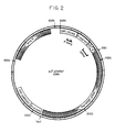
- Figure 2 shows the structure of mammalian COS-7 cell expression vector pJT-2/NRSF.
- the Rat NRSF cDNA insert is the clone 44 cDNA sequence of Figure 5 (SEQ ID NO:4).
- Figure 3 demonstrates stimulation of human neu receptor tyrosine phosphorylation by recombinant NRSF.
- Human MDA-MB-453 breast carcinoma cells were incubated with concentrated conditioned media from COS-7 monkey cells transfected with the indicated partially purified cDNA clones (left panel) or with fully purified cDNA clones (right panel).
- Positive controls comprised 10 ng/ml (left lane) and 100 ng/ml of purified naturally-occurring rat NRSF.
- Negative controls comprised concentrated media conditioned by untransfected COS-7 cells (lane designated “COS”), or by cells transfected with pJT-2 plasmids containing unrelated cDNA (lanes marked as “clone 27" and “clone 29”), and no addition (“NONE”). Lysates were prepared from the stimulated MDA-MB-453 cells and subjected to SDS-PAGE. Shown are autoradiograms of anti-phosphotyrosine Western blots, with the locations of molecular weight marker proteins given in kilodaltons (kDa).
- the following volumes of the COS-7 supernatants were used in a total volume of 0.125 ml PBS containing 0.1% BSA: 0.01 ml (left lane) and 0.1 ml (right lane) for clones 4, 19 and 44.
- the assay of the purified clones was performed in 0.25 ml total volume with 0.2 ml of cell supernatants or as indicated.
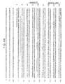
- Figure 4 shows the nucleotide sequence of rat NRSF cDNA and a deduced amino acid sequence. Depicted is the combined nucleotide sequence of four cDNA clones. The beginning of the clone 44 DNA sequence in particular is indicated by an arrow. Nucleotide numbers and amino acid numbers are given in the left and right columns, respectively. Potential sites of N-linked glycoprotein are marked by asterisks, and cysteine residues found in the presumed extracellular domain are encircled. The overlining indicates peptide sequences that were determined directly from purified naturally-occurring NRSF. The underlining indicates the potential transmembrane region, whereas the dashed underlining indicates the polyadenylation sites. The portions of the protein sequence that contain an immunoglobulin (Ig) homology unit and an epidermal growth factor (EGF)-like motif are indicated on the right hand side.
- Ig immunoglobulin
- EGF epidermal growth factor
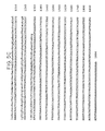
- Figure 5 shows the nucleotide sequence of NRSF cDNA clone 44 separately and a deduced amino acid sequence. Nucleotide numbers are given in the right hand column.
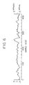
- Figure 6 depicts the hydropathy profile of the precursor of the rat neu receptor stimulating factor.
- Figure 7 shows the alignment of the amino acid sequence of the EGF-like domain and the flanking carboxy terminal sequence of rat NRSF (SEQ ID NO:5) with representative members of the EGF family. Alignment and numbering begin at the most amino terminal cysteine residue of the EGF motifs. Amino acid residues are indicated by the single letter code. Dashes indicate gaps that were introduced for maximal alignment. Residues are boxed to indicate their identity with the corresponding amino acids of NRSF. The underlines indicate the amino terminal portions of the putative transmembrane domains that flank some of the EGF motifs at their carboxy terminal side. Asterisks show the carboxy termini.
- TGF ⁇ rat transforming growth factor ⁇
- AR human amphiregulin
- SFGF sheep fibroma virus growth factor
- MEF myxoma virus growth factor
- mouse EGF (SEQ ID NO:10) (Gray, A. et al., Nature 303 :722-725, 1983; Scott, J. et al., Science 221 :236-240, 1983); human heparin-binding EGF (HB-EGF (SEQ ID NO:11); Higashiyama, S. et al., Science 251 : 936-939, 1991); rat schwannoma-derived growth factor (SDGF (SEQ ID NO:12); Kimura, H. et al., Nature 348 :257-260, 1990); and vaccinia virus growth factor (VGF (SEQ ID NO:13); Blomquist, M.D. et al., Proc. Natl. Acad. Sci. USA 81 :7363-7367, 1984).
- Figure 8 shows the alignment of the immunoglobulin (Ig)-like domain of rat NRSF (SEQ ID NO:14) with the fourth Ig-related sequence of the murine neural cell adhesion molecule (SEQ ID NO:15) (NCAM; Barthels, D. et al., EMBO J. 6 : 907-914, 1987), a representative protein of the C2-set of the immunoglobulin superfamily. Residues that are highly conserved in the C2-set are indicated on the lower line, and positions conserved across the superfamily (according to Williams, A., and Barclay, A., Rev. Immunol. 6 :381-405, 1988) are overlined.
- Stretches of amino acids that may be involved in ⁇ -strand formation are heavily underlined and labeled B through F, in analogy with the immunoglobulin domains. Boxes indicate identical residues in NRSF and NCAM. Dashes (gaps) were introduced for maximal alignment.
- Figure 9 is a schematic presentation of the presumed secondary structure and membrane orientation of the precursor of the neu receptor simulating factor. Positions corresponding to the immunoglobulin (Ig)-domain and epidermal growth factor (EGF) motif are shown by thick lines and their cysteine residues are indicated by circles. The disulfide linkage of the Ig domain was directly demonstrated by amino acid sequence analysis (Example 1D), and the secondary structure of the EGF-domain is based on the homology with the EGF family. Also shown are the three cysteine residues found in the transmembrane domain. Arrows mark the processing sites of the precursor protein at the amino terminus (based on N-terminal sequencing of rat NRSF protein) and a putative proteolytic site close to the plasma membrane. The branched line indicates the location of a proven N-glycosylation site and the short vertical lines represent presumed sites of O-glycosylation.
- Ig immunoglobulin
- EGF epidermal growth factor
- Figure 10 depicts a receptor competition assay using radiolabeled naturally-occurring neu receptor stimulating factor purified from ras-transformed rat fibroblast conditioned media (“125I-NRSF”) .
- the labelled NRSF was incubated with human MDA-MB-453 breast carcinoma cells in the absence ("NONE") or presence of conditioned media from COS-7 cells expressing either recombinant neu receptor stimulating factor ("C-NRSF”) or recombinant TGF ⁇ ("C-TGF ⁇ ").
- Figure 11 depicts another receptor competition assay.
- 125I-labeled naturally-occurring NRSF was incubated with human MDA-MB-453 cells in the absence ("NONE") or presence of unlabeled "cold” naturally-occurring NRSF ("NRSF"; 200 ng/ml), or media conditioned by COS-7 cells that were transfected either with the NRSF expression plasmid of Fig. 2 ("C-NRSF") or with a TGF ⁇ -encoding vector ("C-TGF ⁇ ").
- C-NRSF NRSF expression plasmid of Fig. 2
- TGF ⁇ -encoding vector C-TGF ⁇ "
- Figure 12 shows autoradiograms obtained from Northern blots of mRNA isolated from cultured cells (panels A and C) or freshly isolated tissue of an adult rat (panel B), using clone 44 NRSF cDNA as a probe. The autoradiograms were obtained after exposure to film for 3 hours (panel A) or twenty-four hours (panels B and C). Molecular weights are shown in kilobases. Molecular weight estimation was performed by using a mixture of marker molecules from GIBCO BRL Life Technologies, Inc. (Gaithersburg, MD).
- DNA sequences of this invention are valuable as products useful in effecting the large scale synthesis of NRSF by a variety of recombinant techniques.
- DNA sequences provided by the invention are useful in generating new and useful viral and circular plasmid DNA vectors, new and useful transformed and transfected procaryotic and eucaryotic host cells (including bacterial and yeast cells and mammalian cells grown in culture), and new and useful methods for cultured growth of such host cells capable of the expression of NRSF and its related products.
- DNA sequences of the invention are also suitable materials for use as probes in isolating human cDNA and genomic DNA encoding NRSF and other genes encoding related proteins, including cDNA and genomic DNA sequences of other mammalian species.
- DNA sequences may also be useful in various alternative methods of protein synthesis (e.g., in insect host cells) or in genetic therapy in humans and other mammals.
- DNA sequences of the invention are expected to be useful in developing transgenic mammalian species which may serve as eucaryotic "hosts" for production of NRSF and NRSF products in quantity. See, generally, Palmiter et al., Science 222, 809-814 (1983). Diagnostic applications of NRSF DNA sequences of this invention are also possible, such as for the detection of alterations of genes and of mRNA structure and expression levels.
- the recombinant NRSF of this invention is characterized by being the product of procaryotic or eucaryotic host expression (e.g., by bacterial, yeast, higher plant, insect or mammalian cells in culture) of exogenous DNA sequences obtained by genomic or cDNA cloning or by gene synthesis, particularly with use of the exogenous DNA sequence carried on an autonomously replicating DNA plasmid or viral vector in accordance with conventional techniques.
- procaryotic or eucaryotic host expression e.g., by bacterial, yeast, higher plant, insect or mammalian cells in culture
- exogenous DNA sequences obtained by genomic or cDNA cloning or by gene synthesis, particularly with use of the exogenous DNA sequence carried on an autonomously replicating DNA plasmid or viral vector in accordance with conventional techniques.
- the product of expression in vertebrate [e.g., non-human mammalian (e.g. COS or CHO) and avian] cells is free of association with any human proteins.
- the recombinant NRSF of the invention may be glycosylated with mammalian or other eucaryotic carbohydrates or may be non-glycosylated.
- the recombinant NRSF of the invention may also include an initial methionine amino acid residue (at position -1).
- the present invention also embraces products such as polypeptide analogs of NRSF.
- Such analogs include fragments of NRSF.
- genes encoding related polypeptides having primary structures which differ from that herein specified for in terms of the identity or location of one or more residues (e.g., substitutions, terminal and intermediate additions and deletions).
- modifications of cDNA and genomic nucleotide sequences can be readily accomplished by well-known site-directed mutagenesis techniques and employed to generate analogs and derivatives of NRSF.
- Such products share one or more of the biological properties of naturally-occurring NRSF but may differ in others.
- products of the invention include those which are foreshortened by, e.g., deletions; or those which are more stable to hydrolysis (and, therefore, may have more pronounced or longer-lasting effects than naturally-occurring NRSF); or which have been altered to delete one or more potential sites for O-glycosylation and/or N-glycosylation or which have one or more cysteine residues deleted or replaced by other residues, e.g., alanine or serine residues, and are potentially more easily isolated in active form from microbial systems; or which have one or more tyrosine residues replaced by phenylalanine and bind more or less readily to target proteins or to receptors on target cells.
- polypeptide fragments duplicating only a part of the continuous amino acid sequence or secondary conformations within NRSF which fragments may possess one property of NRSF and not others. It is noteworthy that the described activities are not necessary for any one or more of the products of the invention to have therapeutic utility or utility in other contexts, such as in NRSF antagonism.
- Competitive antagonists may be quite useful in, for example, cases of overproduction of NRSF.
- the present invention also includes that class of polypeptides encoded by portions of the DNA complementary to the protein-coding strand of the human cDNA or genomic DNA sequences of NRSF.
- compositions useful in treating biological disorders associated with neu receptor expression comprising therapeutically effective amounts of polypeptide products of the invention together with suitable diluents, preservatives, solubilizers, emulsifiers, adjuvants and/or carriers useful in recombinant NRSF therapy.
- suitable diluents preservatives, solubilizers, emulsifiers, adjuvants and/or carriers useful in recombinant NRSF therapy.
- a "therapeutically effective amount” as used herein refers to that amount which provides therapeutic effect for a given condition and administration regiment.
- compositions include diluents of various buffer content (e.g., Tris-HCl, acetate, phosphate), pH and ionic strength; additives such as detergents and solubilizing agents (e.g., Tween 80, Polysorbate 80), anti-oxidants (e.g., ascorbic acid, sodium metabisulfite), preservatives (e.g., Thimerosol, benzyl alcohol) and bulking substances (e.g., lactose, mannitol); covalent attachment of polymers to the polypeptide to prolong in vivo half-life and to enhance potency (e.g., water-soluble polymers as polyethylene glycol and copolymers of polyethylene glycol and polypropylene glycol, see Davis et al., U.S.
- Patent No. 4,179,3357 incorporation of the material into particulate preparations of polymeric compounds such as polylactic acid, polyglycolic acid, etc. or into liposomes. Such compositions will influence the physical state, stability, rate of in vivo release, and rate of in vivo clearance of recombinant NRSF.
- the non-naturally-occurring neu receptor stimulating factor of this invention is expected to be useful, alone or in combination with other therapy, in treating diseases and conditions involving cells which express the neu receptor on their surfaces.
- the recombinant NRSFs of this invention are useful as growth inhibitor and differentiation factors for certain human carcinoma cells and will be applicable to the therapeutic treatment of various types of tumors related to neu receptor expression, including breast, ovarian, prostate and gastric carcinomas.
- the factor may also be combined with substances such as radiolabeled molecules, toxins, cytokines, and other compounds useful in tumor treatment, in order to increase localization of these compounds on human tumors expressing high levels of the neu receptor.
- substances such as radiolabeled molecules, toxins, cytokines, and other compounds useful in tumor treatment, in order to increase localization of these compounds on human tumors expressing high levels of the neu receptor.
- an effective amount of recombinant NRSF or analog or derivative is an amount that is effective to alter cellular proliferation and differentiation, or to prevent, lessen the worsening of, alleviate or cure the condition for which the polypeptide is administered.
- the activity of the present polypeptide may be enhanced or supplemented with use of one or more additional biologically active agents which are known to be useful in treating the same condition for which the polypeptide of the invention is being administered, e.g., IL-2 for cancer therapy, platelet-derived growth factors, epidermal growth factor, fibroblast growth factors, and the like for wound healing, and so forth.
- additional biologically active agents which are known to be useful in treating the same condition for which the polypeptide of the invention is being administered, e.g., IL-2 for cancer therapy, platelet-derived growth factors, epidermal growth factor, fibroblast growth factors, and the like for wound healing, and so forth.
- the invention is illustrated in the following examples, which are not intended to be limiting.
- Biological materials employed in these examples were obtained as follows.
- the monoclonal antibody (Ab-3) to the carboxy terminus of the neu receptor was from Oncogene Science (Uniondale, NY).
- a monoclonal antibody to phosphotyrosine, PY20, was obtained from Amersham (Arlington Heights, IL).
- a mouse monoclonal antibody to human casein (types ⁇ and ⁇ ) was a gift from R.C. Coombs (Charing Cross Medical School, London).
- AU-565 human breast carcinoma cells were obtained from the Cell Culture Laboratory, Naval Supply Center (Oakland, CA).
- Ratl-EJ cell line (ATCC CRL 10984) was generated by transfection of the human EJ ras oncogene into Rat1 fibroblasts as described by Peles, E. et al. in Cell 69 :205-216, 1992 and by Land, H. et al., in Nature 304 :596-602, 1983.
- MDA-MB-231 ATCC HTB 26
- MDA-MB-453 ATCC HTB 131
- Hs 294T ATCC HTB 140
- SK-BR-3 ATCC HTB 30
- HT-1080 ATCC CCL 121
- BALB/c 3T3 ATCC CRL 6587
- COS-7 ATCC CRL 1651
- Cells were cultured as recommended by the American Type Culture Collection, or in Dulbecco's modified Eagle medium (GIBCO, Grand Island, NY) supplemented with 10% fetal bovine serum (Hyclone, Logan, Utah).
- the resulting peptide mixture was separated by reverse-phase HPLC and monitored at 215 nm using a Vydac C4 micro column (2.1 mm i.d. x 15 cm, 300 ⁇ ) and an HP 1090 liquid chromatographic system equipped with a diode-array detector and a workstation (Fig. 1).
- the column was equilibrated with 0.1% trifluoroacetic acid (mobile phase A) and elution was affected with a linear gradient from 0-55% mobile phase B (90% acetonitrile in 0.1% trifluoroacetic acid) over seventy minutes.
- the flow rate was 0.2 ml/min and the column temperature was controlled at 25°C.
- the amino acid sequences of the peptides corresponding to the three major eluted fractions were determined by automated Edman degradation. Amino acid sequence analyses of the peptides were performed with a Model 477 protein sequencer (Applied Biosystems, Inc., Foster City, CA) equipped with an on-line phenylthiohydantoinyl (PTH) amino acid analyzer and a Model 900 data analysis system (Hunkapiller, M.W. et al., Methods of Protein Microcharacterization, Humana Press, Clifton, NJ, pages 223-247, 1986). The protein was loaded onto a trifluoroacetic acid-treated glass fiber disc precycled with polybrene and NaCl.
- the PTH-amino acid analysis was performed with a micro liquid chromatography system (Model 120) using dual syringe pumps and reverse-phase (C-18) narrow bore columns (Applied Biosystems, 2.1 mm x 250 mm).
- the first fraction (T13.3) yielded two distinguishable major sequence signals (signal ratio 5:1) with the length of three and four amino acids (the sequences are indicated in Fig. 1).
- the second fraction (T21.8) gave a single sequence, starting at residue number 9 of the previously determined primary N-terminal amino acid sequence (Peles et al., supra; the eighth residue is arginine).
- T21.8 thus confirmed the information obtained from the N-terminal sequence analysis of the whole protein and also assigned an aspartate to position 17, that was previously reported as an unassigned residue.
- the third fraction (T27.3) gave three distinct sequencing signals up to cycle 12 of Edman sequencing, and one clear sequence from cycle 13 through 24, suggesting that this third peptide is even longer.
- the pJT-2 plasmid vector was derived from the V19.8 vector (ATCC 68124).
- V19.8 vector ATCC 68124
- the HindIII and SacII cloning sites of V19.8 were changed into SalI and NotI sites by using synthetic oligonucleotide linkers to yield the PJT-2 vector.
- Plasmids containing cDNA inserts were transformed into DH10B E. coli cells by electroporation (Dower, W.J. et al., Nucleic Acids Res. 16 :6127-6145, 1988).
- the hybridization solution contained 6 x SSC, 50 mM sodium-phosphate (pH 6.8), 0.1% sodium-pyrophosphate, 2 x Denhardt's solution, 50 ⁇ g/ml salmon sperm DNA and 20% formamide (for probe 1) or no formamide (for probe 2). Hybridization was carried out for fourteen hours at 42°C (for probe 1) or 37°C (for probe 2). The filters were washed at either 50°C with 0.5 x SSC/ 0.2% SDS/ 2 mM EDTA (for probe 1) or at 37°C with 2 x SSC/ 0.2% SDS/ 2 mM EDTA (for probe 2). Autoradiography of the filters gave ten clones that hybridized with both probes. These clones were purified by re-plating and probe hybridization as previously described.
- the clones were transiently expressed in COS-7 monkey cells.
- the cDNA clones were inserted into the pJT-2 eukaryotic expression vector under the control of the SV40 promoter and 3'-flanked with SV40 termination and poly-adenylation signals.
- the resulting plasmids including the pJT-2/NRSF plasmid containing clone 44 cDNA (Fig. 2), were used to transfect COS-7 cells by electroporation as follows.
- the resulting conditioned media were then evaluated for their ability to stimulate tyrosine phosphorylation of the neu receptor in MDA-MB-453 human breast tumor cells as follows.
- the conditioned media were filtered through a 0.2-micron sterile filter unit (Costar, Cambridge, MA) and concentrated sixteen-fold by using a Centriprep 10 unit (Amicon, Beverly, MA).
- the concentrated media were diluted in phosphate-buffered saline (PBS) that contained 0.1% bovine serum albumin and were then added to individual wells of a forty eight-well dish that contained 3 x 10⁵ MDA-MB-453 cells per well.
- PBS phosphate-buffered saline
- clone 44 was selected for further analysis by DNA sequencing.
- Clone 44 cDNA was primarily sequenced using a 373A automated DNA sequencer and "Taq DyeDeoxyTM Terminator” cycle sequencing kits from Applied Biosystems, Inc. (Foster City, CA), in accordance with the manufacturer's instructions. Some of the sequencing was performed using 35S-dATP (Amersham) and "SequenaseTM” kits from United States Biochemicals (Cleveland, Ohio), following manufacturer's instructions. Both strands of the cDNA of clone 44 cDNA were sequenced using synthetic oligonucleotides as primers.
- Nucleotide sequence analysis of the cDNA insert of clone 44 yielded a 1894 bp sequence that contained a 436 amino acid long open reading frame that extends to the 5' end of the cDNA. Downstream of the 3' end of this reading frame is a 594 base-long untranslated stretch. The latter includes a poly(A) tail preceded by a polyadenylation signal (AAATAAA). Because no stop codon and recognizable signal peptide were found at the amino terminus of the longest open reading frame, the nucleotide sequences of three other independent positive cDNA clones were analyzed in the same manner. All three clones added to the 5' end a 292 bp sequence that included an in-frame stop codon, suggesting that clone 44 is a 5' truncated cDNA that lacks 0.3 kilobases.
- the combined nucleotide sequence of the cDNA clones is presented in Figure 4 and the clone 44 cDNA sequence is given in Figure 5.
- the combined sequence spans 2,186 base pairs, including a poly(A) tail, and contains an open reading frame of four hundred and twenty-two residues if the amino terminal methionine is considered to be the initiator codon.
- Hydropathy analysis (Fig. 6) revealed no prominent hydrophobic sequence at the amino terminus of the protein that could function as a signal peptide for protein secretion (von Hijne, G., J. Mol . Biol. 184 :99-105, 1985). However, this analysis showed the presence of a highly hydrophobic stretch of twenty-three amino acids (underlined in Fig.
- telomere sequences that were determined directly from the purified protein (overlined in Fig. 4). They all perfectly matched the sequence deduced from the cDNA except for threonine residue 137, which was assigned as isoleucine in peptide T27.3 (Fig. 1). Five different cDNA clones encoded a threonine residue in the corresponding position, indicating that this change may not be due to an isoform of the protein.
- the predicted protein sequence includes four potential sites for N-linked glycosylation, all of which reside amino-terminally to the transmembrane region (indicated by asterisks in Fig. 4).
- a predicted overall structure for the NRSF precursor is presented in Figure 9.
- NRSF precursor is expected to accumulate on the surface of cells expressing high levels of NRSF mRNA.
- Membrane-bound precursor proteins have been found for other members of the EGF growth factor family.
- Brachmann, R., et al. in Cell 56 : 691-700, 1989 demonstrated the presence of transforming growth factor ⁇ on the surface of cells expressing transforming growth factor ⁇ mRNA.
- Molecules that specifically bind NRSF could selectively direct therapeutic molecules to cells overexpressing NRSF.
- monoclonal antibodies raised against recombinant NRSF and conjugated to therapeutic molecules could selectively localize the therapeutic molecule on the surface of cells expressing high levels of membrane-bound NRSF.
- Such cells would include tumor cells with an activated ras gene.
- Antibody conjugates could be constructed using chemotherapeutic compounds, cytokines, toxins, lymphokines or radionuclides.
- the radiolabeled NRSF (10 picomolar) was incubated for sixty minutes at 4°C with monolayers of MDA-MB-453 cells that were grown in a 24-well dish (Costar). This incubation was also performed in the presence of conditioned media from transfected COS-7 cells. Unbound 125I-NRSF was removed by three washes with phosphate buffered saline (PBS), and the cells were solubilized in 0.1 N NaOH solution containing 0.1% SDS. Radioactivity was determined with a ⁇ -counter.
- PBS phosphate buffered saline
- conditioned medium containing recombinant NRSF expressed from the pJT-2/NRSF expression vector (Fig. 2) in COS-7 cells reduced the total 125I-NRSF binding by approximately 50%.
- COS-7 conditioned medium from cells transfected with a pJT-2 expression vector containing a rat TGF ⁇ cDNA (Blasband, A. et al., Mol. Cell Biol. 10: 2111-2121, 1990) was employed.
- the TGF ⁇ conditioned medium did not inhibit 125I-NRSF binding.
- the partial inhibitory effect of the recombinant NRSF conditioned medium may be attributed to its relatively low concentration in the binding assay. In addition, it could be due to high non-specific ligand binding.
- a covalent crosslinking assay was employed. This was performed by allowing MDA-MB-453 cells to bind 125I-NRSF in the presence of transfected COS-7 cell conditioned media, as above.
- the chemical crosslinking reagent bis (sulfosuccinimidyl) suberate (BS3, Pierce) was added at 1 mM final concentration after one hour of binding at 4°C, followed by a brief wash with PBS. Following forty-five minutes of incubation at 22°C, the monolayers were incubated for ten minutes with quenching buffer (100 mM glycine in PBS, pH 7.4).
- the cells were then washed twice with ice-cold PBS, lysed in lysis buffer, and the neu protein was immunoprecipitated with monoclonal antibody Ab-3 according to the protocol described in Peles, E. et al., EMBO J. 10 :2077-2086 (1991).
- the extensively washed immunocomplexes were resolved by gel electrophoresis (6% acrylamide) and autoradiography.
- the results of this analysis indicated that the recombinant NRSF, unlike recombinant TGF ⁇ , was able to displace most of the receptor-bound 125I-NRSF. This result confirmed that the clone 44 cDNA encodes a functional NRSF molecule.
- hybridization was carried out for fourteen hours at 42°C, followed by washing for thirty minutes at 60°C with 0.2 x SSC, 0.1% SDS and 2 mM EDTA.
- the filters were exposed to Kodak XAR x-ray film with an intensifier screen at -70°C for the indicated periods of time.
- Figure 12 shows that three bands were visualized in the Northern blots with poly(A) selected RNA of Rat1-EJ fibroblasts. Their molecular sizes corresponded to 6.8, 2.6 and 1.7 kilobases. After further autoradiography, two additional species of mRNA became visible (not shown).
- the relative level of expression of NRSF in normal Rat1 or 3T3 fibroblasts was significantly lower than in the ras-transformed cells, in agreement with the earlier observation that correlated the neu receptor stimulatory activity with transformation by an oncogenic ras gene (Yarden, Y., and Weinberg, R., Proc. Natl. Acad. Sci. USA 86: 3179-3188, 1989).
- NRSF mRNAs were also detected in brain tissue. In addition to the mRNAs described above, these tissues express a variant mRNA that is 3.4 kb in size.
- the neu receptor which is stimulated by NRSF, is present in human fetal spinal cord and brain (Quirke, P. et al., Br. J. Cancer 60 : 64-69, 1989), which suggests that expression of both NRSF and neu receptor modulates the growth and differentiation of neural tissue.
- Oncogenic activation of the neu receptor gene in rat neuroblastomas (Bargmann, C. et al., Nature 319: 226-230, 1986), further implicates the neu receptor and NRSF in neural tissue growth regulation.
- NRSF expression by virtue of its neu receptor stimulatory activity, may also regulate the growth and differentiation of other cell types.
- the neu receptor is found on the surface of normal differentiated human epithelial cells of the gastrointestinal, respiratory, urinary and reproductive tracts, as well as skin, and is also expressed in corresponding human fetal tissues (Press, M. et al., Oncogene 5 : 953-962, 1990) .
- Figure 12, panel B demonstrates tissue-specific regulation of NRSF mRNA expression levels in adult rat tissues. Positive tissues include lung, ovary and stomach. Relatively low amounts of the middle-size transcript were displayed by the skin, kidney and heart. The liver, spleen and muscle did not contain detectable NRSF mRNA.
- panel B also shows tissue-specific variation in the relative proportion of the different NRSF mRNA species.
- Each variant mRNA could encode variant NRSF proteins with different biological properties.
- Natural NRSF may regulate cell proliferation and differentiation through tissue-specific variations in NRSF mRNA structure and expression levels.
- Administration of the non-natural NRSF of this invention particularly to injured or diseased tissues lacking normal levels of natural NRSF protein, can provide neu receptor stimulation and may modulate cell growth and differentiation in the treated tissue.
- heart, stomach, brain, spinal cord, ovary, lung, kidney and skin tissues express naturally-occurring NRSF and would be expected to respond to treatment with the recombinant NRSF of this invention.
- Abnormal NRSF or neu receptor expression may lead to autocrine interactions that contribute to neoplasia (Browder, T. et al., Cancer Cells 1: 9-17, 1989; Yarden, Y., and Ullrich, A., Ann. Rev. Biochem. 57: 443-448, 1988).
- Overexpression or amplification of the neu receptor gene occurs in human tumors derived from ovary, lung and stomach tissues (Slamon, D. et al., Science 244: 707-712, 1989; Tal, M. et al., Cancer Res. 48: 1517-1520, 1988; Cline, M. et al., Cancer 60: 2669 et seq., 1987).
- FIG. 12 panel B shows that human tumor cells can overexpress NRSF mRNA.
- Figure 12, panel C shows that human tumor cells can overexpress NRSF mRNA.
- An initial survey of human tumor cell lines found that HT-1080 fibrosarcoma cells, Hs 294T melanoma cells and MDA-MB-231 mammary adenocarcinoma cells express elevated NRSF mRNA levels ( Figure 12, panel C).
- Perturbations in the expression of either the neu receptor or its cognate neu receptor stimulatory factor can apparently alter normal cell growth and differentiation, with pathological consequences such as tumor development.
- the recombinant NRSF of this invention may be used to restore a balanced interaction between neu receptor and NRSF in cases where natural NRSF is deficient, or when the neu receptor is overexpressed.
- NRSF Abnormal tumor cell expression of NRSF may result from activated ras oncogenes.
- NRSF mRNA expression increases dramatically in cells transformed with a ras oncogene ( Figure 12, panel A).
- Activated ras genes are found in many carcinoma cell lines and in human tumors from several organs, including colon, lung, gall bladder, urinary bladder, and pancreas carcinomas, as well as melanomas, sarcomas and leukemias (Pimmental, E., Oncogenes , 2nd Edition, Volume II, CRC Press, Boca Raton, FL, 1989).
- Activated ras mutations are also found in premalignant tissues, such as human colorectal adenomas, and are thought to contribute to tumorigenesis (Fearon, E., and Vogelstein, B., Cell 61: 759-767, 1990). Elevated NRSF expression in malignant and premalignant human tissues, particularly those containing activated ras genes, can be found with the NRSF DNA and NRSF mRNA detection methods of this invention. Other methods can also identify elevated NRSF expression in human tissues. These methods, known to those skilled in the art of analyzing protein and mRNA expression, include immunohistochemical assays, radioimmunoassays, enzyme-linked immunoabsorbent assays, and polymerase chain reaction assays. The identification of premalignant and malignant cells overexpressing NRSF is particularly useful for the diagnosis of human cancer.
Landscapes
- Health & Medical Sciences (AREA)
- Chemical & Material Sciences (AREA)
- Life Sciences & Earth Sciences (AREA)
- Organic Chemistry (AREA)
- General Health & Medical Sciences (AREA)
- Medicinal Chemistry (AREA)
- Engineering & Computer Science (AREA)
- Bioinformatics & Cheminformatics (AREA)
- General Chemical & Material Sciences (AREA)
- Nuclear Medicine, Radiotherapy & Molecular Imaging (AREA)
- Chemical Kinetics & Catalysis (AREA)
- Pharmacology & Pharmacy (AREA)
- Animal Behavior & Ethology (AREA)
- Public Health (AREA)
- Veterinary Medicine (AREA)
- Zoology (AREA)
- Genetics & Genomics (AREA)
- Biotechnology (AREA)
- Wood Science & Technology (AREA)
- Biochemistry (AREA)
- Biomedical Technology (AREA)
- Gastroenterology & Hepatology (AREA)
- Molecular Biology (AREA)
- Neurology (AREA)
- Virology (AREA)
- Tropical Medicine & Parasitology (AREA)
- General Engineering & Computer Science (AREA)
- Toxicology (AREA)
- Neurosurgery (AREA)
- Biophysics (AREA)
- Microbiology (AREA)
- Proteomics, Peptides & Aminoacids (AREA)
- Peptides Or Proteins (AREA)
- Preparation Of Compounds By Using Micro-Organisms (AREA)
- Medicines That Contain Protein Lipid Enzymes And Other Medicines (AREA)
- Micro-Organisms Or Cultivation Processes Thereof (AREA)
- Measuring Or Testing Involving Enzymes Or Micro-Organisms (AREA)
- Saccharide Compounds (AREA)
Applications Claiming Priority (2)
| Application Number | Priority Date | Filing Date | Title |
|---|---|---|---|
| US87743192A | 1992-04-29 | 1992-04-29 | |
| US877431 | 1992-04-29 |
Publications (2)
| Publication Number | Publication Date |
|---|---|
| EP0583050A2 true EP0583050A2 (de) | 1994-02-16 |
| EP0583050A3 EP0583050A3 (de) | 1994-10-05 |
Family
ID=25369947
Family Applications (1)
| Application Number | Title | Priority Date | Filing Date |
|---|---|---|---|
| EP9393303308A Withdrawn EP0583050A3 (de) | 1992-04-29 | 1993-04-28 | Rekombinanter, stimulierender Faktor für den "neu"-Rezeptor. |
Country Status (11)
| Country | Link |
|---|---|
| EP (1) | EP0583050A3 (de) |
| JP (1) | JPH07506488A (de) |
| KR (1) | KR950701379A (de) |
| CN (1) | CN1080959A (de) |
| AU (1) | AU4228693A (de) |
| CA (1) | CA2134564A1 (de) |
| FI (1) | FI945085A7 (de) |
| MX (1) | MX9302475A (de) |
| NO (1) | NO944112L (de) |
| WO (1) | WO1993022424A1 (de) |
| ZA (1) | ZA933021B (de) |
Cited By (1)
| Publication number | Priority date | Publication date | Assignee | Title |
|---|---|---|---|---|
| WO1994028133A1 (en) * | 1993-05-21 | 1994-12-08 | Amgen Inc. | Recombinant neu differentiation factors |
Families Citing this family (21)
| Publication number | Priority date | Publication date | Assignee | Title |
|---|---|---|---|---|
| US7285531B1 (en) | 1991-04-10 | 2007-10-23 | Acorda Therapeutics, Inc. | Method for prophylaxis or treatment of a nervous system, pathophysiological condition involving a glial growth factor sensitive cell by administration of a glial growth factor |
| US7135456B1 (en) | 1991-04-10 | 2006-11-14 | Acorda Therapeutics, Inc. | Glial mitogenic factors, their preparation and use |
| US7094749B1 (en) | 1991-04-10 | 2006-08-22 | Acorda Therapeutics, Inc. | Glial mitogenic factors, their preparation and use |
| US7319019B1 (en) | 1991-04-10 | 2008-01-15 | Acorda Therapeutics, Inc. | Glial mitogenic factors lacking an N-terminal signal sequence |
| US5530109A (en) * | 1991-04-10 | 1996-06-25 | Ludwig Institute For Cancer Research | DNA encoding glial mitogenic factors |
| IL101943A0 (en) * | 1991-05-24 | 1992-12-30 | Genentech Inc | Structure,production and use of heregulin |
| US5834229A (en) * | 1991-05-24 | 1998-11-10 | Genentech, Inc. | Nucleic acids vectors and host cells encoding and expressing heregulin 2-α |
| US5367060A (en) * | 1991-05-24 | 1994-11-22 | Genentech, Inc. | Structure, production and use of heregulin |
| US5633161A (en) | 1995-03-29 | 1997-05-27 | Millennium Pharmaceuticals, Inc. | Murine gene fomy030 coding for tumor progression inhibitor |
| US6794185B1 (en) | 1995-03-29 | 2004-09-21 | Millennium Pharmaceuticals, Inc. | fohy030 nucleic acid molecules |
| US6825320B1 (en) | 1995-03-29 | 2004-11-30 | Millenium Pharmaceuticals, Inc. | FOHY03 polypeptides |
| US6251597B1 (en) | 1996-03-29 | 2001-06-26 | Millennium Pharmaceuticals, Inc. | Methods for detecting fohy030 |
| US6312909B1 (en) | 1996-03-29 | 2001-11-06 | Millennium Pharmaceuticals, Inc. | Compositions and methods for the diagnosis prevention and treatment of tumor progression |
| DE69732711T2 (de) | 1996-07-12 | 2006-03-16 | Genentech, Inc., South San Francisco | Gamma-heregulin |
| US6387638B1 (en) * | 1997-02-10 | 2002-05-14 | Genentech, Inc. | Heregulin variants |
| US6136558A (en) * | 1997-02-10 | 2000-10-24 | Genentech, Inc. | Heregulin variants |
| US20010023241A1 (en) | 1998-02-04 | 2001-09-20 | Sliwkowski Mark X. | Use of heregulin as a growth factor |
| JP4314113B2 (ja) * | 2001-07-31 | 2009-08-12 | ウェイン・ステイト・ユニバーシティ | ヘパラン硫酸プロテオグリカンに対する標的化のための、ニューレグリンヘパリン結合ドメインとのハイブリッドタンパク質 |
| SG170021A1 (en) | 2006-02-23 | 2011-04-29 | Novocell Inc | Compositions and methods useful for culturing differentiable cells |
| JP5390624B2 (ja) | 2008-11-04 | 2014-01-15 | バイアサイト インク | 幹細胞集合体懸濁液組成物、その分化方法 |
| AU2013248265B2 (en) | 2012-11-08 | 2018-11-01 | Viacyte, Inc. | Scalable primate pluripotent stem cell aggregate suspension culture and differentiation thereof |
Family Cites Families (2)
| Publication number | Priority date | Publication date | Assignee | Title |
|---|---|---|---|---|
| EP0505148A1 (de) * | 1991-03-17 | 1992-09-23 | Yeda Research And Development Company Limited | Neu-Rezeptor stimulierender Faktor |
| IL101943A0 (en) * | 1991-05-24 | 1992-12-30 | Genentech Inc | Structure,production and use of heregulin |
-
1993
- 1993-04-28 WO PCT/US1993/004128 patent/WO1993022424A1/en not_active Ceased
- 1993-04-28 FI FI945085A patent/FI945085A7/fi not_active Application Discontinuation
- 1993-04-28 EP EP9393303308A patent/EP0583050A3/de not_active Withdrawn
- 1993-04-28 CA CA002134564A patent/CA2134564A1/en not_active Abandoned
- 1993-04-28 MX MX9302475A patent/MX9302475A/es unknown
- 1993-04-28 JP JP5519566A patent/JPH07506488A/ja active Pending
- 1993-04-28 KR KR1019940703860A patent/KR950701379A/ko not_active Withdrawn
- 1993-04-28 AU AU42286/93A patent/AU4228693A/en not_active Abandoned
- 1993-04-29 CN CN93106970A patent/CN1080959A/zh active Pending
- 1993-04-29 ZA ZA933021A patent/ZA933021B/xx unknown
-
1994
- 1994-10-28 NO NO944112A patent/NO944112L/no unknown
Cited By (1)
| Publication number | Priority date | Publication date | Assignee | Title |
|---|---|---|---|---|
| WO1994028133A1 (en) * | 1993-05-21 | 1994-12-08 | Amgen Inc. | Recombinant neu differentiation factors |
Also Published As
| Publication number | Publication date |
|---|---|
| KR950701379A (ko) | 1995-03-23 |
| EP0583050A3 (de) | 1994-10-05 |
| ZA933021B (en) | 1993-11-12 |
| CA2134564A1 (en) | 1993-11-11 |
| MX9302475A (es) | 1994-05-31 |
| NO944112D0 (no) | 1994-10-28 |
| JPH07506488A (ja) | 1995-07-20 |
| FI945085L (fi) | 1994-10-28 |
| NO944112L (no) | 1994-12-29 |
| FI945085A0 (fi) | 1994-10-28 |
| WO1993022424A1 (en) | 1993-11-11 |
| CN1080959A (zh) | 1994-01-19 |
| FI945085A7 (fi) | 1994-10-28 |
| AU4228693A (en) | 1993-11-29 |
Similar Documents
| Publication | Publication Date | Title |
|---|---|---|
| EP0583050A2 (de) | Rekombinanter, stimulierender Faktor für den "neu"-Rezeptor | |
| TOYODA et al. | Distribution of mRNA for human epiregulin, a differentially expressed member of the epidermal growth factor family | |
| Wen et al. | Structural and functional aspects of the multiplicity of Neu differentiation factors | |
| EP2107109B1 (de) | Rekombinanter Vaskularer Endothelzellen Wachstumsfaktor D (VEGF-D) | |
| CA2267651C (en) | Method of stimulating growth of melanocyte precursor cells with stem cell factor polypeptide | |
| EP0423980B1 (de) | Stammzellenfaktor | |
| US7731959B2 (en) | Antagonists of neuropilin receptor function and use thereof | |
| EP0599274A1 (de) | HER4, menschlicher Rezeptor Tyrosine Kinase von der epidermalen Wachstumsfaktor Rezeptor Familie | |
| WO1994028133A1 (en) | Recombinant neu differentiation factors | |
| WO1994028133A9 (en) | Recombinant neu differentiation factors | |
| HUT52545A (en) | Genetic process for production of amphiregulin | |
| IE912135A1 (en) | Mast cell growth factor | |
| Wu | Tumorigenesis of A431 human epidermoid carcinoma cell | |
| AU2003261493B2 (en) | Stem Cell Factor | |
| HK1010397B (en) | Stem cell factor | |
| SK97999A3 (en) | Dna sequence, a polypeptide having hematopoietic biological property, the use thereof and pharmaceutical composition |
Legal Events
| Date | Code | Title | Description |
|---|---|---|---|
| PUAI | Public reference made under article 153(3) epc to a published international application that has entered the european phase |
Free format text: ORIGINAL CODE: 0009012 |
|
| AK | Designated contracting states |
Kind code of ref document: A2 Designated state(s): AT BE CH DE DK ES FR GB GR IE IT LI LU MC NL PT SE |
|
| PUAL | Search report despatched |
Free format text: ORIGINAL CODE: 0009013 |
|
| AK | Designated contracting states |
Kind code of ref document: A3 Designated state(s): AT BE CH DE DK ES FR GB GR IE IT LI LU MC NL PT SE |
|
| 17P | Request for examination filed |
Effective date: 19950424 |
|
| STAA | Information on the status of an ep patent application or granted ep patent |
Free format text: STATUS: THE APPLICATION HAS BEEN WITHDRAWN |
|
| 18W | Application withdrawn |
Withdrawal date: 19960307 |