EP0603735A2 - Anticorps monoclonaux qui déterminent un antigène unique du complèxe de récepteurs antigènes sur les B-cellules humaines - Google Patents
Anticorps monoclonaux qui déterminent un antigène unique du complèxe de récepteurs antigènes sur les B-cellules humaines Download PDFInfo
- Publication number
- EP0603735A2 EP0603735A2 EP93120245A EP93120245A EP0603735A2 EP 0603735 A2 EP0603735 A2 EP 0603735A2 EP 93120245 A EP93120245 A EP 93120245A EP 93120245 A EP93120245 A EP 93120245A EP 0603735 A2 EP0603735 A2 EP 0603735A2
- Authority
- EP
- European Patent Office
- Prior art keywords
- cells
- human
- leukemia
- antigen
- cell
- Prior art date
- Legal status (The legal status is an assumption and is not a legal conclusion. Google has not performed a legal analysis and makes no representation as to the accuracy of the status listed.)
- Withdrawn
Links
Images


Classifications
-
- C—CHEMISTRY; METALLURGY
- C07—ORGANIC CHEMISTRY
- C07K—PEPTIDES
- C07K14/00—Peptides having more than 20 amino acids; Gastrins; Somatostatins; Melanotropins; Derivatives thereof
- C07K14/435—Peptides having more than 20 amino acids; Gastrins; Somatostatins; Melanotropins; Derivatives thereof from animals; from humans
- C07K14/705—Receptors; Cell surface antigens; Cell surface determinants
-
- A—HUMAN NECESSITIES
- A61—MEDICAL OR VETERINARY SCIENCE; HYGIENE
- A61P—SPECIFIC THERAPEUTIC ACTIVITY OF CHEMICAL COMPOUNDS OR MEDICINAL PREPARATIONS
- A61P35/00—Antineoplastic agents
-
- A—HUMAN NECESSITIES
- A61—MEDICAL OR VETERINARY SCIENCE; HYGIENE
- A61P—SPECIFIC THERAPEUTIC ACTIVITY OF CHEMICAL COMPOUNDS OR MEDICINAL PREPARATIONS
- A61P37/00—Drugs for immunological or allergic disorders
-
- C—CHEMISTRY; METALLURGY
- C07—ORGANIC CHEMISTRY
- C07K—PEPTIDES
- C07K16/00—Immunoglobulins [IG], e.g. monoclonal or polyclonal antibodies
- C07K16/18—Immunoglobulins [IG], e.g. monoclonal or polyclonal antibodies against material from animals or humans
- C07K16/28—Immunoglobulins [IG], e.g. monoclonal or polyclonal antibodies against material from animals or humans against receptors, cell surface antigens or cell surface determinants
- C07K16/30—Immunoglobulins [IG], e.g. monoclonal or polyclonal antibodies against material from animals or humans against receptors, cell surface antigens or cell surface determinants from tumour cells
- C07K16/3061—Blood cells
-
- A—HUMAN NECESSITIES
- A61—MEDICAL OR VETERINARY SCIENCE; HYGIENE
- A61K—PREPARATIONS FOR MEDICAL, DENTAL OR TOILETRY PURPOSES
- A61K38/00—Medicinal preparations containing peptides
Definitions
- This invention relates to three novel monoclonal antibodies (mAbs) termed SN8, SN8a and SN8b which are directed toward individually different epitopes of the same antigen molecule, which is primarily expressed on B-cell type B prolymphocytic leukemia (PLL) cells, B non-Hodgkins' lymphoma (NHL) cells, B acute lymphoblastic leukemia (ALL), and to methods of using the monoclonal antibodies, in whole or in part, for the diagnosis and therapy of various human leukemia-lymphomas (HLL), especially PLL and NHL.
- PLL B-cell type B prolymphocytic leukemia
- NHL B non-Hodgkins' lymphoma
- ALL B acute lymphoblastic leukemia
- the monoclonal antibodies have been deposited with the American Type Culture Collection ("ATCC")ö 12 301 Parklawn Drive Rockville, Maryland 20852, USA on July 20, 1993 and have been given the following ATCC Designations: Antibody SN8 (Hybridoma Cell Line 3A2-2E7) HB 11413 Antibody SN8a (Hydridoma Cell LIne 3B3-1D2) HB 11411 Antibody SN8b (Hybridoma Cell Line Q6-1D5) HB 11412
- Prolymphocytic leukemia was initially reported in 1974 by Galton et al. As a variant of chronic lymphocytic leukemia (CLL), but with distinct clinical and laboratory features ("Prolymphocytic Leukemia", British J. Haematol , 27; 7, 1974). The distinction between PLL and CLL is clinically important because PLL is a relentlessly progressive disease with a worse prognosis and usually resistant to the chemotherapy that is often effective in CLL (Catovsky, "Prolymphocytic and hairy cell leukemias", in Henderson ES, Lister TA (eds): Leukemia , Philadelphia, PA, Saunders, 1990, p. 639).
- B PLL hairy cell leukemia
- SN8 was capable of effectively distinguishing B PLL from B CLL as well as from hairy cell leukemia (HCL) cell specimens.
- SN8a and SN8b also showed a selective reactivity with B PLL and B NHL samples.
- these mAbs reacted with higher percentages of CLL samples compared to SN8.
- the degree of PLL selectivity of these mAbs is less than that of SN8.
- SN8 antigen is a novel heterodimer antigen which constitutes the human B cell antigen receptor complex together with cell membrane immunoglobulin.
- Antigen receptors on B lymphocytes play a central role in the immune regulation in animals and humans.
- the mb-1 and B29 products are termed Ig- ⁇ and Ig- ⁇ , respectively.
- Ig- ⁇ A variant form of the Ig- ⁇ was called Ig- ⁇ .
- the disulfide-linked heterodimer ⁇ - ⁇ molecules on B cells are noncovalently associated with cell membrane immunoglobulins (mIgs).
- mIgs cell membrane immunoglobulins
- the present invention discloses the generation and characterization of three mAbs termed SN8, SN8a and sN8b that were generated by immunizing two mice with an isolated PLL antigen preparation. These mAbs, particularly SN8, showed a highly selective reactivity with B PLL and B NHL.
- the surface antigen defined by the SN8 series mAbs was identified as a covalently-linked heterodimer (gp49/40). Comparison of SN8 series mAbs and SN8 antigen with the reported mAbs and their antigens suggests that SN8 series mAbs define a novel B-cell antigen.
- Nakamura et al. used the phrase " putative human equivalent of the mouse B29 protein" because they had neither characterized the antigen chemically nor determined the antigen's amino acid sequence. Also, it appears that no amino acid sequence of the human B29 or mb-1 proteins has been reported prior to the present invention.
- the present mAbs show a highly selective reactivity as well as high binding avidity (SN8 and SN8b) and are of IgG1 isotype, they may be useful for the diagnosis of B leukemia/lymphoma, for studying the pathogenesis of PLL and perhaps for studying normal human B cell differentiation.
- ricin A-chain (RA) conjugates of the three mAbs are all effective for the specific killing of SN8 antigenexpressing cells. Furthermore, binding of these mAbs to the cell surface antigen induced no significant (SN8 and SN8b) or only a small (SN8a) down-regulation of antigen expression. The results suggest the potential of these mAbs as a specific delivery vehicle of cytotoxic agents to the SN8 antigen-expressing target cells.
- the B cell antigen receptor complex plays a central role in the immune regulation in the human. Therefore, SN8 series mAbs will have good potential for the diagnosis and therapy of human diseases involving immunological functions as well as for the immunological manipulation of human B cells, e.g., inducing immune unresponsiveness.
- the invention comprises leukemia lymphoma reactive monoclonal antibodies directed toward individually different epitopes of the same antigen molecule which, relative to normal peripheral blood cells, strongly reacts with one or more leukemia lymphoma cell specimens from the group consisting of B prolymphocytic leukemia cells, B non-Hodgkin's lymphoma cells, B chronic lymphocytic leukemia cells, B hairy cell leukemia cells, and B acute lymphoblastic leukemia cells.
- mAbs three new IgG1- ⁇ monoclonal antibodies (mAbs), termed SN8, SN8a and SN8b, were generated by the use of an unconventional approach, i.e., utilizing an isolated B PLL antigen preparation to immunize mice.
- mAbs, particularly SN8 showed a highly selective reactivity to B PLL and B non-Hodgkin's lymphoma among various human leukemia-lymphoma specimens tested, e.g., SN8 was capable of effectively distinguishing B PLL from B CLL as well as from HCL cell specimens.
- the invention further comprises monoclonal antibodies which are directly or indirectly attached or complexed with a compound having a site suitable for attachment or complexing therewith which compound is selected from the group consisting of drugs, toxins or fragments thereof, growth suppressing biological response modifiers, enzymes, liposomes, radioactive agents and antibodies.
- the invention also includes the methods for using the monoclonal antibodies for detecting and treating leukemia/lymphoma disease, as well as methods for preparation of the monoclonal antibodies and kits containing the same are also within the scope of the invention.
- the invention also relates to the isolation and chemical identification of the human homologues of the murine mb-1 and B29 proteins of B cell antigen receptor complex.
- the amino-terminal amino acid sequences of the mb-1 and B29 gene products were determined using the antigen which was isolated using the monoclonal antibody SN8.
- Figure 1 Reactivity of SN8 with fresh (uncultured) HLL cells as determined by a cellular radioimmunoassay (RIA).
- RIA radioimmunoassay
- Pre-B ALL was included in the group of non-T/non-B ALL in the figure.
- Abbreviations used in the figure CLL, chronic lymphocytic leukemia; PLL, prolymphocytic leukemia; HCL, hairy cell leukemia; NHL, non-Hodgkin's lymphoma; ALL, acute lymphoblastic leukemia; AML, acute myelocytic leukemia; AMOL, acute monocytic leukemia; AMMOL, acute myelo-monocytic leukemia; CML, chronic myelocytic leukemia.
- the types of NHL specimens used are as follows: 2 diffuse small cleaved cell (SN8+, SN8a+, SN8b+; SN8+), 1 diffuse large cell (SN8+, SN8a+, SN8b+), 2 diffuse mixed cell (SN8+, SN8a+, SN8b+; SN8+), 3 follicular small cleaved cell (SN8+, SN8a+, SN8b+; SN8+; SN8 ⁇ ), 3 small lymphocytic (SN8 ⁇ , SN8a ⁇ , SN8b ⁇ ; SN8+; SN8a+; SN8 ⁇ , SN8a ⁇ ) and 1 follicular mixed cell (SN8+) lymphoma.
- Figure 2 FACS analysis of SN8 reactivity with various human cells.
- Target cells were allowed to react with SN8 or an isotype-matching control mouse IgG (MOPC 195variant; IgG1- ⁇ ) and stained with fluorescence-conjugated F(ab')2 of sheep anti-mouse Ig antibodies.
- the three bone marrow specimens shown were obtained from three different ALL patients in remission and mononuclear cells were isolated for use in the test.
- the three B cell specimens were individually isolated from the peripheral blood of three healthy donors.
- the B PLL and B CLL specimens were uncultured cell specimens.
- the fluorescence intensity is on a log scale.
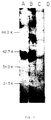
- FIG. 3 SDS-PAGE of immunoprecipitates from a 125I-labeled PLL antigen preparation.
- Applicants used SN8 (lanes A and E), SN8a (lane B and F), SN8b (lanes C and G) and control IgG (MOPC 195variant; lanes D and H).
- Samples were analyzed after being unreduced (lanes A, B, C and D) or reduced with dithiothreitol (DTT) (lanes E, F, G, and H).
- BioRad M r marker proteins ⁇ the heavy chain of human IgG were used after reduction as references.
- FIG 4 Western blot analysis of the epitopes defined by SN8 series mAbs.
- a leukemia antigen preparation was reduced with 50 mM dithiothreitol and subjected to SDS-PAGE.
- the separated proteins in the gels were transferred to nitrocellulose membranes. After blocking with normal goat serum, the membranes were incubated with SN8 (lane A), SN8a (lane B), SN8b (lane C) or control IgG (lane D).
- the membranes were further treated with 125I-F(ab')2 of affinity purified goat anti-mouse IgG antibodies.
- the marker proteins (shown in K daltons) are described in the legend to Figure 3 except for soybean trypsin inhibitor (21.5 K).
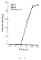
- FIG. 5 Competitive binding between mAbs to SN8 antigen on BALL-1 cells as measured by a cellular RIA.
- BALL-1 cells were incubated with serial dilutions of individual SN8 series mAbs or an isotype-matching control IgG for 1 h at 4°C.
- 125I-SN8 was then added, and the incubation was continued for an additional 1 h.
- Abscissa is ng of the purified SN8 series mAbs and control IgG.
- Radioactivity of 125I-SN8 bound to BALL-1 was in the range between 5.7 and 6.1 x 10³ cpm when BALL-1 was preincubated with 102 to 103 ng of the control IgG or SN8b. The radioactivity was 331 and 190 cpm, respectively, when BALL-1 was preincubated with 103 ng of SN8 or SN8a.
- FIG. 7 Cytotoxic activities of RA conjugates of SN8 series mAbs as measured by a protein synthesis inhibition assay.
- BALL-1 panel A
- gp49/40-negative MOLT-4 control; panel B
- the cells were pulsed with 3H-leucine and incubated for 4 h.
- the pulsed cells were harvested on glass fiber filters using a multiple semiautomatic cell harvester (type 7010; Skatron Inc., Sterling, VA) and the 3H radioactivity was determined in a liquid scintillation spectrometer. Protein synthesis in the conjugate-treated cells is expressed as the percent of [3H]leucine incorporated into control cells not exposed to conjugate.
- SN8-RA, SN8a-RA, SN8b-RA and control conjugate are indicated by circles, triangles, squares and X, respectively, in the figure.
- FIG. 8 Western blot analysis of the SN8 antigen purified from BALL-1 cells by immunoaffinity chromatography. Proteins recognized by SN8 were visualized on Kodak X-OMAT AR film using enhanced chemiluminescence (Amersham). The SN8 column eluate was examined on 8% SDS-polyacrylamide gels under nonreduced conditions (A) or after reduction with 0.1M DTT (B) (see Materials and Methods for details). Arrows indicate SN8 antigen specific bands. BioRad M r marker proteins were used as references and indicated in kD.
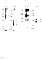
- FIG. 9 Radioimmunoprecipitation of the SN8 antigen from a 125I-labeled antigen preparation from BALL-1 (panel A) and a 125I-labeled B PLL antigen preparation (panel B).
- An isotype-matched murine IgG (IgG1- ⁇ ) was used as a control against SN8 (lanes 2 and 4 in panels A and B).
- the immunoprecipitates were unreduced (lanes 1 and 2 in panels A and B) or reduced with DTT (lanes 3 and 4 in both panels) before they were subjected to SDS-PAGE. Arrows depict specifically immunoprecipitated proteins which were visualized by autoradiography. M r marker proteins (BioRad) are indicated in kD.
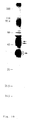
- FIG. 10 Coomassie Blue visualization of the immunoaffinity purified SN8 antigen.
- the SN8 antigen was purified from detergent lysate of BALL-1 cells by repeated immunoaffinity chromatography. The sample was concentrated, reduced with DTT, subjected to SDS-PAGE, and transferred to a PVDF membrane by electroblotting (see Materials and Methods for details). The transferred proteins were visualized by staining with Coomassie Blue R-250.
- the arrows indicate the protein bands containing the human mb-1 protein and B29 protein, respectively, as revealed by amino-terminal amino acid sequence analyses.
- the diffuse 37kD B29 protein band was divided into two portions as indicated in the figure and separately analyzed by amino acid sequencing. An identical amino-terminal amino acid sequence was obtained from the two samples by the sequence analyses.
- Figure 11 Amino-terminal amino acid sequences of the human B29 and mb-1 proteins.
- the amino-terminal sequences of the mouse B29 and mb-1 proteins are shown after alignment to the corresponding human homologues to maximize the homology.
- Identical amino acid residues between the corresponding human and mouse proteins are underlined. Shifts introduced in the sequence to maximize alignment are designated by a dash.
- mAbs Three new IgG1- ⁇ monoclonal antibodies (mAbs), termed SN8, SN8a and SN8b, were generated by the use of an unconventional approach, i.e., utilizing an isolated B PLL antigen preparation to immunize mice.
- HCL hairy cell leukemia
- the cell surface antigen defined by the three mAbs was determined to be a covalently-linked heterodimeric glycoprotein complex (gp49/40) consisting of a 49,000 dalton component ( ⁇ chain) and a 40,000 dalton component ( ⁇ chain) and was subsequently identified as the human homologues of the murine mb-1 and B29 gene products.
- Epitope comparison showed that the epitope defined by SN8 (SN8 epitope) is in close proximity to SN8a epitope but in a distant position from SN8b epitope.
- B B-cell
- HLL human leukemia-lymphoma
- PLL prolymphocytic leukemia
- NHL non-Hodgkin's lymphoma
- CLL chronic lymphocytic leukemia
- HCL hairy cell leukemia
- ALL acute lymphoblastic leukemia
- mAb monoclonal antibody
- RIA radioimmunoassay
- SDS-PAGE sodium dodecyl sulfate polyacrylamide gel electrophoresis
- RA ricin A chain
- FACS fluorescence activated cell sorter
- FITC fluorescein isothiocyanate
- LcH Lens culinaris lectin
- RCA Ricinis communis lectin
- Ig immunoglobulin
- WBC white blood cell
- PBL peripheral blood lymphocytes
- FBS fetal bovine serum
- HLA-DR human major histocompatibility complex
- Mononuclear and blast cells were isolated from the cell suspensions of these specimens by centrifugation on a Ficoll-Paque gradient. Normal (or nearly normal) bone marrow specimens were from patients who were in remission and had a morphologically normal bone marrow. Mononuclear cells were isolated from the bone marrow aspirates by Ficoll-Paque gradient centrifugation.
- B cells, T cells, granulocytes, monocytes, erythrocytes and platelets of normal peripheral blood were isolated from buffy coat preparations of healthy volunteers (Haruta et al. "Distinct human leukemia-associated cell surface glycoprotein GP160 defined by monoclonal antibody SN6", Proc. Natl. Acad. Sci., USA , 83: 7898, 1986).
- L-[3H]leucine and leucine-free medium were purchased from ICN Biomedicals, Inc. (Irvine, CA) and GIBCO Laboratories (Grand Island, NY) respectively. Ricin A chain (RA) was obtained from Inland Laboratories (Austin, TX).
- mAbs SN3 Fukukawa et al. "New monoclonal antibodies SN3, SN3a and SN3b directed to sialic acid of glycoprotein on human non-T leukemia cells" Exp Hematol 14: 850, 1986) (anti-CD24), SN4 (Luo et al.
- mAbs directed toward Leu1(CD5), Leu4(CD3), Leu12(CD19), Leu16(CD20) and HLA-DR were purchased from Becton Dickinson (Mountain View, CA).
- Anti-human immunoglobulin ⁇ chain mAb was obtained from AMAC, Inc. (Westbrook, ME).
- Fluorescein isothiocyanate (FITC)-labeled F(ab')2 fragments of goat anti-human immunoglobulin (Ig) chain (each of ⁇ , ⁇ , ⁇ , ⁇ and ⁇ chains) antibodies were purchased from TAGO, Inc. (Burlingame, CA).
- Antigen Preparation from Leukemia Cell Membranes. Antigen was prepared from the cell membranes of leukemia cells derived from a patient with B PLL.
- the cell surface phenotype of the B PLL cells was Ig ⁇ +, Ig ⁇ , Ig ⁇ +, Ig ⁇ , Ig ⁇ , CD3 ⁇ , CD5 ⁇ , CD9+, CD20+, CD24+, GP160 ⁇ and HLA-DR+.
- the LcH-bound and RCA-bound glycoconjugates were individually eluted, combined and subjected to passive immunoaffinity chromatography. To this end, the combined glycoproteins were passed through three serially connected immunoadsorbent columns. These immunoadsorbents consisted of anti-HLA class I mAb (B3-3D1), anti-HLA-DR mAb (G4-3A7), and rabbit anti-normal human peripheral blood lymphocyte antibodies, all coupled to Sepharose CL-4B. Materials in the pass-through fractions were pooled and concentrated.
- Monoclonal antibody was generated by immunizing two BALB/c mice with the isolated antigen preparation. Immunization of the mice was carried out as described previously (Seon et al., Proc. Natl. Acad. Sci. USA , 80: 845, 1983). Cell fusion, hybridoma screening, cloning and mAb class determination was then performed.
- RIA Cellular Radioimmunoassay
- FACS Analysis Details of the cellular RIA which was used for determining reactivity of mAbs with various cultured and uncultured cells were described previously (Seon et al. "Monoclonal antibody SN2 defining a human T-cell leukemia-associated cell surface glycoprotein", J. Immunol ., 132: 2089, 1984). It should be noted that Fc receptors on the target cells are blocked with human IgG during the assay. In selected cases, the reactivity of mAbs with various cell specimens was also determined by FACS analysis.
- Radioimmunoprecipitation and Sodium Dodecyl Sulfate Polyacrylamide Gel Electrophoresis SDS-PAGE.
- the PLL antigen preparation (see above) and a LcH-bound glycoprotein preparation of PLL cells were separately radiolabeled with 125I using an Iodo-gen coated Minisorp tube.
- the two radiolabeled preparations were used separately for immunoprecipitation and SDS-PAGE.
- An autoradiograph was prepared by using Kodak X-OMAT AR film and X-Omatic intensifying screen (Matsusaki et al. "Molecular nature of a cell membrane antigen specific for human T-cell acute lymphoblastic leukemia", Cancer Res ., 47: 4283, 1987).
- Tris-HC1 buffer pH 7.4
- Tris-saline-BSA 0.9% NaC1 and 5% bovine serum albumin
- Tris-saline-BSA Tris-saline-BSA containing an isotype-matching control murine IgG
- the membranes were washed with 10 mM Tris buffer (pH 7.4) containing 0.9% NaC1 and 0.05% NP-40 (Tris-saline-NP40), treated with 125I-labeled F(ab')2 of affinity-purified goat anti-mouse IgG antibodies, and washed with Tris-saline-NP40.
- the membranes were dried and autoradiograph was prepared as described above.
- mAbs were generated by immunizing two mice with an isolated leukemia antigen preparation (see Materials and Methods ). Initial characterization of primary hybridoma cultures and cloned hybridomas was carried out by testing against normal human peripheral blood lymphocytes (PBL), PLL cells and selected human cell lines by means of a cellular RIA. Hybridoma clones 3A2-2E7 and 3B3-1D2 derived from mouse 1 and Q6-1D5 from mouse 2 produced IgG1- ⁇ mAbs which showed a selective reactivity with PLL cells and some B HLL cell lines. These mAbs were designated SN8, SN8a and SN8b, respectively.
- Reactivity with Fresh (uncultured) HLL Cells Reactivity of SN8 series mAbs with uncultured HLL cell specimens was determined by a cellular RIA and, in selected cases, also by FACS analysis.
- SN8 showed significant reactivity with all of the 8 B PLL, 9 of the 12 B NHL, the one B ALL, and one of the 5 hairy cell leukemia specimens tested. SN8 did not show significant reactivity with any of the specimens of non-T/non-B ALL, T ALL and myeloid/monocytic leukemias. Among the 23 B CLL specimens tested, only 3 showed weak but significant reactivity with SN8.
- SN8 shows a selective reactivity with PLL compared to CLL and hairy cell leukemia which are both closely related to PLL in the differentiation pathway of B cell ontogeny.
- the results of a cellular RIA were supported by FACS analysis, some of which are presented in Fig. 2 where over 90% of a PLL cell specimen reacted with SN8 whereas virtually none of the cells of the two CLL specimens were reactive.
- SN8a and SN8b showed a selective reactivity with B PLL and B NHL samples as SN8 did. However, these mAbs reacted with higher percentages (i.e., 30.0 and 31.3%, respectively) of CLL samples compared to SN8 (13.6%). Thus, the degree of PLL selectivity of these mAbs is less than that of SN8.
- B cells, T cells, monocytes, granulocytes, erythrocytes and platelets were isolated from the peripheral blood of three healthy donors and tested for reactivity with SN8 series mAbs by a cellular RIA. These mAbs showed a moderate reactivity with B cell specimens but no significant reactivity with other cell specimens. Therefore, the reactivities of SN8 series mAbs with B cells were further tested by FAGS analysis.
- the FACS analysis results of SN8 are shown in Fig. 2. A subpopulation (6.0, 17.4 and 24.2%, respectively) of the B cell preparations derived from the three different donors reacted with SN8 (Fig. 2 D, E and F). Two (Nos.
- SN8 series mAbs were tested for their reactivities with normal (or nearly normal) bone marrow specimens by a cellular RIA and FACS analysis; these bone marrow specimens were obtained from 4 different ALL patients in remission.
- a leukemia antigen preparation from cell membrane glycoproteins of PLL cells was labeled with 125I and used for immunoprecipitation with SN8 series mAbs and an isotype-matching control murine IgG (MOPC 195variant).
- each of the SN8, SN8a and SN8b immunoprecipitates showed a single component of approximately 81,000 daltons (lanes A, B and C), whereas no significant component was immunoprecipitated by the control IgG (lane D).
- each mAb immunoprecipitate showed two components of approximately 49,000 ( ⁇ chain) and 40,000 ( ⁇ chain) daltons (lanes E, F and G), whereas the control IgG did not precipitate any significant component (lane H). Therefore, the antigen defined by the SN8 series mAbs consists of two polypeptide chains covalently linked by disulfide bond(s).
- a competitive binding assay was carried out to compare the epitopes defined by SN8 series mAbs (Fig. 5). Preincubation of the SN8 antigen-expressing BALL-1 cells (see Table 1) with SN8 or SN8a completely blocked the subsequent binding of 125I-SN8 at the maximum. However, preincubation with SN8b or an isotype-matching control murine IgG did not inhibit the 125I-SN8 binding at all. These results indicate that the epitopes defined by SN8 and SN8a are in close proximity to each other but distant from the epitope defined by SN8b. These results are consistent with the above finding that both SN8 and SN8a bound to the same component ( ⁇ chain) in the Western immunoblotting.
- Circulating Antigen in the Plasma of HLL Patients may bind an administered mAb and thereby inhibit the therapeutic efficacy of the administered mAb and immunoconjugate. Therefore, Applicant tested for circulating SN8 antigen in the plasma of HLL patients and healthy individuals (control) by using mAbs SN8, SN8a and SN8b and a solid phase RIA. No significant amount of SN8 antigen was detected in any of the plasma samples derived from 7 different B NHL patients and 5 different B CLL patients. Similarly, no significant SN8 antigen was detected in the plasma samples derived from 5 different healthy individuals.
- Binding of antibody to a cell surface antigen may induce antigenic modulation (Ritz et al. "Modulation of human acute lymphoblastic leukemia antigen induced by monoclonal antibody in vitro" J. Immunol ., 125: 1506, 1980) and down-regulation of antigen expression which may make antigentargeting by antibody difficult.
- Applicant tested the effect of binding of SN8, SN8a and SN8b to BALL-1 on the expression of the SN8 antigen.
- IC50 50% inhibitory concentrations (IC50) of SN8a-RA and SN8b-RA against BALL-1 were 2.6 x 10 ⁇ 9 and 3.0 X 10 ⁇ 9 M, respectively (solid lines in Fig. 7A).
- IC50 50% inhibitory concentrations
- the three RA conjugates did not inhibit the protein synthesis of control MOLT-4 cells at any of the concentrations tested (Fig. 7B).
- NH4C1 (10 mM) enhanced the cytotoxic activities of immunotoxins against BALL-1 but not against MOLT-4.
- IC50 of SN8-RA, SN8a-RA and SN8b-RA against BALL-1 were 1.0 x 10 ⁇ 10, 1.2 x 10 ⁇ 10 and 4.8 x 10 ⁇ 10 M, respectively, in the presence of 10 mM NH4C1 (broken lines in Fig. 7A).
- the control RA conjugate did not show significant cytotoxicity against either BALL-1 or MOLT-4 in the absence or in the presence of NH4C1.
- RA conjugates of SN8, SN8a and SN8b were tested for cytotoxicity against Daudi, a lymphoma cell line (see Table 1).
- IC50 of SN8-RA, SN8a-RA and SN8b-RA were 8.2 x 10 ⁇ 10, 6.7 x 10 ⁇ 10 and 2.4 x 10 ⁇ 9 M, respectively.
- cytotoxic activities of these conjugates against Daudi were potentiated by NH4C1; IC50 in the presence of 10 mM NH4C1 were found to be 3.1 x 10 ⁇ 12 4.9 x 10 ⁇ 12 and 1.3 x 10 ⁇ 11 M, respectively.
- the epitope defined by new mAb SN8 is detected on malignant B cells with a restricted stage of maturation, as well as on a small population of normal blood cells in the peripheral blood of healthy individuals.
- SN8 reacted with all of the 8 B PLL specimens and 9 of the 12 B NHL specimens tested but not with most of B CLL, HCL and non-T/non-B ALL (including pre-B ALL) specimens tested (Fig. 1).
- SN8 reacted with the one uncultured B ALL specimen tested as well as with one of the two B ALL cell lines tested (Fig. 1 and Table 1). It appears that SN8 is capable of effectively discriminating B PLL from B CLL and HCL as well as from non-T/non-B ALL.
- mAbs defining FMC-7 (Zola, "The surface antigens of human B lymphocytes", Immunol. Today , 8:308, 1987) and CD22 (Knapp et al. (eds): Leucocyte Typing IV: White Cell Differentiation Antigens, Oxford, England, Oxford University Press, 1989) appear to be able to discriminate B PLL from some cases of B CLL but not from HCL; it should be noted that molecular properties of the antigens defined by these mAbs are different from the antigen defined by SN8.
- B CLL, B PLL and HCL are closely related in the differentiation pathway of B cell ontogeny while non-T/non-B ALL derives from normal counterparts at earlier stages of B cell ontogeny by malignant transformation and clonal expansion.
- B ALL is closely related to B lymphomas (Magrath et al. "Bone marrow involvement in Burkitt's lymphoma and its relationship to acute B-cell leukemia” Leukemia Res ., 4: 33, 1979) and its phenotype corresponds to that of relatively mature B cells.
- B NHL consists of a heterogeneous group of malignant B cells with varying degrees of maturation but the normal counterparts in the majority of cases of B NHL appear to be relatively mature B cells (Jaffe, "The role of immunophenotypic markers in the classification of non-Hodgkin's lymphomas" Seminars Oncol ., 17: 11, 1990). Phenotypic and genetic analyses suggest that B PLL derives from normal counterparts by malignant transformation at a later developmental stage than B CLL. For instance, B PLL cells express higher density of cell surface Ig than B CLL cells. Furthermore, Luzzatto et al.
- SN8 reacted well with B PLL but poorly with HCL, B CLL and non-T/non-B ALL (Fig. 1).
- the results indicate that SN8 defines an epitope which is associated with a relatively narrow range of B cell maturation.
- the epitopes defined by SN8a and SN8b appear to be associated with slightly wider ranges of B cell maturation than SN8 epitope.
- Applicant would like to point out that the present mAbs were generated using an unconventional approach, i.e., by immunizing animals with an HLL antigen preparation rather than with intact HLL cells.
- Applicant developed a novel system for isolating immunologically active HLL associated cell membrane antigen mixtures (Seon et al. "Human T cell leukemia antigens on the cell membranes: Purification, molecular characterization, and preparation of specific antisers", J. Immunol , 127: 2580, 1981).
- this system was applied to isolating a B PLL associated cell membrane antigen preparation which was used for generating mAbs.
- SN8 series mAbs appear to be different from those previously reported mAbs in the antibody specificity and/or in the molecular nature of the antigen defined.
- the SN8 antigen defined by the present mAbs appears to be different from any of the reported CD series antigens.
- CD72 shows some similarity to the SN8 antigen; CD72 is a heterodimer of 43,000 and 39,000 dalton components.
- CD72 is a heterodimer of 43,000 and 39,000 dalton components.
- the smaller molecular size of the heavier component of CD72 compared to the ⁇ chain (gp49) of SN8 antigen there are distinct differences in the specificity between anti-CD72 mAb and SN8 series mAbs.
- anti-CD72 mAb reacted with 44% (7 of the 16 specimens tested) of non-T/non-B ALL specimens, all (3/3) of the HCL specimens, and NALM-6, a pre-B ALL cell line.
- SN8 series mAbs did not react with any of the non-T/non-B ALL specimens tested (i.e., 0/13, 0/7 and 0/4, respectively, for SN8, SN8a and SN8b), did not react with the majority of the HCL specimens (1/5, 1/4 and 0/3, respectively), and did not react with NALM-6 (see Fig. 1 and Table 1).
- CD22 is a single polypeptide chain antigen with a molecular weight of 135,000.
- the data presented here suggests the usefulness of SN8 series mAbs, particularly SN8, for diagnosis of HLL and follow-up of B PLL and B NHL.
- the data also suggests the usefulness of these mAbs for diagnosis and follow-up of normal B cells in patients with immunological disorders.
- Another important application of these mAbs may be their utilization as a specific delivery vehicle of a cytotoxic agent(s) to malignant and normal B cell targets (Vitetta et al. "Redesigning nature's poison to create anti-tumor reagents" Science , 238:1098, 1987).
- these mAbs were conjugated with ricin A chain (RA), and the in vitro cytotoxic activities of the generated immunotoxins were determined. All of the three immunotoxins killed the SN8 antigen-expressing HLL cells effectively while showing no significant cytotoxicity against control cells. Thus, these immunotoxins showed specific cytotoxicity and each of SN8 series mAbs bound to the target antigen on HLL cells was effectively internalized into the cells. However, the binding of these mAbs to HLL cells did not cause (SN8 and SN8b) or caused only a small degree (SN8a) of down-regulation of antigen expression.
- RA ricin A chain
- genetically engineered chimeric antibodies or fragments thereof of SN8, Sn8a and SN8b can be directly or indirectly attached or complexed with a compound having a site suitable for attachment or complexing therewith which compound is selected from the group consisting of drugs, toxins or fragments thereof, growth suppressing biological response modifiers, enzymes, liposomes, radioactive agents, photodynamic agents and antibodies (BioTechniques, Vol. 4, No. 3:214-220, 1986).
- Binding of antigen to the antigen receptor on B cells initiates complicated cascades of signal transduction which may lead to proliferation, differentiation or programmed cell death of the B cells. Furthermore, cross-linking of the antigen receptor by anti-Ig antibodies results in rapid tyrosine phosphorylation of substrate proteins suggesting that the B cell antigen receptor belongs to a subgroup of the tyrosine kinase receptor family (Gold et al. "Stimulation of protein tyrosine phosphorylation by the B-lymphocyte antigen receptor", Nature , 345:810, 1990).
- mIgs cell membrane immunoglobulins
- B lymphocyte antigen receptors are non-covalently associated with a disulfide-linked, inducibly phosphorylated glycoprotein complex
- EMBO J . 9:441, 1990 on a murine myeloma variant and murine normal B cells revealed that the mIgM is non-covalently associated with a disulfide-linked heterodimer consisting of a 32-34 kD and a 37-39 kD subunit.
- murine Ig- ⁇ and Ig- ⁇ were identified as the products of mb-1 and B29 genes, respectively (Sakaguchi et al. "B lymphocyte lineage restricted expression of mb-1, a gene with CD3-like structural properties" EMBO J ., 7:3457, 1988; Hermanson et al. "B29: A member of the immunoglobulin gene superfamily exclusively expressed on B-lineage cells” Proc. Natl. Acad. Sci. USA , 85:6890, 1988; Hombach et al. "Identification of the genes encoding the IgM- ⁇ and Ig- ⁇ components of the IgM antigen receptor complex by amino-terminal sequencing" Eur. J. Immunol , 20:2795, 1990). Most of the information about the structures and functions of the mIg associated heterodimer have been derived from studies of murine B cells and murine myeloma variants.
- Applicant has determined that the amino-terminal amino acid sequences of the two subunits of a disulfidelinked heterodimeric antigen that was isolated from human B leukemia cells using a mAb termed SN8, specific for an extracellular epitope of the Ig- ⁇ component of the heterodimer.
- AEBSF 4-(2-aminoethyl)-benzenesulfonylfluoride
- CAPS 3-(cyclohexylamino)-1-propanesulfonic acid
- PVDF polyvinylidenedifluoride
- DTT dithiothreitol
- mAb SN8 was recently generated in Applicant's laboratory by an unconventional approach, i.e., immunizing mice with a glycoprotein antigen preparation isolated from B PLL cells rather than immunizing mice with intact cells.
- SN7 (Okazaki et al., "A new mAb reactive with human malignant and normal B cells", manuscript in preparation)
- B3-3D1 anti-human MHC I mAb
- IgG1- ⁇ control murine IgG
- BALL-1 a B ALL cell line
- RPMI 1640 medium supplemented with 4% FCS, penicillin (100units/ml) and streptomycin (50 ⁇ g/ml).
- BALL-1 expresses both mIgM- ⁇ and mIgD- ⁇ .
- Peripheral blood cells derived from a patient with B PLL were obtained at the Roswell Park Cancer Institute clinics. This B PLL cell specimen expressed mIgM- ⁇ .
- AminoLink coupling gel a 4% cross-linked beaded agarose support, was purchased from Pierce (Rockford, IL).
- ECL Enhanced chemiluminescence
- BALL-1 Isolation of ⁇ Heterodimer from BALL-1.
- 1.65 x 10¹⁰ BALL-1 cells were lysed for 1 h at 4°C, with gentle shaking, in 160ml of 20mM Tris-HC1 buffer, pH 8.0, containing 0.15M NaC1, 1% (vol/vol) Triton X-100, 1mM iodoacetamide, Trasylol (100 KIU/ml), 1 ⁇ M leupeptin and 0.1mM AEBSF.
- the lysate was centrifuged at 3,000 x g for 20min at 4°C.
- immunoadsorbents consisted of MOPC 195variant (4ml gel; 2.1mg IgG/ml gel), anti-HLA class I mAb (B3-3D1) (5ml gel; 3.7mg IgG/ml gel), and mAb SN7 (6ml gel; 3.3mg IgG/ml gel), all coupled to AminoLink coupling beaded gel (Pierce).
- the passthrough materials from the three immunoadsorbent columns were applied to two serially connected immunoadsorbent columns consisting of MOPC 195variant-agarose (4ml gel; 2.1mg IgG/ml gel) and mAb SN8-agarose (4ml gel; 1.5mg IgG/ml gel).
- the columns were washed with 20 mM Tris-HC1, pH 8.0, containing 0.15M NaC1 and 1% Renex 30, a nonionic detergent.
- Radioimmunonrecipitation and SDS-PAGE Protein samples were radiolabeled with 125I by using IODO-GEN (Pierce)-coated Minisorp tubes. Immunoprecipitation of 125I-labeled ⁇ heterodimer from the radiolabeled samples was carried out using SN8 conjugated to AminoLink agarose or Pansorbin (Calbiochem) which was coated with rabbit anti-mouse IgG and SN8.
- IgG- ⁇ control IgG MOPC 195variant
- IgG- ⁇ irrelevant mAb SN11 (Takeuchi et al., manuscript in preparation)
- aminoLink agarose or Pansorbin an appropriate matrix
- the resulting pellets were washed twice with Tris-HC1, pH 8.0, containing Trasylol (100 KIU/ml), 2mM EDTA, 0.05% NaN3, 0.1% BSA and 0.5% taurocholate (Tris-BSA-TC) and twice with Tris-BSA-Renex (Tris-BSA containing 0.5% Renex 30 instead of 0.5% taurocholate).
- Tris-HC1 buffer pH 6.8.
- the radiolabeled antigens present in the washed immunoprecipitates were released from the agarose beads by boiling for 3 min in the presence of 2% SDS with or without 0.2M DTT. The released antigens were analyzed by SDS-PAGE.
- 0.2M DTT is included in the buffer.
- the separated proteins in SDS-PAGE are electroblotted in 10mM CAPS, pH 11.0, containing 10% methanol to a PVDF membrane (Immobilon-P, Millipore), for 45 min at 500 mA.
- the membrane is incubated overnight at 4°C in 0.01M PBS, pH 7.0, containing 1% nonfat dry milk (blocking buffer). From this point, all steps are carried out at room temperature.
- the blocked membrane is incubated for 1 h with SN8 ascites that was diluted 1,000 fold with blocking buffer containing 0.1% Tween 20.
- the membrane is next washed three times (each wash for 10 min) with blocking buffer containing 0.5% Tween 20.
- the eluate from the SN8 immunoadsorbent column was placed in a Spectrapor dialysis tubing (MWCO 50,000) (Spectrum, Los Angeles, CA) and dialyzed against four daily changes of 20mM Tris-HC1 buffer, pH8.0, containing 0.5% deoxycholate and Trasylol (25 KIU/ml) and two daily changes of 20mM Tris-HC1 buffer, pH 8.0, containing 0.5% taurocholate.
- the dialyzed sample was concentrated (20 to 30 fold) using a Centricon-30 microconcentrator (Amicon).
- Reduced proteins separated in SDS-PAGE were transferred to a PVDF membrane using the same conditions as those described above in Western blot analysis except that 0.1mM thioglycolic acid was included in the CAPS transfer buffer. After electroblotting, the membrane was washed, stained with Coomassie Blue R-250, and destained as described by Matsudaira ("Sequence from picomole quantities of proteins electroblotted onto polyvinylidene difluoride membranes" J. Biol. Chem. , 262:10035, 1987). The stained protein bands were individually cut out with a clean razor.
- NH2-terminal amino acid sequencing of the stained protein was performed with a protein sequencer model 477A with a blot cartridge (Applied Biosystems, Foster City, CA) according to the manufacturer's program, BLOTT-1. The sequences obtained were compared with all reported sequences in the Gen Bank database (Release 73.0, September 1992).
- Fig. 9 panel A
- the SN8 immunoprecipitate (Fig. 9A, lane 1) migated on gel electrophoresis as a diffuse single major band at the area of 73-85 kD and another diffused band of higher M r (probably aggregates), whereas no significant material was present in the immunoprecipitate of an isotype-matched murine control IgG (Fig. 9A, lane 2).
- the SN8 immunoprecipitate showed three components of approximately 47, 37 and 33 kD (Fig.
- Fig. 9A lane 3
- Fig. 9A lane 4
- the 37 and 33 kD components in Fig. 9A correspond to the major and minor components, respectively, detected in the above Western blotting (Fig. 8B).
- Fig. 9 panel B
- the immunoprecipitated SN8 antigen from the 125I-labeled B PLL antigen preparation is shown.
- the SN8 immunoprecipitate (Fig. 9B, lane 1) showed a single major component of approximately 80 kD
- the SN8 immunoprecipitate showed two components of approximately 49 kD and 40 kD (Fig. 9B, lane 3).
- the control IgG did not precipitate any significant component under either unreduced or reduced conditions (Fig.
- the amino-terminal amino acid sequences of the individual components of the heterodimer In order to identify the protein detected by SN8, Applicant determined the amino-terminal amino acid sequences of the individual components of the heterodimer. To this end, the SN8 antigen purified from BALL-1 cells by immunoaffinity chromatography was concentrated, reduced, and subjected to SDS-PAGE. The separated proteins in SDS-PAGE were electroblotted to a PVDF transfer membrane (Immobilon-P, Millipore) and the transferred proteins were detected by staining with Coomassie Blue R-250 (Fig. 10). In a parallel experiment, the ⁇ chain component on the transfer membrane was detected by Western blot analysis. The 37kD component detected by Coomassie Blue staining and Western blotting was observed as a diffuse band.
- this protein band appeared to be composed of two closely situated protein bands. Therefore, this diffuse protein band on PVDF membranes was cut into two portions, the upper and lower portions, as indicated in Fig. 10; they were analyzed separately by amino-terminal amino acid sequencing using an amino acid sequencer. An identical amino-terminal sequence was obtained for the first 14 amino acid residues for the two protein samples. In addition, arginine was identified for the 17th position for the lower band material. The sequence is presented in Fig. 11.
- the amino-terminal sequence for the first 14 amino acid residues of the 37 kD ⁇ chain agreed completely to a portion (position 29 to 42) of the deduced amino acid sequence from a recently determined nucleotide sequence of a human B29 cDNA clone; this cDNA clone was selected from a human tonsil cDNA library by screening with a 32P-labeled probe consisting of a 1153-bp DNA fragment from a full-length murine B29 cDNA clone.
- the deduced amino acid residues for the positions 15, 16 and 17 were a half cystine, serine and arginine, respectively. Muller et al.
- the amino-terminal sequence of the human homologue of the murine B29 protein is Ala-Arg-Ser-Glu-Asp-Arg-Tyr- indicating that the human B29 homologue has a 28-amino acid leader sequence and an extracellular domain of 131 amino acids.
- the amino-terminal sequence of the human homologue of the murine B29 protein is compared with that of the murine B29 protein (Hombach et al. "Identification of the genes encoding the IgM- ⁇ and Ig- ⁇ components of the IgM antigen receptor complex by amino-terminal sequencing" Eur. J. Immunol , 20:2795, 1990).
- the amino-terminal amino acid sequence of the murine mb-1 protein is also presented in Fig. 11.
- the comparison shows that the majority of the conserved amino acid residues are hydrophobic amino acids which include leucine 1, 10, 14, proline 7, valine 12 and alanine 18.
- Such conservation of hydrophobic amino acid residues are not observed in the amino-termini of the human and mouse B29 proteins (Fig. 11).
- the determined amino-terminal amino acid sequence of the human mb-1 protein is consistent with the predicted length (32 amino acid residues) of the leader sequence and the predicted amino-terminal amino acid sequence of the human mb-1 gene product.
- FIGs. 10 and 11 demonstrate the molecular heterogeneity of the human mb-1 protein and perhaps also the human B29 protein.
- Campbell et al. reported a similar difference in the migration on SDS-PAGE between two murine mb-1 proteins, termed pp32 and pp33, each of which was associated with either mIgM or mIgD on murine B cells. They attributed this difference to the differential glycosylation of the proteins.
- the human B29 and mb-1 proteins that were used for amino acid sequencing in the present study were isolated from BALL-1 cells that expressed both mIgM and mIgD. Therefore, the Applicant's observation of the molecular heterogeneity of the human mb-1 protein appears to be consistent with that of Campbell et al.
- the putative B29 and mb-1 protein homologues from human B cells were detected as a 37-40 kD and a 47-50 kD component, respectively, under reduced conditions. Under unreduced conditions, they were detected as a heterodimer of a 82-95 kD antigen.
- Applicant detected a heterodimeric antigen composed of a 37-40 kD and a 47-49 kD component in the antigen preparations from two human B leukemia samples by using radioimmunoprecipitation followed by SDS-PAGE and Western blot analysis.
- Applicant isolated the non-radiolabeled human homologues and chemically identified them as the human B29 and mb-1 proteins by determining the amino-terminal amino acid sequences of the individual proteins. This appears to be the first unequivocal chemical identification of the human B29 and mb-1 proteins.
- Applicant generated three mAbs, termed SN8, SN8a and SN8b, by an unconventional approach, i.e., immunizing mice with an antigen preparation isolated from B PLL cells rather than immunizing mice with intact cells.
- the antigen preparation was isolated from the cell membrane glycoproteins using Applicant's previously developed procedure (Seon et al. "Human T cell leukemia antigens on the cell membranes: Purification, molecular characterization, and preparation of specific antisera" J. Immunol . 127:2580, 1981).
- the antigen was determined to be a disulfide-linked heterodimeric glycoprotein complex with an approximate M r of 81 kD; the complex was composed of 49 kD (termed ⁇ chain) and a 40 kD ( ⁇ chain) component.
- Western blot analyses revealed that the epitopes defined by SN8 and SN8a reside on the ⁇ chain while SN8b epitope could not be localized to an individual chain.
Landscapes
- Health & Medical Sciences (AREA)
- Life Sciences & Earth Sciences (AREA)
- Chemical & Material Sciences (AREA)
- Organic Chemistry (AREA)
- Immunology (AREA)
- General Health & Medical Sciences (AREA)
- Cell Biology (AREA)
- Medicinal Chemistry (AREA)
- Proteomics, Peptides & Aminoacids (AREA)
- Molecular Biology (AREA)
- Biochemistry (AREA)
- Genetics & Genomics (AREA)
- Biophysics (AREA)
- Pharmacology & Pharmacy (AREA)
- Veterinary Medicine (AREA)
- Toxicology (AREA)
- Chemical Kinetics & Catalysis (AREA)
- General Chemical & Material Sciences (AREA)
- Nuclear Medicine, Radiotherapy & Molecular Imaging (AREA)
- Gastroenterology & Hepatology (AREA)
- Animal Behavior & Ethology (AREA)
- Public Health (AREA)
- Zoology (AREA)
- Hematology (AREA)
- Bioinformatics & Cheminformatics (AREA)
- Engineering & Computer Science (AREA)
- Preparation Of Compounds By Using Micro-Organisms (AREA)
- Peptides Or Proteins (AREA)
- Medicines Containing Antibodies Or Antigens For Use As Internal Diagnostic Agents (AREA)
- Micro-Organisms Or Cultivation Processes Thereof (AREA)
Applications Claiming Priority (2)
| Application Number | Priority Date | Filing Date | Title |
|---|---|---|---|
| US994946 | 1992-12-22 | ||
| US07/994,946 US5644033A (en) | 1992-12-22 | 1992-12-22 | Monoclonal antibodies that define a unique antigen of human B cell antigen receptor complex and methods of using same for diagnosis and treatment |
Publications (2)
| Publication Number | Publication Date |
|---|---|
| EP0603735A2 true EP0603735A2 (fr) | 1994-06-29 |
| EP0603735A3 EP0603735A3 (en) | 1996-07-03 |
Family
ID=25541249
Family Applications (1)
| Application Number | Title | Priority Date | Filing Date |
|---|---|---|---|
| EP93120245A Withdrawn EP0603735A3 (en) | 1992-12-22 | 1993-12-16 | Monoclonal antibodies that define a unique antigen of human b-cell antigen receptor complex. |
Country Status (4)
| Country | Link |
|---|---|
| US (1) | US5644033A (fr) |
| EP (1) | EP0603735A3 (fr) |
| JP (1) | JPH07126300A (fr) |
| CA (1) | CA2110852A1 (fr) |
Cited By (14)
| Publication number | Priority date | Publication date | Assignee | Title |
|---|---|---|---|---|
| WO2009013619A3 (fr) * | 2007-07-25 | 2009-04-30 | Philogen Spa | Antigène associé aux cancers du poumon et aux lymphomes |
| EP2260858A3 (fr) * | 2003-11-06 | 2011-12-07 | Seattle Genetics, Inc. | Composés de monométhylvaline capable de conjugaison aux lignads. |
| US8222377B2 (en) | 2007-10-30 | 2012-07-17 | Philogen, S.P.A. | Antigen associated with rheumatoid arthritis |
| US8263041B2 (en) | 2007-04-02 | 2012-09-11 | Philogen, S.P.A. | Antigen associated with the neovasculature of tumour metastases |
| US8679488B2 (en) | 2009-08-05 | 2014-03-25 | Philogen S.P.A. | Targeting of bone marrow neovasculature |
| US9527907B2 (en) | 2009-01-07 | 2016-12-27 | Philogen S.P.A. | Antigens associated with endometriosis, psoriatic arthritis and psoriasis |
| US9695232B2 (en) | 2012-10-03 | 2017-07-04 | Philogen S.P.A. | Anti-ED-A immunoconjugates for inflammatory bowel disease |
| US10011652B2 (en) | 2013-12-12 | 2018-07-03 | Umc Utrecht Holding B.V. | Immunoglobulin-like molecules directed against fibronectin-EDA |
| US10202442B2 (en) | 2007-07-25 | 2019-02-12 | Philogen S.P.A. | Antigen associated with lung cancers and lymphomas |
| US10494432B2 (en) | 2007-07-16 | 2019-12-03 | Genentech, Inc. | Anti-CD79B antibodies and immunoconjugates and methods of use |
| US10544218B2 (en) | 2008-01-31 | 2020-01-28 | Genentech, Inc. | Anti-CD79B antibodies and immunoconjugates and methods of use |
| US10981987B2 (en) | 2007-07-16 | 2021-04-20 | Genentech, Inc. | Humanized anti-CD79b antibodies and immunoconjugates and methods of use |
| US11000510B2 (en) | 2014-09-23 | 2021-05-11 | Genentech, Inc. | Methods of using anti-CD79b immunoconjugates |
| CN113788895A (zh) * | 2021-10-14 | 2021-12-14 | 陈洪栋 | 一种磷酸化兔多克隆抗体及其制备方法和应用 |
Families Citing this family (80)
| Publication number | Priority date | Publication date | Assignee | Title |
|---|---|---|---|---|
| US5641672A (en) * | 1993-06-09 | 1997-06-24 | The Trustees Of Columbia University In The City Of New York | Cloning and uses of the genetic locus bcl-6 |
| AU1457199A (en) | 1997-11-12 | 1999-05-31 | University Of Pittsburgh | Isolation, characterization, and identification of dendritic like cells and methods of using same |
| US6503509B1 (en) * | 1999-02-25 | 2003-01-07 | National Jewish Medical & Research Center | Method for receptor desensitization |
| US20110045005A1 (en) | 2001-10-19 | 2011-02-24 | Craig Crowley | Compositions and methods for the treatment of tumor of hematopoietic origin |
| US20090068178A1 (en) * | 2002-05-08 | 2009-03-12 | Genentech, Inc. | Compositions and Methods for the Treatment of Tumor of Hematopoietic Origin |
| US20110042260A1 (en) * | 2003-04-10 | 2011-02-24 | Craig Crowley | Compositions and methods for the treatment of tumor of hematopoietic origin |
| KR101200133B1 (ko) | 2004-06-01 | 2012-11-13 | 제넨테크, 인크. | 항체 약물 접합체 및 방법 |
| US20100111856A1 (en) | 2004-09-23 | 2010-05-06 | Herman Gill | Zirconium-radiolabeled, cysteine engineered antibody conjugates |
| EP1791565B1 (fr) | 2004-09-23 | 2016-04-20 | Genentech, Inc. | Anticorps et conjugues produits avec de la cysteine |
| WO2011031870A1 (fr) | 2009-09-09 | 2011-03-17 | Centrose, Llc | Conjugués médicamenteux ciblés à visée extracellulaire |
| PH12012501836A1 (en) | 2010-04-15 | 2013-02-04 | Medimmune Ltd | Pyrrolobenzodiazepines and conjugates thereof |
| KR101839163B1 (ko) | 2010-06-08 | 2018-03-15 | 제넨테크, 인크. | 시스테인 조작된 항체 및 접합체 |
| US20120121615A1 (en) | 2010-11-17 | 2012-05-17 | Flygare John A | Alaninyl maytansinol antibody conjugates |
| CA2833212C (fr) | 2011-05-12 | 2020-06-09 | Genentech, Inc. | Reaction multiple de surveillance du procede lc-ms/ms pour detecter les anticorps therapeutiques dans les echantillons d'animaux par des peptides de signature du cadre |
| EA026827B1 (ru) | 2011-10-14 | 2017-05-31 | Медимьюн Лимитед | Пирролбензодиазепины и их конъюгаты |
| WO2013130093A1 (fr) | 2012-03-02 | 2013-09-06 | Genentech, Inc. | Biomarqueurs pour un traitement à base de composés chimiothérapeutiques anti-tubuline |
| ES2660029T3 (es) | 2012-10-12 | 2018-03-20 | Medimmune Limited | Conjugados de anticuerpo-pirrolobenzodiazepinas |
| RS56520B1 (sr) | 2012-10-12 | 2018-02-28 | Adc Therapeutics Sa | Pirolobenzodiazepin-anti-cd22 konjugati antitela |
| LT2906296T (lt) | 2012-10-12 | 2018-06-11 | Adc Therapeutics Sa | Pirolobenzodiazepino-antikūno konjugatai |
| HUE042731T2 (hu) | 2012-10-12 | 2019-07-29 | Adc Therapeutics Sa | Pirrolobenzodiazepin-antitest konjugátumok |
| WO2014057114A1 (fr) | 2012-10-12 | 2014-04-17 | Adc Therapeutics Sàrl | Conjugués pyrrolobenzodiazepine-anticorps anti-psma |
| US10751346B2 (en) | 2012-10-12 | 2020-08-25 | Medimmune Limited | Pyrrolobenzodiazepine—anti-PSMA antibody conjugates |
| CA2885340C (fr) | 2012-10-12 | 2016-11-08 | Spirogen Sarl | Pyrrolobenzodiazepines et leurs conjugues |
| US9562049B2 (en) | 2012-12-21 | 2017-02-07 | Medimmune Limited | Pyrrolobenzodiazepines and conjugates thereof |
| EP2935273A1 (fr) | 2012-12-21 | 2015-10-28 | MedImmune Limited | Dimères de pyrrolobenzodiazépines asymétriques à utiliser dans le traitement de maladies prolifératives et auto-immunes |
| KR102066318B1 (ko) | 2013-03-13 | 2020-01-14 | 메디뮨 리미티드 | 피롤로벤조디아제핀 및 그의 컨쥬게이트 |
| KR102066319B1 (ko) | 2013-03-13 | 2020-01-14 | 메디뮨 리미티드 | 피롤로벤조디아제핀 및 그의 컨쥬게이트 |
| TWI680766B (zh) | 2013-03-13 | 2020-01-01 | 英商梅迪繆思有限公司 | 吡咯并苯并二氮呯及其共軛物 |
| EP3033111B1 (fr) | 2013-08-12 | 2019-03-13 | Genentech, Inc. | Conjugués anticorps-médicament dimérique 1-(chlorométhyl)-2,3-dihydro-1 h-benzo [e]indole, et méthodes d'utilisation et de traitement |
| WO2015052534A1 (fr) | 2013-10-11 | 2015-04-16 | Spirogen Sàrl | Conjugués anticorps-pyrrolobenzodiazépine |
| EP3054986B1 (fr) | 2013-10-11 | 2019-03-20 | Medimmune Limited | Conjugués anticorps-pyrrolobenzodiazépine |
| GB201317982D0 (en) | 2013-10-11 | 2013-11-27 | Spirogen Sarl | Pyrrolobenzodiazepines and conjugates thereof |
| US10010624B2 (en) | 2013-10-11 | 2018-07-03 | Medimmune Limited | Pyrrolobenzodiazepine-antibody conjugates |
| CN105828840B (zh) | 2013-12-16 | 2020-08-04 | 基因泰克公司 | 1-(氯甲基)-2,3-二氢-1H-苯并[e]吲哚二聚体抗体-药物缀合物化合物及使用和治疗方法 |
| SG11201604905WA (en) | 2013-12-16 | 2016-07-28 | Genentech Inc | Peptidomimetic compounds and antibody-drug conjugates thereof |
| KR102354207B1 (ko) | 2013-12-16 | 2022-01-20 | 제넨테크, 인크. | 펩티드모방체 화합물 및 그의 항체-약물 접합체 |
| CN106687141A (zh) | 2014-09-10 | 2017-05-17 | 麦迪穆有限责任公司 | 吡咯并苯并二氮杂卓及其缀合物 |
| CA2957354A1 (fr) | 2014-09-12 | 2016-03-17 | Genentech, Inc. | Anticorps et conjugues modifies genetiquement avec de la cysteine |
| GB201416112D0 (en) | 2014-09-12 | 2014-10-29 | Medimmune Ltd | Pyrrolobenzodiazepines and conjugates thereof |
| JP6622293B2 (ja) | 2014-09-12 | 2019-12-18 | ジェネンテック, インコーポレイテッド | アントラサイクリンジスルフィド中間体、抗体−薬物複合体、及び方法 |
| CR20170099A (es) | 2014-09-17 | 2017-07-19 | Genentech Inc | Pirrolobenzodiazepinas y conjugados de anticuerpos-disulfuro de las mismas |
| US10780096B2 (en) | 2014-11-25 | 2020-09-22 | Adc Therapeutics Sa | Pyrrolobenzodiazepine-antibody conjugates |
| WO2016090050A1 (fr) | 2014-12-03 | 2016-06-09 | Genentech, Inc. | Composés d'amine quaternaire et conjugués anticorps-médicament de ceux-ci |
| GB201506402D0 (en) | 2015-04-15 | 2015-05-27 | Berkel Patricius H C Van And Howard Philip W | Site-specific antibody-drug conjugates |
| GB201506411D0 (en) | 2015-04-15 | 2015-05-27 | Bergenbio As | Humanized anti-axl antibodies |
| AU2016331931C1 (en) | 2015-10-02 | 2020-01-16 | Genentech, Inc. | Pyrrolobenzodiazepine antibody drug conjugates and methods of use |
| JP6621919B2 (ja) | 2015-10-16 | 2019-12-18 | ジェネンテック, インコーポレイテッド | 束縛ジスルフィド薬物コンジュゲート |
| JP6852065B2 (ja) | 2015-10-20 | 2021-03-31 | ジェネンテック, インコーポレイテッド | カリケアマイシン−抗体−薬物コンジュゲート及び使用方法 |
| GB201601431D0 (en) | 2016-01-26 | 2016-03-09 | Medimmune Ltd | Pyrrolobenzodiazepines |
| GB201602356D0 (en) | 2016-02-10 | 2016-03-23 | Medimmune Ltd | Pyrrolobenzodiazepine Conjugates |
| GB201602359D0 (en) | 2016-02-10 | 2016-03-23 | Medimmune Ltd | Pyrrolobenzodiazepine Conjugates |
| CN121431692A (zh) | 2016-03-25 | 2026-01-30 | 豪夫迈·罗氏有限公司 | 多路总抗体和抗体缀合的药物量化测定法 |
| GB201607478D0 (en) | 2016-04-29 | 2016-06-15 | Medimmune Ltd | Pyrrolobenzodiazepine Conjugates |
| EP3458101B1 (fr) | 2016-05-20 | 2020-12-30 | H. Hoffnabb-La Roche Ag | Conjugués anticorps-protac et procédés d'utilisation |
| CN109313200B (zh) | 2016-05-27 | 2022-10-04 | 豪夫迈·罗氏有限公司 | 用于表征位点特异性抗体-药物缀合物的生物分析性方法 |
| JP7043425B2 (ja) | 2016-06-06 | 2022-03-29 | ジェネンテック, インコーポレイテッド | シルベストロール抗体-薬物コンジュゲート及び使用方法 |
| WO2018031662A1 (fr) | 2016-08-11 | 2018-02-15 | Genentech, Inc. | Promédicaments de pyrrolobenzodiazépine et conjugués d'anticorps de ceux-ci |
| JP7050770B2 (ja) | 2016-10-05 | 2022-04-08 | エフ・ホフマン-ラ・ロシュ・アクチェンゲゼルシャフト | 抗体薬物コンジュゲートの調製方法 |
| GB201617466D0 (en) | 2016-10-14 | 2016-11-30 | Medimmune Ltd | Pyrrolobenzodiazepine conjugates |
| GB201702031D0 (en) | 2017-02-08 | 2017-03-22 | Medlmmune Ltd | Pyrrolobenzodiazepine-antibody conjugates |
| HRP20210979T1 (hr) | 2017-02-08 | 2021-09-17 | Adc Therapeutics Sa | Konjugati pirolobenzodiazepin-protutijelo |
| PT3612537T (pt) | 2017-04-18 | 2022-08-29 | Medimmune Ltd | Conjugados de pirrolobenzodiazepina |
| MX2019012464A (es) | 2017-04-20 | 2019-12-11 | Adc Therapeutics Sa | Terapia combinada con un conjugado de anticuerpo y farmaco anti-axl. |
| BR112019026564A2 (pt) | 2017-06-14 | 2020-06-30 | Adc Therapeutics Sa | regimes de dosagem para a administração de um adc anti-cd19 |
| SMT202200225T1 (it) | 2017-08-18 | 2022-07-21 | Medimmune Ltd | Coniugati di pirrolobenzodiazepina |
| WO2019060398A1 (fr) | 2017-09-20 | 2019-03-28 | Ph Pharma Co., Ltd. | Analogues de thailanstatine |
| GB201803342D0 (en) | 2018-03-01 | 2018-04-18 | Medimmune Ltd | Methods |
| GB201806022D0 (en) | 2018-04-12 | 2018-05-30 | Medimmune Ltd | Pyrrolobenzodiazepines and conjugates thereof |
| GB201814281D0 (en) | 2018-09-03 | 2018-10-17 | Femtogenix Ltd | Cytotoxic agents |
| IL282441B2 (en) | 2018-10-24 | 2025-07-01 | Hoffmann La Roche | Conjugated chemical inducers of degradation and methods of use |
| JP2022513198A (ja) | 2018-12-10 | 2022-02-07 | ジェネンテック, インコーポレイテッド | Fc含有タンパク質への部位特異的コンジュゲーションのための光架橋性ペプチド |
| GB201901197D0 (en) | 2019-01-29 | 2019-03-20 | Femtogenix Ltd | G-A Crosslinking cytotoxic agents |
| EP3938372B1 (fr) | 2019-03-15 | 2023-10-25 | MedImmune Limited | Dimères d'azétidobenzodiazépine et conjugués les comprenant destinés à être utilisés dans le traitement du cancer |
| GB2597532A (en) | 2020-07-28 | 2022-02-02 | Femtogenix Ltd | Cytotoxic compounds |
| JP2024541058A (ja) | 2021-11-03 | 2024-11-06 | ハンジョウ ディーエーシー バイオテック シーオー.,エルティディ. | 抗体の特異的共役 |
| WO2024081806A2 (fr) * | 2022-10-12 | 2024-04-18 | Health Research, Inc. | Hétérogénéité de cd79 humain et amélioration synergique de l'activité antitumorale par cociblage de cd79b et de cd79a dans le traitement de tumeurs à lymphocytes b |
| CN120417934A (zh) | 2022-12-23 | 2025-08-01 | 基因泰克公司 | Cereblon降解剂缀合物及其用途 |
| CN121263210A (zh) | 2023-04-17 | 2026-01-02 | 沛科生物公司 | 抗体和抗体-药物偶联物以及使用方法和合成工艺及中间体 |
| WO2026006688A2 (fr) | 2024-06-28 | 2026-01-02 | Firefly Bio, Inc. | Conjugués d'anticorps de dégradation et leurs utilisations |
| WO2026006689A2 (fr) | 2024-06-28 | 2026-01-02 | Firefly Bio, Inc. | Conjugués anticorps-dégradeur de bcl-xl et leurs utilisations |
Family Cites Families (2)
| Publication number | Priority date | Publication date | Assignee | Title |
|---|---|---|---|---|
| IL94389A0 (en) * | 1989-06-01 | 1991-03-10 | Health Research Inc | Monoclonal antibody reactive to a unique antigen widely present on various human leukemia and lymphoma cells and method of using same for diagnosis and treatment |
| WO1992007574A1 (fr) * | 1990-10-25 | 1992-05-14 | Tanox Biosystems, Inc. | Glycoproteines associees a des immunoglobulines a liaison membranaire et utilisees comme cibles anticorpales sur des cellules b |
-
1992
- 1992-12-22 US US07/994,946 patent/US5644033A/en not_active Expired - Lifetime
-
1993
- 1993-12-07 CA CA002110852A patent/CA2110852A1/fr not_active Abandoned
- 1993-12-16 EP EP93120245A patent/EP0603735A3/en not_active Withdrawn
- 1993-12-22 JP JP5324084A patent/JPH07126300A/ja active Pending
Cited By (35)
| Publication number | Priority date | Publication date | Assignee | Title |
|---|---|---|---|---|
| EP2260858A3 (fr) * | 2003-11-06 | 2011-12-07 | Seattle Genetics, Inc. | Composés de monométhylvaline capable de conjugaison aux lignads. |
| US10808039B2 (en) | 2003-11-06 | 2020-10-20 | Seattle Genetics Inc. | Monomethylvaline compounds capable of conjugation to ligands |
| US10414826B2 (en) | 2003-11-06 | 2019-09-17 | Seattle Genetics, Inc. | Monomethylvaline compounds capable of conjugation to ligands |
| US8557780B2 (en) | 2003-11-06 | 2013-10-15 | Seattle Genetics, Inc. | Monomethylvaline compounds capable of conjugation to ligands |
| IL255015A (en) * | 2003-11-06 | 2018-02-28 | Seattle Genetics Inc | Monomethylvaline compounds capable of attaching to ligands |
| US8703714B2 (en) | 2003-11-06 | 2014-04-22 | Seattle Genetics, Inc. | Monomethylvaline compounds capable of conjugation to ligands |
| US9181347B2 (en) | 2007-04-02 | 2015-11-10 | Philogen S.P.A. | Antigen associated with the neovasculature of tumour metastases |
| US8263041B2 (en) | 2007-04-02 | 2012-09-11 | Philogen, S.P.A. | Antigen associated with the neovasculature of tumour metastases |
| US8481684B2 (en) | 2007-04-02 | 2013-07-09 | Philogen S.P.A. | Antigen associated with the neovasculature of tumour metastases |
| US9896503B2 (en) | 2007-04-02 | 2018-02-20 | Philogen S.P.A. | Antigen associated with the neovasculature of tumour metastases |
| US10494432B2 (en) | 2007-07-16 | 2019-12-03 | Genentech, Inc. | Anti-CD79B antibodies and immunoconjugates and methods of use |
| US10981987B2 (en) | 2007-07-16 | 2021-04-20 | Genentech, Inc. | Humanized anti-CD79b antibodies and immunoconjugates and methods of use |
| US11866496B2 (en) | 2007-07-16 | 2024-01-09 | Genentech, Inc. | Humanized anti-CD79B antibodies and immunoconjugates and methods of use |
| USRE48558E1 (en) | 2007-07-16 | 2021-05-18 | Genentech, Inc. | Anti-CD79B antibodies and immunoconjugates and methods of use |
| EA017995B1 (ru) * | 2007-07-25 | 2013-04-30 | Филоджен С.П.А. | Антиген, связанный с вариантами рака легкого и лимфомами |
| EP2612869A3 (fr) * | 2007-07-25 | 2013-10-30 | Philogen S.p.A. | Antigène associé aux cancers du poumon et lymphomes |
| WO2009013619A3 (fr) * | 2007-07-25 | 2009-04-30 | Philogen Spa | Antigène associé aux cancers du poumon et aux lymphomes |
| US10202442B2 (en) | 2007-07-25 | 2019-02-12 | Philogen S.P.A. | Antigen associated with lung cancers and lymphomas |
| AU2014203612B2 (en) * | 2007-07-25 | 2015-12-03 | Philogen S.P.A. | An antigen associated with lung cancers and lymphomas |
| US10385121B2 (en) | 2007-10-30 | 2019-08-20 | Philogen S.P.A. | Antigen associated with rheumatoid arthritis |
| US9556257B2 (en) | 2007-10-30 | 2017-01-31 | Philogen S.P.A. | Antigen associated with rheumatoid arthritis |
| US8222377B2 (en) | 2007-10-30 | 2012-07-17 | Philogen, S.P.A. | Antigen associated with rheumatoid arthritis |
| US10544218B2 (en) | 2008-01-31 | 2020-01-28 | Genentech, Inc. | Anti-CD79B antibodies and immunoconjugates and methods of use |
| US10112992B2 (en) | 2009-01-07 | 2018-10-30 | Philogen S.P.A. | Antigens associated with endometriosis, psoriatic arthritis and psoriasis |
| US9527907B2 (en) | 2009-01-07 | 2016-12-27 | Philogen S.P.A. | Antigens associated with endometriosis, psoriatic arthritis and psoriasis |
| US8679488B2 (en) | 2009-08-05 | 2014-03-25 | Philogen S.P.A. | Targeting of bone marrow neovasculature |
| US9446124B2 (en) | 2009-08-05 | 2016-09-20 | Philochem Ag | Targeting of bone marrow neovasculature |
| US10239939B2 (en) | 2012-10-03 | 2019-03-26 | Philogen S.P.A. | Anti-ED-A immunoconjugates for inflammatory bowel disease |
| US9695232B2 (en) | 2012-10-03 | 2017-07-04 | Philogen S.P.A. | Anti-ED-A immunoconjugates for inflammatory bowel disease |
| US10723789B2 (en) | 2013-12-12 | 2020-07-28 | Umc Utrecht Holding B.V. | Immunoglobulin-like molecules directed against fibronectin-EDA |
| US10011652B2 (en) | 2013-12-12 | 2018-07-03 | Umc Utrecht Holding B.V. | Immunoglobulin-like molecules directed against fibronectin-EDA |
| US11000510B2 (en) | 2014-09-23 | 2021-05-11 | Genentech, Inc. | Methods of using anti-CD79b immunoconjugates |
| US12016842B2 (en) | 2014-09-23 | 2024-06-25 | Genentech, Inc. | Methods of using anti-CD79b immunoconjugates |
| CN113788895A (zh) * | 2021-10-14 | 2021-12-14 | 陈洪栋 | 一种磷酸化兔多克隆抗体及其制备方法和应用 |
| CN113788895B (zh) * | 2021-10-14 | 2023-09-15 | 陕西健吉跃生物科技有限公司 | 一种兔多克隆抗体及其制备方法和应用 |
Also Published As
| Publication number | Publication date |
|---|---|
| CA2110852A1 (fr) | 1994-06-23 |
| JPH07126300A (ja) | 1995-05-16 |
| EP0603735A3 (en) | 1996-07-03 |
| US5644033A (en) | 1997-07-01 |
Similar Documents
| Publication | Publication Date | Title |
|---|---|---|
| US5644033A (en) | Monoclonal antibodies that define a unique antigen of human B cell antigen receptor complex and methods of using same for diagnosis and treatment | |
| Dörken et al. | HD39 (B3), a B lineage-restricted antigen whose cell surface expression is limited to resting and activated human B lymphocytes. | |
| US5223426A (en) | Monoclonal antibodies reactive with defined regions of the t-cell antigen receptor | |
| Spertini et al. | Induction of human T cell proliferation by a monoclonal antibody to CD5 | |
| Nakamura et al. | Human T cell activation. IV. T cell activation and proliferation via the early activation antigen EA 1. | |
| Routledge et al. | A humanized monovalent CD3 antibody which can activate homologous complement | |
| Okazaki et al. | Three new monoclonal antibodies that define a unique antigen associated with prolymphocytic leukemia/non-Hodgkin‧ s lymphoma and are effectively internalized after binding to the cell surface antigen | |
| Tsujisaki et al. | Fine specificity and idiotype diversity of the murine anti-HLA-A2, A28 monoclonal antibodies CR11–351 and KS1 | |
| US5998172A (en) | Anti-CD6 ligand antibodies | |
| US5723437A (en) | CD6 ligand | |
| JPH10509948A (ja) | T細胞受容体ペプチドに対するヒト抗体およびそれらの調製方法 | |
| AU722388B2 (en) | CD6 ligand | |
| US5407805A (en) | Monoclonal antibody reactive to various human leukemia and lymphoma cells and methods of using same for diagnosis and treatment | |
| EP0503646A1 (fr) | Anticorps monocloneaux dressés contre l'antigène-3 associé à la fonction lymphocyte | |
| De Tribolet et al. | Brain tumor-associated antigens | |
| US5441871A (en) | Monoclonal antibody reactive to human leukemia and lymphoma cells and methods of using same for diagnosis and treatment | |
| Torimoto et al. | Activation of T cells through a T cell-specific epitope of CD45 | |
| Takeuchi et al. | Monoclonal antibody SN10 which shows a highly selective reactivity with human B leukemia-lymphoma and is effectively internalized into cells | |
| IE921810A1 (en) | Me20: monoclonal antibodies and antigen for human melanoma | |
| Takahashi et al. | An epitope on the transferrin receptor preferentially exposed during tumor progression in human lymphoma is close to the ligand binding site | |
| Okazaki et al. | Three new monoclonal antibodies that define a unique antigen | |
| JP2788979B2 (ja) | 新規抗体及びその用途 | |
| Leeuwenberg | Functional studies on monoclonal antibodies directed against the T cell differentiation antigens CD3 and CD7 | |
| WINCHESTER et al. | ANTI-IgM-MEDIATED B CELL SIGNALING | |
| NAKAMURA et al. | HUMAN T CELL ACTIVATION |
Legal Events
| Date | Code | Title | Description |
|---|---|---|---|
| PUAI | Public reference made under article 153(3) epc to a published international application that has entered the european phase |
Free format text: ORIGINAL CODE: 0009012 |
|
| AK | Designated contracting states |
Kind code of ref document: A2 Designated state(s): DE FR GB NL SE |
|
| PUAL | Search report despatched |
Free format text: ORIGINAL CODE: 0009013 |
|
| AK | Designated contracting states |
Kind code of ref document: A3 Designated state(s): DE FR GB NL SE |
|
| STAA | Information on the status of an ep patent application or granted ep patent |
Free format text: STATUS: THE APPLICATION IS DEEMED TO BE WITHDRAWN |
|
| 18D | Application deemed to be withdrawn |
Effective date: 19970104 |