EP0612325B1 - Rekombinantes core-streptavidin - Google Patents
Rekombinantes core-streptavidin Download PDFInfo
- Publication number
- EP0612325B1 EP0612325B1 EP92922212A EP92922212A EP0612325B1 EP 0612325 B1 EP0612325 B1 EP 0612325B1 EP 92922212 A EP92922212 A EP 92922212A EP 92922212 A EP92922212 A EP 92922212A EP 0612325 B1 EP0612325 B1 EP 0612325B1
- Authority
- EP
- European Patent Office
- Prior art keywords
- streptavidin
- core streptavidin
- core
- recombinant
- vector
- Prior art date
- Legal status (The legal status is an assumption and is not a legal conclusion. Google has not performed a legal analysis and makes no representation as to the accuracy of the status listed.)
- Expired - Lifetime
Links
Images

Classifications
-
- C—CHEMISTRY; METALLURGY
- C07—ORGANIC CHEMISTRY
- C07K—PEPTIDES
- C07K14/00—Peptides having more than 20 amino acids; Gastrins; Somatostatins; Melanotropins; Derivatives thereof
- C07K14/195—Peptides having more than 20 amino acids; Gastrins; Somatostatins; Melanotropins; Derivatives thereof from bacteria
- C07K14/36—Peptides having more than 20 amino acids; Gastrins; Somatostatins; Melanotropins; Derivatives thereof from bacteria from Actinomyces; from Streptomyces (G)
Definitions
- Streptavidin is naturally a tetrameric protein, with each subunit being 159 amino acids long and having a molecular weight of 16,450 Da. Streptavidin is obtained from the culture filtrate from Streptomyces Avidinii (Chaiet, L. et al., Antimicrob. Agents Chemother. 3 (1963), 28-32). Streptavidin, like the homologous protein avidin isolated from the egg protein, is characterized by an exceptionally strong non-covalent bond to the water-soluble vitamin H (d-biotin). It binds one molecule of biotin per streptavidin subunit, i.e. 4 molecules of biotin to the tetramer. The dissociation constant of this bond is 10 -15 mol / l.
- streptavidin derived from Streptomyces avidinii is not glycosylated, contains no sulfur-containing amino acids and has a lower isoelectric point at around pH 6.5.
- the avidin-biotin system, and in particular the streptavidin-biotin system is already widely used in diagnostics and molecular biology (Wilchek, M. and Bayer, EA, Methods Enzymol. 184 (1990), 5-13 and 14-45 ).
- biotin-streptavidin system An example of the use of the biotin-streptavidin system is that one fixes biotin or streptavidin to target molecules, for example analytes, or to a surface, for example a reagent vessel surface, chromatographic media or biopolymers, the fixing of the target molecule for example, it can be verified.
- target molecules for example analytes
- a surface for example a reagent vessel surface, chromatographic media or biopolymers
- Streptavidin is usually expressed as a secreted protein the culture filtrate from Streptomyces avidinii isolated. This Streptavidin preparations are, however, regarding the N and / or C-terminal amino acid sequence is heterogeneous, indicating proteolysis during fermentation (long fermentation times) or / and the purification is due (Argarana, C.E. et al., Nucl. Acids Res. 14 (1986) 1871-1882; Bayer, E.A. et al., Methods Enzymol. 184: 80-89 (1990); Pähler, A. et al., J. Biol. Chem. 262 (1987) 13933-13937; Hendrickson, W.A.
- Another disadvantage of the native streptavidin is that it during fermentation and processing at its N and C terminus is processed proteolytically limited. So get you usually have a mixture of different degradation products, being the end product of this proteolytic processing a "core" protein with about 125 to 127 amino acids (Pähler, A. et al., J. Biol. Chem. 262 (1987) from 13,933 to 13,937; Bayer, E.A. et al., Biochem. J. 259 (1989) 369-376).
- the proteolytic processing improves the binding behavior from streptavidin to biotin conjugates (Bayer, E.A. et al., Biochem. J. 259 (1989) 369-376).
- the mixture of degradation products obtained does not show any precisely reproducible binding behavior, so that falsifications of the measurement results are possible.
- streptavidin The recombinant production of streptavidin is described in EP-B 0 198 015.
- the S. avidinii signal sequence not completely cleaved in E. coli, causing the N-terminus of the molecule compared 13 additional amino acids to native streptavidin contains.
- the object of the present invention was to achieve a homogeneous To provide streptavidin mutant protein (core streptavidin), where the disadvantages of the prior art are at least partially eliminated.
- Another goal of present invention was the development of a safe, simple, reproducible and economical manufacturing process for a homogeneous core streptavidin that for the various analytical and preparative applications and is particularly suitable for diagnostic tests.
- the process according to the invention succeeds in producing a core streptavidin to get which is homogeneous and therefore in is particularly suitable for diagnostic applications.
- the core streptavidin according to the invention can optionally be N-terminal contain a methionine residue.
- the production of the DNA coding for core streptavidin, the transformation of suitable host cells and their cultivation, and the expression and recovery of the recombinant product can be carried out by the methods familiar to the person skilled in the art, as are typically described in "Molecular Cloning", T. Maniatis, IF Fritsch and J. Sambrook, Cold Spring Harbor Laboratory.
- the DNA coding for core streptavidin can be produced starting from the streptavidin structural gene (available, for example, from British Biotechnology limited) by shortening the 3 'end by a DNA fragment which is responsible for the C-terminal amino acid sequence encoded and shortened the 5 'end by a DNA fragment that is for the N-terminal amino acid sequence coded.
- the selection of the expression vector depends on the selected host cell.
- suitable vectors are known to the person skilled in the art and need not to be explained in more detail here.
- a microorganism e.g. on Prokaryote or yeast used.
- the host cell becomes known per se Transformed with a recombinant plasmid, the one or more copies of those used according to the invention Contains core streptavidin-encoding DNA.
- Is preferred a recombinant expression plasmid is used, which has a multi-copy origin of replication, i.e. in a transformed cell with at least 20, preferably with at least 100 copies per cell.
- Multi-copy origin of replication is suitable, for example the origin of replication of the plasmid pUC19.
- the DNA coding for the core streptavidin in the transformed Host cell under the control of a well regulated Promoter is preferably in the transformed host cell has an additional plasmid, which is the overexpression of a repressor protein causes the adjustable promoter during the growing phase of the cells inactivated.
- the method of the invention can then be used to express the Core streptavidin-encoding DNA by adding a den regulatable promoter activating inductor.
- the expression conditions are in a manner known per se preferably adjusted so that the recombinant core streptavidin in the form of the so-called “Inclusion Bodies", since these separate easily from soluble cell components to let.
- the "Inclusion Bodies” obtained in this way can then also solubilized in a manner known per se and closed soluble biologically active core streptavidin naturalized are, for example according to that described in EP-A 253 823 Method.
- the natured core streptavidin is then appropriately one Subject to fine cleaning, preferably by chromatography.
- chromatography Affinity chromatography materials, ion exchangers or Materials for hydrophobic chromatography.
- Another object of the invention is a recombinant Vector containing at least one copy of the one defined above contains recombinant DNA.
- the base vector is appropriately a Plasmid, but viral vectors can also be used.
- the vector of the invention preferably has one multi-copy origin of replication. It is also located in vector according to the invention encoding core streptavidin DNA preferably under the control of an adjustable Promoter.
- Another object of the invention is a host cell, those with the recombinant DNA defined above or the latter containing vector is transformed.
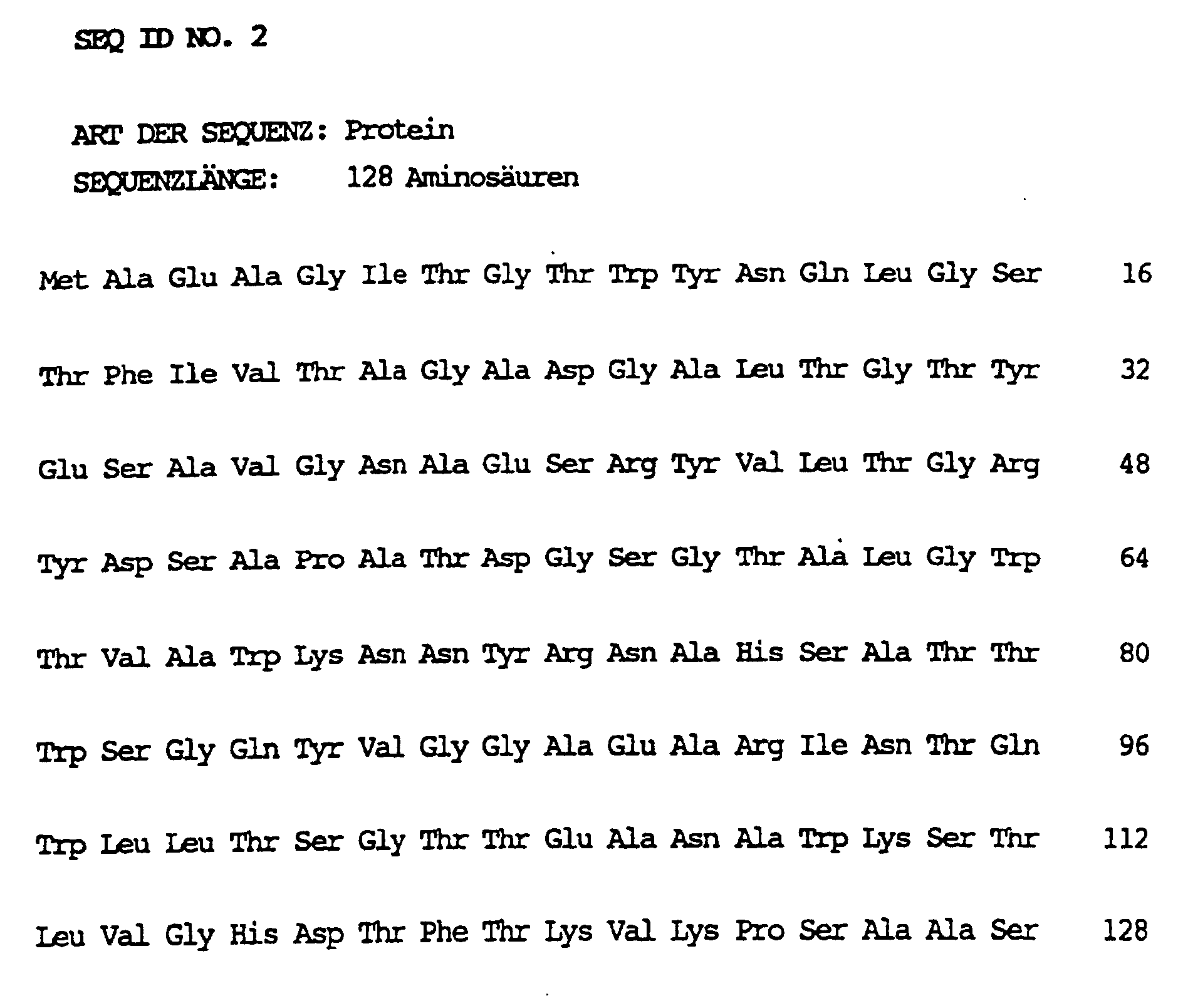
- the invention also relates to recombinant Core streptavidin, which is characterized by the in SEQ ID NO. 2 listed Amino acid sequence with or without N-terminal Methionine residue is labeled.
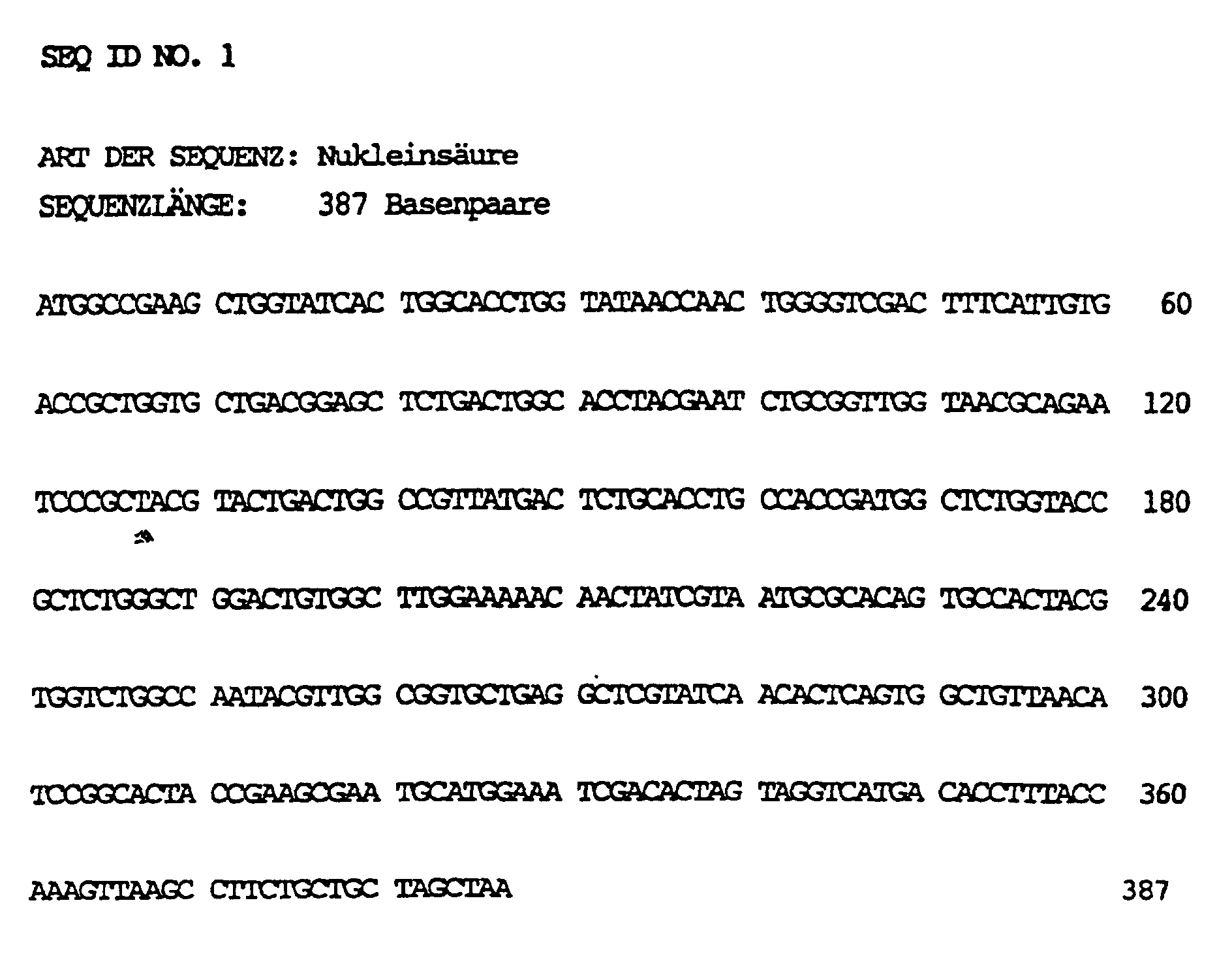
- SEQ ID NO. 1 the DNA sequence of core streptavidin
- SEQ ID NO. 2 shows the protein sequence of core streptavidin with an N-terminal methionine residue.
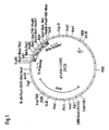
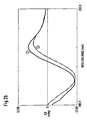
- 1 shows the plasmid map of the core streptavidin expression plasmid pSAM-Core. 2 shows circular dichroism (CD) spectra of core streptavidin according to the invention (2a) or of conventional core streptavidin from S. avidinii (2b) in each case without (curve 1) and with biotin (curve 2).
- CD circular dichroism
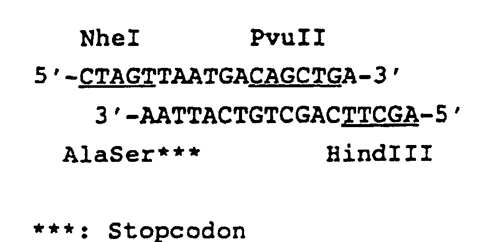
- the 3 'end of the native streptavidin structural gene was truncated by a DNA fragment that is for the C-terminal amino acid sequence coded.
- the plasmid pUC18-BBG9 (pUC18 vector with inserted streptavidin gene, British Biotechnology Limited (WO 89/03422) with the restriction endonucleases NheI and HindIII was digested, the approx. 2.7 kBp long NheI / HindIII fragment and isolated with the synthetic DNA fragment ligated (construction: pUC18-BBG9-C).
- the desired DNA sequence was confirmed by DNA sequencing.
- the plasmid pD-NX is a derivative of the plasmid pQE-6 (pDS56 / RBSII, Ncol) from Diagen (Düsseldorf), from which the promoterless chloramphenicol acetyltransferase gene (CAT) was removed.
- the plasmid pDS56 / RBSII, Ncol with the restriction endonucleases Digests NheI and XbaI which is about 2.6 kBp long NheI / XbaI vector fragment isolated and the compatible ends the NheI and XbaI interface linked by ligation (Construction: pD-NX).
- the 5 'end of the native streptavidin structural gene was truncated by a DNA fragment that corresponds to the N-terminal amino acid sequence coded.
- the plasmid prepared as described above was used digested pUC18-BBG9-C with the restriction endonuclease PstI, the 3 'overhanging end of the PstI site with T4 DNA polymerase digested, the DNA cleaved with HindIII, the approx. 390 bp PstI (blunt) / HindIII-streptavidin fragment isolated and into the approximately 2.6 kbp long NcoI (blunt) / HindIII pD-NX Vector fragment after filling in the 5 'overhanging end the NcoI interface with Klenow polymerase, ligated (construction: pSA-core).
- Fig. 1 shows the plasmid map of pSAM core.
- the plasmid pSAM-core is a plasmid mixture with pUBS500 deposited under DSM 6720.
- E. coli K12 strain RM82 (DSM 5445) (a methionine revertant from ED 8654, Murray, NE et al., Mol. Gen. Genet. 150 (1977) 53-61) was used to express core streptavidin the core-streptavidin expression plasmid pSAM-core (ampicillin resistance) and the lacI q repressor plasmid pUBS500 (kanamycin resistance, preparation and description see: EP-A 0 373 365).
- the RM82 / pUBS500 / pSAM-core cells were placed in DYT medium (1% (w / v) yeast extract, 1% (w / v) Bacto Tryptone, Difco, and 0.5% NaCl) with 50 mg / l ampicillin and 50 mg / l kanamycin up to an optical density at 550 nm of 0.6-0.9 and then induced with IPTG (1-5 mmol / l final concentration). After an induction phase of 4-8 hours the cells were harvested by centrifugation, with 25 mmol / l Tris-HCl buffer, pH 7.5, washed and until further processing stored at -20 ° C.
- DYT medium 1% (w / v) yeast extract, 1% (w / v) Bacto Tryptone, Difco, and 0.5% NaCl
- IPTG 1-5 mmol / l final concentration
- the cell pellet from 1 ml of centrifuged growth medium (RM82 / pUBS500 / pSAM-core cells) was in 0.25 ml 10 mmol / l Phosphate buffer, pH 6.8, and 1 mmol / l EDTA and resuspended the cells are disrupted by ultrasound treatment.
- To The supernatant was centrifuged with 1/5 volume 5xSDS sample buffer (1x SDS sample buffer: 50 mmol / l Tris-HCl, pH 6.8, 1% SDS, 1% mercaptoethanol, 10% glycerin, 0.001% bromophenol blue) added.
- the insoluble cell debris fraction was found in 0.3 ml 1xSDS sample buffer resuspended with 6 - 8 M urea, the samples incubated for 5 minutes at 95 ° C and centrifuged. After that, the proteins were replaced by SDS-polyacrylamide Gel electrophoresis (PAGE) separated (Laemmli, U.K., Nature 227 (1970) 680-685) and with Coomassie Brilliant Blue R dye stained.
- PAGE SDS-polyacrylamide Gel electrophoresis
- electrophoretically separated proteins were found Nitrocellulose filter transferred, fixed (Towbin, H. et al., Proc. Natl. Acad. Sci. 76 (1979) 4350) and the streptavidin immunoreactive Proteins with a specific anti-streptavidin antiserum detected from sheep.
- the core streptavidin protein synthesized in E. coli was homogeneous and became exclusively in the insoluble cell debris fraction found (IB's). Through SDS-PAGE and Western biotech could not see truncated core streptavidin fragments be detected.
- the expression level for core streptavidin was 30-50% based on the total E. coli protein.
- the protein sequence of the core streptavidin is in SEQ ID NO. 2 shown.
- E. coli RM82 / pUBS500 / pSAM-core cells were suspended in 200 ml of 0.1 mol / l Tris-HCl, pH 7.0, at 0 ° C., mixed with 60 mg of lysozyme and for 20 minutes at 0 ° C incubated. After addition of 2 mmol / l MgCl 2 and 1 mg / 100 ml DNase (Boehringer Mannheim GmbH, cat. No. 104159), the cells were completely disrupted mechanically by means of high pressure dispersion, after which the DNA was digested at 25 ° C. in 30 minutes.
- DNase Boehringer Mannheim GmbH, cat. No. 104159
- the digestion solution was then mixed with 100 ml of 60 mmol / l EDTA, 6% Triton X100 and 1.5 mol / l NaCl, pH 7.0 and incubated at 0 ° C. for a further 30 minutes.
- the insoluble constituents were then sedimented by centrifugation with a Sorvall centrifuge.
- the pellet was dissolved in 200 ml of 0.1 mol / l Tris-HCl, 20 mmol / l EDTA, pH 6.5 resuspended and incubated for 30 minutes at 25 ° C; the IB preparation was then isolated by centrifugation.
- the renaturation was carried out at 25 ° C. by adding 20 times 2 ml each of core streptavidin solubilisate (40 mg / ml) in 1.6 1 0.1 mol / l sodium phosphate, 5 mmol / l EDTA, pH 7.0 at intervals of 20 minutes.
- the renaturation approach can be carried out by membrane filtration concentrated and / or to remove guanidine HCl dialyzed against a desired buffer.
- Core streptavidin from renaturation approaches can be used if necessary using chromatographic methods known to those skilled in the art are to be further cleaned.
- the renaturation mixture was concentrated in a stirred cell to 120 ml by ultrafiltration, centrifuged to remove undissolved residues and then dialyzed against 20 mmol / l Tris-HCl, pH 8.5 (2 ⁇ 10 1, 24 hours, 4 ° C.).
- the bound material was eluted by a gradient from 0 to 150 mmol / l NaCl in equilibration buffer (10 SV, 1 SV / h). volume activity C Prot SA ml Einh./ml mg / ml Einh./mg dialysate 180 34.8 2 17.4 Q eluate 200 24.8 1.3 19.4
- the homogeneity and purity of natured cleaned Core streptavidin was determined by SDS-PAGE (Laemmli, U.K., Nature 227 (1970) 680-685).
- Naturally purified core streptavidin was homogeneous (single band) in the SDS-PAGE (molecular weight: approx. 13 500 Da) with a purity of> 98% with approx. 70 - 90% processing of the N-terminal methionine.
- the activity of core streptavidin was determined by titration with Biotin in pursuit of those caused by complex formation Change in absorption at 233 nm was determined. 1 unit is defined as the binding of 1 ⁇ g biotin through the sample.
- the protein was determined by measuring the absorbance at 280 nm.
- the measured specific activity (19.4 units / mg protein) corresponds to a binding stoichiometry of biotin: core strept avidin ⁇ 1 (based on the monomer).
- FIG. 2a Spectra of purified core streptavidin from E. coli (FIG. 2a) and spectra of conventionally produced core streptavidin from S. avidinii (FIG. 2b) cannot be distinguished.
Landscapes
- Chemical & Material Sciences (AREA)
- Organic Chemistry (AREA)
- Health & Medical Sciences (AREA)
- Proteomics, Peptides & Aminoacids (AREA)
- Biochemistry (AREA)
- Biophysics (AREA)
- General Health & Medical Sciences (AREA)
- Genetics & Genomics (AREA)
- Medicinal Chemistry (AREA)
- Molecular Biology (AREA)
- Life Sciences & Earth Sciences (AREA)
- Gastroenterology & Hepatology (AREA)
- Micro-Organisms Or Cultivation Processes Thereof (AREA)
- Preparation Of Compounds By Using Micro-Organisms (AREA)
- Peptides Or Proteins (AREA)
- Medicines Containing Antibodies Or Antigens For Use As Internal Diagnostic Agents (AREA)
- Medicines That Contain Protein Lipid Enzymes And Other Medicines (AREA)
- Investigating Or Analysing Biological Materials (AREA)
- Nitrogen Condensed Heterocyclic Rings (AREA)
- Detergent Compositions (AREA)
- Replacement Of Web Rolls (AREA)
- Measuring Or Testing Involving Enzymes Or Micro-Organisms (AREA)
Description


| E. coli K12 RM 82 | DSM 5445 am 02.10.1991 |
| pSAM-core/pUBS 500 | DSM 6720 am 20.09.1991 |
SEQ ID NO. 1 die DNA-Sequenz von Core-Streptavidin,
SEQ ID NO. 2 die Proteinsequenz von Core-Streptavidin mit einem N-terminalen Methioninrest.
Fig. 1 zeigt die Plasmidkarte des Core-Streptavidin-Expressionsplasmids pSAM-Core.
Fig. 2 zeigt Circulardichroismus (CD)-Spektren von erfindungsgemäßem Core-Streptavidin (2a) bzw. von konventionellem Core-Streptavidin aus S. avidinii (2b) jeweils ohne (Kurve 1) und mit Biotin (Kurve 2).


| Volumen | Aktivität | CProt | SA | |
| ml | Einh./ml | mg/ml | Einh./mg | |
| Dialysat | 180 | 34,8 | 2 | 17,4 |
| Q-Eluat | 200 | 24,8 | 1,3 | 19,4 |
Claims (21)
- Verfahren zur Gewinnung von rekombinantem Core-Streptavidin, worin man Wirtszellen mit einer für ein Core-Streptavidin kodierenden DNA transformiert, eine Kultivierung der transformierten Wirtszellen unter geeigneten Bedingungen durchführt, eine Expression der für das Core-Streptavidin kodierenden DNA bewirkt und das rekombinante Core-Streptavidin aus den Wirtszellen oder dem Kulturmedium gewinnt,
dadurch gekennzeichnet, daß man eine für Core-Streptavidin kodierende DNA verwendet, welche(a) die in SEQ ID NO. 1 gezeigte Nukleotidsequenz oder(b) eine der Nukleotidsequenz (a) im Rahmen der Degeneration des genetischen Codes entsprechende Nukleotidsequenz aufweist. - Verfahren nach Anspruch 1,
dadurch gekennzeichnet, daß man eine mikrobielle Wirtszelle verwendet. - Verfahren nach Anspruch 2,
dadurch gekennzeichnet, daß man eine prokaryontische Wirtszelle verwendet. - Verfahren nach Anspruch 3,
dadurch gekennzeichnet, daß man E.coli als Wirtszelle verwendet. - Verfahren nach Anspruch 1,
dadurch gekennzeichnet, daß man eine eukaryontische Wirtszelle verwendet. - Verfahren nach Anspruch 5,
dadurch gekennzeichnet, daß man Hefe als Wirtszelle verwendet. - Verfahren nach einem der Ansprüche 1 bis 6,
dadurch gekennzeichnet, daß man die Wirtszelle mit einem rekombinanten Plasmid transformiert, das mindestens eine Kopie der für das Core-Streptavidin kodierenden DNA enthält. - Verfahren nach Anspruch 7,
dadurch gekennzeichnet, daß man ein rekombinantes Plasmid mit einem "multi copy" Replikationsursprung verwendet. - Verfahren nach einem der vorhergehenden Ansprüche,
dadurch gekennzeichnet, daß sich die für das Core-Streptavidin kodierende DNA in der transformierten Wirtszelle unter Kontrolle eines regulierbaren Promotors befindet. - Verfahren nach Anspruch 9,
dadurch gekennzeichnet, daß man die Expression der für das Core-Streptavidin kodierenden DNA durch Zugabe eines Induktors für den regulierbaren Promotor bewirkt. - Verfahren nach einem der vorhergehenden Ansprüche,
dadurch gekennzeichnet, daß man das rekombinante Core-Streptavidin aus den Wirtszellen in Form von Inclusion Bodies gewinnt und anschließend eine Solubilisierung und Naturierung der Inclusion Bodies zu löslichem, biologisch aktivem Core-Streptavidin durchführt. - Verfahren nach Anspruch 11,
dadurch gekennzeichnet, daß man das naturierte Core-Streptavidin chromatographisch reinigt. - Rekombinante DNA,
dadurch gekennzeichnet, daß sie für ein Core-Streptavidin kodiert und(a) die in SEQ ID NO. 1 gezeigte Nukleotidsequenz oder(b) eine der Nukleotidsequenz (a) im Rahmen der Degeneration des genetischen Codes entsprechende Nukleotidsequenz aufweist. - Rekombinanter Vektor,
dadurch gekennzeichnet, daß er mindestens eine Kopie einer DNA nach Anspruch 13 enthält. - Vektor nach Anspruch 14,
dadurch gekennzeichnet, daß er ein Plasmid ist. - Vektor nach Anspruch 15,
dadurch gekennzeichnet, daß er einen "multi copy" Replikationsursprung besitzt. - Vektor nach einem der Ansprüche 14 bis 16,
dadurch gekennzeichnet, daß sich die für das Core-Streptavidin kodierende DNA unter Kontrolle eines regulierbaren Promotors befindet. - Plasmid pSAM-core DSM 6720
- Wirtszelle,
dadurch gekennzeichnet, daß sie mit einer DNA nach Anspruch 13 oder einem Vektor nach einem der Ansprüche 14 bis 18 transformiert ist. - Rekombinantes Core-Streptavidin,
dadurch gekennzeichnet, daß esaufweist.(a) die in SEQ ID NO. 2 gezeigte Aminosäuresequenz und/oder(b) die in SEQ ID NO. 2 gezeigte Aminosäuresequenz ohne den N-terminalen Met-Rest - Verwendung eines rekombinanten Core-Streptavidins nach Anspruch 20 für diagnostischer in vitro Testverfahren und Affinitätschromatographie.
Applications Claiming Priority (3)
| Application Number | Priority Date | Filing Date | Title |
|---|---|---|---|
| DE4135543A DE4135543A1 (de) | 1991-10-28 | 1991-10-28 | Rekombinantes core-streptavidin |
| DE4135543 | 1991-10-28 | ||
| PCT/EP1992/002463 WO1993009144A1 (de) | 1991-10-28 | 1992-10-28 | Rekombinantes core-streptavidin |
Publications (2)
| Publication Number | Publication Date |
|---|---|
| EP0612325A1 EP0612325A1 (de) | 1994-08-31 |
| EP0612325B1 true EP0612325B1 (de) | 2003-05-02 |
Family
ID=6443603
Family Applications (1)
| Application Number | Title | Priority Date | Filing Date |
|---|---|---|---|
| EP92922212A Expired - Lifetime EP0612325B1 (de) | 1991-10-28 | 1992-10-28 | Rekombinantes core-streptavidin |
Country Status (11)
| Country | Link |
|---|---|
| US (2) | US5489528A (de) |
| EP (1) | EP0612325B1 (de) |
| JP (1) | JP2621025B2 (de) |
| KR (1) | KR100203512B1 (de) |
| AT (1) | ATE239036T1 (de) |
| AU (1) | AU659240B2 (de) |
| CA (1) | CA2122135C (de) |
| DE (2) | DE4135543A1 (de) |
| ES (1) | ES2199218T3 (de) |
| FI (1) | FI941947A0 (de) |
| WO (1) | WO1993009144A1 (de) |
Families Citing this family (28)
| Publication number | Priority date | Publication date | Assignee | Title |
|---|---|---|---|---|
| CA2184386C (en) * | 1994-03-05 | 2005-10-18 | Rosemarie Kientsch-Engel | Interference eliminating agent for application in immunoassays |
| WO1996024606A1 (en) * | 1995-02-09 | 1996-08-15 | University Of Washington | Modified-affinity streptavidin |
| AU5917796A (en) * | 1995-04-11 | 1997-04-09 | Trustees Of Boston University | Streptavidin mutants |
| DE19637718A1 (de) * | 1996-04-01 | 1997-10-02 | Boehringer Mannheim Gmbh | Rekombinante inaktive Core-Streptavidin Mutanten |
| US6492492B1 (en) | 1998-04-03 | 2002-12-10 | University Of Washington | Circularly permuted biotin binding proteins |
| US20040009535A1 (en) | 1998-11-27 | 2004-01-15 | Celltech R&D, Inc. | Compositions and methods for increasing bone mineralization |
| RU2153535C1 (ru) * | 1999-05-14 | 2000-07-27 | Государственный научно-исследовательский институт генетики и селекции промышленных микроорганизмов | Рекомбинантная плазмидная днк psav27, кодирующая синтез растворимого стрептавидина из streptomyces avidinii, и бактериальный штамм escherichia coli - продуцент растворимого стрептавидина из streptomyces avidinii |
| US7927602B2 (en) * | 2002-07-23 | 2011-04-19 | University Of Louisville Research Foundation, Inc. | Fas ligand-avidin/streptavidin fusion proteins |
| US7270960B2 (en) | 2001-08-29 | 2007-09-18 | Pacific Northwest Research Institute | Diagnosis of ovarian carcinomas |
| US7381409B2 (en) | 2003-06-16 | 2008-06-03 | Celltech R&D, Inc. | Antibodies specific for sclerostin and methods of screening and use therefor |
| US7265205B2 (en) * | 2005-02-11 | 2007-09-04 | Uti Limited Partnership | Monomeric streptavidin muteins |
| US7704708B2 (en) * | 2006-02-13 | 2010-04-27 | Uti Limited Partnership | Monomeric streptavidin muteins |
| EP2005185B1 (de) * | 2006-03-22 | 2010-10-20 | Viral Logic Systems Technology Corp. | Verfahren zur identifizierung von polypeptid-targets |
| US8377448B2 (en) * | 2006-05-15 | 2013-02-19 | The Board Of Trustees Of The Leland Standford Junior University | CD47 related compositions and methods for treating immunological diseases and disorders |
| US20080131431A1 (en) * | 2006-05-15 | 2008-06-05 | Viral Logic Systems Technology Corp. | CD47 related compositions and methods for treating immunological diseases and disorders |
| GB0622956D0 (en) * | 2006-11-17 | 2006-12-27 | Univ Birmingham | Molecular detection system |
| US8993714B2 (en) * | 2007-10-26 | 2015-03-31 | Imiplex Llc | Streptavidin macromolecular adaptor and complexes thereof |
| EP2147683A1 (de) | 2008-07-25 | 2010-01-27 | DKFZ Deutsches Krebsforschungszentrum | Konstrukt und Verfahren zur Aufnahme von Cargomolekülen in eine Zelle |
| US9102526B2 (en) * | 2008-08-12 | 2015-08-11 | Imiplex Llc | Node polypeptides for nanostructure assembly |
| DE102008053892A1 (de) | 2008-10-30 | 2010-05-06 | Fachhochschule Gelsenkirchen | Medizinisches Implantat mit biofunktionalisierter Oberfläche |
| CN102325884B (zh) | 2009-02-20 | 2013-11-20 | 国立大学法人东京大学 | 低免疫原性链霉抗生物素蛋白及其应用 |
| WO2010132363A1 (en) | 2009-05-11 | 2010-11-18 | Imiplex Llc | Method of protein nanostructure fabrication |
| EP2563396A2 (de) * | 2010-04-30 | 2013-03-06 | Augmenta Biologicals LLC | Abgabeproteine |
| WO2013018836A1 (ja) * | 2011-08-01 | 2013-02-07 | 日本たばこ産業株式会社 | 生物学的試料中の物質を検出する工程において、非特異的結合を抑制する方法、当該方法に使用するための剤 |
| EP3511413B1 (de) | 2014-07-25 | 2022-09-07 | Theravectys | Lentivirale vektoren zur regulierten expression eines chimären antigenrezeptormoleküls |
| EP3697809A1 (de) | 2017-10-20 | 2020-08-26 | Institut Curie | Für rush angepasste dap10/12-basierte cars |
| US20200283485A1 (en) | 2017-10-20 | 2020-09-10 | Institut Curie | Hook fusion protein for regulating the cellular trafficking of a target protein |
| CN116082472A (zh) * | 2022-12-29 | 2023-05-09 | 四川安可瑞新材料技术有限公司 | 一种核心链霉亲和素及其制备方法及应用 |
Family Cites Families (4)
| Publication number | Priority date | Publication date | Assignee | Title |
|---|---|---|---|---|
| DE3583940D1 (de) * | 1984-10-02 | 1991-10-02 | Harry M Meade | Herstellung von streptavidinaehnlichen polypeptiden. |
| US4839293A (en) * | 1986-02-24 | 1989-06-13 | The Trustees Of Columbia University In The City Of New York | DNA encoding streptavidin, streptavidin produced therefrom, fused polypeptides which include amino acid sequences present in streptavidin and uses thereof |
| GB8723661D0 (en) * | 1987-10-08 | 1987-11-11 | British Bio Technology | Synthetic gene |
| US5328985A (en) * | 1991-07-12 | 1994-07-12 | The Regents Of The University Of California | Recombinant streptavidin-protein chimeras useful for conjugation of molecules in the immune system |
-
1991
- 1991-10-28 DE DE4135543A patent/DE4135543A1/de not_active Withdrawn
-
1992
- 1992-10-28 DE DE59209981T patent/DE59209981D1/de not_active Expired - Lifetime
- 1992-10-28 EP EP92922212A patent/EP0612325B1/de not_active Expired - Lifetime
- 1992-10-28 JP JP5508140A patent/JP2621025B2/ja not_active Expired - Lifetime
- 1992-10-28 AU AU28754/92A patent/AU659240B2/en not_active Ceased
- 1992-10-28 WO PCT/EP1992/002463 patent/WO1993009144A1/de not_active Ceased
- 1992-10-28 AT AT92922212T patent/ATE239036T1/de active
- 1992-10-28 ES ES92922212T patent/ES2199218T3/es not_active Expired - Lifetime
- 1992-10-28 KR KR1019940701409A patent/KR100203512B1/ko not_active Expired - Fee Related
- 1992-10-28 US US08/211,833 patent/US5489528A/en not_active Expired - Lifetime
- 1992-10-28 CA CA002122135A patent/CA2122135C/en not_active Expired - Lifetime
-
1994
- 1994-04-27 FI FI941947A patent/FI941947A0/fi unknown
-
1995
- 1995-05-04 US US08/434,718 patent/US5672691A/en not_active Expired - Lifetime
Non-Patent Citations (1)
| Title |
|---|
| SANO T. AND CANTOR C.R.: "Expression vectors for streptavidin-containing chimeric proteins", BIOCHEMICAL AND BIOPHYSICAL RESEARCH COMMUNICATIONS, vol. 176, no. 2, 1991, pages 571 - 577 * |
Also Published As
| Publication number | Publication date |
|---|---|
| FI941947A7 (fi) | 1994-04-27 |
| US5672691A (en) | 1997-09-30 |
| CA2122135C (en) | 1999-12-21 |
| FI941947A0 (fi) | 1994-04-27 |
| ES2199218T3 (es) | 2004-02-16 |
| JPH06510434A (ja) | 1994-11-24 |
| AU659240B2 (en) | 1995-05-11 |
| CA2122135A1 (en) | 1993-05-13 |
| DE4135543A1 (de) | 1993-04-29 |
| KR100203512B1 (ko) | 1999-06-15 |
| US5489528A (en) | 1996-02-06 |
| ATE239036T1 (de) | 2003-05-15 |
| AU2875492A (en) | 1993-06-07 |
| JP2621025B2 (ja) | 1997-06-18 |
| WO1993009144A1 (de) | 1993-05-13 |
| EP0612325A1 (de) | 1994-08-31 |
| DE59209981D1 (de) | 2003-06-05 |
Similar Documents
| Publication | Publication Date | Title |
|---|---|---|
| WO1993009144A1 (de) | Rekombinantes core-streptavidin | |
| DE3781765T2 (de) | Verfahren zur herstellung eines polypeptides. | |
| DE69228705T2 (de) | Methode zur rückfaltung von igf-i in eine aktive konformation | |
| DE69233008T2 (de) | Fusionierung von Peptiden und Proteinen mit Thioredoxin und thioredoxin-ähnlichen Molekülen | |
| EP0099084B1 (de) | Verfahren zur Herstellung von Interferon und Expressionsvektoren dafür | |
| US5608036A (en) | Enhanced secretion of polypeptides | |
| DE3880739T2 (de) | Expression von menschlichem proapolipoprotein a-1. | |
| DE68925138T2 (de) | Verfahren zur Herstellung von Proteinen durch Verwendung von CKS-Fusionsproteinen | |
| DE69024652T2 (de) | Expression von tetanustoxinfragment c | |
| EP0510658B1 (de) | Verbesserung der Ausbeute bei der Sekretion von disulfidverbrückten Proteinen | |
| EP0229998B1 (de) | Fusionsproteine mit eukaryotischem Ballastanteil | |
| DE3686579T2 (de) | Prokaryotisches expressionssystem. | |
| DE69010096T2 (de) | Expression und Sekretion von reifem menschlichem beta-Interleukin-1 in Bacillus subtilis und Mittel und Verfahren zu deren Ausführung. | |
| DE3887856T2 (de) | Methode zur Herstellung von natürlichen, menschlichen Wachstumshormon in reiner Form. | |
| DE69310540T2 (de) | Verhindern von spaltung von n-terminalen aminosäuren durch endogene aminopeptidasen während die expression von fremden genen in bakterien | |
| DE3687112T2 (de) | Verfahren zur ausscheidung von genprodukten in das wachstumsmedium von gram-negativen bakterien. | |
| EP0161629A1 (de) | Signalpeptid für die Exkretion von Peptiden in Streptomyceten | |
| US5656458A (en) | Expression and processing of amino terminus acetylated FGF's in yeast | |
| DE4002636A1 (de) | Expression von hiv1- und 2-polypeptiden und deren verwendung | |
| EP0289936B1 (de) | Verfahren zur Herstellung von Fremdproteinen in Streptomyceten | |
| DE68915204T2 (de) | Biosynthetisches Verfahren zur Herstellung von chemischen Verbindungen. | |
| CA2002210C (en) | Expression and processing of authentic fgf's in yeast | |
| DE3786947T2 (de) | Mikrobiologisches Verfahren zur Gewinnung eines Proteins durch Züchtung eines mutanten Bakterienstammes. | |
| EP0627006B1 (de) | Hybrid-dna, die eine untereinheit eines bakteriellen toxins mit einer haemolysin kodiert | |
| RU2238323C2 (ru) | Способ получения белков в трансформированных дрожжевых клетках |
Legal Events
| Date | Code | Title | Description |
|---|---|---|---|
| PUAI | Public reference made under article 153(3) epc to a published international application that has entered the european phase |
Free format text: ORIGINAL CODE: 0009012 |
|
| 17P | Request for examination filed |
Effective date: 19940425 |
|
| AK | Designated contracting states |
Kind code of ref document: A1 Designated state(s): AT BE CH DE DK ES FR GB GR IE IT LI LU NL SE |
|
| RAP3 | Party data changed (applicant data changed or rights of an application transferred) |
Owner name: ROCHE DIAGNOSTICS GMBH |
|
| 17Q | First examination report despatched |
Effective date: 19990628 |
|
| GRAH | Despatch of communication of intention to grant a patent |
Free format text: ORIGINAL CODE: EPIDOS IGRA |
|
| RIC1 | Information provided on ipc code assigned before grant |
Free format text: 7C 07K 14/36 A, 7C 12N 15/31 B |
|
| RIC1 | Information provided on ipc code assigned before grant |
Free format text: 7C 07K 14/36 A, 7C 12N 15/31 B |
|
| RAP1 | Party data changed (applicant data changed or rights of an application transferred) |
Owner name: ROCHE DIAGNOSTICS GMBH |
|
| GRAH | Despatch of communication of intention to grant a patent |
Free format text: ORIGINAL CODE: EPIDOS IGRA |
|
| GRAA | (expected) grant |
Free format text: ORIGINAL CODE: 0009210 |
|
| AK | Designated contracting states |
Designated state(s): AT BE CH DE DK ES FR GB GR IE IT LI LU NL SE |
|
| PG25 | Lapsed in a contracting state [announced via postgrant information from national office to epo] |
Ref country code: NL Free format text: LAPSE BECAUSE OF FAILURE TO SUBMIT A TRANSLATION OF THE DESCRIPTION OR TO PAY THE FEE WITHIN THE PRESCRIBED TIME-LIMIT Effective date: 20030502 Ref country code: IE Free format text: LAPSE BECAUSE OF NON-PAYMENT OF DUE FEES Effective date: 20030502 |
|
| REG | Reference to a national code |
Ref country code: GB Ref legal event code: FG4D Free format text: NOT ENGLISH |
|
| REG | Reference to a national code |
Ref country code: CH Ref legal event code: EP |
|
| REG | Reference to a national code |
Ref country code: CH Ref legal event code: NV |
|
| REF | Corresponds to: |
Ref document number: 59209981 Country of ref document: DE Date of ref document: 20030605 Kind code of ref document: P |
|
| REG | Reference to a national code |
Ref country code: IE Ref legal event code: FG4D Free format text: GERMAN |
|
| GBT | Gb: translation of ep patent filed (gb section 77(6)(a)/1977) | ||
| PG25 | Lapsed in a contracting state [announced via postgrant information from national office to epo] |
Ref country code: SE Free format text: LAPSE BECAUSE OF FAILURE TO SUBMIT A TRANSLATION OF THE DESCRIPTION OR TO PAY THE FEE WITHIN THE PRESCRIBED TIME-LIMIT Effective date: 20030802 Ref country code: GR Free format text: LAPSE BECAUSE OF FAILURE TO SUBMIT A TRANSLATION OF THE DESCRIPTION OR TO PAY THE FEE WITHIN THE PRESCRIBED TIME-LIMIT Effective date: 20030802 Ref country code: DK Free format text: LAPSE BECAUSE OF FAILURE TO SUBMIT A TRANSLATION OF THE DESCRIPTION OR TO PAY THE FEE WITHIN THE PRESCRIBED TIME-LIMIT Effective date: 20030802 |
|
| NLV1 | Nl: lapsed or annulled due to failure to fulfill the requirements of art. 29p and 29m of the patents act | ||
| PGFP | Annual fee paid to national office [announced via postgrant information from national office to epo] |
Ref country code: IE Payment date: 20031023 Year of fee payment: 12 |
|
| PGFP | Annual fee paid to national office [announced via postgrant information from national office to epo] |
Ref country code: LU Payment date: 20031027 Year of fee payment: 12 |
|
| PGFP | Annual fee paid to national office [announced via postgrant information from national office to epo] |
Ref country code: BE Payment date: 20031211 Year of fee payment: 12 |
|
| REG | Reference to a national code |
Ref country code: IE Ref legal event code: FD4D Ref document number: 0612325E Country of ref document: IE |
|
| ET | Fr: translation filed | ||
| REG | Reference to a national code |
Ref country code: ES Ref legal event code: FG2A Ref document number: 2199218 Country of ref document: ES Kind code of ref document: T3 |
|
| PLBE | No opposition filed within time limit |
Free format text: ORIGINAL CODE: 0009261 |
|
| STAA | Information on the status of an ep patent application or granted ep patent |
Free format text: STATUS: NO OPPOSITION FILED WITHIN TIME LIMIT |
|
| 26N | No opposition filed |
Effective date: 20040203 |
|
| PG25 | Lapsed in a contracting state [announced via postgrant information from national office to epo] |
Ref country code: LU Free format text: LAPSE BECAUSE OF NON-PAYMENT OF DUE FEES Effective date: 20041028 |
|
| PG25 | Lapsed in a contracting state [announced via postgrant information from national office to epo] |
Ref country code: BE Free format text: LAPSE BECAUSE OF NON-PAYMENT OF DUE FEES Effective date: 20041031 |
|
| BERE | Be: lapsed |
Owner name: ROCHE DIAGNOSTICS G.M.B.H. Effective date: 20041031 |
|
| BERE | Be: lapsed |
Owner name: ROCHE DIAGNOSTICS G.M.B.H. Effective date: 20041031 |
|
| REG | Reference to a national code |
Ref country code: CH Ref legal event code: PFA Owner name: ROCHE DIAGNOSTICS GMBH Free format text: ROCHE DIAGNOSTICS GMBH#SANDHOFER STRASSE 116#68305 MANNHEIM (DE) -TRANSFER TO- ROCHE DIAGNOSTICS GMBH#SANDHOFER STRASSE 116#68305 MANNHEIM (DE) |
|
| PGFP | Annual fee paid to national office [announced via postgrant information from national office to epo] |
Ref country code: AT Payment date: 20100923 Year of fee payment: 19 |
|
| PGFP | Annual fee paid to national office [announced via postgrant information from national office to epo] |
Ref country code: DE Payment date: 20101029 Year of fee payment: 19 |
|
| PGFP | Annual fee paid to national office [announced via postgrant information from national office to epo] |
Ref country code: IT Payment date: 20101016 Year of fee payment: 19 |
|
| PGFP | Annual fee paid to national office [announced via postgrant information from national office to epo] |
Ref country code: FR Payment date: 20111005 Year of fee payment: 20 Ref country code: GB Payment date: 20110930 Year of fee payment: 20 |
|
| PGFP | Annual fee paid to national office [announced via postgrant information from national office to epo] |
Ref country code: CH Payment date: 20111026 Year of fee payment: 20 Ref country code: ES Payment date: 20111018 Year of fee payment: 20 |
|
| REG | Reference to a national code |
Ref country code: DE Ref legal event code: R071 Ref document number: 59209981 Country of ref document: DE |
|
| REG | Reference to a national code |
Ref country code: DE Ref legal event code: R071 Ref document number: 59209981 Country of ref document: DE |
|
| REG | Reference to a national code |
Ref country code: CH Ref legal event code: PL |
|
| REG | Reference to a national code |
Ref country code: GB Ref legal event code: PE20 Expiry date: 20121027 |
|
| REG | Reference to a national code |
Ref country code: AT Ref legal event code: MK07 Ref document number: 239036 Country of ref document: AT Kind code of ref document: T Effective date: 20121028 |
|
| PG25 | Lapsed in a contracting state [announced via postgrant information from national office to epo] |
Ref country code: GB Free format text: LAPSE BECAUSE OF EXPIRATION OF PROTECTION Effective date: 20121027 |
|
| REG | Reference to a national code |
Ref country code: ES Ref legal event code: FD2A Effective date: 20130719 |
|
| PG25 | Lapsed in a contracting state [announced via postgrant information from national office to epo] |
Ref country code: ES Free format text: LAPSE BECAUSE OF EXPIRATION OF PROTECTION Effective date: 20121029 |