EP0617431A2 - Appareil d'analyse de rayon-X - Google Patents
Appareil d'analyse de rayon-X Download PDFInfo
- Publication number
- EP0617431A2 EP0617431A2 EP94302171A EP94302171A EP0617431A2 EP 0617431 A2 EP0617431 A2 EP 0617431A2 EP 94302171 A EP94302171 A EP 94302171A EP 94302171 A EP94302171 A EP 94302171A EP 0617431 A2 EP0617431 A2 EP 0617431A2
- Authority
- EP
- European Patent Office
- Prior art keywords
- ray
- sample
- rays
- optical element
- detector
- Prior art date
- Legal status (The legal status is an assumption and is not a legal conclusion. Google has not performed a legal analysis and makes no representation as to the accuracy of the status listed.)
- Granted
Links
Images






Classifications
-
- C—CHEMISTRY; METALLURGY
- C09—DYES; PAINTS; POLISHES; NATURAL RESINS; ADHESIVES; COMPOSITIONS NOT OTHERWISE PROVIDED FOR; APPLICATIONS OF MATERIALS NOT OTHERWISE PROVIDED FOR
- C09K—MATERIALS FOR MISCELLANEOUS APPLICATIONS, NOT PROVIDED FOR ELSEWHERE
- C09K11/00—Luminescent materials, e.g. electroluminescent or chemiluminescent
- C09K11/08—Luminescent materials, e.g. electroluminescent or chemiluminescent containing inorganic luminescent materials
- C09K11/77—Luminescent materials, e.g. electroluminescent or chemiluminescent containing inorganic luminescent materials containing rare earth metals
- C09K11/7766—Luminescent materials, e.g. electroluminescent or chemiluminescent containing inorganic luminescent materials containing rare earth metals containing two or more rare earth metals
- C09K11/7767—Chalcogenides
- C09K11/7769—Oxides
- C09K11/7771—Oxysulfides
-
- C—CHEMISTRY; METALLURGY
- C09—DYES; PAINTS; POLISHES; NATURAL RESINS; ADHESIVES; COMPOSITIONS NOT OTHERWISE PROVIDED FOR; APPLICATIONS OF MATERIALS NOT OTHERWISE PROVIDED FOR
- C09K—MATERIALS FOR MISCELLANEOUS APPLICATIONS, NOT PROVIDED FOR ELSEWHERE
- C09K11/00—Luminescent materials, e.g. electroluminescent or chemiluminescent
- C09K11/08—Luminescent materials, e.g. electroluminescent or chemiluminescent containing inorganic luminescent materials
- C09K11/77—Luminescent materials, e.g. electroluminescent or chemiluminescent containing inorganic luminescent materials containing rare earth metals
- C09K11/7766—Luminescent materials, e.g. electroluminescent or chemiluminescent containing inorganic luminescent materials containing rare earth metals containing two or more rare earth metals
- C09K11/7767—Chalcogenides
- C09K11/7769—Oxides
-
- G—PHYSICS
- G21—NUCLEAR PHYSICS; NUCLEAR ENGINEERING
- G21K—HANDLING OF PARTICLES OR IONISING RADIATION NOT OTHERWISE PROVIDED FOR; IRRADIATION DEVICES; GAMMA RAY OR X-RAY MICROSCOPES
- G21K4/00—Conversion screens for the conversion of the spatial distribution of X-rays or particle radiation into visible images, e.g. fluoroscopic screens
-
- G—PHYSICS
- G21—NUCLEAR PHYSICS; NUCLEAR ENGINEERING
- G21K—HANDLING OF PARTICLES OR IONISING RADIATION NOT OTHERWISE PROVIDED FOR; IRRADIATION DEVICES; GAMMA RAY OR X-RAY MICROSCOPES
- G21K4/00—Conversion screens for the conversion of the spatial distribution of X-rays or particle radiation into visible images, e.g. fluoroscopic screens
- G21K2004/06—Conversion screens for the conversion of the spatial distribution of X-rays or particle radiation into visible images, e.g. fluoroscopic screens with a phosphor layer
Definitions
- the present invention relates to an X-ray apparatus using diffraction X-rays, fluorescent X-rays, or transmission X-rays to identify or chemically analyse a microarea of a sample non-destructively, eg. by scanning over a line or area of the sample surface.
- Japanese Patent Provisional Publication No. SH059-72052 discloses one of examples of conventional X-ray analysing apparatus.
- a hollow capillary whose inside cross section is a circle, an ellipse or a polygon and whose inner surface is parallel and mirror-finished is extended from an X-ray source to a sample fixed at the centre of a sample stage composed of a triaxial precise movement mechanism and a biaxial rotational mechanism which are combined.
- An X-ray diffraction image and fluorescent X-rays of a microarea of the sample are received by a film or a position sensitive proportional counter or a guide tube for measuring fluorescent X-rays.
- the hollow capillary extending from the X-ray source is brought close to a vicinity of the sample, and a thin X-ray beam approximately as then as an internal diameter of the hollow capillary is directed on the sample.
- the fluorescent X-ray emitted from the sample is received by the guide tube for measuring fluorescent X-rays and measured by a semiconductor detector arranged at the end of the guide tube.
- the diffraction X-rays are measured by the film or the position sensitive proportional counter.
- Another prior art arrangement is, for example, disclosed by the Japanese Patent Provisional Publication No. SH061-22240.
- This prior art arrangement is composed of: a collimator for getting a thin X-ray beam incident on a minute part of a sample; a detachable spectrocrystal which is provided so that fluorescent X-rays emitted from a sample surface may be incident thereon; and an incident position sensitive X-ray detector in an arc provided so as to move around either a point which is symmetric to the microarea with respect to the surface of the spectrocrystal or the microarea.
- a tip of the collimator is brought to close vicinity to the sample, a fine X-ray beam is formed and the beam irradiates the microarea of the sample.
- the microarea emits fluorescent X-rays, which are reflected by the spectrocrystal and whose energy is measured from a difference in incident position by the position sensitive X-ray detector which is moved in an arc around the point symmetric to an X-ray irradiation area with respect to the spectrocrystal.
- diffraction X-rays are measured by the incident position sensitive X-ray detector which is moved in an arc, around the X-ray irradiation area.
- a manipulator for manipulating a thin X-ray beam In a manipulator for manipulating a thin X-ray beam, the intensity of X-rays attenuates considerably depending on the energy and optical distance therefrom even when the beam energy is 5 keV or more. Therefore, to fabricate an X-ray optical system by using a visible light optical alignment bench for an X-ray optical element is difficult, so that a manipulator for exclusive use with X-ray optical systems to control the attenuation of the X-rays in the air to a minimum using vacuum technology has been used.
- An exemplary known X-ray mirror manipulator is constructed as shown in figure 5. That is, it comprises a mirror mount tube 43 formed integral with the X-ray mirror, six vacuum-sealed micrometers 40, a vacuum tube 46 for fixing the micrometers in vacuum, a vacuum bellows 45 for securing the rectilinear movement of the mirror mount tube in the longitudinal direction, a linear stage mechanism 41, vacuum flanges and supports 39, and an X-ray window 42 for separating the atmosphere from a vacuum.
- the manipulator for an X-ray optical element performs bidirectional gates and bidirectional rectilinear movements on the inserted mirror mount tube with respect to the vacuum tubes by means of the six micrometers, and linearly moves the vacuum tube with respect to the flanges and supports by using the rectilinear stage mechanism. Accordingly, it is shown in figure 5 that the X-ray mirror in the mirror mount tube in a vacuum-sealed state can be aligned in pentaaxial directions by the operation through the X-ray window in the atmosphere.
- X-ray position detectors are of two types depending on the way they detect fluorescent X-rays, transmission X-rays or X-ray diffraction intensity: a pulse counting type in which X-ray light quanta are counted one by one and an integral counting type in which the intensity of X-rays received by the detector for a predetermined time period is integrated for counting.
- Typical examples of position sensitive detectors having a position detection function capable of recording one-dimensionally or two-dimensionally location distribution of the X-ray density are a one-dimensional and a two-dimensional proportional counter tube, an X-ray film, an imaging plate, an X-ray television, a CCD X-ray sensor.
- the proportional counter tube which is one of the pulse counter type detectors has the excellent features of one-dimensional or two-dimensional position resolving power and also energy resolving power.
- the integral counting type detector is excellent in position resolving power for position detection, and an X-ray film is one of the examples which is most frequently used.
- the Japanese Patent Provisional Publication No. SH059-72052 discloses an apparatus in which a hollow capillary leads X-rays from an X-ray source to the centre of a sample stage. Since this hollow capillary is a non-image forming optical element, it has a very large aberration. Further, when some distance of a few mm or more is kept between a sample and the hollow capillary, it is impossible to form X-rays into a thin X-ray beam having a diameter of the order of a few ⁇ m.
- the inner diameter of the capillary which is equivalent to an X-ray outgoing pupil should be not more than the size of the area to be measured and also that a tip of the capillary should be arranged in the close vicinity of the sample stage.
- the tip of the capillary is in the vicinity of the sample stage, not only the size of a sample is restricted but also a rotational angle of the sample stage is restricted. If the thickness of the hollow capillary is made smaller in order to avoid the restriction on the rotational angle, the hollow capillary transmits the X-rays and the effective diameter of the X-ray beam exceeds the required value, which results in the problem that a microarea cannot be analysed.
- a triaxial precise rectilinear movement mechanism is incorporated in a rotational mechanism, so that the size of the sample stage becomes smaller and a movement distance of the triaxial precise rectilinear movement mechanism is as short as a few millimetres or less.
- an apparatus disclosed by the Japanese Patent Provisional Publication No. SH061-22240 uses a collimator for applying a thin X-ray beam from an X-ray source to a sample surface.
- a microarea of the sample irradiated with the X-rays depends on an inner diameter of the collimator and a distance between the collimator and the sample surface.
- obtaining a minimum diameter of the X-ray beam requires the collimator to have a small inner diameter and also to have the collimator and the sample surface in close vicinity. Therefore, also in this case, a rotational angle of the sample surface is restricted by the collimator itself, thereby being unable to have a large rotational angle for the sample surface.
- the embodiment of this publication has only one rotational axis of a sample, it is difficult to measure diffraction X-rays when the number of crystal grains is small in the microarea irradiated with the X-rays.
- the samples to be measured are necessarily small ones.
- the reason is, as mentioned above, that the sample scanning distance of the sample stage is short and that sample gate rotation is restricted by the collimator or the hollow capillary. Because these X-ray optical elements perform bidirectional rectilinear movements and utilise bidirectional gates with 6 micrometers, they waste degrees of freedom of movement.
- they gate the X-ray mirror only by the rectilinear mechanism of the micrometers it is extremely difficult to operate the rectilinear components and the gate components independently of each other with respect to the pentaaxial alignment components of the X-ray mirror, and therefore it is difficult to perform alignment of the X-ray mirror.
- a method for detecting X-ray diffraction patterns depends on the wavelength of X-rays to be used and the kind of the X-rays, for example, characteristic X-ray and white X-rays.
- a one dimensional or two dimensional proportional counter tube which is a pulse counting detector has a defect that counting loss due to dead time occurs at high counting rates and moreover the proportion resolving power is not sufficient.
- X-ray films and X-ray televisions which are integral counting detectors have defects of low sensitivity, a narrow dynamic range and poor linearity. These defects have been alleviated by an imaging plate, but after an X-ray image is recorded on the imaging plate, a He-Ne laser is used and a focused laser beam two-dimensionally scans over the imaging plate to read an X-ray latent image of the above X-ray image.
- the fluorescent intensity of the image is measured by a photo multiplier tube, and its output is multiplied by a logarithmic amplifier. Further, the output is converted into numeric values by A/D convertor, and then the image is recomposed by a computer. As a result the reading tends to take a long time and the required size of apparatus is increased.
- an object of the present invention is to provide an improved microarea measuring apparatus, preferably having a high spatial resolving power and a high sensitivity.
- an X-ray generator is combined with a manipulator which has an X-ray optical element with a condensing and imaging function and an operation distance of 1 cm or more to provide a three-dimensional space between the X-ray optical element and a sample scanning table.
- This space makes it possible to insert an X-ray spatial filter for separating a desired wavelength and collimating between the X-ray optical element and the sample scanning table.
- a mechanism capable of large rotations and large gates, and thereon a three-dimensional rectilinear mechanism which can operate precise movements and moves at least 1 cm is attached.
- the three-dimensional rectilinear mechanism has a through hole provided in its part through which an X-ray optical axis passes.
- An X-ray detecting section is concentric with a rotational axis of the sample scanning table.
- the X-ray detecting section is a combination of a rectilinear mechanism for moving the section in a predetermined distance in a direction of the rotational axis of the sample scanning table, and another rectilinear mechanism for moving the section in a direction orthogonal to the rotational axis direction; which is further combined with an X-ray position detector and/or an X-ray energy detector.
- the X-ray position detector is preferably a two-dimensional X-ray detector for detecting a small amount of X-rays which is composed of an X-ray fluorescent surface made of an X-ray fluorescent material which includes terbium-doped gadolinium oxide sulphide and whose surface density is preferably 5 mg/cm2 - 50 mg/cm2, optionally an image intensifier tube and optionally a CCD.
- the fluorescent surface is preferably coupled to a tapered fibre plate in order to vary the area of a surface to be detected, and the image intensifier tube and the CCD (if used) are coupled by means of the fibre plate to make the best light efficiency.
- the above structure uses an X-ray optical element which has a condensing and imaging function and a movement distance of 1 cm or more, it is possible to arbitrarily change the position of the sample in a space between the X-ray optical element and the sample scanning table.
- the sample of approximately 1 cm or less in half width can be freely gate-rotated. Therefore, the use of the X-ray optical element with a longer operation distance makes it possible to gate-rotate further larger samples arbitrarily.
- the sample is scanned in a distance of 1 cm or more in three directions, so that an area equivalent to almost all the surface of the attached sample can be scanned and a sample of approximately 1 cm thick or less can be given non-destructive measurement.
- the use of the X-ray position detector with the above structure enables highly sensitive measurement.
- the use of a spatial filter facilitates to make an X-ray beam monochromatic and to change the size of an aperture of the X-ray optical element.
- a reduction ratio and an X-ray source diameter depending on the X-ray optical element determines the X-ray beam diameter. Therefore, the use of a large reduction ratio and a small X-ray source diameter makes it possible to irradiate the sample with an X-ray beam which is far more precise compared with the above prior art.
- X-rays penetrating the sample on the sample scanning table can be measured when the X-ray detecting section is rotationally moved along the X-ray axis.
- An X-ray detector can be placed at a desired position with the sample at the centre by a detector moving mechanism attached to the X-ray detecting section.
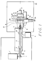
- Figure 1 is a top plan view showing the structure of an X-ray analysing apparatus.
- Figure 2 is a side view showing a structure in the vicinity of a sample scanning table.
- Figure 3 is a sectional view showing the surroundings of an X-ray optical element in an X-ray optical element alignment mechanism.
- X-ray generator 1' is a reflection type X-ray generator, and generates X-rays from an X-ray target 1.
- the focal spot size of the X-ray generator 1' can be varied within a range between 10 ⁇ m and a few hundred ⁇ m. Further, the X-ray target can be exchanged with various kinds of targets such as Cu, Fe and W.
- Generated X-rays 3 pass through a pipe 6 evacuated with a vacuum pump unit 4.
- a vacuum gate valve can divide the pipe 6 into an X-ray generator side and a vacuum.
- the X-rays 3 passing through the pipe 6 impinge on an X-ray optical element manipulator 7.
- the X-ray optical element manipulator 7 is composed of a bidirectional rectilinear mechanism and a biaxial gate mechanism. By these mechanisms, an X-ray optical element 30 (figure 3) is aligned in four axial directions except an X-ray axis 2. Alignment sensitivity is kept to 1 ⁇ m during a parallel movement of the X-ray optical element and 5 seconds or less during a gate, so that the X-ray optical element can be precisely positioned.
- This X-ray optical element manipulator 7 has an X-ray window 8 for separating a vacuum from an atmosphere incorporated therein so that the X-ray optical element 30 attached inside a tube 7' can be exchanged in the atmosphere.
- the X-rays 3 incident on the X-ray optical element 30 form a minute X-ray spot on a focal plane on the X-ray axis in the atmosphere, that is, on a sample surface 9 on a sample scanning table 10.
- the X-ray optical element available for this embodiment is a small X-ray optical element capable of condensing and imaging X-ray energy of approximately 5 keV or more such as a transmission zone plate, a total reflection mirror, a multiplier film mirror, and a curved crystal for example.
- X-ray optical element 30 there is used, for example, an X-ray converging mirror made of pyrex glass in the shape of a rotational elliptic surface.
- the X-ray converging mirror has a focal length of about 1 meter and an operation distance of about 70 mm, and reduces the X-ray generating area by a factor of ten for imaging.
- the incident angle of the X-rays is 5 m rad, and the X-ray reflecting surface inside the X-ray converging mirror 30 is coated with platinum. Therefore, the X-ray reflecting surface reflects the X-rays of which X-ray energy is below approximately 10 keV, at a reflectance of 80% or more.
- the X-ray converging mirror 30 is 1.6 mm at most in inside diameter gently decreasing to 1.2 mm, approximately 8 mm ⁇ in outside diameter, and approximately 40 mm in length, and is in a shape of a glass tube divided lengthwise.
- the X-ray converging mirror 30 is fixed within the stainless tube 7' of 10 mm ⁇ outside diameter so as to house the outside diameter of the X-ray converging mirror 30.
- the X-ray converging mirror 30 and the holder tube 7' of the X-ray converging mirror are fixed by placing an end of the tube 7' and an end of an outgoing pupil neatly side by side and are adhered to each other with an adhesive.
- the tube 7' incorporated with the X-ray converging mirror 30 is attached on the X-ray optical element manipulator section 7 by use of a screw thread cut on the tube 7'.
- a sample scanning table 10 (figure 1) is composed of a rectilinear stage 11 (figure 2) for moving the sample scanning table 10 in the direction of the X-ray axis, a rotational stage 12 for rotating an X-ray detecting section 21, a rotational stage 13 for rotating the sample scanning table 10, a bracket 14, a goniostage 15, a bracket 16, rectilinear stages 17, 18 and 19, and a large sample holder 20.
- a function of the rectilinear stage 11 is to adjust the centre axis of the scanning rotational stage 13 and the goniostage 15 to the focal point of the X-ray converging mirror 30.
- the range of travel of the linear stage 11 is approximately 80 mm and its position accuracy is 1 ⁇ m.
- the rotational stage 12 rotates the X-ray detecting section 21 independently of the sample scanning table rotational stage 13.
- the X-ray detecting section 21 can rotate within a range of approximately 300° except an angle obstructed by the X-ray optical element alignment mechanism 7, and the rotational angle can set with a rotational accuracy of 1/100 deg.
- the X-ray detecting section 21 includes a moving mechanism and an X-ray detector.
- the moving mechanism is composed of a stage 24 having a rectilinear travel of approximately 250 mm or more and a rectilinear stage 25 capable of moving approximately 20 mm in a direction perpendicular to both a rotational direction of the sample table and a rotational centre direction of the table.
- Apparati available as X-ray detectors include a semiconductor detector having an energy resolving power of incident X-rays, a gas flow proportional counter tube, and a detector in which an X-ray fluorescent substrate having a positional resolving power of the incident X-rays and an image intensifier are combined.
- the rotational stage 13 for rotating the sample scanning table 10 can rotate through 300° and its rotational angle accuracy is 1/1000 deg.
- the goniostage 15 on the rotational stage 13 can rotate through ⁇ 45° and its rotational accuracy is 1/1000 deg.
- the goniostage 15 and the rotational stage 13 have a common rotational centre. Therefore, the bracket 14 is provided between the two stages 13 and 15 to accurately adjust rotational axes of the two stages 13 and 15.
- the rectilinear stages for three directions 17, 18 and 19, and the bracket 14 which are combined are provided on the goniostage 15 on the scanning sample rotational stage 13.
- the rectilinear stage 17 is provided on the goniostage 15.
- the rectilinear stage 17 has a moving distance of 25 mm, resolving power of 0.1 ⁇ m and reproducibility of i 1 ⁇ m, and can move and adjust the sample surface 9 to the focal point of the X-ray converging mirror, that is, the rotational centre. Therefore, it is possible to mount a sample of approximately 20 mm in thickness or less on the sample scanning table.
- This rectilinear stage 17 is driven by a side motor.
- the bracket 16 is mounted on the rectilinear stage 17 so that the rectilinear stages 18 and 19 moving along the other two axes can move perpendicularly to the direction of travel of the rectilinear stage 17. Performance of the stages 18 and 19 is equivalent to that of the rectilinear stage 17 including resolving power and position reproducibility.
- the moving distances of both the stages are around 50 mm, and respective motors are mounted for driving the stages in rectilinear directions respectively.
- a through hole of 32 mm ⁇ in diameter is made in a part, through which the X-ray axis 2 passes, of the bidirectional rectilinear stages 18 and 19 provided vertically on the goniostage 15 and the bracket 16 for supporting the stages.
- a large sample for example, a silicon wafer of 152.4 mm (6 inch) diameter can be mounted on the large sample holder 20.
- the large sample holder 20 includes a rectilinear mechanism capable of moving 70 mm and a rotational mechanism capable of rotating through 360°. Therewith, it is possible to scan all over the surface of a large sample. All the stages except the X-ray detecting section 21 and the large sample holder 20 can be remotely controlled.
- the distance between the X-ray emitting section and the sample surface to be measured is about 70 mm.
- Effective energy of the X-rays converged on the sample surface ranges approximately from 5 keV to 10 keV.
- the X-ray energy of 5 keV or less is absorbed by a space of about 150 mm between the X-ray window 8 and the focal point on the X-ray surface.
- the X-ray generating spot in the X-ray generator 1' is concentrated to 20 to 30 ⁇ m in diameter.
- the X-ray spot formed by the X-ray converging mirror 30 has a maximum diameter of about 5 ⁇ m.
- the X-ray generating spot diameter is reduced to 1/10 of the diameter of the X-ray converging mirror, but not to the X-ray converging spot diameter.
- the cause is surface roughness and any error in the shape of the X-ray converging mirror 30.
- An A l wiring pattern on a silicon wafer of 101.6 mm (4 inch) diameter is used for the sample 9.
- the A l wiring is about 2 ⁇ m thick and has pitches of 65 ⁇ m width.
- the sample 9 is fixed on the sample scanning table with the large sample holder 20.
- As Cu is used as the X-ray target, characteristic X-rays of Cu having energy of 8 keV and high intensity peak irradiate the sample surface.
- the A l wiring is regarded as a thin film in a polycrystalline state, so that the grid interval of the A l surface showing the largest X-ray diffraction intensity (111) is assumed to be the grid interval d of the A l wiring on the silicon wafer.
- the X-ray wavelength ⁇ converged on the sample is chiefly characteristic X-rays of Cu, so that a value of a sample angle ⁇ which satisfies Bragg's Law is obtained.
- a sample angle is regarded as 0°.
- ⁇ 19.24°, to which the sample rotational angle is adjusted.
- a fluorescent substance Gd2O2S:Tb is applied to a fibre plate to form a fluorescent surface and the fibre plate is fixed to an image intensifier with an adhesive having the same optical refractive index as that of the fibre plate.
- the angle of the X-ray detector is adjusted to an angle twice as large as the rotational angle of the sample. The intensity of the diffraction X-rays incident on the X-ray detector from the A l wiring is very weak.
- the X-ray detector surface is moved close to the sample surface so that a distance between the two surfaces is 40 mm or less.
- integration for 30 seconds is performed on each spot. Scanning is performed at intervals of 5 ⁇ m over 500 spots on the sample surface, thereby measuring a characteristic line of Cu diffracted on the surfaces of the samples arranged at intervals of 65 ⁇ m respectively.
- a Ni filter is inserted between the X-ray converging mirror 30 and the sample surface 9 of the embodiment 1 to convert the X-rays into monochromatic radiation so that the X-ray detector can measure a clearer diffraction spot.
- Cu is used for the X-ray target 1 and the sample rotational angle is set at about 3°, smaller than that of the embodiment 1 to observe a reflected Lave image of the A l wiring.
- the large sample holder 20 is removed from the scanning sample table so that the sample surface is vertical to the X-ray axis 2.
- an A l thin film having an organic thin film as a supporting body.
- the sample is directly attached to the rectilinear stage 19.
- the X-ray detector is placed behind the sample surface on the X-ray axis to observe a transmission Lave image.
- Embodiment 2 discloses a method for measuring fluorescent X-rays of a microarea.
- the X-ray converging mirror used in embodiment 1 is utilised as the X-ray optical element. Therefore, the distance between the X-ray outgoing section of the tube 7' and the sample surface to be measured is approximately 70 mm.
- the conditions of the X-ray generator are, as in embodiment 1, that the acceleration voltage is 50 keV, and Cu is used for the X-ray target.
- the maximum diameter of the X-ray converging spot formed by the X-ray converging mirror 30 is about 5 ⁇ m.
- the energy of the X-rays converged on the sample surface is predominantly in the range of approximately 5 keV and 10 keV.
- the X-ray detector there is used a gas-flow proportional counter tube using PR gas.
- Stainless mesh of 127 ⁇ m pitch constituting the sample is fixed on the large sample holder 20.
- the sample 9 is rotated through about 30° by the rotational stage so that the proportional counter tube is placed within a range of 40 mm from the sample.
- the rectilinear movement stages 18 and 19 are used to scan two-dimensionally an area of 1 mm2 on the sample and measure a mesh image formed by fluorescent X-rays of Fe.
- a method utilising a semiconductor X-ray detector can be used.
- the semiconductor X-ray detector has remarkably high resolving power, so that it is possible to measure separately the respective fluorescent X-ray energies of plural elements whose atomic numbers are close.
- a Si wafer of 101.6 mm (4 inch) diameter is utilised as a sample and fixed on the large sample holder 20 to detect a minute amount of the element in the vicinity of the surface.
- the rotational angle of the sample table is set at 1° or less so that the X-rays are grazingly incident on the sample surface.
- a monochromatic filter (a Ni thin film of 25 ⁇ m thickness in this case) is placed ahead of the sample table. Further, the collimator is arranged between the X-ray converging mirror 30 and the sample surface.
- the X-ray converging mirror used in this embodiment is characterised by a large divergent angle of the X-rays, approximately 0.6°. Therefore, the collimator is used to restrict the effective area of the X-ray reflective surface of the X-ray converging mirror, that is, a surface portion of the X-ray converging mirror 30 so that the convergent angle of the X-ray converging beam is 0.1°.
- the semiconductor is placed within 20 mm of the sample surface.
- the fluorescent X-rays emitted from the sample surface are measured by varying the rotational angle of the sample formed by the rotational stage 13 starting with 0°. This measurement detects contamination caused by Fe in a silicon wafer sample at depths up to 10 nm (100 ⁇ ).
- Embodiment 3 relates to a method for measuring transmission X-rays of the microarea.
- the X-ray converging mirror used in embodiments 1 and 2 is applied as the X-ray optical element. Therefore, the distance between the X-ray outgoing section of the tube 7' and the sample surface to be measured is approximately 70 mm.
- the conditions of the X-ray generator are that the acceleration voltage is 50 kV and that Cu is used for the X-ray target.
- the maximum diameter of the X-ray converging spot formed with the X-ray converging mirror 30 is about 5 ⁇ m.
- the efficient energy of the X-rays converged on the sample surface ranges from 5 keV to 10 keV.
- the X-ray detector there is used a gas flow proportional counter tube.
- the large sample holder 20 is removed from the sample scanning table and the sample is directly attached to the rectilinear stage 19 in order to utilise a through hole made on the rectilinear stages 13 and 15, and the bracket 16 of the sample scanning table 10.
- a stainless mesh with 127 meshes is used as the sample.
- the proportional counter tube is moved on the X-ray axis 2 by the rotational stage 12 and drawn to a position within the extremely close vicinity of a back face of the bracket 16 of the sample scanning table.
- the rectilinear stages 18 and 19 are used to two-dimensionally scan the samples of 1 mm2 at an interval of 10 ⁇ m.
- the characteristic X-rays of Cu are measured with the proportional counter tube and the mesh image of the Cu is obtained from the change in intensity of the transmission X-rays.
- An apparatus having high resolving power for example, a semiconductor X-ray detector is used in a method for measuring the transmission X-rays. It is possible to determine an element composition of the sample by detecting a change in energy intensity of the incident X-rays which are incident on the sample and a change in energy intensity of the transmission X-rays. When the sample has no change in the transmission X-rays, the sample is tilted with the rotational stage 13 and the goniostage 15. It is also possible to guess an approximate inside structure in one direction of the sample from the change in the transmission X-ray intensity in a particular direction of the sample.
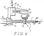
- FIG. 4 is a schematic diagram of a structure of an X-ray optical element manipulator in accordance with the invention.
- a vacuum flange 27 which serves as a reference surface facing a vacuum flange 26 is provided with a gasket channel.
- Compact linear stages 28 and 29 are fixed in an intersecting state at a location separated from the X-ray optical axis 2 of the vacuum flange 27.
- Each of the rectilinear stages has independent travel of 10 mm or more, a sensitivity of 1 ⁇ m, and both yawing and pitching of 10 ⁇ 4 rad or less, and with the incorporation of an operation micrometer having a minimum readout of 1 ⁇ m, high-precision positioning can be performed.
- a bracket 31 is fixed on the combined stages, and a compact rectilinear stage 36 is fixed on the bracket.
- the performance of this stage is the same as that of the above-mentioned compact stage, but because a micrometer head is attached transversely to the rectilinear stage, the travel of the stage is not restricted by the micrometer head.
- a triaxial gate table 32 is fixed to this stage 36.
- This gate table has a mechanism for moving the centre of a movable carriage along a spherical surface having a diameter corresponding to the distance from the table itself to the movable carriage and for inclining the movable carriage in two directions, and a mechanism for rotating the movable carriage in a plane parallel to the table itself, the rotational angle thereof being ⁇ 2.5° and sensitivity 3 seconds or less.
- a bracket 33 having a hole drilled therein so as not to become an obstacle to the X-ray optical axis is provided over the gate table. Respective O-ring grooves for maintaining vacuum are formed at both sides of the bracket.
- a flexible vacuum tube 34 connected to the vacuum flange 27 is connected on one side of the bracket, and on the other side thereof, an X-ray window 8 and mirror mount tubes 7' and 7 are connected.
- a Mylar (Registered Trade Mark) film of 25 ⁇ m thickness in the form of an organic high polymer transparent film is pasted above the stainless steel plate so as to cover the hole so that the Mylar film is not broken due to the pressure difference between the vacuum and the atmosphere.
- the mirror mount tube is divided into the tube main body 7' which is integrated with the X-ray mirror and the portion 7 which supports the tube, and a tap is cut in the connecting portion so that these two portions are easily detached.
- the portion 7, which supports the tube is attached to the bracket 8 so as to sandwich the X-ray window, thereby providing a vacuum section up to the X-ray window.
- the X-ray optical element manipulator is constructed as above, X-rays permeate the X-ray window, and X-rays having energy of 5 keV or more and usable in the atmosphere permeate the X-ray window with a transmittance of 95 % or more. Further, because the force applied on the vacuum portion due to the atmospheric pressure pulls the mirror mount tube toward the vacuum flange 27.
- the vacuum components are designed such that the above pulling force in the direction of the flange 26 is 3 kg weight or less.
- the inner diameters of the vacuum flexible tube 34 and the bracket 33 are set at 10 mm ⁇ or less, and to enable a stable alignment to be made, even if the tube is pulled by this level of force, the rectilinear stages 28, 29 and 36 and the gate table 32 have a lock mechanism with a load rating of 3 kg weight or more in the direction of the force application, and the brackets 31 and 33 are made of stainless steel of high rigidity.
- the mirror mount tube 7' displaces the vacuum flange 27 and an actual fluctuation width is obtained by subtracting the fluctuation width of the vacuum flange from the fluctuation width of the mirror mount tube. Accordingly, it has been found from figures 6A and 7C that the fluctuation of the mirror mount tube relative to the vacuum flange is a fluctuation of 0.5 ⁇ m width or less in the three directions.
- the method for aligning the X-ray mirror using this manipulator is, for example, such that an alignment is first performed using visible light, and then another alignment is performed using X-rays of the wavelength to be used.
- the X-ray mirror is about 10 mm ⁇ in outside diameter and 40 mm in length and is intended to form a minute converged spot at a focal position with 5 keV or more X-rays.
- visible light is made to coincide with the X-ray optical axis 2 using a mirror, and the visible light is reflected by the X-ray mirror in the mirror mount tube 35.
- the visible light to converge on the X-ray optical axis 2 is viewed on white paper, and if it does not converge, first the axial displacement from the X-ray optical axis of the X-ray mirror is corrected by means of the gate mechanism of the gate table 7.
- the light reflected from the X-ray mirror is moved to a desired location by the rectilinear tables 28, 29 and 36.
- the axial displacement of the X-ray mirror is examined in the manner as described previously and corrected, and again, the reflected light is moved by the rectilinear stages and such operation is repeated until the moving mechanisms do not move. Then, the X-rays to be used are introduced into the X-ray optical axis, and the converged X-rays formed by the X-ray mirror are observed by using a high-sensitivity X-ray two-dimensional position detector or the like. If the axis of the X-ray mirror or the X-ray converging position has been corrected, adjustment is performed in the same manner as in the case of visible light until the moving mechanisms do not move, and lastly, each of the moving mechanisms is locked.
- Embodiment 5 relates to an X-ray diffraction position detector.
- Figure 8 is a structural diagram of a detector in accordance with the invention.
- An X-ray fluorescent surface 48, and an image intensifier tube 50 and a CCD 52 are coupled with a fibre plate 49 to maximise the optical detection efficiency.
- the X-ray fluorescent surface used in this invention was made by a precipitation method, it can be also formed by a physical or chemical film forming method such as a coating method, deposition film forming method such as a coating method, deposition and sputtering by use of positional resolving power and a coating film thickness.
- the X-ray fluorescent surface was designed so that the surface has high sensitivity and high position resolving power when the wavelength of the diffracted X-rays is within the range 0.07 nm to 0.2 nm (0.7 ⁇ to 2 ⁇ ).
- the surface was formed on a fibre plate of 2 mm diameter and 3 mm thickness by the precipitation method with a fluorescent material, Gd202S:Tb.
- the thickness of the fluorescent surface can be determined from the relationship between the amount of X-rays absorbed by the fluorescent material Gd202S:Tb and the X-ray wavelength to be used, and is determined by the following equation:
- I/I0 exp [ - ⁇ (p/p0)t], where I/I0 is the transmittance of the X-rays of the fluorescent material, ⁇ is a linear absorption coefficient of the used X-rays, p0 is a density of the fluorescent material Gd202S:Tb, p is the filling rate (packing density) of the fluorescent material, and t is the thickness of the fluorescent surface.
- ⁇ a linear absorption coefficient of the used X-rays
- p0 is a density of the fluorescent material Gd202S:Tb
- p the filling rate (packing density) of the fluorescent material
- t is the thickness of the fluorescent surface.
- grain sizes of the fluorescent material were arranged to be almost uniform and actually were approximately 3 ⁇ m.
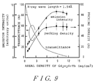
- Figure 9 shows a relationship between the transmittance of the X-rays, with respect to a surface density of the Gd202S:Tb fluorescent surface, a relationship between the packing density and the same, and a relationship between an emission intensity of the X-ray fluorescent and the same respectively which are observed when the X-ray wavelength is 1.54 ⁇ .
- the surface density is 5 mg/cm2 or less
- the packing density is as low as 10% to 40% and the absorption of the X-rays and the emission efficiency of the fluorescence become small.
- the filling rate packing density
- the irradiation direction of the X-rays and the direction of emission of visible light are opposite when the surface density exceeds approximately 10 mg/cm2, so that if the film thickness of the X-ray fluorescent surface exceeds a predetermined value, the surface does not transmit the emitted light and the emission intensity apparently decreases. Therefore, in order to detect an X-ray diffraction pattern with an X-ray wavelength of 0.154 nm (1.54 ⁇ ), the optimum surface density is between 8 mg/cm2 and 10 mg/cm2.
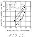
- Figure 10 shows results of investigating sensitivity of the X-ray diffraction one-dimensional position detector having a structure shown in figure 8 by use of the X-ray fluorescent surface.
- Figure 10 shows results of investigating a relationship between the X-ray intensity of the X-ray diffraction two-dimensional position detector having the Gd202S:Tb fluorescent surface and its response intensity at the wavelength of 0.154 nm (1.54 ⁇ ).
- each response intensity of two kinds of fluorescent surfaces and an X-ray CCD detector in addition to the Gd202S:Tb fluorescent surface was plotted. It was found out that when the surface density of the fluorescent screen is about 10 mg/cm2, an X-ray photon responds to one pixel and the fluorescent screen has a dynamic range of about 2.5 digits. The positional resolving power was about 20 ⁇ m.
- embodiment 5 discloses a case when the X-ray wavelength is 1.54 ⁇ .
- Figure 11 shows the surface density of the fluorescent material Gd202S:Tb which is observed when the fluorescent intensity of the fluorescent screen with respect to the X-ray wavelength becomes strongest.
- the fluorescent surface has a surface density ranging from 5 mg/cm2 to 50 mg/cm2.
- the response sensitivity of the detector and the dynamic range were the same as those of a case that the X-ray wavelength is 0.154 nm (1.54 ⁇ ), but the position resolving power ranged between 15 ⁇ m and 60 ⁇ m because the thickness of the fluorescent surface depends on the used X-ray wavelength.
- the light receiving area can be expanded by combining the X-ray fluorescent surface 48 and a tapered fibre plate 53.
- Figure 12 is a schematic diagram showing a combination of a tapered fibre plate 53 having a reduction rate of approximately one third and the X-ray fluorescent surface Gd202S:Tb.
- a case 54 has the X-ray fluorescent surface 48 and the tapered fibre plate 53 therein.
- an effective diameter of the image intensifier tube is 18 mm, so that a diameter of the effective X-ray fluorescent surface is 54 mm.
- light lost due to the tapered fibre plate is about 30% of that of the X-ray diffraction two-dimensional detector having a structure shown in figure 8, which considerably enhances the sensitivity.
- the position resolving power increases as the reciprocal of the reduction rate, it becomes approximately 60 ⁇ m when the X-ray wavelength is 0.154 nm (1.54 ⁇ ) as in embodiment 5. Further, when the X-ray wavelength is between 0.07 nm and 0.2 nm (0.7 ⁇ and 2 ⁇ ) the position resolving power ranges from 45 ⁇ m to 150 ⁇ m. In embodiments 5 and 6, there was used an image intensifier tube of 18 mm effective diameter.
- an image intensifier tube of approximately 40 mm effective diameter is used and combined with a tapered fibre plate with a reduction rate between one third and one fifth, the diameter of the X-ray receiving surface becomes between 120 mm and 200 mm, whereby it becomes possible to detect a large area X-ray diffraction pattern.
- embodiments 5 and 6 are referred to as embodiments of an X-ray diffraction position detector, the use of this position detector also enables intensity measurement and two dimensional observation of fluorescent X-rays and transmitted X-rays.
- the present invention by using the same apparatus structure as the above with transmission X-rays, diffraction X-rays, and fluorescent X-rays, it is possible to identify the sample material and measure the chemical composition of the material in a microarea easily and with high sensitivity.
Landscapes
- Chemical & Material Sciences (AREA)
- Engineering & Computer Science (AREA)
- Inorganic Chemistry (AREA)
- Materials Engineering (AREA)
- Organic Chemistry (AREA)
- Physics & Mathematics (AREA)
- General Engineering & Computer Science (AREA)
- High Energy & Nuclear Physics (AREA)
- Analysing Materials By The Use Of Radiation (AREA)
- Measurement Of Radiation (AREA)
Applications Claiming Priority (6)
| Application Number | Priority Date | Filing Date | Title |
|---|---|---|---|
| JP67135/93 | 1993-03-25 | ||
| JP6713593A JPH06281798A (ja) | 1993-03-25 | 1993-03-25 | X線ミラー用マニピュレータ |
| JP91688/93 | 1993-04-19 | ||
| JP09168893A JP3197104B2 (ja) | 1993-04-19 | 1993-04-19 | X線解析装置 |
| JP91683/93 | 1993-04-19 | ||
| JP05091683A JP3111269B2 (ja) | 1993-04-19 | 1993-04-19 | X線回折用二次元検出器 |
Publications (3)
| Publication Number | Publication Date |
|---|---|
| EP0617431A2 true EP0617431A2 (fr) | 1994-09-28 |
| EP0617431A3 EP0617431A3 (fr) | 1994-12-21 |
| EP0617431B1 EP0617431B1 (fr) | 1997-05-14 |
Family
ID=27299360
Family Applications (1)
| Application Number | Title | Priority Date | Filing Date |
|---|---|---|---|
| EP94302171A Expired - Lifetime EP0617431B1 (fr) | 1993-03-25 | 1994-03-25 | Appareil d'analyse de rayon-X |
Country Status (3)
| Country | Link |
|---|---|
| US (2) | US5612987A (fr) |
| EP (1) | EP0617431B1 (fr) |
| DE (1) | DE69403129T2 (fr) |
Cited By (2)
| Publication number | Priority date | Publication date | Assignee | Title |
|---|---|---|---|---|
| CN109983326A (zh) * | 2016-11-09 | 2019-07-05 | Imec 非营利协会 | 用于组合的stem和eds断层摄影的装备 |
| WO2024061574A1 (fr) * | 2022-09-23 | 2024-03-28 | Carl Zeiss Smt Gmbh | Ensemble système optique |
Families Citing this family (6)
| Publication number | Priority date | Publication date | Assignee | Title |
|---|---|---|---|---|
| JP3531908B2 (ja) * | 1999-02-17 | 2004-05-31 | キヤノン株式会社 | 撮像装置、放射線検出装置および画像処理システム |
| US6577705B1 (en) * | 2001-04-02 | 2003-06-10 | William Chang | Combinatorial material analysis using X-ray capillary optics |
| WO2004031753A1 (fr) * | 2002-09-30 | 2004-04-15 | Applied Materials Israel, Ltd. | Systeme d'inspection a angle de visionnement oblique |
| RU2242748C1 (ru) * | 2003-08-19 | 2004-12-20 | Общество с ограниченной ответственностью "Институт рентгеновской оптики" | Детектирующий узел для рентгеновских дифракционных измерений |
| US7978820B2 (en) * | 2009-10-22 | 2011-07-12 | Panalytical B.V. | X-ray diffraction and fluorescence |
| AU2020306067A1 (en) | 2019-06-26 | 2022-01-27 | Seaman Paper Company of Massachusetts, Inc. | Stretchable shipping/pallet wrap and method for use |
Family Cites Families (13)
| Publication number | Priority date | Publication date | Assignee | Title |
|---|---|---|---|---|
| US3725704A (en) * | 1971-01-28 | 1973-04-03 | Lockheed Aircraft Corp | Rare earth phosphors for x-ray conversion screens |
| BE786323A (fr) * | 1971-07-16 | 1973-01-15 | Eastman Kodak Co | Ecran renforcateur et produit radiographique le |
| DE2907160C2 (de) * | 1979-02-23 | 1986-09-25 | Siemens AG, 1000 Berlin und 8000 München | Röntgen-Pulverdiffraktometer |
| US4446568A (en) * | 1981-06-05 | 1984-05-01 | California Institute Of Technology | Versatile focusing radiation analyzer |
| EP0102085B1 (fr) * | 1982-08-30 | 1989-11-29 | Fuji Photo Film Co., Ltd. | Ecran renforçateur radiographique et procédé pour sa préparation |
| FR2574583B1 (fr) * | 1984-12-07 | 1989-04-28 | Commissariat Energie Atomique | Ecran radioluminescent en couche epaisse, son procede de fabrication et dispositif pour la mise en oeuvre du procede |
| US4739172A (en) * | 1986-01-08 | 1988-04-19 | Tokyo Shibaura Denki Kabushiki Kaisha | Fiber optic phosphor screen and a method of manufacturing the same |
| US4821303A (en) * | 1986-12-05 | 1989-04-11 | The Dow Chemical Company | Combined thermal analyzer and x-ray diffractometer |
| JP2742415B2 (ja) * | 1987-11-27 | 1998-04-22 | 株式会社日立製作所 | X線分析装置 |
| US5127039A (en) * | 1991-01-16 | 1992-06-30 | The United States Of America As Represented By The United States Department Of Energy | Sample holder for X-ray diffractometry |
| US5216252A (en) * | 1991-06-20 | 1993-06-01 | Thomas Jefferson University | Binary screen, system and method for single pulse dual energy radiography |
| US5373544A (en) * | 1992-08-12 | 1994-12-13 | Siemens Aktiengesellschaft | X-ray diffractometer |
| US5367172A (en) * | 1993-06-01 | 1994-11-22 | E. I. Du Pont De Nemours And Company | Radiological system employing phosphors of different densities |
-
1994
- 1994-03-25 DE DE69403129T patent/DE69403129T2/de not_active Expired - Lifetime
- 1994-03-25 EP EP94302171A patent/EP0617431B1/fr not_active Expired - Lifetime
-
1995
- 1995-12-08 US US08/569,861 patent/US5612987A/en not_active Expired - Lifetime
-
1996
- 1996-11-14 US US08/749,943 patent/US5739542A/en not_active Expired - Lifetime
Cited By (2)
| Publication number | Priority date | Publication date | Assignee | Title |
|---|---|---|---|---|
| CN109983326A (zh) * | 2016-11-09 | 2019-07-05 | Imec 非营利协会 | 用于组合的stem和eds断层摄影的装备 |
| WO2024061574A1 (fr) * | 2022-09-23 | 2024-03-28 | Carl Zeiss Smt Gmbh | Ensemble système optique |
Also Published As
| Publication number | Publication date |
|---|---|
| US5739542A (en) | 1998-04-14 |
| US5612987A (en) | 1997-03-18 |
| DE69403129T2 (de) | 1997-08-28 |
| EP0617431B1 (fr) | 1997-05-14 |
| DE69403129D1 (de) | 1997-06-19 |
| EP0617431A3 (fr) | 1994-12-21 |
Similar Documents
| Publication | Publication Date | Title |
|---|---|---|
| TWI890901B (zh) | 三維x射線成像系統 | |
| US7680243B2 (en) | X-ray measurement of properties of nano-particles | |
| EP0423803B1 (fr) | Appareil à fluorescence par rayons X utilisant la réflexion totale | |
| EP0615123B1 (fr) | Procede et dispositif d'analyse en surface | |
| US6052431A (en) | X-ray converging mirror for an energy-dispersive fluorescent X-ray system | |
| CN112654861A (zh) | 小角度x射线散射测量 | |
| US20100172470A1 (en) | Three-dimensional contents determination method using transmitted x-ray | |
| EP0377446A2 (fr) | Procédé et appareil pour l'analyse de surface | |
| KR20080045156A (ko) | 랍스터 아이 엑스레이 이미징 시스템 및 그 제조 방법 | |
| CA2785413A1 (fr) | Instrument de diffraction de rayons x | |
| US6577705B1 (en) | Combinatorial material analysis using X-ray capillary optics | |
| WO1995022758A1 (fr) | Appareil d'analyse a rayons x | |
| US20120140888A1 (en) | X-ray diffraction instrument | |
| EP0617431A2 (fr) | Appareil d'analyse de rayon-X | |
| Pizzini et al. | Instrumentation for glancing angle x‐ray absorption spectroscopy on the Synchrotron Radiation Source | |
| JPH06258260A (ja) | X線回折装置 | |
| CN109839397B (zh) | 同步辐射共聚焦荧光实验装置中共聚焦微元尺寸测量方法 | |
| JP2002528859A (ja) | 毛細管光学系を含むx源を有するx線照射装置 | |
| JP3197104B2 (ja) | X線解析装置 | |
| JP5346916B2 (ja) | エネルギーフィルタ及び角度フィルタを行う回折アナライザシステムを備えた試料のx線解析装置 | |
| EP1049928B1 (fr) | Appareil d'analyse aux rayons x dans des conditions de sortie rasante | |
| JP2001201599A (ja) | X線を案内するための器具 | |
| Dhez et al. | Tests Of Short Period X-Ray Multilayer Mirrors Using A Position Sensitive Proportional Counter | |
| JP2002333409A (ja) | X線応力測定装置 | |
| JPH0543080B2 (fr) |
Legal Events
| Date | Code | Title | Description |
|---|---|---|---|
| PUAI | Public reference made under article 153(3) epc to a published international application that has entered the european phase |
Free format text: ORIGINAL CODE: 0009012 |
|
| AK | Designated contracting states |
Kind code of ref document: A2 Designated state(s): DE FR GB |
|
| PUAL | Search report despatched |
Free format text: ORIGINAL CODE: 0009013 |
|
| AK | Designated contracting states |
Kind code of ref document: A3 Designated state(s): DE FR GB |
|
| 17P | Request for examination filed |
Effective date: 19950227 |
|
| 17Q | First examination report despatched |
Effective date: 19960122 |
|
| GRAG | Despatch of communication of intention to grant |
Free format text: ORIGINAL CODE: EPIDOS AGRA |
|
| GRAH | Despatch of communication of intention to grant a patent |
Free format text: ORIGINAL CODE: EPIDOS IGRA |
|
| GRAH | Despatch of communication of intention to grant a patent |
Free format text: ORIGINAL CODE: EPIDOS IGRA |
|
| GRAA | (expected) grant |
Free format text: ORIGINAL CODE: 0009210 |
|
| AK | Designated contracting states |
Kind code of ref document: B1 Designated state(s): DE FR GB |
|
| REF | Corresponds to: |
Ref document number: 69403129 Country of ref document: DE Date of ref document: 19970619 |
|
| ET | Fr: translation filed | ||
| PLBE | No opposition filed within time limit |
Free format text: ORIGINAL CODE: 0009261 |
|
| STAA | Information on the status of an ep patent application or granted ep patent |
Free format text: STATUS: NO OPPOSITION FILED WITHIN TIME LIMIT |
|
| 26N | No opposition filed | ||
| REG | Reference to a national code |
Ref country code: GB Ref legal event code: IF02 |
|
| PGFP | Annual fee paid to national office [announced via postgrant information from national office to epo] |
Ref country code: FR Payment date: 20130325 Year of fee payment: 20 Ref country code: GB Payment date: 20130320 Year of fee payment: 20 Ref country code: DE Payment date: 20130320 Year of fee payment: 20 |
|
| REG | Reference to a national code |
Ref country code: DE Ref legal event code: R071 Ref document number: 69403129 Country of ref document: DE |
|
| REG | Reference to a national code |
Ref country code: GB Ref legal event code: PE20 Expiry date: 20140324 |
|
| PG25 | Lapsed in a contracting state [announced via postgrant information from national office to epo] |
Ref country code: GB Free format text: LAPSE BECAUSE OF EXPIRATION OF PROTECTION Effective date: 20140324 Ref country code: DE Free format text: LAPSE BECAUSE OF EXPIRATION OF PROTECTION Effective date: 20140326 |