EP0628633A1 - Vaccin gB-gD récombinant contre l'herpès simplex - Google Patents
Vaccin gB-gD récombinant contre l'herpès simplex Download PDFInfo
- Publication number
- EP0628633A1 EP0628633A1 EP94202224A EP94202224A EP0628633A1 EP 0628633 A1 EP0628633 A1 EP 0628633A1 EP 94202224 A EP94202224 A EP 94202224A EP 94202224 A EP94202224 A EP 94202224A EP 0628633 A1 EP0628633 A1 EP 0628633A1
- Authority
- EP
- European Patent Office
- Prior art keywords
- fragment
- hsv
- sequence
- dna
- gene
- Prior art date
- Legal status (The legal status is an assumption and is not a legal conclusion. Google has not performed a legal analysis and makes no representation as to the accuracy of the status listed.)
- Granted
Links
- 229960005486 vaccine Drugs 0.000 title claims abstract description 32
- 208000009889 Herpes Simplex Diseases 0.000 title description 2
- 239000012634 fragment Substances 0.000 claims abstract description 242
- 108090000288 Glycoproteins Proteins 0.000 claims abstract description 32
- 102000003886 Glycoproteins Human genes 0.000 claims abstract description 32
- 238000000034 method Methods 0.000 claims abstract description 26
- 241000700584 Simplexvirus Species 0.000 claims abstract description 11
- 108020004414 DNA Proteins 0.000 claims description 66
- 210000004027 cell Anatomy 0.000 claims description 62
- 240000004808 Saccharomyces cerevisiae Species 0.000 claims description 49
- 230000014509 gene expression Effects 0.000 claims description 33
- 102100022717 Atypical chemokine receptor 1 Human genes 0.000 claims description 14
- 101000678879 Homo sapiens Atypical chemokine receptor 1 Proteins 0.000 claims description 14
- 210000004978 chinese hamster ovary cell Anatomy 0.000 claims description 8
- 230000028993 immune response Effects 0.000 claims description 3
- 238000004519 manufacturing process Methods 0.000 abstract description 7
- 108090000623 proteins and genes Proteins 0.000 description 106
- 239000013612 plasmid Substances 0.000 description 73
- 102000004169 proteins and genes Human genes 0.000 description 54
- 150000001413 amino acids Chemical group 0.000 description 44
- 102000006602 glyceraldehyde-3-phosphate dehydrogenase Human genes 0.000 description 43
- 108020004445 glyceraldehyde-3-phosphate dehydrogenase Proteins 0.000 description 43
- 108090000765 processed proteins & peptides Proteins 0.000 description 42
- 241000700588 Human alphaherpesvirus 1 Species 0.000 description 39
- 102000004196 processed proteins & peptides Human genes 0.000 description 37
- 108091026890 Coding region Proteins 0.000 description 36
- 229940024606 amino acid Drugs 0.000 description 33
- 238000001502 gel electrophoresis Methods 0.000 description 33
- 229920001184 polypeptide Polymers 0.000 description 33
- 101150074155 DHFR gene Proteins 0.000 description 25
- 108020004999 messenger RNA Proteins 0.000 description 24
- 239000013598 vector Substances 0.000 description 24
- 108020004705 Codon Proteins 0.000 description 23
- 101150036031 gD gene Proteins 0.000 description 23
- 238000010276 construction Methods 0.000 description 22
- 230000002209 hydrophobic effect Effects 0.000 description 21
- 125000003729 nucleotide group Chemical group 0.000 description 21
- 230000029087 digestion Effects 0.000 description 20
- 239000002773 nucleotide Substances 0.000 description 20
- 241000701074 Human alphaherpesvirus 2 Species 0.000 description 19
- 108091028043 Nucleic acid sequence Proteins 0.000 description 17
- 239000000523 sample Substances 0.000 description 16
- ROHFNLRQFUQHCH-YFKPBYRVSA-N L-leucine Chemical compound CC(C)C[C@H](N)C(O)=O ROHFNLRQFUQHCH-YFKPBYRVSA-N 0.000 description 14
- 230000003248 secreting effect Effects 0.000 description 14
- FAPWRFPIFSIZLT-UHFFFAOYSA-M Sodium chloride Chemical compound [Na+].[Cl-] FAPWRFPIFSIZLT-UHFFFAOYSA-M 0.000 description 13
- IXKSXJFAGXLQOQ-XISFHERQSA-N WHWLQLKPGQPMY Chemical compound C([C@@H](C(=O)N[C@@H](CC=1C2=CC=CC=C2NC=1)C(=O)N[C@@H](CC(C)C)C(=O)N[C@@H](CCC(N)=O)C(=O)N[C@@H](CC(C)C)C(=O)N1CCC[C@H]1C(=O)NCC(=O)N[C@@H](CCC(N)=O)C(=O)N[C@@H](CC(O)=O)C(=O)N1CCC[C@H]1C(=O)N[C@@H](CCSC)C(=O)N[C@@H](CC=1C=CC(O)=CC=1)C(O)=O)NC(=O)[C@@H](N)CC=1C2=CC=CC=C2NC=1)C1=CNC=N1 IXKSXJFAGXLQOQ-XISFHERQSA-N 0.000 description 13
- 241000699670 Mus sp. Species 0.000 description 12
- 239000013604 expression vector Substances 0.000 description 12
- 102000002260 Alkaline Phosphatase Human genes 0.000 description 11
- 108020004774 Alkaline Phosphatase Proteins 0.000 description 11
- 241000700605 Viruses Species 0.000 description 11
- 238000012217 deletion Methods 0.000 description 11
- 230000037430 deletion Effects 0.000 description 11
- 239000012528 membrane Substances 0.000 description 11
- 238000012545 processing Methods 0.000 description 11
- 108091032973 (ribonucleotides)n+m Proteins 0.000 description 10
- 108010017826 DNA Polymerase I Proteins 0.000 description 10
- 102000004594 DNA Polymerase I Human genes 0.000 description 10
- 102000004190 Enzymes Human genes 0.000 description 10
- 108090000790 Enzymes Proteins 0.000 description 10
- 238000010367 cloning Methods 0.000 description 10
- 238000009396 hybridization Methods 0.000 description 10
- 238000002360 preparation method Methods 0.000 description 10
- QKNYBSVHEMOAJP-UHFFFAOYSA-N 2-amino-2-(hydroxymethyl)propane-1,3-diol;hydron;chloride Chemical compound Cl.OCC(N)(CO)CO QKNYBSVHEMOAJP-UHFFFAOYSA-N 0.000 description 9
- LFQSCWFLJHTTHZ-UHFFFAOYSA-N Ethanol Chemical compound CCO LFQSCWFLJHTTHZ-UHFFFAOYSA-N 0.000 description 9
- 108010076504 Protein Sorting Signals Proteins 0.000 description 9
- DBMJMQXJHONAFJ-UHFFFAOYSA-M Sodium laurylsulphate Chemical compound [Na+].CCCCCCCCCCCCOS([O-])(=O)=O DBMJMQXJHONAFJ-UHFFFAOYSA-M 0.000 description 9
- 238000003776 cleavage reaction Methods 0.000 description 9
- 238000002474 experimental method Methods 0.000 description 9
- 108091033319 polynucleotide Proteins 0.000 description 9
- 102000040430 polynucleotide Human genes 0.000 description 9
- 239000002157 polynucleotide Substances 0.000 description 9
- 230000001105 regulatory effect Effects 0.000 description 9
- 230000007017 scission Effects 0.000 description 9
- 230000028327 secretion Effects 0.000 description 9
- FBOZXECLQNJBKD-ZDUSSCGKSA-N L-methotrexate Chemical compound C=1N=C2N=C(N)N=C(N)C2=NC=1CN(C)C1=CC=C(C(=O)N[C@@H](CCC(O)=O)C(O)=O)C=C1 FBOZXECLQNJBKD-ZDUSSCGKSA-N 0.000 description 8
- 239000003153 chemical reaction reagent Substances 0.000 description 8
- 239000000499 gel Substances 0.000 description 8
- 238000003018 immunoassay Methods 0.000 description 8
- 229960000485 methotrexate Drugs 0.000 description 8
- 239000000047 product Substances 0.000 description 8
- 108091008146 restriction endonucleases Proteins 0.000 description 8
- 238000013518 transcription Methods 0.000 description 8
- 230000035897 transcription Effects 0.000 description 8
- 102000053602 DNA Human genes 0.000 description 7
- KCXVZYZYPLLWCC-UHFFFAOYSA-N EDTA Chemical compound OC(=O)CN(CC(O)=O)CCN(CC(O)=O)CC(O)=O KCXVZYZYPLLWCC-UHFFFAOYSA-N 0.000 description 7
- 241000588724 Escherichia coli Species 0.000 description 7
- 101150112014 Gapdh gene Proteins 0.000 description 7
- 238000004458 analytical method Methods 0.000 description 7
- 238000005119 centrifugation Methods 0.000 description 7
- 239000002299 complementary DNA Substances 0.000 description 7
- 208000015181 infectious disease Diseases 0.000 description 7
- 238000002955 isolation Methods 0.000 description 7
- 230000004048 modification Effects 0.000 description 7
- 238000012986 modification Methods 0.000 description 7
- 239000002953 phosphate buffered saline Substances 0.000 description 7
- 239000002243 precursor Substances 0.000 description 7
- 239000011780 sodium chloride Substances 0.000 description 7
- 210000005253 yeast cell Anatomy 0.000 description 7
- 108020005065 3' Flanking Region Proteins 0.000 description 6
- 238000002965 ELISA Methods 0.000 description 6
- 241001131785 Escherichia coli HB101 Species 0.000 description 6
- 241001465754 Metazoa Species 0.000 description 6
- 238000000636 Northern blotting Methods 0.000 description 6
- 101150052131 Pgap2 gene Proteins 0.000 description 6
- 239000002671 adjuvant Substances 0.000 description 6
- 210000004962 mammalian cell Anatomy 0.000 description 6
- 239000000463 material Substances 0.000 description 6
- 239000006152 selective media Substances 0.000 description 6
- 230000002103 transcriptional effect Effects 0.000 description 6
- 210000003501 vero cell Anatomy 0.000 description 6
- XLYOFNOQVPJJNP-UHFFFAOYSA-N water Substances O XLYOFNOQVPJJNP-UHFFFAOYSA-N 0.000 description 6
- 101100028791 Caenorhabditis elegans pbs-5 gene Proteins 0.000 description 5
- 241000283707 Capra Species 0.000 description 5
- 230000003053 immunization Effects 0.000 description 5
- 238000010166 immunofluorescence Methods 0.000 description 5
- 238000002372 labelling Methods 0.000 description 5
- 239000012139 lysis buffer Substances 0.000 description 5
- 238000013507 mapping Methods 0.000 description 5
- 239000000203 mixture Substances 0.000 description 5
- 238000001556 precipitation Methods 0.000 description 5
- 238000003786 synthesis reaction Methods 0.000 description 5
- 108091003079 Bovine Serum Albumin Proteins 0.000 description 4
- 102000012410 DNA Ligases Human genes 0.000 description 4
- 108010061982 DNA Ligases Proteins 0.000 description 4
- 108700039691 Genetic Promoter Regions Proteins 0.000 description 4
- 241000701068 Human herpesvirus 2 strain 333 Species 0.000 description 4
- ONIBWKKTOPOVIA-BYPYZUCNSA-N L-Proline Chemical compound OC(=O)[C@@H]1CCCN1 ONIBWKKTOPOVIA-BYPYZUCNSA-N 0.000 description 4
- ROHFNLRQFUQHCH-UHFFFAOYSA-N Leucine Natural products CC(C)CC(N)C(O)=O ROHFNLRQFUQHCH-UHFFFAOYSA-N 0.000 description 4
- TWRXJAOTZQYOKJ-UHFFFAOYSA-L Magnesium chloride Chemical compound [Mg+2].[Cl-].[Cl-] TWRXJAOTZQYOKJ-UHFFFAOYSA-L 0.000 description 4
- 108700026244 Open Reading Frames Proteins 0.000 description 4
- 241000283973 Oryctolagus cuniculus Species 0.000 description 4
- 239000011543 agarose gel Substances 0.000 description 4
- 238000003556 assay Methods 0.000 description 4
- 230000015572 biosynthetic process Effects 0.000 description 4
- 238000004113 cell culture Methods 0.000 description 4
- 230000000295 complement effect Effects 0.000 description 4
- 230000002950 deficient Effects 0.000 description 4
- 238000001962 electrophoresis Methods 0.000 description 4
- 238000002649 immunization Methods 0.000 description 4
- 238000000338 in vitro Methods 0.000 description 4
- 238000011534 incubation Methods 0.000 description 4
- 239000002609 medium Substances 0.000 description 4
- YBYRMVIVWMBXKQ-UHFFFAOYSA-N phenylmethanesulfonyl fluoride Chemical compound FS(=O)(=O)CC1=CC=CC=C1 YBYRMVIVWMBXKQ-UHFFFAOYSA-N 0.000 description 4
- 229920002401 polyacrylamide Polymers 0.000 description 4
- 229960002429 proline Drugs 0.000 description 4
- 238000000746 purification Methods 0.000 description 4
- 238000011160 research Methods 0.000 description 4
- ATHGHQPFGPMSJY-UHFFFAOYSA-N spermidine Chemical compound NCCCCNCCCN ATHGHQPFGPMSJY-UHFFFAOYSA-N 0.000 description 4
- UCSJYZPVAKXKNQ-HZYVHMACSA-N streptomycin Chemical compound CN[C@H]1[C@H](O)[C@@H](O)[C@H](CO)O[C@H]1O[C@@H]1[C@](C=O)(O)[C@H](C)O[C@H]1O[C@@H]1[C@@H](NC(N)=N)[C@H](O)[C@@H](NC(N)=N)[C@H](O)[C@H]1O UCSJYZPVAKXKNQ-HZYVHMACSA-N 0.000 description 4
- 238000013519 translation Methods 0.000 description 4
- 238000011144 upstream manufacturing Methods 0.000 description 4
- WEVYAHXRMPXWCK-UHFFFAOYSA-N Acetonitrile Chemical compound CC#N WEVYAHXRMPXWCK-UHFFFAOYSA-N 0.000 description 3
- WSFSSNUMVMOOMR-UHFFFAOYSA-N Formaldehyde Chemical compound O=C WSFSSNUMVMOOMR-UHFFFAOYSA-N 0.000 description 3
- JZDHUJAFXGNDSB-WHFBIAKZSA-N Glu-Ala Chemical compound OC(=O)[C@H](C)NC(=O)[C@@H](N)CCC(O)=O JZDHUJAFXGNDSB-WHFBIAKZSA-N 0.000 description 3
- WQZGKKKJIJFFOK-GASJEMHNSA-N Glucose Natural products OC[C@H]1OC(O)[C@H](O)[C@@H](O)[C@@H]1O WQZGKKKJIJFFOK-GASJEMHNSA-N 0.000 description 3
- FFEARJCKVFRZRR-BYPYZUCNSA-N L-methionine Chemical compound CSCC[C@H](N)C(O)=O FFEARJCKVFRZRR-BYPYZUCNSA-N 0.000 description 3
- 108010052285 Membrane Proteins Proteins 0.000 description 3
- OKKJLVBELUTLKV-UHFFFAOYSA-N Methanol Chemical compound OC OKKJLVBELUTLKV-UHFFFAOYSA-N 0.000 description 3
- 125000001429 N-terminal alpha-amino-acid group Chemical group 0.000 description 3
- 239000000020 Nitrocellulose Substances 0.000 description 3
- 108091092724 Noncoding DNA Proteins 0.000 description 3
- 108020004682 Single-Stranded DNA Proteins 0.000 description 3
- 238000002105 Southern blotting Methods 0.000 description 3
- AVKUERGKIZMTKX-NJBDSQKTSA-N ampicillin Chemical compound C1([C@@H](N)C(=O)N[C@H]2[C@H]3SC([C@@H](N3C2=O)C(O)=O)(C)C)=CC=CC=C1 AVKUERGKIZMTKX-NJBDSQKTSA-N 0.000 description 3
- 229960000723 ampicillin Drugs 0.000 description 3
- 230000003321 amplification Effects 0.000 description 3
- 238000000137 annealing Methods 0.000 description 3
- 239000000427 antigen Substances 0.000 description 3
- 108091007433 antigens Proteins 0.000 description 3
- 102000036639 antigens Human genes 0.000 description 3
- 230000001580 bacterial effect Effects 0.000 description 3
- 239000001506 calcium phosphate Substances 0.000 description 3
- 229910000389 calcium phosphate Inorganic materials 0.000 description 3
- 235000011010 calcium phosphates Nutrition 0.000 description 3
- 210000000805 cytoplasm Anatomy 0.000 description 3
- 238000001514 detection method Methods 0.000 description 3
- VHJLVAABSRFDPM-QWWZWVQMSA-N dithiothreitol Chemical compound SC[C@@H](O)[C@H](O)CS VHJLVAABSRFDPM-QWWZWVQMSA-N 0.000 description 3
- 239000012894 fetal calf serum Substances 0.000 description 3
- 238000011049 filling Methods 0.000 description 3
- 239000008103 glucose Substances 0.000 description 3
- 108010049041 glutamylalanine Proteins 0.000 description 3
- 230000002163 immunogen Effects 0.000 description 3
- 238000001114 immunoprecipitation Methods 0.000 description 3
- 238000001727 in vivo Methods 0.000 description 3
- 229930182817 methionine Natural products 0.000 description 3
- 229920001220 nitrocellulos Polymers 0.000 description 3
- 238000003199 nucleic acid amplification method Methods 0.000 description 3
- 239000008188 pellet Substances 0.000 description 3
- 230000008488 polyadenylation Effects 0.000 description 3
- 102000005962 receptors Human genes 0.000 description 3
- 108020003175 receptors Proteins 0.000 description 3
- 230000010076 replication Effects 0.000 description 3
- 238000002271 resection Methods 0.000 description 3
- 210000002966 serum Anatomy 0.000 description 3
- 230000002194 synthesizing effect Effects 0.000 description 3
- 230000009466 transformation Effects 0.000 description 3
- QORWJWZARLRLPR-UHFFFAOYSA-H tricalcium bis(phosphate) Chemical compound [Ca+2].[Ca+2].[Ca+2].[O-]P([O-])([O-])=O.[O-]P([O-])([O-])=O QORWJWZARLRLPR-UHFFFAOYSA-H 0.000 description 3
- JKMHFZQWWAIEOD-UHFFFAOYSA-N 2-[4-(2-hydroxyethyl)piperazin-1-yl]ethanesulfonic acid Chemical compound OCC[NH+]1CCN(CCS([O-])(=O)=O)CC1 JKMHFZQWWAIEOD-UHFFFAOYSA-N 0.000 description 2
- 229920001817 Agar Polymers 0.000 description 2
- 229920000936 Agarose Polymers 0.000 description 2
- 240000003291 Armoracia rusticana Species 0.000 description 2
- 235000011330 Armoracia rusticana Nutrition 0.000 description 2
- IJGRMHOSHXDMSA-UHFFFAOYSA-N Atomic nitrogen Chemical compound N#N IJGRMHOSHXDMSA-UHFFFAOYSA-N 0.000 description 2
- 241000894006 Bacteria Species 0.000 description 2
- 239000003298 DNA probe Substances 0.000 description 2
- 238000001712 DNA sequencing Methods 0.000 description 2
- 102000016928 DNA-directed DNA polymerase Human genes 0.000 description 2
- 108010014303 DNA-directed DNA polymerase Proteins 0.000 description 2
- 238000012286 ELISA Assay Methods 0.000 description 2
- 102100031780 Endonuclease Human genes 0.000 description 2
- 241000701959 Escherichia virus Lambda Species 0.000 description 2
- 108700007698 Genetic Terminator Regions Proteins 0.000 description 2
- DHMQDGOQFOQNFH-UHFFFAOYSA-N Glycine Chemical compound NCC(O)=O DHMQDGOQFOQNFH-UHFFFAOYSA-N 0.000 description 2
- 239000007995 HEPES buffer Substances 0.000 description 2
- 241000700320 Herpes simplex virus (type 1 / strain Patton) Species 0.000 description 2
- 101900111625 Human herpesvirus 1 Envelope glycoprotein D Proteins 0.000 description 2
- 229930182821 L-proline Natural products 0.000 description 2
- 108090001090 Lectins Proteins 0.000 description 2
- 102000004856 Lectins Human genes 0.000 description 2
- NPBGTPKLVJEOBE-IUCAKERBSA-N Lys-Arg Chemical compound NCCCC[C@H](N)C(=O)N[C@H](C(O)=O)CCCNC(N)=N NPBGTPKLVJEOBE-IUCAKERBSA-N 0.000 description 2
- 241000699666 Mus <mouse, genus> Species 0.000 description 2
- 230000004988 N-glycosylation Effects 0.000 description 2
- 108091034117 Oligonucleotide Proteins 0.000 description 2
- 229930182555 Penicillin Natural products 0.000 description 2
- JGSARLDLIJGVTE-MBNYWOFBSA-N Penicillin G Chemical compound N([C@H]1[C@H]2SC([C@@H](N2C1=O)C(O)=O)(C)C)C(=O)CC1=CC=CC=C1 JGSARLDLIJGVTE-MBNYWOFBSA-N 0.000 description 2
- ONIBWKKTOPOVIA-UHFFFAOYSA-N Proline Natural products OC(=O)C1CCCN1 ONIBWKKTOPOVIA-UHFFFAOYSA-N 0.000 description 2
- 108020005067 RNA Splice Sites Proteins 0.000 description 2
- 108020004511 Recombinant DNA Proteins 0.000 description 2
- 108091081024 Start codon Proteins 0.000 description 2
- 108010022394 Threonine synthase Proteins 0.000 description 2
- IQFYYKKMVGJFEH-XLPZGREQSA-N Thymidine Chemical compound O=C1NC(=O)C(C)=CN1[C@@H]1O[C@H](CO)[C@@H](O)C1 IQFYYKKMVGJFEH-XLPZGREQSA-N 0.000 description 2
- DTQVDTLACAAQTR-UHFFFAOYSA-N Trifluoroacetic acid Chemical compound OC(=O)C(F)(F)F DTQVDTLACAAQTR-UHFFFAOYSA-N 0.000 description 2
- 239000008272 agar Substances 0.000 description 2
- 238000004166 bioassay Methods 0.000 description 2
- 238000012754 cardiac puncture Methods 0.000 description 2
- 239000013592 cell lysate Substances 0.000 description 2
- 238000012512 characterization method Methods 0.000 description 2
- 230000002860 competitive effect Effects 0.000 description 2
- 230000001086 cytosolic effect Effects 0.000 description 2
- 238000010586 diagram Methods 0.000 description 2
- 102000004419 dihydrofolate reductase Human genes 0.000 description 2
- 229940042399 direct acting antivirals protease inhibitors Drugs 0.000 description 2
- 201000010099 disease Diseases 0.000 description 2
- 208000037265 diseases, disorders, signs and symptoms Diseases 0.000 description 2
- BNIILDVGGAEEIG-UHFFFAOYSA-L disodium hydrogen phosphate Chemical compound [Na+].[Na+].OP([O-])([O-])=O BNIILDVGGAEEIG-UHFFFAOYSA-L 0.000 description 2
- 229910000397 disodium phosphate Inorganic materials 0.000 description 2
- 235000019800 disodium phosphate Nutrition 0.000 description 2
- 239000000975 dye Substances 0.000 description 2
- -1 e.g. Inorganic materials 0.000 description 2
- 230000000694 effects Effects 0.000 description 2
- 239000000839 emulsion Substances 0.000 description 2
- 238000005516 engineering process Methods 0.000 description 2
- 239000002532 enzyme inhibitor Substances 0.000 description 2
- 230000006870 function Effects 0.000 description 2
- 230000004927 fusion Effects 0.000 description 2
- 101150029683 gB gene Proteins 0.000 description 2
- 230000002068 genetic effect Effects 0.000 description 2
- 230000002414 glycolytic effect Effects 0.000 description 2
- LEQAOMBKQFMDFZ-UHFFFAOYSA-N glyoxal Chemical compound O=CC=O LEQAOMBKQFMDFZ-UHFFFAOYSA-N 0.000 description 2
- 230000012010 growth Effects 0.000 description 2
- 238000004128 high performance liquid chromatography Methods 0.000 description 2
- 230000001900 immune effect Effects 0.000 description 2
- 230000005847 immunogenicity Effects 0.000 description 2
- 239000003112 inhibitor Substances 0.000 description 2
- 238000002347 injection Methods 0.000 description 2
- 239000007924 injection Substances 0.000 description 2
- 230000003834 intracellular effect Effects 0.000 description 2
- 239000002523 lectin Substances 0.000 description 2
- 230000003902 lesion Effects 0.000 description 2
- 230000004807 localization Effects 0.000 description 2
- 210000003141 lower extremity Anatomy 0.000 description 2
- 229910001629 magnesium chloride Inorganic materials 0.000 description 2
- 230000014759 maintenance of location Effects 0.000 description 2
- 238000010369 molecular cloning Methods 0.000 description 2
- 238000002703 mutagenesis Methods 0.000 description 2
- 231100000350 mutagenesis Toxicity 0.000 description 2
- 238000006386 neutralization reaction Methods 0.000 description 2
- 230000003472 neutralizing effect Effects 0.000 description 2
- 239000002245 particle Substances 0.000 description 2
- 244000052769 pathogen Species 0.000 description 2
- 229940049954 penicillin Drugs 0.000 description 2
- 239000000137 peptide hydrolase inhibitor Substances 0.000 description 2
- 150000008300 phosphoramidites Chemical class 0.000 description 2
- 230000004481 post-translational protein modification Effects 0.000 description 2
- 239000011541 reaction mixture Substances 0.000 description 2
- 238000012216 screening Methods 0.000 description 2
- 239000000243 solution Substances 0.000 description 2
- 241000894007 species Species 0.000 description 2
- 229940063673 spermidine Drugs 0.000 description 2
- 238000010561 standard procedure Methods 0.000 description 2
- 229960005322 streptomycin Drugs 0.000 description 2
- 239000000758 substrate Substances 0.000 description 2
- 229940031626 subunit vaccine Drugs 0.000 description 2
- 239000006228 supernatant Substances 0.000 description 2
- 239000000725 suspension Substances 0.000 description 2
- 210000001519 tissue Anatomy 0.000 description 2
- 238000001890 transfection Methods 0.000 description 2
- 229940125575 vaccine candidate Drugs 0.000 description 2
- 230000003612 virological effect Effects 0.000 description 2
- 238000001262 western blot Methods 0.000 description 2
- GZCWLCBFPRFLKL-UHFFFAOYSA-N 1-prop-2-ynoxypropan-2-ol Chemical compound CC(O)COCC#C GZCWLCBFPRFLKL-UHFFFAOYSA-N 0.000 description 1
- BHNQPLPANNDEGL-UHFFFAOYSA-N 2-(4-octylphenoxy)ethanol Chemical compound CCCCCCCCC1=CC=C(OCCO)C=C1 BHNQPLPANNDEGL-UHFFFAOYSA-N 0.000 description 1
- 108020005029 5' Flanking Region Proteins 0.000 description 1
- 102000013563 Acid Phosphatase Human genes 0.000 description 1
- 108010051457 Acid Phosphatase Proteins 0.000 description 1
- 102000007698 Alcohol dehydrogenase Human genes 0.000 description 1
- 108010021809 Alcohol dehydrogenase Proteins 0.000 description 1
- 241000024188 Andala Species 0.000 description 1
- 239000004475 Arginine Substances 0.000 description 1
- 101150076489 B gene Proteins 0.000 description 1
- 235000014469 Bacillus subtilis Nutrition 0.000 description 1
- DWRXFEITVBNRMK-UHFFFAOYSA-N Beta-D-1-Arabinofuranosylthymine Natural products O=C1NC(=O)C(C)=CN1C1C(O)C(O)C(CO)O1 DWRXFEITVBNRMK-UHFFFAOYSA-N 0.000 description 1
- UXVMQQNJUSDDNG-UHFFFAOYSA-L Calcium chloride Chemical compound [Cl-].[Cl-].[Ca+2] UXVMQQNJUSDDNG-UHFFFAOYSA-L 0.000 description 1
- 101000909256 Caldicellulosiruptor bescii (strain ATCC BAA-1888 / DSM 6725 / Z-1320) DNA polymerase I Proteins 0.000 description 1
- OKTJSMMVPCPJKN-UHFFFAOYSA-N Carbon Chemical compound [C] OKTJSMMVPCPJKN-UHFFFAOYSA-N 0.000 description 1
- 102000014914 Carrier Proteins Human genes 0.000 description 1
- 108010078791 Carrier Proteins Proteins 0.000 description 1
- 241000700198 Cavia Species 0.000 description 1
- 241000700199 Cavia porcellus Species 0.000 description 1
- 241000282693 Cercopithecidae Species 0.000 description 1
- 208000035473 Communicable disease Diseases 0.000 description 1
- RYGMFSIKBFXOCR-UHFFFAOYSA-N Copper Chemical compound [Cu] RYGMFSIKBFXOCR-UHFFFAOYSA-N 0.000 description 1
- 241000699800 Cricetinae Species 0.000 description 1
- 241000699802 Cricetulus griseus Species 0.000 description 1
- 108010008286 DNA nucleotidylexotransferase Proteins 0.000 description 1
- 102100029764 DNA-directed DNA/RNA polymerase mu Human genes 0.000 description 1
- 108090000204 Dipeptidase 1 Proteins 0.000 description 1
- 108010016626 Dipeptides Proteins 0.000 description 1
- 239000006144 Dulbecco’s modified Eagle's medium Substances 0.000 description 1
- 101150084418 EGF gene Proteins 0.000 description 1
- 206010014612 Encephalitis viral Diseases 0.000 description 1
- 108010042407 Endonucleases Proteins 0.000 description 1
- 102400001368 Epidermal growth factor Human genes 0.000 description 1
- 101800003838 Epidermal growth factor Proteins 0.000 description 1
- 241001524679 Escherichia virus M13 Species 0.000 description 1
- 208000004729 Feline Leukemia Diseases 0.000 description 1
- 241000233866 Fungi Species 0.000 description 1
- 239000004471 Glycine Substances 0.000 description 1
- 208000001688 Herpes Genitalis Diseases 0.000 description 1
- 101500025419 Homo sapiens Epidermal growth factor Proteins 0.000 description 1
- 108010001336 Horseradish Peroxidase Proteins 0.000 description 1
- 239000007836 KH2PO4 Substances 0.000 description 1
- 102000003960 Ligases Human genes 0.000 description 1
- 108090000364 Ligases Proteins 0.000 description 1
- 108010090054 Membrane Glycoproteins Proteins 0.000 description 1
- 102000012750 Membrane Glycoproteins Human genes 0.000 description 1
- 102000018697 Membrane Proteins Human genes 0.000 description 1
- 102000003792 Metallothionein Human genes 0.000 description 1
- 108090000157 Metallothionein Proteins 0.000 description 1
- 101000966481 Mus musculus Dihydrofolate reductase Proteins 0.000 description 1
- 101710163270 Nuclease Proteins 0.000 description 1
- 108020005187 Oligonucleotide Probes Proteins 0.000 description 1
- 108010038807 Oligopeptides Proteins 0.000 description 1
- 102000015636 Oligopeptides Human genes 0.000 description 1
- 229910019142 PO4 Inorganic materials 0.000 description 1
- 241000609499 Palicourea Species 0.000 description 1
- 108091005804 Peptidases Proteins 0.000 description 1
- 102000035195 Peptidases Human genes 0.000 description 1
- 239000001888 Peptone Substances 0.000 description 1
- 108010080698 Peptones Proteins 0.000 description 1
- 102000001105 Phosphofructokinases Human genes 0.000 description 1
- 108010069341 Phosphofructokinases Proteins 0.000 description 1
- 108091000080 Phosphotransferase Proteins 0.000 description 1
- 108010021757 Polynucleotide 5'-Hydroxyl-Kinase Proteins 0.000 description 1
- 102000008422 Polynucleotide 5'-hydroxyl-kinase Human genes 0.000 description 1
- 101000902592 Pyrococcus furiosus (strain ATCC 43587 / DSM 3638 / JCM 8422 / Vc1) DNA polymerase Proteins 0.000 description 1
- 102000013009 Pyruvate Kinase Human genes 0.000 description 1
- 108020005115 Pyruvate Kinase Proteins 0.000 description 1
- 108010092799 RNA-directed DNA polymerase Proteins 0.000 description 1
- 239000012722 SDS sample buffer Substances 0.000 description 1
- 229920002684 Sepharose Polymers 0.000 description 1
- 238000012300 Sequence Analysis Methods 0.000 description 1
- VMHLLURERBWHNL-UHFFFAOYSA-M Sodium acetate Chemical compound [Na+].CC([O-])=O VMHLLURERBWHNL-UHFFFAOYSA-M 0.000 description 1
- 108010008038 Synthetic Vaccines Proteins 0.000 description 1
- 239000008049 TAE buffer Substances 0.000 description 1
- 108091036066 Three prime untranslated region Proteins 0.000 description 1
- 102000005924 Triose-Phosphate Isomerase Human genes 0.000 description 1
- 108700015934 Triose-phosphate isomerases Proteins 0.000 description 1
- 229920004890 Triton X-100 Polymers 0.000 description 1
- 239000013504 Triton X-100 Substances 0.000 description 1
- 208000025865 Ulcer Diseases 0.000 description 1
- 108020005202 Viral DNA Proteins 0.000 description 1
- 238000002835 absorbance Methods 0.000 description 1
- HGEVZDLYZYVYHD-UHFFFAOYSA-N acetic acid;2-amino-2-(hydroxymethyl)propane-1,3-diol;2-[2-[bis(carboxymethyl)amino]ethyl-(carboxymethyl)amino]acetic acid Chemical compound CC(O)=O.OCC(N)(CO)CO.OC(=O)CN(CC(O)=O)CCN(CC(O)=O)CC(O)=O HGEVZDLYZYVYHD-UHFFFAOYSA-N 0.000 description 1
- 239000012190 activator Substances 0.000 description 1
- 239000012082 adaptor molecule Substances 0.000 description 1
- 238000001042 affinity chromatography Methods 0.000 description 1
- WNROFYMDJYEPJX-UHFFFAOYSA-K aluminium hydroxide Chemical compound [OH-].[OH-].[OH-].[Al+3] WNROFYMDJYEPJX-UHFFFAOYSA-K 0.000 description 1
- 125000000539 amino acid group Chemical group 0.000 description 1
- 230000000890 antigenic effect Effects 0.000 description 1
- 238000013459 approach Methods 0.000 description 1
- 239000011324 bead Substances 0.000 description 1
- 230000008901 benefit Effects 0.000 description 1
- PXXJHWLDUBFPOL-UHFFFAOYSA-N benzamidine Chemical compound NC(=N)C1=CC=CC=C1 PXXJHWLDUBFPOL-UHFFFAOYSA-N 0.000 description 1
- IQFYYKKMVGJFEH-UHFFFAOYSA-N beta-L-thymidine Natural products O=C1NC(=O)C(C)=CN1C1OC(CO)C(O)C1 IQFYYKKMVGJFEH-UHFFFAOYSA-N 0.000 description 1
- 230000003115 biocidal effect Effects 0.000 description 1
- 239000003139 biocide Substances 0.000 description 1
- 239000003181 biological factor Substances 0.000 description 1
- 230000037396 body weight Effects 0.000 description 1
- 238000009835 boiling Methods 0.000 description 1
- 229940098773 bovine serum albumin Drugs 0.000 description 1
- 239000000872 buffer Substances 0.000 description 1
- 239000007975 buffered saline Substances 0.000 description 1
- 210000004899 c-terminal region Anatomy 0.000 description 1
- 239000001110 calcium chloride Substances 0.000 description 1
- 235000011148 calcium chloride Nutrition 0.000 description 1
- 229910001628 calcium chloride Inorganic materials 0.000 description 1
- 229940041514 candida albicans extract Drugs 0.000 description 1
- 150000001720 carbohydrates Chemical class 0.000 description 1
- 229910052799 carbon Inorganic materials 0.000 description 1
- 239000006143 cell culture medium Substances 0.000 description 1
- 239000002771 cell marker Substances 0.000 description 1
- 210000000170 cell membrane Anatomy 0.000 description 1
- 239000013553 cell monolayer Substances 0.000 description 1
- 230000010307 cell transformation Effects 0.000 description 1
- 210000004671 cell-free system Anatomy 0.000 description 1
- 239000001913 cellulose Substances 0.000 description 1
- 229920002678 cellulose Polymers 0.000 description 1
- 230000008859 change Effects 0.000 description 1
- 238000006243 chemical reaction Methods 0.000 description 1
- 239000007795 chemical reaction product Substances 0.000 description 1
- 210000003837 chick embryo Anatomy 0.000 description 1
- 235000019365 chlortetracycline Nutrition 0.000 description 1
- 239000013599 cloning vector Substances 0.000 description 1
- 238000000975 co-precipitation Methods 0.000 description 1
- 238000012790 confirmation Methods 0.000 description 1
- 230000001268 conjugating effect Effects 0.000 description 1
- 238000007796 conventional method Methods 0.000 description 1
- 229910052802 copper Inorganic materials 0.000 description 1
- 239000010949 copper Substances 0.000 description 1
- 229960003964 deoxycholic acid Drugs 0.000 description 1
- KXGVEGMKQFWNSR-LLQZFEROSA-N deoxycholic acid Chemical compound C([C@H]1CC2)[C@H](O)CC[C@]1(C)[C@@H]1[C@@H]2[C@@H]2CC[C@H]([C@@H](CCC(O)=O)C)[C@@]2(C)[C@@H](O)C1 KXGVEGMKQFWNSR-LLQZFEROSA-N 0.000 description 1
- 230000001419 dependent effect Effects 0.000 description 1
- 230000001627 detrimental effect Effects 0.000 description 1
- 238000011161 development Methods 0.000 description 1
- 238000000502 dialysis Methods 0.000 description 1
- 239000005546 dideoxynucleotide Substances 0.000 description 1
- LOKCTEFSRHRXRJ-UHFFFAOYSA-I dipotassium trisodium dihydrogen phosphate hydrogen phosphate dichloride Chemical compound P(=O)(O)(O)[O-].[K+].P(=O)(O)([O-])[O-].[Na+].[Na+].[Cl-].[K+].[Cl-].[Na+] LOKCTEFSRHRXRJ-UHFFFAOYSA-I 0.000 description 1
- 239000012153 distilled water Substances 0.000 description 1
- 210000002472 endoplasmic reticulum Anatomy 0.000 description 1
- 239000003623 enhancer Substances 0.000 description 1
- 229940116977 epidermal growth factor Drugs 0.000 description 1
- 230000029142 excretion Effects 0.000 description 1
- 239000013613 expression plasmid Substances 0.000 description 1
- 239000000284 extract Substances 0.000 description 1
- 238000000605 extraction Methods 0.000 description 1
- 239000011536 extraction buffer Substances 0.000 description 1
- GNBHRKFJIUUOQI-UHFFFAOYSA-N fluorescein Chemical compound O1C(=O)C2=CC=CC=C2C21C1=CC=C(O)C=C1OC1=CC(O)=CC=C21 GNBHRKFJIUUOQI-UHFFFAOYSA-N 0.000 description 1
- MHMNJMPURVTYEJ-UHFFFAOYSA-N fluorescein-5-isothiocyanate Chemical compound O1C(=O)C2=CC(N=C=S)=CC=C2C21C1=CC=C(O)C=C1OC1=CC(O)=CC=C21 MHMNJMPURVTYEJ-UHFFFAOYSA-N 0.000 description 1
- MKXKFYHWDHIYRV-UHFFFAOYSA-N flutamide Chemical compound CC(C)C(=O)NC1=CC=C([N+]([O-])=O)C(C(F)(F)F)=C1 MKXKFYHWDHIYRV-UHFFFAOYSA-N 0.000 description 1
- 229930182830 galactose Natural products 0.000 description 1
- 201000004946 genital herpes Diseases 0.000 description 1
- 239000011521 glass Substances 0.000 description 1
- 230000034659 glycolysis Effects 0.000 description 1
- 230000013595 glycosylation Effects 0.000 description 1
- 238000006206 glycosylation reaction Methods 0.000 description 1
- 229940015043 glyoxal Drugs 0.000 description 1
- 230000007773 growth pattern Effects 0.000 description 1
- 238000011597 hartley guinea pig Methods 0.000 description 1
- 238000010438 heat treatment Methods 0.000 description 1
- 229910001385 heavy metal Inorganic materials 0.000 description 1
- 208000002672 hepatitis B Diseases 0.000 description 1
- 229940116978 human epidermal growth factor Drugs 0.000 description 1
- 239000000815 hypotonic solution Substances 0.000 description 1
- 230000036039 immunity Effects 0.000 description 1
- 239000003999 initiator Substances 0.000 description 1
- 238000003780 insertion Methods 0.000 description 1
- 230000037431 insertion Effects 0.000 description 1
- 230000010354 integration Effects 0.000 description 1
- 231100000636 lethal dose Toxicity 0.000 description 1
- 239000003446 ligand Substances 0.000 description 1
- 239000006166 lysate Substances 0.000 description 1
- 238000002803 maceration Methods 0.000 description 1
- 238000012423 maintenance Methods 0.000 description 1
- 239000003550 marker Substances 0.000 description 1
- 244000005700 microbiome Species 0.000 description 1
- 238000002156 mixing Methods 0.000 description 1
- 229910000402 monopotassium phosphate Inorganic materials 0.000 description 1
- 235000019796 monopotassium phosphate Nutrition 0.000 description 1
- 230000035772 mutation Effects 0.000 description 1
- 210000004897 n-terminal region Anatomy 0.000 description 1
- 239000013642 negative control Substances 0.000 description 1
- GVUGOAYIVIDWIO-UFWWTJHBSA-N nepidermin Chemical compound C([C@@H](C(=O)N[C@@H]([C@@H](C)CC)C(=O)NCC(=O)N[C@@H](CCC(O)=O)C(=O)N[C@@H](CCCNC(N)=N)C(=O)N[C@@H](CS)C(=O)N[C@@H](CCC(N)=O)C(=O)N[C@@H](CC=1C=CC(O)=CC=1)C(=O)N[C@@H](CCCNC(N)=N)C(=O)N[C@@H](CC(O)=O)C(=O)N[C@@H](CC(C)C)C(=O)N[C@@H](CCCCN)C(=O)N[C@@H](CC=1C2=CC=CC=C2NC=1)C(=O)N[C@@H](CC=1C2=CC=CC=C2NC=1)C(=O)N[C@@H](CCC(O)=O)C(=O)N[C@@H](CC(C)C)C(=O)N[C@@H](CCCNC(N)=N)C(O)=O)NC(=O)CNC(=O)[C@@H](NC(=O)[C@@H](NC(=O)[C@H](CS)NC(=O)[C@H](CC(N)=O)NC(=O)[C@H](CS)NC(=O)[C@H](C)NC(=O)[C@H](CC=1C=CC(O)=CC=1)NC(=O)[C@H](CCCCN)NC(=O)[C@H](CC(O)=O)NC(=O)[C@H](CC(C)C)NC(=O)[C@H](C)NC(=O)[C@H](CCC(O)=O)NC(=O)[C@@H](NC(=O)[C@H](CC=1C=CC(O)=CC=1)NC(=O)[C@H](CCSC)NC(=O)[C@H](CS)NC(=O)[C@@H](NC(=O)CNC(=O)[C@H](CC(O)=O)NC(=O)[C@H](CC=1NC=NC=1)NC(=O)[C@H](CC(C)C)NC(=O)[C@H](CS)NC(=O)[C@H](CC=1C=CC(O)=CC=1)NC(=O)CNC(=O)[C@H](CC(O)=O)NC(=O)[C@H](CC=1NC=NC=1)NC(=O)[C@H](CO)NC(=O)[C@H](CC(C)C)NC(=O)[C@H]1N(CCC1)C(=O)[C@H](CS)NC(=O)[C@H](CCC(O)=O)NC(=O)[C@H](CO)NC(=O)[C@H](CC(O)=O)NC(=O)[C@H](CO)NC(=O)[C@@H](N)CC(N)=O)C(C)C)[C@@H](C)CC)C(C)C)C(C)C)C1=CC=C(O)C=C1 GVUGOAYIVIDWIO-UFWWTJHBSA-N 0.000 description 1
- 229910052757 nitrogen Inorganic materials 0.000 description 1
- 102000039446 nucleic acids Human genes 0.000 description 1
- 108020004707 nucleic acids Proteins 0.000 description 1
- 150000007523 nucleic acids Chemical class 0.000 description 1
- 235000015097 nutrients Nutrition 0.000 description 1
- 239000002751 oligonucleotide probe Substances 0.000 description 1
- 210000000287 oocyte Anatomy 0.000 description 1
- 230000008520 organization Effects 0.000 description 1
- 210000001672 ovary Anatomy 0.000 description 1
- 230000001717 pathogenic effect Effects 0.000 description 1
- 108010091212 pepstatin Proteins 0.000 description 1
- 229950000964 pepstatin Drugs 0.000 description 1
- FAXGPCHRFPCXOO-LXTPJMTPSA-N pepstatin A Chemical compound OC(=O)C[C@H](O)[C@H](CC(C)C)NC(=O)[C@H](C)NC(=O)C[C@H](O)[C@H](CC(C)C)NC(=O)[C@H](C(C)C)NC(=O)[C@H](C(C)C)NC(=O)CC(C)C FAXGPCHRFPCXOO-LXTPJMTPSA-N 0.000 description 1
- 235000019319 peptone Nutrition 0.000 description 1
- 150000002978 peroxides Chemical class 0.000 description 1
- NBIIXXVUZAFLBC-UHFFFAOYSA-K phosphate Chemical compound [O-]P([O-])([O-])=O NBIIXXVUZAFLBC-UHFFFAOYSA-K 0.000 description 1
- 239000010452 phosphate Substances 0.000 description 1
- 102000020233 phosphotransferase Human genes 0.000 description 1
- 230000001766 physiological effect Effects 0.000 description 1
- 238000007747 plating Methods 0.000 description 1
- GNSKLFRGEWLPPA-UHFFFAOYSA-M potassium dihydrogen phosphate Chemical compound [K+].OP(O)([O-])=O GNSKLFRGEWLPPA-UHFFFAOYSA-M 0.000 description 1
- 239000002244 precipitate Substances 0.000 description 1
- 230000008569 process Effects 0.000 description 1
- 230000000644 propagated effect Effects 0.000 description 1
- 235000019833 protease Nutrition 0.000 description 1
- 150000003212 purines Chemical class 0.000 description 1
- 238000003127 radioimmunoassay Methods 0.000 description 1
- 238000003156 radioimmunoprecipitation Methods 0.000 description 1
- 229940124551 recombinant vaccine Drugs 0.000 description 1
- 238000005215 recombination Methods 0.000 description 1
- 230000006798 recombination Effects 0.000 description 1
- 230000009467 reduction Effects 0.000 description 1
- 230000022532 regulation of transcription, DNA-dependent Effects 0.000 description 1
- 230000004044 response Effects 0.000 description 1
- 210000001995 reticulocyte Anatomy 0.000 description 1
- 238000012163 sequencing technique Methods 0.000 description 1
- 239000002356 single layer Substances 0.000 description 1
- 206010040872 skin infection Diseases 0.000 description 1
- 239000001632 sodium acetate Substances 0.000 description 1
- 235000017281 sodium acetate Nutrition 0.000 description 1
- 238000002415 sodium dodecyl sulfate polyacrylamide gel electrophoresis Methods 0.000 description 1
- 239000007787 solid Substances 0.000 description 1
- 238000002798 spectrophotometry method Methods 0.000 description 1
- 239000000126 substance Substances 0.000 description 1
- 238000006467 substitution reaction Methods 0.000 description 1
- 230000008961 swelling Effects 0.000 description 1
- 238000001308 synthesis method Methods 0.000 description 1
- 229960000814 tetanus toxoid Drugs 0.000 description 1
- 229940104230 thymidine Drugs 0.000 description 1
- 238000004448 titration Methods 0.000 description 1
- 238000012546 transfer Methods 0.000 description 1
- 230000010474 transient expression Effects 0.000 description 1
- 230000014621 translational initiation Effects 0.000 description 1
- PIEPQKCYPFFYMG-UHFFFAOYSA-N tris acetate Chemical compound CC(O)=O.OCC(N)(CO)CO PIEPQKCYPFFYMG-UHFFFAOYSA-N 0.000 description 1
- 231100000397 ulcer Toxicity 0.000 description 1
- 241001529453 unidentified herpesvirus Species 0.000 description 1
- VBEQCZHXXJYVRD-GACYYNSASA-N uroanthelone Chemical group C([C@@H](C(=O)N[C@H](C(=O)N[C@@H](CS)C(=O)N[C@@H](CC(N)=O)C(=O)N[C@@H](CS)C(=O)N[C@H](C(=O)N[C@@H]([C@@H](C)CC)C(=O)NCC(=O)N[C@@H](CC=1C=CC(O)=CC=1)C(=O)N[C@@H](CO)C(=O)NCC(=O)N[C@@H](CC(O)=O)C(=O)N[C@@H](CCCNC(N)=N)C(=O)N[C@@H](CS)C(=O)N[C@@H](CCC(N)=O)C(=O)N[C@@H]([C@@H](C)O)C(=O)N[C@@H](CCCNC(N)=N)C(=O)N[C@@H](CC(O)=O)C(=O)N[C@@H](CC(C)C)C(=O)N[C@@H](CCCNC(N)=N)C(=O)N[C@@H](CC=1C2=CC=CC=C2NC=1)C(=O)N[C@@H](CC=1C2=CC=CC=C2NC=1)C(=O)N[C@@H](CCC(O)=O)C(=O)N[C@@H](CC(C)C)C(=O)N[C@@H](CCCNC(N)=N)C(O)=O)C(C)C)[C@@H](C)O)NC(=O)[C@H](CO)NC(=O)[C@H](CC(O)=O)NC(=O)[C@H](CC(C)C)NC(=O)[C@H](CO)NC(=O)[C@H](CCC(O)=O)NC(=O)[C@@H](NC(=O)[C@H](CC=1NC=NC=1)NC(=O)[C@H](CCSC)NC(=O)[C@H](CS)NC(=O)[C@@H](NC(=O)CNC(=O)CNC(=O)[C@H](CC(N)=O)NC(=O)[C@H](CC(C)C)NC(=O)[C@H](CS)NC(=O)[C@H](CC=1C=CC(O)=CC=1)NC(=O)CNC(=O)[C@H](CC(O)=O)NC(=O)[C@H](CC=1C=CC(O)=CC=1)NC(=O)[C@H](CO)NC(=O)[C@H](CO)NC(=O)[C@H]1N(CCC1)C(=O)[C@H](CS)NC(=O)CNC(=O)[C@H]1N(CCC1)C(=O)[C@H](CC=1C=CC(O)=CC=1)NC(=O)[C@H](CO)NC(=O)[C@@H](N)CC(N)=O)C(C)C)[C@@H](C)CC)C1=CC=C(O)C=C1 VBEQCZHXXJYVRD-GACYYNSASA-N 0.000 description 1
- 108700026220 vif Genes Proteins 0.000 description 1
- 201000002498 viral encephalitis Diseases 0.000 description 1
- 238000003260 vortexing Methods 0.000 description 1
- 238000005406 washing Methods 0.000 description 1
- 239000003643 water by type Substances 0.000 description 1
- 239000012138 yeast extract Substances 0.000 description 1
- 108010082737 zymolyase Proteins 0.000 description 1
Images





Classifications
-
- C—CHEMISTRY; METALLURGY
- C12—BIOCHEMISTRY; BEER; SPIRITS; WINE; VINEGAR; MICROBIOLOGY; ENZYMOLOGY; MUTATION OR GENETIC ENGINEERING
- C12N—MICROORGANISMS OR ENZYMES; COMPOSITIONS THEREOF; PROPAGATING, PRESERVING, OR MAINTAINING MICROORGANISMS; MUTATION OR GENETIC ENGINEERING; CULTURE MEDIA
- C12N15/00—Mutation or genetic engineering; DNA or RNA concerning genetic engineering, vectors, e.g. plasmids, or their isolation, preparation or purification; Use of hosts therefor
- C12N15/09—Recombinant DNA-technology
- C12N15/63—Introduction of foreign genetic material using vectors; Vectors; Use of hosts therefor; Regulation of expression
- C12N15/79—Vectors or expression systems specially adapted for eukaryotic hosts
- C12N15/80—Vectors or expression systems specially adapted for eukaryotic hosts for fungi
- C12N15/81—Vectors or expression systems specially adapted for eukaryotic hosts for fungi for yeasts
-
- C—CHEMISTRY; METALLURGY
- C07—ORGANIC CHEMISTRY
- C07K—PEPTIDES
- C07K14/00—Peptides having more than 20 amino acids; Gastrins; Somatostatins; Melanotropins; Derivatives thereof
- C07K14/005—Peptides having more than 20 amino acids; Gastrins; Somatostatins; Melanotropins; Derivatives thereof from viruses
-
- A—HUMAN NECESSITIES
- A61—MEDICAL OR VETERINARY SCIENCE; HYGIENE
- A61K—PREPARATIONS FOR MEDICAL, DENTAL OR TOILETRY PURPOSES
- A61K39/00—Medicinal preparations containing antigens or antibodies
-
- C—CHEMISTRY; METALLURGY
- C12—BIOCHEMISTRY; BEER; SPIRITS; WINE; VINEGAR; MICROBIOLOGY; ENZYMOLOGY; MUTATION OR GENETIC ENGINEERING
- C12N—MICROORGANISMS OR ENZYMES; COMPOSITIONS THEREOF; PROPAGATING, PRESERVING, OR MAINTAINING MICROORGANISMS; MUTATION OR GENETIC ENGINEERING; CULTURE MEDIA
- C12N2710/00—MICROORGANISMS OR ENZYMES; COMPOSITIONS THEREOF; PROPAGATING, PRESERVING, OR MAINTAINING MICROORGANISMS; MUTATION OR GENETIC ENGINEERING; CULTURE MEDIA dsDNA viruses
- C12N2710/00011—Details
- C12N2710/16011—Herpesviridae
- C12N2710/16611—Simplexvirus, e.g. human herpesvirus 1, 2
- C12N2710/16622—New viral proteins or individual genes, new structural or functional aspects of known viral proteins or genes
Definitions
- the herpes simplex virus is a double stranded DNA virus having a genome of about 150 to 160kbp packaged within an icosahedral nucleocapsid enveloped in a membrane.
- the membrane includes a number of virus-specific glycoproteins, the most abundant of which are gB, gC, gD and gE, where gB and gD are cross-reactive between types 1 and 2.
- amino acid sequence of gB2 may differ from that of gB1 by as much as 20 number percent
- other strains of HSV-1 or of HSV-2 will have gB glycoproteins the same as or similar to gB1 strain Patton or gB2 strain 333, respectively, usually differing by fewer than 5 number percent, more usually differing by fewer than 2 number percent, and frequently differing by fewer than 0.5 number percent amino acids from the amino acid sequence of gB1 strain Patton or gB2 strain 333.
- the first hydrophobic sequence can serve as a signal leader sequence and be joined to DNA sequences coding for unnatural amino acid sequences to provide for transfer from the endoplasmic reticulum through the external membrane, with loss of the signal leader.
- “unnatural” is intended a sequence other than the naturally occurring sequence associated with an indicated flanking sequence.
- the polynucleotide sequence will require the first hydrophobic region, while being free of the second hydrophobic region and normally the second variable polar region.
- the polynucleotide sequence will be free of the sequence coding the secretory leader sequence and may or may not have the second hydrophobic sequence including the second variable polar region.
- the polynucleotide sequence encoding the gB precursor can be restriction mapped and where appropriate restriction sites are found, the sequence may be cleaved at or near the desired site.
- Polypeptides which are immunologically cross-reactive with naturally-occurring glycoprotein D are produced in yeast by recombinant DNA methodology. Production in yeast allows the advantages associated with eukaryotic hosts, e.g., post-translational modification and secretion.
- the polypeptides of the present invention may be produced from relatively short synthetic DNA fragments encoding for at least about nine amino acids to provide haptens useful for eliciting an immune response specific for gD, e.g., for use in vaccine production.
- the cloned DNA fragments (or the RNA produced therefrom) may also find use as hybridization probes in a variety of applications.
- DNA fragments either synthetic or natural, may be used to produce larger polypeptides for use as vaccines, immunogens, immunological reagents, and the like.
- the peptides of the present invention will find particular use in the preparation of vaccines against infection by Herpes Simplex Virus.
- epitopic sites may be predicted by employing the folding rules of Chou and Fasman, Biochemistry (1974) 13 :211-222, in conjunction with an analysis of hydrophobic and hydrophilic regions of the protein (Hopp and Woods, Proc. Natl. Acad. Sci. USA (1981) 78 :3824-3828).
- polypeptides corresponding to amino acids 253-283 and 8-23 in the mature naturally-occurring gD protein will cross-react with antisera raised against the natural gD protein.
- the transcriptional termination regulatory sequence will include a terminator, preferably a terminator balanced with the promoter to provide for proper transcription. Conveniently, the terminator which is naturally found with the promoter may be employed.
- the remaining sequences in the construct including the replication systems for both yeast, e.g., 2 ⁇ plasmid, and bacteria, e.g., Col E1, are well known and amply described in the literature.
- the HSV-1 strain Patton and HSV-2 strain 333 viable stocks are available from Dr. Richard Hyman, Hershey Medical Center, Hershey, Pennsylvania. These viruses can be propagated in Vero cells available from Dr. Evelyn Linnette, Viro Labs, Emeryville, California, or from the American Type Tissue Culture Laboratory, the propagation being performed in accordance with standard procedures.
- a library of HSV-1 Patton Eco RI DNA fragments cloned in the Eco RI site of the plasmid pACYC184 (Chang and Cohen, J. Bacteriology (1978) 134 :1141) can be obtained from Dr. Hyman or be independently prepared in accordance with conventional techniques.
- Two HSV-2 333 clones can also be obtained from Dr. Hyman, namely the Hin dIII fragments H and L inserted into the Hin dIII site of pBR322 (Sutcliffe, Nucleic Acids Research (1978) 5 :2721).
- PAS-immune complexes were collected by centrifugation, washed 3x with lysis buffer lacking BSA and protease inhibitors and once with 0.12M Tris HCl pH 7.0. Immune precipitated proteins were released from PAS by boiling in SDS sample buffer, followed by analysis on 12% polyacrylamide gels. For immune precipitation of labeled proteins from cell media, the media was first clarified by centrifugation and then 1/10 volume of 10x lysis buffer was added and proteins were precipitated as described above.
- the slides were mounted with coverslips using 50% glycerol--100mM Tris HCl, pH 8 and observed in a Leitz microscope equipped with epifluorescent optics. Live cell immunofluorescence was carried out as described above except that the cells were initially washed once in PBS-5% GS directly followed by incubation with the first antibody. Before mounting with coverslips, the live cells were fixed with 5% formaldehyde in PBS. The fluorescein stained cells were photographed using Kodak Ektachrome film (ASA 400).
- ASA 400 Kodak Ektachrome film
- Transformation of dhfr deficient CHO cells with plasmids pSV1/ dhfr , pHS112 and pHS114 was carried out using the procedure of van der Eb and Graham ( Methods in Enz. (1980) 65 :826-839), as modified by Parker and Stark, ( J. of Virol. (1979) 31 :360-369), except that carrier DNA was omitted.
- DNA fragments spanning map coordinates 0.345 to 0.40 within the Eco RI F restriction fragment of the HSV-1 strain Patton (Skare and Summers, Virology (1977) 76 :581-595) were subcloned in the plasmid pBR322. These fragments were prepared from the appropriate restriction digests of the Eco RI F region in the plasmid pACYC184, separated by electrophoresis on a 1% agarose gel in TAE buffer (0.04M Tris-acetate, 0.002M EDTA) and electroeluted as noted above.
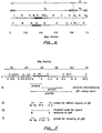
- FIG. 6 A restriction map for the entire HSV-1 genome is shown in Fig. 6, and a more detailed map of the region which was subcloned is shown in Fig. 7. Referring to Fig. 6, the conventional map is shown in the first two lines (Roizman, 1979). The dotted line indicates the L-S junction. The restriction enzyme cleavage map for Eco RI for the prototype isomer arrangement is shown in the third line (Skare and Summers, 1977; Roizman, 1979) with the Eco RI fragment F denoted by the cross-hatched box.
- the restriction enzyme sites shown in the detailed map line (I) are E, Eco RI; B, Bam HI; S, Sal I; P, Pst I; X, Xho I from DeLucca et al ., 1983; N, Nde I; Xn, Xmn I; V, Eco RV.
- the Bst EII site mapped by DeLucca et al ., at 0.355 is missing in this strain and there is a new Pst I site at 0.357.
- Line II shows three plasmid subclones which encompass the gB1 coding region.
- pHS106 which extends from the Bam HI site at 0.345 to the Sal I site at 0.360
- pHS107 which extends from the Sal I site at 0.36 to the Sal I site at 0.388
- pHS108 which is a Bam HI fragment extending from 0.345 to 0.40 map units.
- Line III indicates three probes used for mRNA mapping of gB1; line IV indicates the fragment used for hybrid selection; and line V shows those probes used to locate the gB2 gene (see below).
- the additional restriction sites used to generate these fragments are Nc, Nco I; K, Kpn I; and A, Alu I.
- Hybrid selected translation was performed by hybridizing HSV-1 poly A+ mRNA with a 3.2kb Kpn I- Xho I fragment, which encompasses the region indicated as encoding gB1.
- a 100kd protein similar in size to gB1 from HSV-1 infected Vero cells, was detected. Confirmation of the identity of the 100kd protein was achieved by immunoprecipitation with a gB1-specific monoclonal antibody.
- Several other proteins were also detected by hybrid selection using the Kpn I- Xho I fragment, probably the result of non-specific hybridization of mRNAs due to the high G+C content of the DNA.
- Fig. 8 is a restriction map of a 3.95kb DNA fragment, which extends from a Bam HI restriction site at 0.345 to an Xho I site at 0.373 map units.
- the open reading frame for gB1 is indicated by the box and the direction of transcription is from right to left as shown.
- the actual coding region covers map units 0.348 to 0.367.
- the DNA sequence from the Bam HI site to a non-unique Alu I site at nucleotide number 3640 is shown with the Alu I site indicated by the (A).
- restriction sites shown include B, Bam HI; Bl, Bal I; Bs, Bst EII; K, Kpn I; Nc, Nco I; P, Pst I; Pv, Pvu II; S, Sal I; Sc, Sac I; X, Xho I; Xm, Xma 3. Restriction sites are not shown for the right-hand end from the Alu I site to the terminal Xho I site. Three probes used for mapping the gB2 gene and potential glycosylation sites and hydrophobic anchor and signal regions (solid box) in the product gB1 protein are noted.
- the observed amino acid sequence is characteristic of a membrane protein.
- the first 56 amino acids are primarily hydrophobic, there being no charged residues from residues 11 to 56.
- This hydrophobic amino acid domain precedes a region with a high concentration of charged or hydrophilic amino acids.
- the hydrophobic sequence at the N-terminus may serve as a secretory leader or signal sequence followed by processing signals for cleavage and removal of the secretory leader.
- the hydrophobic region near the C-terminus can serve as a transmembrane integrating sequence for binding the protein to the cell membrane.
- the vector employed is a mammalian expression vector, referred to as pSV1/ dhfr .
- This 5.63kb plasmid contains 2.8kb of E . coli plasmid pBR328 sequences, including the ampicillin-resistance ⁇ -lactamase gene and the origin of replication.
- the vector also contains a selectable mammalian cell marker, the mouse dihydrofolate reductase cDNA gene ( dhfr ) (Nunberg et al ., Cell (1980) 19 :355) linked to the SV40 early promoter, which directs the transcription of dhfr .
- Additional SV40 sequences including t antigen splice donor and splice acceptor sites and the polyadenylation sites for early transcripts, are included downstream from the dhfr gene within a 1.65kb Bgl II- Eco RI fragment.
- pSV1/ dhfr -gB plasmids pHS112 and pHS114 were constructed.
- pSV1/ dhfr was restricted with Bgl II and Hin dIII excising the dhfr cDNA fragment. The resulting fragments are then blunt-ended by filling in the overhangs with the Klenow fragment of DNA polymerase I.
- a Xho I- Kpn I HSV-1 fragment containing the gB gene is isolated from pHS108. A portion is taken and partially digested with Pvu II to generate a DNA sequence lacking the 3'-anchor region. The resulting truncated gB1 gene lacks 580bp from the 3'-end of the gene. Both fragments are then blunt-ended with the Klenow fragment of pol I.
- Each gB blunt-ended fragment is ligated into the Bgl II- Hin dIII restricted pSV1/ dhfr vector to provide two sets each of constructs, with the gB1 gene in opposite orientations.
- the orientations having the Xho I generated terminus proximal to the SV promoter, with the direction of transcription being from the SV promoter to the SV splice sites are selected and designated pHS111 and pHS113 for the complete and truncated gB1 genes, respectively.
- the two plasmids are then completely digested with Eco RI and partially digested with Bam HI to provide a cassette which includes the SV promoter, the gB gene and the SV splice and polyadenylation sites.
- the plasmids are then transfected into Chinese hamster ovary (CHO) cells deficient in dhfr using the calcium phosphate precipitation method as described in Materials and Methods.
- Transfected cells are selected by employing a selective medium lacking thymidine, purines and glycine. Cells were isolated by removal with a Pasteur pipette and propagation in multiwell plates. A number of clones were isolated which were shown to produce gB by immunofluorescence and radioimmunoprecipitation employing a HSV-1 polyclonal antibody or a monoclonal antibody specific for gB.
- Clones were initially selected at 0.01, 0.05, 0.10 and 0.3 ⁇ M MTX. Three clones synthesizing high levels of gB, as detected by immunofluorescence, were isolated from the 0.3 ⁇ M MTX selections. By radioimmune precipitation, these clones, pHS114-0.3 ⁇ M-6, 23 and 25, synthesize 2-3 times more gB during a 1hr labeling with 35S-methionine than the unamplified clone, pHS114-7. Pulse chase experiments indicate that at least 50% of the gB synthesized in these clones during a 1hr pulse is secreted extracellularly by 5hr.
- Yeast expression was developed as follows: Partial N-terminal amino acid analysis of isolated gB1 was found to have glu, ala, and ala for amino acids 2, 5 and 11, indicating that the N-terminal region of the mature protein was at amino acid 151 of the sequence described previously (after the Mbo II site).
- a cassette was prepared employing the glyceraldehyde-3-phosphate-dehydrogenase promoter region and terminator region.
- a yeast gene library was prepared by inserting fragments obtained after partial digestion of total yeast DNA with restriction endonuclease Sau 3A in lambda-phage Charon 28 (Blattner et al ., Science (1977) 196 :161-169). The phage library was screened with DNA complementary to the yeast GAPDH mRNA and the yeast GAPDH gene from one of these clones was subcloned as a 3.5kb Bam HI fragment in the Bam HI site of pBR322 (pGAP-2). The GAPDH promoting-active fragments were isolated from these clones.
- a Hha I- Hin dIII fragment of about 350bp containing the 3' portion of the promoter was obtained by: a) digestion of pGAP-2 with Hin fI to generate an approximately 500bp segment which includes the 3' part of the promoter and a region encoding the N-terminal amino acids of GAPDH; b) resection with Bal 31 to yield a 400bp fragment lacking the GAPDH coding region (3'-terminus 1 base upstream from the ATG initiator codon); c) addition of Hin dIII ]inkers; and d) cleavage with Hha I.
- a Hin dIII Hha I fragment of about 700bp containing the 5' portion of the promoter was ligated to the 350bp Hha I- Hin dIII fragment and treated with Hin dIII.
- the resulting 1,061bp Hin dIII fragment was isolated by gel electrophoresis and cloned in pBR322 (pGAP-347).
- the GAPDH promoter fragment in pGAP347 was isolated by cleavage with Bam HI (within the 5' pBR322 flanking region) and partially with Hin dIII (at the 3' end of the promoter fragment) to provide a 1,407bp fragment containing a 1,061bp region of the GAPDH promoter region and 346bp of pBR322.
- This procedure utilized digestion of 50 ⁇ g of the pGAP-347 with 10 units each of Bam HI and Hin dIII with the resulting fragment purified by preparative gel electrophoresis in 1% agarose.
- a third fragment was a Xho II- Sac II fragment of 1,187bp containing the gB1 coding region.
- a fourth fragment containing the GAPDH terminator fragment (approximately 900bp) was isolated by Sal I- Bam HI digestion of a cloned fragment of the GAPDH gene with its 3' flanking region including the GAPDH termination region, so that a portion of the coding region is included with the termination region.
- the two fragments can be ligated together by means of a Sac II- Sal I adapter
- the Hin dIII- Sal I fragment (0.25 picomoles) was ligated in a single step to the 1407bp Bam HI- Hin dIII GAPDH promoter fragment (0.1 picomoles), the 900bp Sal I- Bam HI terminator (0.1 picomoles) and 0.02 picomoles of Bam HI-digested, phosphatased pBR322 in the presence of T4 DNA ligase.
- Plasmids containing the cassette clones in pBR322 were isolated and the correct nucleotide sequence confirmed by DNA sequencing. This plasmid was then digested with Bam HI and the Bam HI cassette fragment containing the gB1 segment and GAPDH regulatory regions gel isolated and inserted into Bam HI digested, phosphatased pC1/1. Plasmid pC1/1 is a derivative of pJDB219 (Beggs, Nature (1978) 275 :104) in which the region corresponding to bacterial plasmid pMB9 in pJDB219 is replaced by pBR322 in pC1/1.
- the pC1/1 plasmid containing the 1187bp gB1 insert and GAPDH promoter and terminator regions was designated pHS127A. This plasmid was then used to transform the yeast strain S . cerevisiae AB103.1 ( ⁇ , pep 4-3 , leu 2-3 , leu 2-112 , ura 3-52 , his 4-580 ). Transformants were initially grown in 1.0ml of leu ⁇ medium and then 50ml of YEPD inoculated with 0.4ml and grown further to an absorbence of 1-3 at 650nm (12hr).
- the yeast cells were pelleted by centrifugation at 2krpm for 10min at 4°C and resuspended in 50mM Tris-HCl, pH 8, 150mM NaCl, 0.2% Triton X-100, 1mM EDTA and freshly added 1.0mM phenylmethylsulfonyl fluoride and 0.1 ⁇ g/ml pepstatin.
- the cells were repelleted and then resuspended in a volume equal to the packed cell volume in the same buffer.
- An equal volume of acid-washed glass beads (diameter 0.45-0.5mm) was added and the yeast cells disrupted by vortexing at 4°C for 10min total, using 1min intervals.
- the tubes were centrifuged for 15min at 14,000xg at 4°C and the supernatant isolated and analyzed on 10% SDS polyacrylamide gel and blotted onto nitrocellulose paper for Western analysis (Burnett, Anal. Biochemistry (1981) 112 :195).
- a polyclonal antibody (DAKO) to HSV-1 was employed as the primary antibody. Expression of an HSV specific protein was observed at about 44kd, the size expected for the gB fragment.
- the gene for glycoprotein gB2 was located by hybridization with specific DNA probes generated from the gB1 coding region.
- a restriction map was determined for the Hin dIII H fragment of HSV-2 strain 333 which spans map coordinates 0.28-0.39 as shown in Fig. 9.
- a random library of HSV-2 strain 333 sequences cloned into the lambda vector L-47.1 (Leonen and Brammar, Gene (1980) 10 :249) (obtained from Dr. James Casey, Louisiana State University) was generated by a partial Mbo I digestion of the viral DNA and cloned into the Bam HI site of the vector.
- the library was screened for plaque hybridization using a Kpn I- Nde I fragment of gB1 as a probe.
- a positive lambda clone was picked and the lambda DNA prepared and digested with Sph I (Maniatis et al ., Molecular Cloning , Cold Spring Harbor Laboratory, 1982).
- a Southern blot analysis show that the gB2 information was contained within a 4.2kb fragment.
- the Sph I-digested lambda DNA was ligated to Sph I-digested pBR322 and the mixture used to transform E . coli HB101.
- the appropriate transformant was selected by restriction analysis and confirmed by southern blot analysis
- the 5'-end is contained within pHS203, a 2.6kb Xho I- Xho I clone, and the 3'-end within the adjacent 4.2kb Sph I- Sph I fragment, pH206, indicated on the above figure.
- FIG. 10 A more detailed restriction map of the pHS203 Xho I subclone is shown in Fig. 10.
- the figure shows a restriction map of pHS203, a 2.6kb Xho I- Xho I fragment which contains the 5'-end of the gene encoding gB2.
- Line I shows a restriction map which includes the restriction sites for Bl, Bal I.; Nr, Nru I; M, Sma I; P, Pst I; S, Sal I; Sp, Sph I; V, Pvu I; X, Xho I; Xm, Xma III.
- Those restriction sites marked by an asterisk are conserved between the DNA sequences of gB1 and gB2.
- the three fragments indicated on Fig. 10 were prepared as probes to Northern blots of poly A+ mRNA isolated from HSV-2 infected Vero cells 12hr after infection. Two of the probes, the 850bp Xho I to Sma I fragment and the adjacent 1150bp Sma I to Nru I fragment both hybridized to an abundant mRNA of 3.0kb, the expected size and representation frequency expected for the gB2 message. The third probe, the 470bp Nru I- Xho I fragment did not hybridize to the same message.
- the sequence of the fragments is shown as follows in Table 3 including a comparison of the sequence of the 5'-end of gB1 and gB2 for other than fragment DNA a.
- HSV-2 glycoprotein gB has been achieved in COS cells (transient expression) and in CHO cells (stable cell line secreting gB2) transformed with pHS210 alone or cotransformed with pHS210 and a second plasmid containing dhfr .
- Plasmid pHS210 was constructed as follows. The entire gene was subcloned as a 3.8kb Nru I- Bam HI fragment in pBR322 to generate pHS208. See Fig. 11. The Pst I site at the 5' end of the gene, 100bp to the right (downstream) of the Nru I site, was changed to a Hin dIII site by in vitro mutagenesis in M13. A Hin dIII to Pvu fragment of 1.9kb was then inserted into pSV1 which was obtained by digestion of pSV1/ dhfr with Hin dIII and Bgl II. See Figs. 1 and 11. For this cloning step pHS208, was cut with Pvu II and the end repaired to blunt.
- the molecule was then cut with Hin dIII and the 1.9kb Hin dIII - ( Pvu II) fragment isolated by gel electrophoresis.
- pSV1/ dhfr was cut with Bgl II, repaired to blunt, cut with Hin dIII and the 4.85kb Hin dIII-( Bgl II) vector fragment isolated by gel electrophoresis. These two fragments (1.9kb and 4.85kb) were ligated together to generate pHS210 - the expression plasmid.
- Plasmid pHS210 was used directly to transform COS cells. Expression was detected by immunofluorescence using a gB specific monoclonal antibody, F3 A/B, and also using a commercially available polyclonal anti HSV-2 antibody (DAKO) as the primary antibody screen. Plates were coated with the monoclonal antibody. Samples of cell culture medium were added to coated plates, then bound gB2 was detected with the rabbit anti HSV-2 polyclonal antibody (DAKO) followed by horseradish conjugated goat antirabbit IgG.
- DAKO rabbit anti HSV-2 polyclonal antibody
- pHS210 For CHO cell transformation plasmid pHS210 was used along with a second plasmid containing dhfr as a selective marker in a cotransfection protocol. Expression was detected using an ELISA assay in which ELISA plates were coated with F3 A/B specific monoclonal antibody.
- Yeast were transformed and grown using a variety of media including selective medium (yeast nitrogen base without leucine); YEPD medium, containing 1% (w/v) yeast extract, 2% (w/v) peptone and 2% (w/v) glucose, and others as appropriate and/or detailed below. In the case of plating medium contained 2% (w/v) agar and for transformation 3% top agar.
- yeast expression vectors containing synthetic DNA fragments coding for polypeptides A and B of the gD gene .
- the gD-A sequence which encodes amino acids 253-283 (corresponding to amino acids 258-288, as incorrectly numbered by Watson et al ., supra.) of the mature protein, was as follows:
- the gD-B sequence which encodes amino acids 8-23 (corresponding to amino acids 13-28, as incorrectly numbered by Watson et al ., supra .) of the mature protein, was as follows: The sequences each include a Kpn I cohesive end at the 5'-end and a Sal I cohesive end at the 3'-end. Coding for the mature secreted peptide begins after the LysArg processing site.
- the 5'-end of each sequence is a modification of the 3'-end of the naturally-occurring ⁇ -factor secretory leader and processing signal sequence, where the modification consists of a deletion of three glu-ala pairs and a replacement of a leucine by a proline to create a Kpn I site in the nucleotide sequence.
- the 3'-end of each sequence includes two translational stop codons, OP and OC.
- Synthetic DNA fragments having the sequences just described were prepared by synthesizing overlapping ssDNA segments as described by Urdea et al ., Proc. Natl. Acad. Sci. USA (1984) 80 :7461-7465 using the phosphoramidite method of Beaucage and Caruthers, Tetrahedron Lett . (1981) 27 :1859-1862, and annealing and ligating under the following conditions.
- the ssDNA fragments were joined as follows: 50 to 100 pmoles of each segment (except the two segments at the 5'-termini) were 5'-phosphorylated with 5.6 units of T4 polynucleotide kinase (New England Nuclear) in 10mM dithiothreitol (DTT), 1mM ATP, 10mM MgCl2, 100ng/ml spermidine, 50mM Tris-HCl, pH 7.8 (total volume: 20 ⁇ l) for 30 min at 37°C. Additional T4 kinase (5.6 units) was then added and the reaction continued for 30 min at 37°C.
- T4 polynucleotide kinase New England Nuclear
- DTT dithiothreitol
- 1mM ATP 1mM ATP
- 10mM MgCl2 100ng/ml spermidine
- 50mM Tris-HCl pH 7.8 (total volume: 20 ⁇ l)
- the fragments (except for the 5'-termini) were combined, diluted to 40 ⁇ l with water followed by addition of 60 ⁇ l of 1M sodium acetate, 12 ⁇ l of 250mM EDTA, and 5 ⁇ g of 1mg/ml poly-dA. After heating for 5min at 65°C, the 5' terminal pieces were added, followed by 420 ⁇ l of ethanol (100%). The solution was chilled for 20min at -80°C, centrifuged, and the pellet was washed twice with ethanol (100%) and dried. The pellet was redissolved in water (18 ⁇ l), heated to 100°C for 2 minutes and then cooled slowly over 1.5 hours to 25°C in a water bath.
- the annealed fragment pool was ligated in a reaction mixture containing T4 DNA ligase (New England Biolabs, 1200 units) 1mM ATP, 10mM DTT, 10mM MgCl2, 100ng/ml spermidine, and 50mM Tris-HCl, pH 7.8 (30 ⁇ l). After 1 hour at 14°C, the reaction mixture was partially purified by preparative polyacrylamide gel (7%, native) electrophoresis. The DNA was removed from the appropriate gel slice by electroelution and ethanol coprecipitation with poly dA (5 ⁇ g).
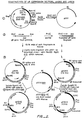
- the synthetic gD-A and gD-B fragments were substituted into a Kpn I/ Sal I digested bacterial cloning plasmid pAB114 ⁇ EGF-24, which plasmid was prepared by cloning a mutagenized fragment of pYEGF-8 into pAB114 (see Fig. 3).
- the plasmids resulting from the insertion were designated pAB114 ⁇ HS109 (gD-A) and pAB114 ⁇ HS110 (gD-B).
- Plasmid pAB101 was obtained from the screening of a random yeast genomic library cloned in YEp24 (Fasiolo et al ., J. Biol. Chem. (1981) 256 :2324) using a synthetic 20-mer oligonucleotide probe (5'-TTAGTACATTGGTTGGCCGG-3') homologous to the published ⁇ -factor coding region (Kurjan and Herskowitz, Abstracts (1981), Cold Springs Harbor meeting on the Molecular Biology of Yeasts, page 242). Plasmid AB11 was obtained by deleting the Hin dIII to Sal I region of pBR322.
- Plasmid pAB112 was digested to completion with Hin dIII, and then religated at low (4 ⁇ g/ml) DNA concentration to produce plasmid pAB113 in which three 63bp Hin dIII fragments were deleted from the ⁇ -factor structural gene, leaving only a single copy of mature ⁇ -factor coding region.
- a Bam HI site was added to plasmid pAB11 by cleavage with Eco RI, filling in of the overhanging ends by the Klenow fragment of DNA polymerase, ligation of Bam HI linkers and religation to obtain a plasmid designated pAB12.
- Plasmid pAB113 was digested with Eco RI, the overhanging ends filled in, and ligated to Bam HI linkers.
- the resulting 1500bp which carries the single copy of the ⁇ -factor gene fragment was gel-purified and ligated to pAB12 which had been digested with Bam HI and treated with alkaline phosphatase to produce pAB114, which contains a 1500bp Bam HI fragment carrying the ⁇ -factor gene.
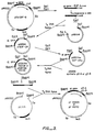
- pYEGF-8 The preparation of pYEGF-8 was as follows: A synthetic sequence for human epidermal growth factor (EGF) was prepared and ligated to pAB112 (described above) which had been previously completely digested with Hin dIII and Sal I to produce pAB201. The Hin dIII site lies within the 3'-end of the ⁇ -factor gene, and the EGF sequence was inserted using appropriate linkers. The resulting plasmid was designated pAB201.
- Plasmid pAB201 (5 ⁇ g) was digested to completion with Eco RI and the resulting fragments were filled in with DNA polymerase I Klenow fragment and ligated to an excess of Bam HI linkers. The resulting 1.75kbp fragment was isolated by preparative gel electrophoresis, and approximately 100ng of this fragment was ligated to 10ng of yeast plasmid pC1/1 (described below) which had been previously digested to completion with restriction enzyme Bam HI and treated with alkaline phosphatase. The ligation mixture of the 1.75 kbp fragment carrying the partial ⁇ -factor gene fused to the EGF gene and pC1/1 was used to transform E .
- DNA from one ampicillin resistant clone (designated pYEGF-8) was used to transform yeast AB103 (genotype: MAT ⁇ , pep 4-3 , leu 2-3 , leu 2-112 , ura 3-52 , his 4-580 , cir°) cells, and transformants selected based on their leu+ phenotype.
- Plasmid pC1/1 is a derivative of pJDB219 (Beggs, Nature (1978) 275 :104) where the region derived from bacterial plasmid pMB9 has been replaced by pBR322.
- the pC1/1 plasmid carries genes for both ampicillin resistance and leucine prototrophy.
- Plasmid pAB114 ⁇ EGF-24 was generated by an in vitro mutagenesis procedure which deleted the sequences coding for the glu-ala processing region in the ⁇ -factor leader. Plasmid pAB114 ⁇ EGF-24 was obtained as follows: a Pst I- Sal I fragment of pYEGF-8 containing the ⁇ -factor leader hEGF fusion was cloned in phage M13 and isolated in single-stranded form. A synthetic 36-mer oligonucleotide primer (5'-GGGGTACCTTTGGATAAAAGAAACTCCGACTCCGAA-3') was used as a primer for the synthesis of the second strand using the Klenow fragment of DNA polymerase I.
- the Bam HI- Bam HI fragment of pAB114 ⁇ HS109 or pAB114 ⁇ HS110 (1588 base pairs for gD-A and 1546 base pairs for gD-B) was excised and ligated into the unique Bam HI site of pC1/1.
- the resulting expression vectors were designated pYHS109 for gD-A and pYHS110 for gD-B.
- Plasmids pYHS109 and pYHS110 were both used to transform yeast strain AB103.1 ( ⁇ , pep 4-3 , leu 2-3 , leu 2-112 , ura 3-52 , his 4-580 , cir°) to leu prototrophy following the procedure of Hinnen et al ., Proc. Natl. Acad. Sci. USA (1978) 75 :1929-1933.
- the transformants were grown in 1 L cultures at 30°C in buffered leucine-deficient media to saturation, corresponding to an absorbance of 5 at 650nm.
- Yeast cell cultures were maintained at saturation for an additional 12 to 24 hrs with shaking at 30°C.
- the cultures were then harvested, the intact yeast cells pelleted by centrifugation at 3000 RPM, and the resulting supernatant media filtered through a 0.22 ⁇ Millipore filter. This fraction was then passed through a C18 reverse phase column, constructed from 8 Seppak units purchased from Waters. The bound material was eluted with 30 ml of 80% (v/v) acetonitrile, 0.1% (v/v) trifluoroacetic acid in water, evaporated to dryness with a Buchii RotoVap and redissolved in 1.6ml of distilled water. This material was separated on a HPLC C18 column monitored at 210nm. The peak corresponding to each respective peptide was collected and its identity confirmed by antigenicity.
- Nucleotide fragments from the naturally-occurring gD gene expressing gD of HSV-1 were also expressed intracellularly in yeast under control of the GAPDH promoter and terminator.
- the gD is entirely contained within a 2.9kb Sac I fragment within the Eco RI fragment of one clone (clone H) isolated from the HSV-1 library. Clone H was obtained from Dr.
- Fig. 4 illustrates the protein coding region (boxed region) and the fragments used for the construction of yeast expression vectors pYHS115, pYHS116, pYHS117, pYHS118 and pYHS119. A description of the construction of each plasmid follows.
- Plasmid pYHS115 contains the gD gene in a glyceraldehyde-3-phosphate dehydrogenase (GAPDH) expression cassette cloned into the Bam HI site of pC1/1 (described hereinabove).
- GPDH glyceraldehyde-3-phosphate dehydrogenase
- the GAPDH expression cassette was constructed as follows. Three fragments were prepared (as described in detail below):
- Fragment (a) was prepared by completely digesting pGAP347 (described below) with Bam HI followed by partial digestion with Hin dIII. The resulting 1407 bp fragment containing 346bp of pBR322 and 1061bp of the GAPDH promoter was isolated by gel electrophoresis.
- PolyA+ RNA was isolated from S . cerevisiae yeast strain A364A. Double-stranded cDNA was synthesized using AMV reverse transcriptase and E . coli DNA polymerase I. Poly-dC-tails were added to the double-stranded cDNA molecule using deoxynucleotide terminal transferase. Poly-dC-tailed cDNA was annealed to poly-dG-tailed pBR322 and used to transform E . coli HB101. 1000 transformants were screened by colony hybridization to labeled PolyA+ RNA, and a subset further examined by restriction endonuclease mapping, and DNA sequencing. Three clones containing GAPDH sequences were isolated from the pool. One clone (pcGAP-9) contained an insert of about 1200 base pairs and was used for further work.
- a yeast gene library was prepared by inserting fragments obtained after partial digestion of total yeast DNA with restriction endonuclease Sau 3A in lambda phage Charon 28, according to Blattner, et al ., Science (1977) 196 :161-169.
- Several fragments containing yeast GAPDH coding sequences were isolated by screening the phage library with labeled DNA from pcGAP-9.
- the yeast GAPDH gene of one of these clones was subcloned in pBR322 as a 2.1kb Hin dIII fragment (pGAP-1). The GAPDH promoter region was isolated from these clones.
- a Hha I- Hin dIII fragment of about 350bp containing the 3' portion of the promoter was obtained by: a) digestion of pGAP-1 with Hin fI to generate an approximately 500bp segment which includes the 3' part of the promoter and a region encoding the N-terminal amino acids of GAPDH; b) resection with Bal 31 to yield a 400bp fragment lacking the GAPDH coding region (3'-terminus one base upstream from the ATG initiator codon); c) addition of Hin dIII linkers; and d) cleavage with Hha I.
- Hin dIII- Hha I fragment of about 700bp containing the 5' portion of the promoter was isolated from pGAP-1, ligated to the 350bp Hha I- Hin dIII fragment and treated with Hin dIII.
- the resulting 1061bp Hin dIII fragment was isolated by gel electrophoresis and cloned in Hin dIII digested, alkaline phosphatase treated pBR322 to produce pGAP347.
- Fragment (b) was obtained as follows. Clone H, isolated from the HSV-1 Patton library was digested with Sac I. A 2.9kb Sac I fragment was purified by gel electrophoresis and subsequently digested with Hin dIII and Nru I. The 1430bp Hin dIII- Nru I fragment containing the gD gene (Fig. 3) was purified by gel electrophoresis, ligated to Nru I- Sal I adaptors of the following sequence: and digested with Sal I.
- Fragment (c) was obtained as follows. A 900bp fragment containing the GAPDH terminator was obtained by Bam HI and Sal I digestion of pUH28 (described under "Construction of pYHS117”) and purification by gel electrophoresis.
- pYHS116 contains a gD gene fragment which has a 600bp deletion at the 5' end of the coding region that comprises most of the signal sequence coding region.
- pYHS116 two fragments were obtained:
- Fragments (a) and (b) were ligated together and subsequently digested with Bam HI.
- the resulting 3.5kb cassette was isolated by gel electrophoresis and ligated to Bam HI cut, alkaline phosphatase treated pC1/1.
- Plasmid pYHS117 contains the same partial gD gene clone in pYHS116, fused in reading frame to 7 extra codons at the 5'-end, which code for the first 7 amino acids of the GAPDH structural gene. To construct pYHS117 two fragments were obtained.
- Plasmid pUH28 contains the coding and 3' non-coding regions of the hepatitis B surface antigen (HBsAg) gene fused in incorrect reading frame to the first 7 codons of the GAPDH structural gene. This fusion is flanked in its 5' end by the GAPDH promoter and in its 3' end by part of the GAPDH coding region followed by the GAPDH terminator.
- This plasmid was constructed so as to have an Nco I site at the 3' end of the first 7 codons of the GAPDH gene with the following sequence: When this Nco I end is ligated to the partial gD fragment (b, described above) the correct reading frame for the gD protein is regenerated.
- the Sal I site used in the preparation of fragment a (described above) is at the 5' region of the GAPDH terminator. Therefore, a deletion of the sAg coding plus non-coding regions and GAPDH coding region was obtained by digesting pUH28 with Nco I and partially with Sal I.
- pUH28 involves cloning of a fragment that contains the HBsAg coding and 607bp of 3' non-coding region prepared from pHBS5-3 Hae2-1 (described below) into the GAPDH containing vector pGAP ' 2 (described below).
- pHBS5-3 Hae2-1 was linearized by Pst I digestion, partially digested with Nco I and a Pst I- Nco I fragment of 1.9kb containing pBR322 sequences, HBsAg coding and 3' sequences was purified by gel electrophoresis.
- This fragment was subsequently digested with Eco RI and a 1.2kb Nco I- Eco RI fragment containing the HBsAg coding and 3' non-coding regions was purified by gel electrophoresis.
- Plasmid pGAP ' 2 was linearized with Xba I and treated with Bal 31 to remove approximately 100bp.
- the plasmid was subsequently digested with Nco I and a vector fragment of about 9kb was purified by gel electrophoresis.
- the Nco I ends of the vector and the 1.2kb Nco I- Eco RI fragment encoding HBsAg were ligated.
- the recessed end was filled in with Klenow and the resulting blunt end was ligated to the blunt end of the vector obtained by Bal 31 digestion to produce pUH28.
- pHBS5-3 Hae2-1 is a plasmid that contains the HBsAg coding region and 607bp of 3' flanking sequences. This plasmid is a derivative of pHBS5-3 which contains the same insert but only 128bp of 3' untranslated region instead of 607bp. Plasmid pHBS5-3 has been previously described in copending U.S. application, serial no. 609,540, filed May 11, 1984, (pp. 13-14). pHBS5-3 Hae2-1 was constructed as follows. The HBV genome (3.2kb) was excised from pHB-3200 (Valenzuela et al ., Nature (1979) 280 :815-819) by restriction digestion with Eco RI.
- the 3.2kb fragment was purified by gel electrophoresis and was recircularized by ligation of the Eco RI sticky ends.
- This closed HBV genome was digested with Hae II, which cuts in the 3' non-coding region. Recessed ends were filled in with Klenow and Hin dIII linkers were ligated.
- the DNA was cut with Hin dIII and subsequently with Xba I, which has a single site in the HBS coding region.
- a 1.2kb Xba I- Hin dIII fragment containing 586 base pairs of the coding sequence of HBV and 607 base pairs of the 3' non-coding region was isolated by gel electrophoresis. This fragment was cloned into pHBS5-3 previously cut with Xba I and Hin dIII and treated with alkaline phosphatase, to yield pHBS5-3 Hae 2-1.
- pGAP-2 is a pBR322 derived vector which contains a Bam HI insert that has the GAPDH coding sequence, 5' and 3' flanking regions. There are two Xba I sites in this plasmid: one in the coding region and one in the 3' flanking sequences.
- pGAP-2' is a derivative of pGAP-2 in which the Xba I site present in the 3' flanking region has been eliminated. For this purpose, 50 ⁇ g of pGAP-2 were partially digested with Xba I, treated with Bal 31 to remove 25 base pairs per end, and ligated. The plasmids were used to transform E . coli HB101 and the transformants were selected for loss of the Xba I site in the 3' flanking region.
- This vector contains a partial gD gene with deletions in two regions: a 600bp deletion in the 5'-end coding region which comprises most of the signal sequence coding region and a 1300bp deletion in the 3'-end coding region which includes most of the anchor sequence coding region. It also contains 7 extra codons from the GAPDH gene coding region fused in reading frame at the 5' end of the gD gene, similar to pYHS117. Plasmid pYHS118 was constructed as follows. pYHS115 was digested with Nco I and Sal I, the resulting 1430bp fragment containing the partial gD was purified by gel electrophoresis and submitted to digestion with Nar I (Fig. 4).
- fragment a 873bp containing 5' end and fragment b : 411bp containing 3' end
- Fragment b was subsequently digested with Sau 96A to yield three fragments which were separated by gel electrophoresis.
- the 87bp Nar - Sau 96A fragment was recovered from the gel and was ligated to Sau 96I- Sal I synthetic adaptors of the following sequence:
- the Nar I-( Sau 96A) Sal I fragment (102bp) was digested with Sal I, purified by gel electrophoresis and ligated with fragment a (previously described).
- Nco I- Sal I fragment (975bp) was ligated to Nco I- Sal I digested and gel purified pUH28 as described under "Construction of pYHS117.”
- the resulting pBR322 derived vector was digested with Bam HI and a 3.1kb fragment containing the gD expression cassette was purified by gel electrophoresis. This cassette was ligated to Bam HI digested, alkaline phosphatase treated pC1/1 to produce pYHS118.
- This vector contains a partial gD gene with deletions in two regions: a 600bp deletion in the 5' end coding region which comprises most of the signal sequence coding region and a 2400bp deletion in the 3' end coding region which includes all the anchor sequence coding region and about 700bp upstream of the anchor sequence. It also contains 7 extra codons from the GAPDH gene coding region fused in reading frame at the 5' end of the gD gene, as pYHS117 and pYHS118. Plasmid pYHS119 was constructed as follows. pYHS115 was digested with Nco I and Sal I, the resulting 1430bp fragment containing the partial gD was purified by gel electrophoresis and subsequently digested with NarI.
- the 873bp Nco I- Nar I fragment was isolated by gel electrophoresis.
- the resulting Nco I- Sal I fragment was ligated to pUH28 which had been previously completely digested with Nco I and partially digested with Sal I and purified by gel electrophoresis as described under "Construction of pYHS117.”
- the resulting pBR322 derived vector was digested with Bam HI and a 2.2kb fragment containing the gD expression cassette was purified by gel electrophoresis. This cassette was ligated to Bam HI digested, alkaline phosphatase treated pC1/1 to produce pYHS119.
- Plasmids pYHS115, 116, 117, 118 and 119 were used to transform yeast strain AB103.1 ( ⁇ , pep 4-3 , leu 2-3 , leu 2-113 , ura 3-52 , his 4-580 , cir°) following the procedure of Hinnen et al ., supra .
- the cultures were then harvested by pelleting the yeast cells at 3000 RPM. Cells were spheroplasted with zymolyase and subsequently osmotically lysed in a hypotonic solution.
- Membranes were spun down in an eppendorf centrifuge, and the pellet was solubilized in 0.1% SDS with protease inhibitors for 16 hours at 4°C. The suspension was centrifuged and total protein, as well as gD specific protein, was determined in both soluble and insoluble fractions. Expression of the gD gene in each of the above described constructions was detected by Western Blot hybridization (Towbin et al ., Proc. Natl. Acad. Sci. USA (1979) 76 :4350).
- protein samples were submitted to SDS-polyacrylamide gel electrophoresis (Laemmli, Nature (1970) 227 :680) and electroblotted onto nitrocellulose filters (Towbin, et al ., supra.).
- the filter was preincubated with goat serum and subsequently treated with a rabbit polyclonal antibody raised against HSV-1 (Dako).
- the filter was then incubated with a second goat anti-rabbit antibody conjugated with horseradish peroxidase (Boehringer-Mannheim) and finally it was incubated with horseradish peroxide color development reagent (Bio-Rad) and washed.
- mice Female Swiss Webster mice (Simonsen) were immunized on day 1 with a 1.1 mixture of recombinant vaccine preparation and complete Freund's adjuvant. One half the dose was administered subcutaneously, the other half intraperitoneally. The mice received either 20 ⁇ g recombinant gD protein or 5 ⁇ g recombinant gB protein. Mice were boosted on day 20 with the same dose and vaccine preparations, but incomplete Freund's adjuvant was used to make the emulsion. They were bled by cardiac puncture on day 28. Total lectin affinity purified Patton strain HSV glycoproteins served as a positive vaccine control, and a recombinant Feline leukemia envelope B protein was used as a negative control. A second boost was given on day 41 and animals were bled again on day 53. Sera from the mouse bleeds were collected and assayed for antibody levels by ELISA and plaque reduction neutralization assay. The results of this experiment are summarized in Table 4 below.
- mice Female Swiss Webster mice were immunized with the vaccine candidates. The first injections were done using 20 ⁇ g recombinant gD or 5 ⁇ g recombinant gB mixed in an equal volume of complete Freund's adjuvant. One half the dose was given intraperitoneally, the other half subcutaneously on day 1. Mice were boosted 2 and 4 weeks later in the same manner with incomplete Freund's adjuvant in the emulsion. Control animals received 20 ⁇ g of lectin purified total HSV glycoproteins previously described or 23 ⁇ g of a control yeast plus CHO cell extract. Mice were bled one week after the final boost by cardiac puncture.
- the initial immunization contained 10-100 ⁇ g of glycoprotein in complete Freund's adjuvant in a volume of 100-200 ⁇ l injected into the hindlimb footpads, followed four weeks later by the same vaccine given intramuscularly to each hindlimb.
- the animals were intravaginally inoculated with the HSV-2 MS strain.
- the animals were evaluated daily for 14 days to determine the severity of primary infection .
- the severity of the primary infection was determined by clinical observation of the lesions. The lesions were scored according to the following manifestation:
- novel vaccines are provided effective against Herpes Simplex Virus Types 1 and 2. These vaccines employ recombinant gB and gD or related polypeptides either separately or together.
- E . coli HB101 strains were deposited at the A.T.C.C., where the plasmid indicates the plasmid employed to transform the strain: pHS203; pHS112; pHS114; pHS127A and pHS206 were deposited on April 4, 1984, and assigned Accession Nos. 39649-39653, respectively; pYHS109 and pYHS118 were deposited on July 11, 1984, and given Accession Nos. 39762 and 39763 respectively.
Landscapes
- Life Sciences & Earth Sciences (AREA)
- Health & Medical Sciences (AREA)
- Genetics & Genomics (AREA)
- Chemical & Material Sciences (AREA)
- Organic Chemistry (AREA)
- Engineering & Computer Science (AREA)
- Wood Science & Technology (AREA)
- Biochemistry (AREA)
- Biotechnology (AREA)
- General Engineering & Computer Science (AREA)
- Molecular Biology (AREA)
- Mycology (AREA)
- Bioinformatics & Cheminformatics (AREA)
- General Health & Medical Sciences (AREA)
- Biomedical Technology (AREA)
- Zoology (AREA)
- Biophysics (AREA)
- Physics & Mathematics (AREA)
- Microbiology (AREA)
- Plant Pathology (AREA)
- Virology (AREA)
- Gastroenterology & Hepatology (AREA)
- Medicinal Chemistry (AREA)
- Proteomics, Peptides & Aminoacids (AREA)
- Medicines Containing Antibodies Or Antigens For Use As Internal Diagnostic Agents (AREA)
- Preparation Of Compounds By Using Micro-Organisms (AREA)
- Micro-Organisms Or Cultivation Processes Thereof (AREA)
- Peptides Or Proteins (AREA)
Applications Claiming Priority (5)
| Application Number | Priority Date | Filing Date | Title |
|---|---|---|---|
| US59778484A | 1984-04-06 | 1984-04-06 | |
| US06/631,669 US4618578A (en) | 1984-07-17 | 1984-07-17 | Expression of glycoprotein D of herpes simplex virus |
| EP85902226A EP0175787B1 (fr) | 1984-04-06 | 1985-04-04 | VACCIN gB-gD RECOMBINANT CONTRE L'HERPES SIMPLEX |
| US597784 | 1990-10-19 | ||
| US631669 | 1990-12-20 |
Related Parent Applications (2)
| Application Number | Title | Priority Date | Filing Date |
|---|---|---|---|
| EP85902226A Division EP0175787B1 (fr) | 1984-04-06 | 1985-04-04 | VACCIN gB-gD RECOMBINANT CONTRE L'HERPES SIMPLEX |
| EP85902226.1 Division | 1985-04-04 |
Publications (2)
| Publication Number | Publication Date |
|---|---|
| EP0628633A1 true EP0628633A1 (fr) | 1994-12-14 |
| EP0628633B1 EP0628633B1 (fr) | 2003-01-08 |
Family
ID=27082907
Family Applications (2)
| Application Number | Title | Priority Date | Filing Date |
|---|---|---|---|
| EP94202224A Expired - Lifetime EP0628633B1 (fr) | 1984-04-06 | 1985-04-04 | Vaccin gB-gD récombinant contre l'herpès simplex |
| EP85902226A Expired - Lifetime EP0175787B1 (fr) | 1984-04-06 | 1985-04-04 | VACCIN gB-gD RECOMBINANT CONTRE L'HERPES SIMPLEX |
Family Applications After (1)
| Application Number | Title | Priority Date | Filing Date |
|---|---|---|---|
| EP85902226A Expired - Lifetime EP0175787B1 (fr) | 1984-04-06 | 1985-04-04 | VACCIN gB-gD RECOMBINANT CONTRE L'HERPES SIMPLEX |
Country Status (6)
| Country | Link |
|---|---|
| EP (2) | EP0628633B1 (fr) |
| AT (2) | ATE118353T1 (fr) |
| CA (1) | CA1337181C (fr) |
| DE (1) | DE3587991T2 (fr) |
| HK (1) | HK107595A (fr) |
| WO (1) | WO1985004587A1 (fr) |
Families Citing this family (24)
| Publication number | Priority date | Publication date | Assignee | Title |
|---|---|---|---|---|
| NZ209308A (en) * | 1983-08-30 | 1991-08-27 | Genentech Inc | Vaccine against hsv involving a truncated membrane-free derivative of a membrane-bound protein |
| US7264817B1 (en) | 1983-08-30 | 2007-09-04 | Genentech, Inc. | Immunogenic composition based on a truncated derivative of a membrane bound protein and process for making it |
| US6100064A (en) * | 1984-04-06 | 2000-08-08 | Chiron Corporation | Secreted viral proteins useful for vaccines and diagnostics |
| US5244792A (en) * | 1984-04-06 | 1993-09-14 | Chiron Corporation | Expression of recombinant glyoprotein B from herpes simplex virus |
| US5171568A (en) * | 1984-04-06 | 1992-12-15 | Chiron Corporation | Recombinant herpes simplex gb-gd vaccine |
| EP0289550B1 (fr) * | 1986-10-20 | 1996-04-10 | Chiron Corporation | Vaccin utilise pour le traitement therapeutique de hsv |
| EP0297924A3 (fr) * | 1987-07-01 | 1990-07-11 | City Of Hope | Virus de la vaccine recombinant exprimant la glycoproteine HSV 1 gB |
| CA1335718C (fr) * | 1988-04-14 | 1995-05-30 | Mary L. Haffey | Expression d'une forme fonctionnelle d'adn polymerase d'herpesvirus dans la levure |
| DE69017769T2 (de) * | 1989-06-27 | 1995-07-13 | Smithkline Beecham Biologicals S.A., Rixensart | Verbindungen. |
| CA2088600C (fr) * | 1990-08-02 | 1999-11-16 | Rae L. Burke | Vaccins antiherpetiques fabriques a partir de l'epitope vp16 du virus |
| ES2150906T3 (es) | 1990-10-04 | 2000-12-16 | Res Corp Technologies Inc | Antigeno del virus varicella-zoster. |
| US5955088A (en) * | 1992-02-03 | 1999-09-21 | Cedars-Sinai Medical Center | Pharmaceutical compsition of herpes simplex virus type-1 (HSV-1), glycoproteins |
| US6193984B1 (en) | 1992-02-03 | 2001-02-27 | Cedars-Sinai Medical Center | Pharmaceutical composition of herpes simplex virus typ-1 (HSV-1) glycoproteins |
| US5679348A (en) * | 1992-02-03 | 1997-10-21 | Cedars-Sinai Medical Center | Immunotherapy for recurrent HSV infections |
| CA2090295A1 (fr) * | 1992-02-03 | 1993-08-04 | Anthony B. Nesburn | Procede pour l'expression de la glycoproteine e du virus de l'herpes simplex de type-1 et methodes d'utilisation |
| US5672349A (en) * | 1992-02-03 | 1997-09-30 | Cedars Sinai Medical Center | Process for the expression of herpes simplex virus type 1 glycoprotein I and methods of use |
| AU1273495A (en) * | 1994-12-06 | 1996-06-26 | Bruschettini S.R.L. | Glycoproteins from herpes virus and vaccines containing them |
| WO1996032962A1 (fr) | 1995-04-21 | 1996-10-24 | The University Of New Mexico | Dosage immunologique pour le virus de l'herpes |
| KR100408817B1 (ko) * | 1995-07-28 | 2004-02-25 | 주식회사 엘지생명과학 | 헤르페스심플렉스바이러스의당단백질을대량발현시키는동물세포주 |
| EP1156781B1 (fr) | 1999-02-26 | 2005-06-08 | Chiron Corporation | Microemulsions avec des macromolecules adsorbees et microparticules |
| JP2004099584A (ja) * | 2002-05-02 | 2004-04-02 | Keio Gijuku | Hsvを用いた抗腫瘍剤 |
| CA2538794C (fr) | 2003-09-12 | 2016-04-19 | Antigenics, Inc. | Vaccin pour le traitement et la prevention de l'infection provoquee par le virus de l'herpes simplex |
| CA3142023A1 (fr) * | 2019-05-28 | 2020-12-03 | Case Western Reserve University | Compositions et procedes de preservation de la methylation de l'adn |
| WO2021242872A1 (fr) | 2020-05-27 | 2021-12-02 | Case Western Reserve University | Compositions et procédés de préservation de la méthylation de l'adn |
Citations (3)
| Publication number | Priority date | Publication date | Assignee | Title |
|---|---|---|---|---|
| EP0100521A2 (fr) * | 1982-07-30 | 1984-02-15 | BEHRINGWERKE Aktiengesellschaft | Procédé pour la préparation d'un antigène d'herpès, moyen approprié à cet effet ainsi qu'un procédé pour sa préparation et l'utilisation de cet antigène |
| EP0168662A1 (fr) * | 1984-06-19 | 1986-01-22 | Juridical Foundation The Chemo-Sero-Therapeutic Research Institute | ADN recombinant inséré avec un gène de virus d'herpès simplex, cellules mammifères transformées avec ledit ADN recombinant et méthode pour la production de protéines dudit |
| EP0170169A1 (fr) * | 1984-07-20 | 1986-02-05 | Juridical Foundation The Chemo-Sero-Therapeutic Research Institute | ADN recombinant, contenant un gène du virus Herpès simplex ou un fragment de celui-ci, levure transformée par cet ADN recombinant, et procédé pour la production de protéines du virus Herpès simplex |
-
1985
- 1985-04-04 AT AT85902226T patent/ATE118353T1/de not_active IP Right Cessation
- 1985-04-04 DE DE3587991T patent/DE3587991T2/de not_active Expired - Lifetime
- 1985-04-04 WO PCT/US1985/000587 patent/WO1985004587A1/fr not_active Ceased
- 1985-04-04 EP EP94202224A patent/EP0628633B1/fr not_active Expired - Lifetime
- 1985-04-04 AT AT94202224T patent/ATE230798T1/de not_active IP Right Cessation
- 1985-04-04 EP EP85902226A patent/EP0175787B1/fr not_active Expired - Lifetime
- 1985-04-09 CA CA000478661A patent/CA1337181C/fr not_active Expired - Lifetime
-
1995
- 1995-07-06 HK HK107595A patent/HK107595A/en not_active IP Right Cessation
Patent Citations (3)
| Publication number | Priority date | Publication date | Assignee | Title |
|---|---|---|---|---|
| EP0100521A2 (fr) * | 1982-07-30 | 1984-02-15 | BEHRINGWERKE Aktiengesellschaft | Procédé pour la préparation d'un antigène d'herpès, moyen approprié à cet effet ainsi qu'un procédé pour sa préparation et l'utilisation de cet antigène |
| EP0168662A1 (fr) * | 1984-06-19 | 1986-01-22 | Juridical Foundation The Chemo-Sero-Therapeutic Research Institute | ADN recombinant inséré avec un gène de virus d'herpès simplex, cellules mammifères transformées avec ledit ADN recombinant et méthode pour la production de protéines dudit |
| EP0170169A1 (fr) * | 1984-07-20 | 1986-02-05 | Juridical Foundation The Chemo-Sero-Therapeutic Research Institute | ADN recombinant, contenant un gène du virus Herpès simplex ou un fragment de celui-ci, levure transformée par cet ADN recombinant, et procédé pour la production de protéines du virus Herpès simplex |
Non-Patent Citations (4)
| Title |
|---|
| J.H. WEIS ET AL.: "AN IMMUNOLOGICALLY ACTIVE CHIMAERIC PROTEIN CONTAINING HERPES SIMPLEX TYPE 1 GLYCOPROTEIN D.", NATURE., vol. 302, no. 5903, 3 March 1983 (1983-03-03), LONDON GB, pages 72 - 74 * |
| L.A. LASKY ET AL.: "PROTECTION OF MICE FROM LETHAL HERPES SIMPLEX VIRUS INFECTION BY VACCINATION WITH A SECRETED FORM OF CLONED GLYCOPROTEIN D.", BIOTECHNOLOGY, vol. 2, no. 6, June 1984 (1984-06-01), NEW YORK US, pages 527 - 532, XP001075097, DOI: doi:10.1038/nbt0684-527 * |
| R.J.WATSON ET AL.: "HERPES SIMPLEX VIRUS TYPE-1 GLYCOPROTEIN D GENE: NUCLEOTIDE SEQUENCE AND EXPRESSION IN ESCHERICHIA COLI.", SCIENCE, vol. 218, 22 October 1982 (1982-10-22), LANCASTER, PA US, pages 381 - 384, XP000995249, DOI: doi:10.1126/science.6289440 * |
| W.L. CHAN: "PROTECTIVE IMMUNIZATION OF MICE WITH SPECIFIC HSV-1 GLYCOPROTEINS.", IMMUNOLOGY, vol. 49, no. 2, June 1983 (1983-06-01), OXFORD, GB, pages 343 - 352 * |
Also Published As
| Publication number | Publication date |
|---|---|
| DE3587991D1 (de) | 1995-03-23 |
| HK107595A (en) | 1995-07-14 |
| CA1337181C (fr) | 1995-10-03 |
| ATE118353T1 (de) | 1995-03-15 |
| EP0175787A4 (fr) | 1988-02-23 |
| EP0175787B1 (fr) | 1995-02-15 |
| DE3587991T2 (de) | 1995-07-20 |
| EP0628633B1 (fr) | 2003-01-08 |
| ATE230798T1 (de) | 2003-01-15 |
| EP0175787A1 (fr) | 1986-04-02 |
| WO1985004587A1 (fr) | 1985-10-24 |
Similar Documents
| Publication | Publication Date | Title |
|---|---|---|
| EP0628633B1 (fr) | Vaccin gB-gD récombinant contre l'herpès simplex | |
| US4618578A (en) | Expression of glycoprotein D of herpes simplex virus | |
| US5747039A (en) | Recombinant herpes simplex gB-gD vaccine | |
| US5244792A (en) | Expression of recombinant glyoprotein B from herpes simplex virus | |
| US5612041A (en) | Recombinant herpes simplex gD vaccine | |
| US7045136B1 (en) | Methods of immunization using recombinant poxviruses having foreign DNA expressed under the control of poxvirus regulatory sequences | |
| EP0139417B2 (fr) | Vaccins à base de protéines liées à des membranes et procédé pour leur préparation | |
| Mackett et al. | Recombinant vaccinia virus induces neutralising antibodies in rabbits against Epstein‐Barr virus membrane antigen gp340. | |
| DE69131200T2 (de) | Herpes simplex virus vp16 impfstoffe | |
| US6024953A (en) | Vaccine against rabies and process for preparation thereof | |
| IE913711A1 (en) | Baculoviral expression system comprising procaryotic leader sequence | |
| HK173996A (en) | Feline leukemia virus vaccine | |
| EP0289550B1 (fr) | Vaccin utilise pour le traitement therapeutique de hsv | |
| DE69535561T2 (de) | NUKLEOTID- UND AMINOSÄURESEQUENZEN gB, gC UND gD DES HERPESVIRUS DES HUNDES UND IHRE VERWENDUNGEN | |
| US6610295B1 (en) | Processes for the production of HCMV glycoproteins, antibodies thereto and HCMV vaccines, and recombinant vectors therefor | |
| JP2007246531A (ja) | Hsvの治療的処置に用いるワクチン | |
| WO1988005817A1 (fr) | Expression de la proteine de circumsporozoite p. falciparum | |
| AU604791B2 (en) | Viral vector and recombinant DNA coding, in particular, for the p25 protein of the virus that is a causal agent of aids, infected cell culture, protein obtained, vaccine and antibodies obtained | |
| Chikateru et al. | Expression of herpes simplex virus glycoprotein B gene in yeast | |
| DE3588244T2 (de) | Rekombinanter Herpes Simplex gB-gD-Impfstoff |
Legal Events
| Date | Code | Title | Description |
|---|---|---|---|
| PUAI | Public reference made under article 153(3) epc to a published international application that has entered the european phase |
Free format text: ORIGINAL CODE: 0009012 |
|
| AC | Divisional application: reference to earlier application |
Ref document number: 175787 Country of ref document: EP |
|
| AK | Designated contracting states |
Kind code of ref document: A1 Designated state(s): AT BE CH DE FR GB IT LI LU NL SE |
|
| 17P | Request for examination filed |
Effective date: 19950519 |
|
| 17Q | First examination report despatched |
Effective date: 20010207 |
|
| GRAG | Despatch of communication of intention to grant |
Free format text: ORIGINAL CODE: EPIDOS AGRA |
|
| GRAG | Despatch of communication of intention to grant |
Free format text: ORIGINAL CODE: EPIDOS AGRA |
|
| GRAH | Despatch of communication of intention to grant a patent |
Free format text: ORIGINAL CODE: EPIDOS IGRA |
|
| GRAH | Despatch of communication of intention to grant a patent |
Free format text: ORIGINAL CODE: EPIDOS IGRA |
|
| GRAA | (expected) grant |
Free format text: ORIGINAL CODE: 0009210 |
|
| RIC1 | Information provided on ipc code assigned before grant |
Free format text: 7C 12N 15/38 A, 7A 61K 39/245 B, 7C 07K 14/03 - |
|
| AC | Divisional application: reference to earlier application |
Ref document number: 175787 Country of ref document: EP |
|
| AK | Designated contracting states |
Kind code of ref document: B1 Designated state(s): AT BE CH DE FR GB IT LI LU NL SE |
|
| PG25 | Lapsed in a contracting state [announced via postgrant information from national office to epo] |
Ref country code: NL Free format text: LAPSE BECAUSE OF FAILURE TO SUBMIT A TRANSLATION OF THE DESCRIPTION OR TO PAY THE FEE WITHIN THE PRESCRIBED TIME-LIMIT Effective date: 20030108 |
|
| REF | Corresponds to: |
Ref document number: 230798 Country of ref document: AT Date of ref document: 20030115 Kind code of ref document: T |
|
| REG | Reference to a national code |
Ref country code: GB Ref legal event code: FG4D |
|
| REG | Reference to a national code |
Ref country code: CH Ref legal event code: EP |
|
| REG | Reference to a national code |
Ref country code: CH Ref legal event code: NV Representative=s name: PATENTANWAELTE SCHAAD, BALASS, MENZL & PARTNER AG |
|
| REF | Corresponds to: |
Ref document number: 3588244 Country of ref document: DE Date of ref document: 20030213 Kind code of ref document: P |
|
| PG25 | Lapsed in a contracting state [announced via postgrant information from national office to epo] |
Ref country code: SE Free format text: LAPSE BECAUSE OF FAILURE TO SUBMIT A TRANSLATION OF THE DESCRIPTION OR TO PAY THE FEE WITHIN THE PRESCRIBED TIME-LIMIT Effective date: 20030408 |
|
| ET | Fr: translation filed | ||
| PLBE | No opposition filed within time limit |
Free format text: ORIGINAL CODE: 0009261 |
|
| STAA | Information on the status of an ep patent application or granted ep patent |
Free format text: STATUS: NO OPPOSITION FILED WITHIN TIME LIMIT |
|
| 26N | No opposition filed |
Effective date: 20031009 |
|
| PGFP | Annual fee paid to national office [announced via postgrant information from national office to epo] |
Ref country code: AT Payment date: 20040319 Year of fee payment: 20 |
|
| PGFP | Annual fee paid to national office [announced via postgrant information from national office to epo] |
Ref country code: GB Payment date: 20040401 Year of fee payment: 20 |
|
| PGFP | Annual fee paid to national office [announced via postgrant information from national office to epo] |
Ref country code: FR Payment date: 20040420 Year of fee payment: 20 |
|
| PGFP | Annual fee paid to national office [announced via postgrant information from national office to epo] |
Ref country code: CH Payment date: 20040423 Year of fee payment: 20 |
|
| PGFP | Annual fee paid to national office [announced via postgrant information from national office to epo] |
Ref country code: LU Payment date: 20040510 Year of fee payment: 20 |
|
| PGFP | Annual fee paid to national office [announced via postgrant information from national office to epo] |
Ref country code: BE Payment date: 20040528 Year of fee payment: 20 |
|
| PGFP | Annual fee paid to national office [announced via postgrant information from national office to epo] |
Ref country code: DE Payment date: 20040601 Year of fee payment: 20 |
|
| PG25 | Lapsed in a contracting state [announced via postgrant information from national office to epo] |
Ref country code: GB Free format text: LAPSE BECAUSE OF EXPIRATION OF PROTECTION Effective date: 20050403 |
|
| REG | Reference to a national code |
Ref country code: GB Ref legal event code: PE20 |
|
| REG | Reference to a national code |
Ref country code: CH Ref legal event code: PL |
|
| BE20 | Be: patent expired |
Owner name: *CHIRON CORP. Effective date: 20050404 |
|
| BE20 | Be: patent expired |
Owner name: *CHIRON CORP. Effective date: 20050404 |