EP0633527A1 - Optischer Röntgencomputertomograph - Google Patents
Optischer Röntgencomputertomograph Download PDFInfo
- Publication number
- EP0633527A1 EP0633527A1 EP93305412A EP93305412A EP0633527A1 EP 0633527 A1 EP0633527 A1 EP 0633527A1 EP 93305412 A EP93305412 A EP 93305412A EP 93305412 A EP93305412 A EP 93305412A EP 0633527 A1 EP0633527 A1 EP 0633527A1
- Authority
- EP
- European Patent Office
- Prior art keywords
- optical
- presumed
- light
- image
- computer tomograph
- Prior art date
- Legal status (The legal status is an assumption and is not a legal conclusion. Google has not performed a legal analysis and makes no representation as to the accuracy of the status listed.)
- Withdrawn
Links
Images



Classifications
-
- G—PHYSICS
- G06—COMPUTING OR CALCULATING; COUNTING
- G06T—IMAGE DATA PROCESSING OR GENERATION, IN GENERAL
- G06T12/00—Tomographic reconstruction from projections
- G06T12/20—Inverse problem, i.e. transformations from projection space into object space
Definitions
- the present invention relates generally to an optical computer tomograph for measuring spatial distributions of optical absorption/scattering coefficients in an object to be probed.
- the fundamental and important parameters to measure are the oxygen quantity in the body organ and the organ's utilization of oxygen.
- Supplying body organs with a sufficient quantity of oxygen is indispensable for the growth ability of new-born infants. If the supply of oxygen is insufficient, the probability that the new-born infant will die is high. Even if the newborn infant lives, serious problems in the body organs may remain as a sequel.
- the insufficiency of oxygen affects every body organ, but especially causes a serious damage in the cerebral tissues.
- a diagnostic apparatus using near infra-red spectroscopy technique has been developed as disclosed, for example, in U.S. Patent No. 4,281,645 issued August 4, 1981.
- This apparatus allows safe continuous monitoring of changes in blood and tissue oxygenation on an intact organ. This is made possible by observing spectral changes in the tissues caused by oxygenated haemoglobin, deoxygenated haemoglobin and cytochrome.
- the absorption spectra of near infrared light (700 to 1300 nm), ⁇ Hb2 and ⁇ Hb by oxyhaemoglobin (HbO2) and deoxyhaemoglobin (Hb), respectively, are different from each other.
- This diagnostic apparatus utilizes the above-described absorption spectra of near infrared light.
- Four near infrared light rays with different wavelengths, ⁇ 1, ⁇ 2, ⁇ 3 and ⁇ 4 (e.g.
- an optical computer tomograph has recently been proposed to observe an oxygen distribution in brain, rather than to simply measure the oxygen quantity in the cerebral tissue.
- a spatial distribution of an optical absorption coefficient is reconstructed using the near infra-red (NIR) rays and an oxygen distribution is obtained upon computation of the measured results.
- NMR CT nuclear magnetic resonance computer tomograph
- a predicted value F( ⁇ k ) of a transilluminated light predicted for the k-th presumed image ⁇ k is computed using a predetermined prediction operator F, wherein k is an integer. Then, on the basis of a set of measurements at N output sites for N input distributions, which are represented by a single vector X , and the predicted value F( ⁇ k ), a difference ⁇ X -F( ⁇ k ) ⁇ is computed wherein when the difference ⁇ X - F( ⁇ k ) ⁇ falls within a predetermined range, the k-th presumed image ⁇ k is determined as a real absorption image.
- an initial predicted value F( ⁇ 1) predicted on the basis of an initial presumed image ⁇ 1 must be given for initial computation.
- the initial image is presumed to be homogeneous in both absorption coefficient ⁇ a1 and scattering coefficient ⁇ s1 , and these coefficients are determined in accordance with data proposed in medical papers or obtained through experiments with animals.
- the absorption/scattering coefficient used for the initial computation is not specific to the individual object to be imaged but common to all the objects falling within the same category, hence accurate reconstruction of the image is not assured.
- an optical computer tomograph which comprises light applying means for applying light onto an object, detecting means for detecting light emerging from a plurality of points of the object and outputting an actual measurement value, first operating means having an input, for computing a predicted value of light which is predicted to be output from the detecting means when data regarding a presumed optical spatial distribution representative of a presumed image of the object is applied to the input of the first operating means, second operating means having a first input applied with the actual measurement value and a second input applied with the predicted value, for computing a difference between the actual measurement value and the predicted value, correcting an error of the presumed image based on the difference therebetween, and applying corrected data representative of a corrected presumed image to the input of the first operating means so that a corrected predicted value of light is computed by the first operating means based on the corrected data, wherein when the difference between the actual measurement value and the corrected predicted value falls within a predetermined range, the corrected presumed image is determined as being a real image of the object, and initial
- the light applying means comprises means for applying impulse light onto the object
- the detecting means comprises a streak camera outputting time-dependent photon density curve as the actual measurement value.
- the initial data determining means determines the data under an assumption that the object is homogeneous in terms of optical absorption coefficient and optical scattering coefficient.
- the degrees of the optical absorption coefficient and the optical scattering coefficient are determined based on characterizations of the time-dependent photo density curve output from the streak camera.
- the optical absorption coefficient and the optical scattering coefficient of the data determined by the initial data determining means are determined if the time-dependent photon density curve output from the streak camera best fits a given curve whose optical absorption coefficient and optical scattering coefficient are known.
- the preferred embodiment of the invention is directed to reconstructing a spatial distribution of an optical absorption coefficient of an object.
- the invention is also applicable to reconstruction of a spatial distribution of an optical scattering coefficient as will be readily apparent from the following description.
- N-number (4 in the embodiment) optical fibers are attached to the object to be imaged in which N-number (4 in the embodiment) optical fibers are used for applying light into the object and the remaining N-number optical fibers are used to detect transilluminated light.
- the object to be imaged is assumed to be homogeneous in both optical absorption coefficient ⁇ a and optical scattering coefficient ⁇ s .
- a time-dependent photon density curve as shown in Fig. 2(B) is obtained through a streak camera.
- This photon density curve is characterized by parameters including a slope of rising curve segment S F , a peak point P and a slope of descendent curve segment S R . These parameters are determined depending on the optical absorption coefficient ⁇ a and the optical scattering coefficient ⁇ s .
- a relationship between the absorption and scattering coefficients ⁇ a and ⁇ s and the parameters of the photon density curve, S R , S F and P, can be predicted from a computer-controlled Monte-Carlo simulation or from actual measurement using a phantom with known ⁇ a and ⁇ s .
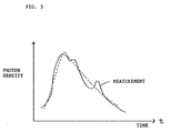
- a time-dependent photon density curve plotted according to actual measurement is shown in Fig. 1(B).
- the curve does not smoothly change due to the presence of light absorbing or light scattering material in the object to be imaged.
- the initial image is determined based on the data obtained through the actual measurement. More specifically, a predicted time-dependent photon density curve which is best fit to the curve obtained through the actual measurement is selected and used as the initial values for the absorption and scattering coefficients ⁇ a and ⁇ s .
- F( ⁇ 1) is given with respect to the incident point ⁇ k and the detection point ⁇ j .
- F( ⁇ 1) is rewritten as F kj ( ⁇ 1) in relation to the associated incident point ⁇ k and the detection point ⁇ j .
- the first error computation is carried out by comparing X kj with F kj ( ⁇ 1).
- the operator A represents a change of predicted value of the detected light resulting from small perturbation ⁇ k of the presumed absorption image, i.e., A( ⁇ k ) ⁇ F( ⁇ k + ⁇ k ) - F( ⁇ k ) (3)
- A( ⁇ k ) ⁇ F( ⁇ k + ⁇ k ) - F( ⁇ k ) (3)
- ⁇ G ⁇ s [ V ( ⁇ V ⁇ )] (5)
- the perturbation ⁇ of the photon density in the interior of the object resulting from the perturbation ⁇ of the absorption image ⁇ a can be computed as it is dependent on parameters of ⁇ , ⁇ and ⁇ a with no perturbations, the Green's function G ⁇ s with no perturbation, and the perturbation ⁇ .
- the perturbation of the detected light is given by - ⁇ V n ⁇ which corresponds to the operator A, so that the following equation is obtained.
- Equation (7) gives perturbation of the detected light at the surface of the object for the perturbation of the presumed image from ⁇ k to ⁇ k + ⁇ k .
- the goal to be attained is to obtain a correction value ⁇ k for the presumed image ⁇ k from a difference between the measured detection light X and the predicted value F( ⁇ k ) of the light to be detected for the k-th presumed image ⁇ k .
- a correction value ⁇ k for the presumed image ⁇ k from a difference between the measured detection light X and the predicted value F( ⁇ k ) of the light to be detected for the k-th presumed image ⁇ k .
- ⁇ k A ⁇ 1 ⁇ X - F( ⁇ k ) ⁇
- the above-described loop is repeatedly carried out, whereby F( ⁇ k ) is in access of X given an appropriate noise inducing circumstance.
- X - F( ⁇ k ) falls below a predetermined level, the ⁇ k at this instant is determined as a reconstructed absorption image.
- Fig. 6 shows a modification of the present invention wherein the absorption image and the scattering image are obtained independently of each other while performing processing for the absorption image and the scattering image alternately. According to such a processing, both the absorption and scattering images close to the real images can be reconstructed as the computations are repeatedly carried out in an appropriate noise circumstance.
- data regarding the spatial distribution of the absorption coefficient can be obtained together with the spatial distribution of the scattering coefficient.
- the accuracy of the former data is greatly improved as compared with the data obtained by the conventional device wherein the spatial distribution of the scattering coefficient is assumed to be constant in obtaining the spatial distribution of the absorption coefficients.
Landscapes
- Physics & Mathematics (AREA)
- General Physics & Mathematics (AREA)
- Engineering & Computer Science (AREA)
- Theoretical Computer Science (AREA)
- Investigating Or Analysing Materials By Optical Means (AREA)
Priority Applications (1)
| Application Number | Priority Date | Filing Date | Title |
|---|---|---|---|
| EP93305412A EP0633527A1 (de) | 1993-07-09 | 1993-07-09 | Optischer Röntgencomputertomograph |
Applications Claiming Priority (1)
| Application Number | Priority Date | Filing Date | Title |
|---|---|---|---|
| EP93305412A EP0633527A1 (de) | 1993-07-09 | 1993-07-09 | Optischer Röntgencomputertomograph |
Publications (1)
| Publication Number | Publication Date |
|---|---|
| EP0633527A1 true EP0633527A1 (de) | 1995-01-11 |
Family
ID=8214468
Family Applications (1)
| Application Number | Title | Priority Date | Filing Date |
|---|---|---|---|
| EP93305412A Withdrawn EP0633527A1 (de) | 1993-07-09 | 1993-07-09 | Optischer Röntgencomputertomograph |
Country Status (1)
| Country | Link |
|---|---|
| EP (1) | EP0633527A1 (de) |
Cited By (4)
| Publication number | Priority date | Publication date | Assignee | Title |
|---|---|---|---|---|
| WO1997033255A1 (en) * | 1996-03-07 | 1997-09-12 | Skoglund B Ulf | Apparatus and method for providing high fidelity reconstruction of an observed sample |
| WO1998024361A3 (en) * | 1996-12-03 | 1998-08-13 | Philips Electronics Nv | Method and apparatus for imaging an interior of a turbid medium |
| WO1999041593A1 (en) * | 1998-02-13 | 1999-08-19 | Art Advanced Research Technologies Inc. | Optical imaging with fitting to an inhomogeneous diffusion model |
| US6332093B1 (en) | 1998-08-06 | 2001-12-18 | Art Recherches Et Technologies Avancees Inc./Art Advanced Research Technologies, Inc. | Scanning module for imaging through scattering media |
Citations (2)
| Publication number | Priority date | Publication date | Assignee | Title |
|---|---|---|---|---|
| WO1991007655A1 (en) * | 1989-11-22 | 1991-05-30 | Singer Imaging, Inc. | Imaging using scattered and diffused radiation |
| US5142372A (en) * | 1990-03-08 | 1992-08-25 | Alfano Robert R | Three-dimensional optical imaging of semi-transparent and opaque objects using ultrashort light pulses, a streak camera and a coherent fiber bundle |
-
1993
- 1993-07-09 EP EP93305412A patent/EP0633527A1/de not_active Withdrawn
Patent Citations (2)
| Publication number | Priority date | Publication date | Assignee | Title |
|---|---|---|---|---|
| WO1991007655A1 (en) * | 1989-11-22 | 1991-05-30 | Singer Imaging, Inc. | Imaging using scattered and diffused radiation |
| US5142372A (en) * | 1990-03-08 | 1992-08-25 | Alfano Robert R | Three-dimensional optical imaging of semi-transparent and opaque objects using ultrashort light pulses, a streak camera and a coherent fiber bundle |
Cited By (5)
| Publication number | Priority date | Publication date | Assignee | Title |
|---|---|---|---|---|
| WO1997033255A1 (en) * | 1996-03-07 | 1997-09-12 | Skoglund B Ulf | Apparatus and method for providing high fidelity reconstruction of an observed sample |
| US6418243B1 (en) | 1996-03-07 | 2002-07-09 | B. Ulf Skoglund | Apparatus and method for providing high fidelity reconstruction of an observed sample |
| WO1998024361A3 (en) * | 1996-12-03 | 1998-08-13 | Philips Electronics Nv | Method and apparatus for imaging an interior of a turbid medium |
| WO1999041593A1 (en) * | 1998-02-13 | 1999-08-19 | Art Advanced Research Technologies Inc. | Optical imaging with fitting to an inhomogeneous diffusion model |
| US6332093B1 (en) | 1998-08-06 | 2001-12-18 | Art Recherches Et Technologies Avancees Inc./Art Advanced Research Technologies, Inc. | Scanning module for imaging through scattering media |
Similar Documents
| Publication | Publication Date | Title |
|---|---|---|
| Brooksby et al. | Magnetic resonance-guided near-infrared tomography of the breast | |
| US8050744B2 (en) | Optical system for measuring metabolism in a body and imaging method | |
| DE69525219T2 (de) | Verfahren und Vorrichtung zur Messung der Streu- und Absorptionsmerkmale eines streuenden Mediums | |
| Hebden et al. | Imaging changes in blood volume and oxygenation in the newborn infant brain using three-dimensional optical tomography | |
| DE69425081T2 (de) | Sensor zur Überwachung der arteriellen Blutströmung | |
| JP3112025B2 (ja) | 生体計測装置 | |
| Ntziachristos et al. | Diffuse optical tomography of highly heterogeneous media | |
| DE69326790T2 (de) | Abbildungsvorrichtung für die rechnergestützte optische Tomographie | |
| JP4043530B2 (ja) | 混濁媒体中の対象を画像化する方法 | |
| US7050533B2 (en) | Method and device for determining the type of fluid in a fluid mass in an object | |
| EP0497021A1 (de) | Oximeter mit Anzeige | |
| JPH0481455B2 (de) | ||
| WO2009077947A2 (en) | Method for detecting the presence of inhomogeneities in an interior of a turbid medium and device for imaging the interior of turbid media | |
| CN110522466A (zh) | 确定患者重量和/或体重指数的方法和装置 | |
| JP2001500978A (ja) | 混濁媒体内で対象を位置決めする方法 | |
| Pogue et al. | Statistical analysis of nonlinearly reconstructed near-infrared tomographic images. I. Theory and simulations | |
| McBride et al. | Strategies for absolute calibration of near infrared tomographic tissue imaging | |
| JP3164112B2 (ja) | 生体計測装置 | |
| Song et al. | Statistical analysis of nonlinearly reconstructed near-infrared tomographic images. II. Experimental interpretation | |
| JPS63277038A (ja) | 診断装置 | |
| US20080269617A1 (en) | Absorption and Scattering Map Reconstruction For Optical Fluorescence Tomography | |
| JP3304559B2 (ja) | 光計測方法および装置 | |
| EP0627698A1 (de) | Optischer Computer-Tomograph | |
| EP1926419B1 (de) | Oxymetrie mit räumlicher auflösung | |
| JP4327017B2 (ja) | 画面再構成方法 |
Legal Events
| Date | Code | Title | Description |
|---|---|---|---|
| PUAI | Public reference made under article 153(3) epc to a published international application that has entered the european phase |
Free format text: ORIGINAL CODE: 0009012 |
|
| AK | Designated contracting states |
Kind code of ref document: A1 Designated state(s): DE FR GB |
|
| STAA | Information on the status of an ep patent application or granted ep patent |
Free format text: STATUS: THE APPLICATION IS DEEMED TO BE WITHDRAWN |
|
| 18D | Application deemed to be withdrawn |
Effective date: 19960201 |