EP0637246B1 - Utilisation de la proteine c-reactive pour la preparation d'un medicament pour le traitement du cancer - Google Patents
Utilisation de la proteine c-reactive pour la preparation d'un medicament pour le traitement du cancer Download PDFInfo
- Publication number
- EP0637246B1 EP0637246B1 EP93910710A EP93910710A EP0637246B1 EP 0637246 B1 EP0637246 B1 EP 0637246B1 EP 93910710 A EP93910710 A EP 93910710A EP 93910710 A EP93910710 A EP 93910710A EP 0637246 B1 EP0637246 B1 EP 0637246B1
- Authority
- EP
- European Patent Office
- Prior art keywords
- mcrp
- crp
- animals
- tumor
- luvets
- Prior art date
- Legal status (The legal status is an assumption and is not a legal conclusion. Google has not performed a legal analysis and makes no representation as to the accuracy of the status listed.)
- Expired - Lifetime
Links
- 206010028980 Neoplasm Diseases 0.000 title claims abstract description 116
- 201000011510 cancer Diseases 0.000 title claims abstract description 44
- 238000011282 treatment Methods 0.000 title claims description 20
- 239000003814 drug Substances 0.000 title claims description 6
- 102100032752 C-reactive protein Human genes 0.000 title abstract description 179
- 108010074051 C-Reactive Protein Proteins 0.000 title abstract description 8
- 238000002360 preparation method Methods 0.000 title description 13
- 239000002502 liposome Substances 0.000 claims abstract description 16
- 239000003937 drug carrier Substances 0.000 claims abstract description 5
- 238000004519 manufacturing process Methods 0.000 claims description 7
- 239000000203 mixture Substances 0.000 claims description 7
- 239000002691 unilamellar liposome Substances 0.000 claims description 5
- 239000004480 active ingredient Substances 0.000 claims description 4
- 238000001514 detection method Methods 0.000 claims description 4
- 238000000034 method Methods 0.000 abstract description 34
- 241000124008 Mammalia Species 0.000 abstract description 30
- 239000012216 imaging agent Substances 0.000 abstract description 4
- 102000004127 Cytokines Human genes 0.000 abstract 1
- 108090000695 Cytokines Proteins 0.000 abstract 1
- 239000003795 chemical substances by application Substances 0.000 abstract 1
- 230000000973 chemotherapeutic effect Effects 0.000 abstract 1
- 150000001875 compounds Chemical class 0.000 abstract 1
- 230000001571 immunoadjuvant effect Effects 0.000 abstract 1
- 239000000568 immunological adjuvant Substances 0.000 abstract 1
- 241001465754 Metazoa Species 0.000 description 99
- 210000004027 cell Anatomy 0.000 description 56
- 238000002560 therapeutic procedure Methods 0.000 description 49
- 230000004614 tumor growth Effects 0.000 description 37
- 230000000694 effects Effects 0.000 description 30
- 239000000872 buffer Substances 0.000 description 29
- 230000037396 body weight Effects 0.000 description 24
- 108090000623 proteins and genes Proteins 0.000 description 22
- FAPWRFPIFSIZLT-UHFFFAOYSA-M Sodium chloride Chemical compound [Na+].[Cl-] FAPWRFPIFSIZLT-UHFFFAOYSA-M 0.000 description 21
- 102000004169 proteins and genes Human genes 0.000 description 21
- GHASVSINZRGABV-UHFFFAOYSA-N Fluorouracil Chemical compound FC1=CNC(=O)NC1=O GHASVSINZRGABV-UHFFFAOYSA-N 0.000 description 20
- 229960002949 fluorouracil Drugs 0.000 description 20
- 150000002632 lipids Chemical class 0.000 description 20
- 241001529936 Murinae Species 0.000 description 19
- 241000282414 Homo sapiens Species 0.000 description 18
- 210000001519 tissue Anatomy 0.000 description 17
- 238000001727 in vivo Methods 0.000 description 15
- 239000002953 phosphate buffered saline Substances 0.000 description 15
- LOKCTEFSRHRXRJ-UHFFFAOYSA-I dipotassium trisodium dihydrogen phosphate hydrogen phosphate dichloride Chemical compound P(=O)(O)(O)[O-].[K+].P(=O)(O)([O-])[O-].[Na+].[Na+].[Cl-].[K+].[Cl-].[Na+] LOKCTEFSRHRXRJ-UHFFFAOYSA-I 0.000 description 14
- 101000942118 Homo sapiens C-reactive protein Proteins 0.000 description 12
- 239000011780 sodium chloride Substances 0.000 description 12
- XLYOFNOQVPJJNP-UHFFFAOYSA-N water Substances O XLYOFNOQVPJJNP-UHFFFAOYSA-N 0.000 description 12
- 206010006187 Breast cancer Diseases 0.000 description 11
- 101000922020 Homo sapiens Cysteine and glycine-rich protein 1 Proteins 0.000 description 11
- 206010027476 Metastases Diseases 0.000 description 11
- 230000000259 anti-tumor effect Effects 0.000 description 11
- 238000003556 assay Methods 0.000 description 11
- 201000008274 breast adenocarcinoma Diseases 0.000 description 11
- 230000012010 growth Effects 0.000 description 11
- 102000051143 human CRP Human genes 0.000 description 11
- 238000005538 encapsulation Methods 0.000 description 10
- 239000012528 membrane Substances 0.000 description 10
- 210000001616 monocyte Anatomy 0.000 description 10
- 241000699670 Mus sp. Species 0.000 description 9
- 239000012530 fluid Substances 0.000 description 9
- 238000000338 in vitro Methods 0.000 description 9
- 210000002540 macrophage Anatomy 0.000 description 9
- 230000009257 reactivity Effects 0.000 description 9
- 239000000499 gel Substances 0.000 description 8
- 238000002347 injection Methods 0.000 description 8
- 239000007924 injection Substances 0.000 description 8
- 210000004379 membrane Anatomy 0.000 description 8
- 238000011160 research Methods 0.000 description 8
- 108091003079 Bovine Serum Albumin Proteins 0.000 description 7
- 230000008859 change Effects 0.000 description 7
- 238000001125 extrusion Methods 0.000 description 7
- 238000011534 incubation Methods 0.000 description 7
- 208000032839 leukemia Diseases 0.000 description 7
- 210000004072 lung Anatomy 0.000 description 7
- 230000009401 metastasis Effects 0.000 description 7
- 239000000523 sample Substances 0.000 description 7
- 241000894007 species Species 0.000 description 7
- QKNYBSVHEMOAJP-UHFFFAOYSA-N 2-amino-2-(hydroxymethyl)propane-1,3-diol;hydron;chloride Chemical compound Cl.OCC(N)(CO)CO QKNYBSVHEMOAJP-UHFFFAOYSA-N 0.000 description 6
- 206010027458 Metastases to lung Diseases 0.000 description 6
- 206010028851 Necrosis Diseases 0.000 description 6
- 108060008682 Tumor Necrosis Factor Proteins 0.000 description 6
- XSQUKJJJFZCRTK-UHFFFAOYSA-N Urea Chemical compound NC(N)=O XSQUKJJJFZCRTK-UHFFFAOYSA-N 0.000 description 6
- 239000004202 carbamide Substances 0.000 description 6
- 230000007423 decrease Effects 0.000 description 6
- 239000011521 glass Substances 0.000 description 6
- 206010061289 metastatic neoplasm Diseases 0.000 description 6
- 230000017074 necrotic cell death Effects 0.000 description 6
- 208000013435 necrotic lesion Diseases 0.000 description 6
- 239000000243 solution Substances 0.000 description 6
- 238000012360 testing method Methods 0.000 description 6
- 102000003390 tumor necrosis factor Human genes 0.000 description 6
- 230000002476 tumorcidal effect Effects 0.000 description 6
- KCXVZYZYPLLWCC-UHFFFAOYSA-N EDTA Chemical compound OC(=O)CN(CC(O)=O)CCN(CC(O)=O)CC(O)=O KCXVZYZYPLLWCC-UHFFFAOYSA-N 0.000 description 5
- 241000283973 Oryctolagus cuniculus Species 0.000 description 5
- 238000002835 absorbance Methods 0.000 description 5
- 229940098773 bovine serum albumin Drugs 0.000 description 5
- 239000011575 calcium Substances 0.000 description 5
- 229910052791 calcium Inorganic materials 0.000 description 5
- AOMZHDJXSYHPKS-UHFFFAOYSA-L disodium 4-amino-5-hydroxy-3-[(4-nitrophenyl)diazenyl]-6-phenyldiazenylnaphthalene-2,7-disulfonate Chemical compound [Na+].[Na+].[O-]S(=O)(=O)C1=CC2=CC(S([O-])(=O)=O)=C(N=NC=3C=CC=CC=3)C(O)=C2C(N)=C1N=NC1=CC=C([N+]([O-])=O)C=C1 AOMZHDJXSYHPKS-UHFFFAOYSA-L 0.000 description 5
- 230000004048 modification Effects 0.000 description 5
- 238000012986 modification Methods 0.000 description 5
- 108090000765 processed proteins & peptides Proteins 0.000 description 5
- 238000010186 staining Methods 0.000 description 5
- 208000010507 Adenocarcinoma of Lung Diseases 0.000 description 4
- OYPRJOBELJOOCE-UHFFFAOYSA-N Calcium Chemical compound [Ca] OYPRJOBELJOOCE-UHFFFAOYSA-N 0.000 description 4
- HEDRZPFGACZZDS-UHFFFAOYSA-N Chloroform Chemical compound ClC(Cl)Cl HEDRZPFGACZZDS-UHFFFAOYSA-N 0.000 description 4
- 201000008808 Fibrosarcoma Diseases 0.000 description 4
- WZUVPPKBWHMQCE-UHFFFAOYSA-N Haematoxylin Chemical compound C12=CC(O)=C(O)C=C2CC2(O)C1C1=CC=C(O)C(O)=C1OC2 WZUVPPKBWHMQCE-UHFFFAOYSA-N 0.000 description 4
- 108010002350 Interleukin-2 Proteins 0.000 description 4
- 241000699666 Mus <mouse, genus> Species 0.000 description 4
- 239000012980 RPMI-1640 medium Substances 0.000 description 4
- 238000000692 Student's t-test Methods 0.000 description 4
- 238000004458 analytical method Methods 0.000 description 4
- 230000000890 antigenic effect Effects 0.000 description 4
- 210000004369 blood Anatomy 0.000 description 4
- 239000008280 blood Substances 0.000 description 4
- 239000001110 calcium chloride Substances 0.000 description 4
- 229910001628 calcium chloride Inorganic materials 0.000 description 4
- 238000002648 combination therapy Methods 0.000 description 4
- 239000002299 complementary DNA Substances 0.000 description 4
- 231100000135 cytotoxicity Toxicity 0.000 description 4
- 230000003013 cytotoxicity Effects 0.000 description 4
- 230000034994 death Effects 0.000 description 4
- 231100000517 death Toxicity 0.000 description 4
- 230000003247 decreasing effect Effects 0.000 description 4
- 238000007912 intraperitoneal administration Methods 0.000 description 4
- 238000001990 intravenous administration Methods 0.000 description 4
- 201000005249 lung adenocarcinoma Diseases 0.000 description 4
- MYWUZJCMWCOHBA-VIFPVBQESA-N methamphetamine Chemical compound CN[C@@H](C)CC1=CC=CC=C1 MYWUZJCMWCOHBA-VIFPVBQESA-N 0.000 description 4
- 239000012071 phase Substances 0.000 description 4
- 239000000047 product Substances 0.000 description 4
- 238000002731 protein assay Methods 0.000 description 4
- 210000002966 serum Anatomy 0.000 description 4
- UCSJYZPVAKXKNQ-HZYVHMACSA-N streptomycin Chemical compound CN[C@H]1[C@H](O)[C@@H](O)[C@H](CO)O[C@H]1O[C@@H]1[C@](C=O)(O)[C@H](C)O[C@H]1O[C@@H]1[C@@H](NC(N)=N)[C@H](O)[C@@H](NC(N)=N)[C@H](O)[C@H]1O UCSJYZPVAKXKNQ-HZYVHMACSA-N 0.000 description 4
- YNJBWRMUSHSURL-UHFFFAOYSA-N trichloroacetic acid Chemical compound OC(=O)C(Cl)(Cl)Cl YNJBWRMUSHSURL-UHFFFAOYSA-N 0.000 description 4
- 239000003656 tris buffered saline Substances 0.000 description 4
- 210000004881 tumor cell Anatomy 0.000 description 4
- 238000005406 washing Methods 0.000 description 4
- UXVMQQNJUSDDNG-UHFFFAOYSA-L Calcium chloride Chemical compound [Cl-].[Cl-].[Ca+2] UXVMQQNJUSDDNG-UHFFFAOYSA-L 0.000 description 3
- 201000009030 Carcinoma Diseases 0.000 description 3
- 241000283073 Equus caballus Species 0.000 description 3
- 108010001336 Horseradish Peroxidase Proteins 0.000 description 3
- 108010002352 Interleukin-1 Proteins 0.000 description 3
- 102000000589 Interleukin-1 Human genes 0.000 description 3
- OKKJLVBELUTLKV-UHFFFAOYSA-N Methanol Chemical compound OC OKKJLVBELUTLKV-UHFFFAOYSA-N 0.000 description 3
- 239000000020 Nitrocellulose Substances 0.000 description 3
- 102000007079 Peptide Fragments Human genes 0.000 description 3
- 108010033276 Peptide Fragments Proteins 0.000 description 3
- HEMHJVSKTPXQMS-UHFFFAOYSA-M Sodium hydroxide Chemical compound [OH-].[Na+] HEMHJVSKTPXQMS-UHFFFAOYSA-M 0.000 description 3
- OUUQCZGPVNCOIJ-UHFFFAOYSA-M Superoxide Chemical compound [O-][O] OUUQCZGPVNCOIJ-UHFFFAOYSA-M 0.000 description 3
- 230000004913 activation Effects 0.000 description 3
- 210000001132 alveolar macrophage Anatomy 0.000 description 3
- 230000002001 anti-metastasis Effects 0.000 description 3
- 210000003567 ascitic fluid Anatomy 0.000 description 3
- DEGAKNSWVGKMLS-UHFFFAOYSA-N calcein Chemical compound O1C(=O)C2=CC=CC=C2C21C1=CC(CN(CC(O)=O)CC(O)=O)=C(O)C=C1OC1=C2C=C(CN(CC(O)=O)CC(=O)O)C(O)=C1 DEGAKNSWVGKMLS-UHFFFAOYSA-N 0.000 description 3
- 230000010261 cell growth Effects 0.000 description 3
- 238000006243 chemical reaction Methods 0.000 description 3
- KRKNYBCHXYNGOX-UHFFFAOYSA-N citric acid Chemical compound OC(=O)CC(O)(C(O)=O)CC(O)=O KRKNYBCHXYNGOX-UHFFFAOYSA-N 0.000 description 3
- 101150006779 crp gene Proteins 0.000 description 3
- 239000008367 deionised water Substances 0.000 description 3
- 229910021641 deionized water Inorganic materials 0.000 description 3
- 230000000951 immunodiffusion Effects 0.000 description 3
- 238000010253 intravenous injection Methods 0.000 description 3
- 238000005259 measurement Methods 0.000 description 3
- 230000001394 metastastic effect Effects 0.000 description 3
- 229920001220 nitrocellulos Polymers 0.000 description 3
- 229960002378 oftasceine Drugs 0.000 description 3
- 210000003200 peritoneal cavity Anatomy 0.000 description 3
- YHHSONZFOIEMCP-UHFFFAOYSA-O phosphocholine Chemical compound C[N+](C)(C)CCOP(O)(O)=O YHHSONZFOIEMCP-UHFFFAOYSA-O 0.000 description 3
- 229950004354 phosphorylcholine Drugs 0.000 description 3
- 229920003023 plastic Polymers 0.000 description 3
- 239000004033 plastic Substances 0.000 description 3
- 229920000724 poly(L-arginine) polymer Polymers 0.000 description 3
- 230000008569 process Effects 0.000 description 3
- 239000012064 sodium phosphate buffer Substances 0.000 description 3
- 238000007920 subcutaneous administration Methods 0.000 description 3
- 238000013519 translation Methods 0.000 description 3
- 210000002700 urine Anatomy 0.000 description 3
- YBJHBAHKTGYVGT-ZKWXMUAHSA-N (+)-Biotin Chemical compound N1C(=O)N[C@@H]2[C@H](CCCCC(=O)O)SC[C@@H]21 YBJHBAHKTGYVGT-ZKWXMUAHSA-N 0.000 description 2
- QTBSBXVTEAMEQO-UHFFFAOYSA-N Acetic acid Chemical compound CC(O)=O QTBSBXVTEAMEQO-UHFFFAOYSA-N 0.000 description 2
- APKFDSVGJQXUKY-KKGHZKTASA-N Amphotericin-B Natural products O[C@H]1[C@@H](N)[C@H](O)[C@@H](C)O[C@H]1O[C@H]1C=CC=CC=CC=CC=CC=CC=C[C@H](C)[C@@H](O)[C@@H](C)[C@H](C)OC(=O)C[C@H](O)C[C@H](O)CC[C@@H](O)[C@H](O)C[C@H](O)C[C@](O)(C[C@H](O)[C@H]2C(O)=O)O[C@H]2C1 APKFDSVGJQXUKY-KKGHZKTASA-N 0.000 description 2
- 206010003445 Ascites Diseases 0.000 description 2
- 206010003571 Astrocytoma Diseases 0.000 description 2
- IJGRMHOSHXDMSA-UHFFFAOYSA-N Atomic nitrogen Chemical compound N#N IJGRMHOSHXDMSA-UHFFFAOYSA-N 0.000 description 2
- 238000011725 BALB/c mouse Methods 0.000 description 2
- 241000283707 Capra Species 0.000 description 2
- 238000011765 DBA/2 mouse Methods 0.000 description 2
- 102000004190 Enzymes Human genes 0.000 description 2
- 108090000790 Enzymes Proteins 0.000 description 2
- LFQSCWFLJHTTHZ-UHFFFAOYSA-N Ethanol Chemical compound CCO LFQSCWFLJHTTHZ-UHFFFAOYSA-N 0.000 description 2
- WQZGKKKJIJFFOK-GASJEMHNSA-N Glucose Natural products OC[C@H]1OC(O)[C@H](O)[C@@H](O)[C@@H]1O WQZGKKKJIJFFOK-GASJEMHNSA-N 0.000 description 2
- 241000282412 Homo Species 0.000 description 2
- 241000713772 Human immunodeficiency virus 1 Species 0.000 description 2
- 206010020751 Hypersensitivity Diseases 0.000 description 2
- 108060003951 Immunoglobulin Proteins 0.000 description 2
- ZDXPYRJPNDTMRX-VKHMYHEASA-N L-glutamine Chemical compound OC(=O)[C@@H](N)CCC(N)=O ZDXPYRJPNDTMRX-VKHMYHEASA-N 0.000 description 2
- 229930182816 L-glutamine Natural products 0.000 description 2
- 229930182555 Penicillin Natural products 0.000 description 2
- JGSARLDLIJGVTE-MBNYWOFBSA-N Penicillin G Chemical compound N([C@H]1[C@H]2SC([C@@H](N2C1=O)C(O)=O)(C)C)C(=O)CC1=CC=CC=C1 JGSARLDLIJGVTE-MBNYWOFBSA-N 0.000 description 2
- 108020004511 Recombinant DNA Proteins 0.000 description 2
- 206010039491 Sarcoma Diseases 0.000 description 2
- 241000193998 Streptococcus pneumoniae Species 0.000 description 2
- 230000006978 adaptation Effects 0.000 description 2
- 238000001042 affinity chromatography Methods 0.000 description 2
- 208000026935 allergic disease Diseases 0.000 description 2
- 230000007815 allergy Effects 0.000 description 2
- 150000001413 amino acids Chemical group 0.000 description 2
- APKFDSVGJQXUKY-INPOYWNPSA-N amphotericin B Chemical compound O[C@H]1[C@@H](N)[C@H](O)[C@@H](C)O[C@H]1O[C@H]1/C=C/C=C/C=C/C=C/C=C/C=C/C=C/[C@H](C)[C@@H](O)[C@@H](C)[C@H](C)OC(=O)C[C@H](O)C[C@H](O)CC[C@@H](O)[C@H](O)C[C@H](O)C[C@](O)(C[C@H](O)[C@H]2C(O)=O)O[C@H]2C1 APKFDSVGJQXUKY-INPOYWNPSA-N 0.000 description 2
- 229960003942 amphotericin b Drugs 0.000 description 2
- 239000002246 antineoplastic agent Substances 0.000 description 2
- 239000008346 aqueous phase Substances 0.000 description 2
- AFYNADDZULBEJA-UHFFFAOYSA-N bicinchoninic acid Chemical compound C1=CC=CC2=NC(C=3C=C(C4=CC=CC=C4N=3)C(=O)O)=CC(C(O)=O)=C21 AFYNADDZULBEJA-UHFFFAOYSA-N 0.000 description 2
- 230000004071 biological effect Effects 0.000 description 2
- 230000008033 biological extinction Effects 0.000 description 2
- 238000001574 biopsy Methods 0.000 description 2
- 239000000969 carrier Substances 0.000 description 2
- 238000004113 cell culture Methods 0.000 description 2
- 239000006285 cell suspension Substances 0.000 description 2
- 239000002738 chelating agent Substances 0.000 description 2
- 239000003153 chemical reaction reagent Substances 0.000 description 2
- HVYWMOMLDIMFJA-DPAQBDIFSA-N cholesterol Chemical compound C1C=C2C[C@@H](O)CC[C@]2(C)[C@@H]2[C@@H]1[C@@H]1CC[C@H]([C@H](C)CCCC(C)C)[C@@]1(C)CC2 HVYWMOMLDIMFJA-DPAQBDIFSA-N 0.000 description 2
- 230000000295 complement effect Effects 0.000 description 2
- 238000011109 contamination Methods 0.000 description 2
- 230000001419 dependent effect Effects 0.000 description 2
- 238000011161 development Methods 0.000 description 2
- 201000010099 disease Diseases 0.000 description 2
- 208000037265 diseases, disorders, signs and symptoms Diseases 0.000 description 2
- 239000012153 distilled water Substances 0.000 description 2
- 239000006167 equilibration buffer Substances 0.000 description 2
- 230000001747 exhibiting effect Effects 0.000 description 2
- 238000002474 experimental method Methods 0.000 description 2
- 239000012894 fetal calf serum Substances 0.000 description 2
- 239000011888 foil Substances 0.000 description 2
- 239000012634 fragment Substances 0.000 description 2
- 230000006870 function Effects 0.000 description 2
- 239000008103 glucose Substances 0.000 description 2
- 102000018358 immunoglobulin Human genes 0.000 description 2
- 238000007918 intramuscular administration Methods 0.000 description 2
- 238000002955 isolation Methods 0.000 description 2
- 230000003902 lesion Effects 0.000 description 2
- 238000012417 linear regression Methods 0.000 description 2
- 210000004185 liver Anatomy 0.000 description 2
- 210000005228 liver tissue Anatomy 0.000 description 2
- 210000003141 lower extremity Anatomy 0.000 description 2
- 208000037841 lung tumor Diseases 0.000 description 2
- 210000004698 lymphocyte Anatomy 0.000 description 2
- 230000007246 mechanism Effects 0.000 description 2
- 201000001441 melanoma Diseases 0.000 description 2
- 239000003607 modifier Substances 0.000 description 2
- 210000000822 natural killer cell Anatomy 0.000 description 2
- 230000001338 necrotic effect Effects 0.000 description 2
- 238000002559 palpation Methods 0.000 description 2
- 229940049954 penicillin Drugs 0.000 description 2
- 102000013415 peroxidase activity proteins Human genes 0.000 description 2
- 108040007629 peroxidase activity proteins Proteins 0.000 description 2
- 230000001681 protective effect Effects 0.000 description 2
- 238000000746 purification Methods 0.000 description 2
- 239000011347 resin Substances 0.000 description 2
- 229920005989 resin Polymers 0.000 description 2
- 230000028327 secretion Effects 0.000 description 2
- 210000002027 skeletal muscle Anatomy 0.000 description 2
- 238000002415 sodium dodecyl sulfate polyacrylamide gel electrophoresis Methods 0.000 description 2
- 210000000952 spleen Anatomy 0.000 description 2
- 239000012192 staining solution Substances 0.000 description 2
- 229960005322 streptomycin Drugs 0.000 description 2
- 239000000758 substrate Substances 0.000 description 2
- 238000001356 surgical procedure Methods 0.000 description 2
- 201000000596 systemic lupus erythematosus Diseases 0.000 description 2
- 239000008399 tap water Substances 0.000 description 2
- 235000020679 tap water Nutrition 0.000 description 2
- 231100000331 toxic Toxicity 0.000 description 2
- 230000002588 toxic effect Effects 0.000 description 2
- 231100000419 toxicity Toxicity 0.000 description 2
- 230000001988 toxicity Effects 0.000 description 2
- 231100000041 toxicology testing Toxicity 0.000 description 2
- LENZDBCJOHFCAS-UHFFFAOYSA-N tris Chemical compound OCC(N)(CO)CO LENZDBCJOHFCAS-UHFFFAOYSA-N 0.000 description 2
- 238000002562 urinalysis Methods 0.000 description 2
- NHBKXEKEPDILRR-UHFFFAOYSA-N 2,3-bis(butanoylsulfanyl)propyl butanoate Chemical compound CCCC(=O)OCC(SC(=O)CCC)CSC(=O)CCC NHBKXEKEPDILRR-UHFFFAOYSA-N 0.000 description 1
- KJDSORYAHBAGPP-UHFFFAOYSA-N 4-(3,4-diaminophenyl)benzene-1,2-diamine;hydron;tetrachloride Chemical compound Cl.Cl.Cl.Cl.C1=C(N)C(N)=CC=C1C1=CC=C(N)C(N)=C1 KJDSORYAHBAGPP-UHFFFAOYSA-N 0.000 description 1
- 206010048998 Acute phase reaction Diseases 0.000 description 1
- 108010062271 Acute-Phase Proteins Proteins 0.000 description 1
- 102000011767 Acute-Phase Proteins Human genes 0.000 description 1
- 229920000936 Agarose Polymers 0.000 description 1
- AOMZHDJXSYHPKS-DROYEMJCSA-L Amido Black 10B Chemical compound [Na+].[Na+].[O-]S(=O)(=O)C1=CC2=CC(S([O-])(=O)=O)=C(\N=N\C=3C=CC=CC=3)C(O)=C2C(N)=C1\N=N\C1=CC=C(N(=O)=O)C=C1 AOMZHDJXSYHPKS-DROYEMJCSA-L 0.000 description 1
- VHUUQVKOLVNVRT-UHFFFAOYSA-N Ammonium hydroxide Chemical compound [NH4+].[OH-] VHUUQVKOLVNVRT-UHFFFAOYSA-N 0.000 description 1
- 238000011728 BALB/c nude (JAX™ mouse strain) Methods 0.000 description 1
- 238000011729 BALB/c nude mouse Methods 0.000 description 1
- 238000000035 BCA protein assay Methods 0.000 description 1
- 208000035143 Bacterial infection Diseases 0.000 description 1
- 101100289995 Caenorhabditis elegans mac-1 gene Proteins 0.000 description 1
- 101100298998 Caenorhabditis elegans pbs-3 gene Proteins 0.000 description 1
- 101100191768 Caenorhabditis elegans pbs-4 gene Proteins 0.000 description 1
- 101100245381 Caenorhabditis elegans pbs-6 gene Proteins 0.000 description 1
- CURLTUGMZLYLDI-UHFFFAOYSA-N Carbon dioxide Chemical compound O=C=O CURLTUGMZLYLDI-UHFFFAOYSA-N 0.000 description 1
- KRKNYBCHXYNGOX-UHFFFAOYSA-K Citrate Chemical compound [O-]C(=O)CC(O)(CC([O-])=O)C([O-])=O KRKNYBCHXYNGOX-UHFFFAOYSA-K 0.000 description 1
- 206010009944 Colon cancer Diseases 0.000 description 1
- 108020004414 DNA Proteins 0.000 description 1
- 238000012286 ELISA Assay Methods 0.000 description 1
- 241001635598 Enicostema Species 0.000 description 1
- 208000001382 Experimental Melanoma Diseases 0.000 description 1
- 108010010803 Gelatin Proteins 0.000 description 1
- 208000009329 Graft vs Host Disease Diseases 0.000 description 1
- 206010061218 Inflammation Diseases 0.000 description 1
- 239000000232 Lipid Bilayer Substances 0.000 description 1
- 102000003820 Lipoxygenases Human genes 0.000 description 1
- 108090000128 Lipoxygenases Proteins 0.000 description 1
- 206010025323 Lymphomas Diseases 0.000 description 1
- 206010027457 Metastases to liver Diseases 0.000 description 1
- 206010027626 Milia Diseases 0.000 description 1
- 238000011887 Necropsy Methods 0.000 description 1
- 101100471831 Neurospora crassa (strain ATCC 24698 / 74-OR23-1A / CBS 708.71 / DSM 1257 / FGSC 987) rpl-5 gene Proteins 0.000 description 1
- 208000030852 Parasitic disease Diseases 0.000 description 1
- 241001494479 Pecora Species 0.000 description 1
- 241000009328 Perro Species 0.000 description 1
- 239000002202 Polyethylene glycol Substances 0.000 description 1
- 239000004372 Polyvinyl alcohol Substances 0.000 description 1
- 241000700159 Rattus Species 0.000 description 1
- 241000283984 Rodentia Species 0.000 description 1
- 206010040047 Sepsis Diseases 0.000 description 1
- 201000005010 Streptococcus pneumonia Diseases 0.000 description 1
- 210000001744 T-lymphocyte Anatomy 0.000 description 1
- 239000007983 Tris buffer Substances 0.000 description 1
- 206010054094 Tumour necrosis Diseases 0.000 description 1
- 208000036142 Viral infection Diseases 0.000 description 1
- 208000027418 Wounds and injury Diseases 0.000 description 1
- JLPULHDHAOZNQI-JLOPVYAASA-N [(2r)-3-hexadecanoyloxy-2-[(9e,12e)-octadeca-9,12-dienoyl]oxypropyl] 2-(trimethylazaniumyl)ethyl phosphate Chemical compound CCCCCCCCCCCCCCCC(=O)OC[C@H](COP([O-])(=O)OCC[N+](C)(C)C)OC(=O)CCCCCCC\C=C\C\C=C\CCCCC JLPULHDHAOZNQI-JLOPVYAASA-N 0.000 description 1
- SXEHKFHPFVVDIR-UHFFFAOYSA-N [4-(4-hydrazinylphenyl)phenyl]hydrazine Chemical compound C1=CC(NN)=CC=C1C1=CC=C(NN)C=C1 SXEHKFHPFVVDIR-UHFFFAOYSA-N 0.000 description 1
- 229960000583 acetic acid Drugs 0.000 description 1
- 239000002253 acid Substances 0.000 description 1
- 230000001154 acute effect Effects 0.000 description 1
- 230000004658 acute-phase response Effects 0.000 description 1
- 208000009956 adenocarcinoma Diseases 0.000 description 1
- 230000002411 adverse Effects 0.000 description 1
- 230000002776 aggregation Effects 0.000 description 1
- 238000004220 aggregation Methods 0.000 description 1
- 230000000735 allogeneic effect Effects 0.000 description 1
- XAGFODPZIPBFFR-UHFFFAOYSA-N aluminium Chemical compound [Al] XAGFODPZIPBFFR-UHFFFAOYSA-N 0.000 description 1
- 229910052782 aluminium Inorganic materials 0.000 description 1
- 239000012491 analyte Substances 0.000 description 1
- 238000010171 animal model Methods 0.000 description 1
- 238000005349 anion exchange Methods 0.000 description 1
- 230000001093 anti-cancer Effects 0.000 description 1
- 210000003719 b-lymphocyte Anatomy 0.000 description 1
- 230000001580 bacterial effect Effects 0.000 description 1
- 208000022362 bacterial infectious disease Diseases 0.000 description 1
- CADWTSSKOVRVJC-UHFFFAOYSA-N benzyl(dimethyl)azanium;chloride Chemical compound [Cl-].C[NH+](C)CC1=CC=CC=C1 CADWTSSKOVRVJC-UHFFFAOYSA-N 0.000 description 1
- 229960002685 biotin Drugs 0.000 description 1
- 235000020958 biotin Nutrition 0.000 description 1
- 239000011616 biotin Substances 0.000 description 1
- 230000000903 blocking effect Effects 0.000 description 1
- 210000000459 calcaneus Anatomy 0.000 description 1
- 239000002775 capsule Substances 0.000 description 1
- 235000011089 carbon dioxide Nutrition 0.000 description 1
- 230000003915 cell function Effects 0.000 description 1
- 210000000170 cell membrane Anatomy 0.000 description 1
- 239000013553 cell monolayer Substances 0.000 description 1
- 230000004663 cell proliferation Effects 0.000 description 1
- 210000002421 cell wall Anatomy 0.000 description 1
- 230000001413 cellular effect Effects 0.000 description 1
- 238000005119 centrifugation Methods 0.000 description 1
- 230000009920 chelation Effects 0.000 description 1
- 238000002512 chemotherapy Methods 0.000 description 1
- 229940044683 chemotherapy drug Drugs 0.000 description 1
- 235000012000 cholesterol Nutrition 0.000 description 1
- 238000004587 chromatography analysis Methods 0.000 description 1
- 201000010897 colon adenocarcinoma Diseases 0.000 description 1
- 208000029742 colonic neoplasm Diseases 0.000 description 1
- 230000001609 comparable effect Effects 0.000 description 1
- 230000006957 competitive inhibition Effects 0.000 description 1
- 238000012258 culturing Methods 0.000 description 1
- 230000001461 cytolytic effect Effects 0.000 description 1
- 229940127089 cytotoxic agent Drugs 0.000 description 1
- 230000006378 damage Effects 0.000 description 1
- 238000004925 denaturation Methods 0.000 description 1
- 230000036425 denaturation Effects 0.000 description 1
- 238000000326 densiometry Methods 0.000 description 1
- 239000003599 detergent Substances 0.000 description 1
- 238000003745 diagnosis Methods 0.000 description 1
- 238000000502 dialysis Methods 0.000 description 1
- 238000009792 diffusion process Methods 0.000 description 1
- 239000012470 diluted sample Substances 0.000 description 1
- XEYBRNLFEZDVAW-ARSRFYASSA-N dinoprostone Chemical compound CCCCC[C@H](O)\C=C\[C@H]1[C@H](O)CC(=O)[C@@H]1C\C=C/CCCC(O)=O XEYBRNLFEZDVAW-ARSRFYASSA-N 0.000 description 1
- 238000010494 dissociation reaction Methods 0.000 description 1
- 230000005593 dissociations Effects 0.000 description 1
- 238000009826 distribution Methods 0.000 description 1
- 231100000673 dose–response relationship Toxicity 0.000 description 1
- 229940079593 drug Drugs 0.000 description 1
- 239000012149 elution buffer Substances 0.000 description 1
- 239000002158 endotoxin Substances 0.000 description 1
- 238000005516 engineering process Methods 0.000 description 1
- YQGOJNYOYNNSMM-UHFFFAOYSA-N eosin Chemical compound [Na+].OC(=O)C1=CC=CC=C1C1=C2C=C(Br)C(=O)C(Br)=C2OC2=C(Br)C(O)=C(Br)C=C21 YQGOJNYOYNNSMM-UHFFFAOYSA-N 0.000 description 1
- 210000003743 erythrocyte Anatomy 0.000 description 1
- 238000011156 evaluation Methods 0.000 description 1
- 238000000855 fermentation Methods 0.000 description 1
- 230000004151 fermentation Effects 0.000 description 1
- 210000002950 fibroblast Anatomy 0.000 description 1
- GNBHRKFJIUUOQI-UHFFFAOYSA-N fluorescein Chemical compound O1C(=O)C2=CC=CC=C2C21C1=CC=C(O)C=C1OC1=CC(O)=CC=C21 GNBHRKFJIUUOQI-UHFFFAOYSA-N 0.000 description 1
- MHMNJMPURVTYEJ-UHFFFAOYSA-N fluorescein-5-isothiocyanate Chemical compound O1C(=O)C2=CC(N=C=S)=CC=C2C21C1=CC=C(O)C=C1OC1=CC(O)=CC=C21 MHMNJMPURVTYEJ-UHFFFAOYSA-N 0.000 description 1
- 210000002683 foot Anatomy 0.000 description 1
- 230000002538 fungal effect Effects 0.000 description 1
- 239000008273 gelatin Substances 0.000 description 1
- 229920000159 gelatin Polymers 0.000 description 1
- 235000019322 gelatine Nutrition 0.000 description 1
- 235000011852 gelatine desserts Nutrition 0.000 description 1
- 239000012362 glacial acetic acid Substances 0.000 description 1
- 208000024908 graft versus host disease Diseases 0.000 description 1
- 210000000224 granular leucocyte Anatomy 0.000 description 1
- 230000003370 grooming effect Effects 0.000 description 1
- 244000144993 groups of animals Species 0.000 description 1
- 230000036541 health Effects 0.000 description 1
- 238000010438 heat treatment Methods 0.000 description 1
- 238000004128 high performance liquid chromatography Methods 0.000 description 1
- 210000004408 hybridoma Anatomy 0.000 description 1
- 230000036571 hydration Effects 0.000 description 1
- 238000006703 hydration reaction Methods 0.000 description 1
- 238000007654 immersion Methods 0.000 description 1
- 230000008105 immune reaction Effects 0.000 description 1
- 230000002055 immunohistochemical effect Effects 0.000 description 1
- 239000007943 implant Substances 0.000 description 1
- 238000002513 implantation Methods 0.000 description 1
- 208000015181 infectious disease Diseases 0.000 description 1
- 230000002458 infectious effect Effects 0.000 description 1
- 230000008595 infiltration Effects 0.000 description 1
- 238000001764 infiltration Methods 0.000 description 1
- 230000004054 inflammatory process Effects 0.000 description 1
- 230000002401 inhibitory effect Effects 0.000 description 1
- 238000011221 initial treatment Methods 0.000 description 1
- 230000000977 initiatory effect Effects 0.000 description 1
- 208000014674 injury Diseases 0.000 description 1
- 239000002198 insoluble material Substances 0.000 description 1
- 238000002642 intravenous therapy Methods 0.000 description 1
- 238000011835 investigation Methods 0.000 description 1
- 230000000302 ischemic effect Effects 0.000 description 1
- 230000002147 killing effect Effects 0.000 description 1
- 231100000518 lethal Toxicity 0.000 description 1
- 231100000636 lethal dose Toxicity 0.000 description 1
- 230000001665 lethal effect Effects 0.000 description 1
- 230000004807 localization Effects 0.000 description 1
- 229920002521 macromolecule Polymers 0.000 description 1
- 230000003211 malignant effect Effects 0.000 description 1
- 238000007726 management method Methods 0.000 description 1
- 239000003550 marker Substances 0.000 description 1
- 201000006512 mast cell neoplasm Diseases 0.000 description 1
- 208000006971 mastocytoma Diseases 0.000 description 1
- 239000000463 material Substances 0.000 description 1
- 230000000813 microbial effect Effects 0.000 description 1
- 230000009826 neoplastic cell growth Effects 0.000 description 1
- 238000006386 neutralization reaction Methods 0.000 description 1
- 210000000440 neutrophil Anatomy 0.000 description 1
- 229910052757 nitrogen Inorganic materials 0.000 description 1
- 231100000252 nontoxic Toxicity 0.000 description 1
- 230000003000 nontoxic effect Effects 0.000 description 1
- 210000000633 nuclear envelope Anatomy 0.000 description 1
- 238000011275 oncology therapy Methods 0.000 description 1
- 210000000056 organ Anatomy 0.000 description 1
- 239000003960 organic solvent Substances 0.000 description 1
- 230000004783 oxidative metabolism Effects 0.000 description 1
- 230000007170 pathology Effects 0.000 description 1
- 210000005259 peripheral blood Anatomy 0.000 description 1
- 239000011886 peripheral blood Substances 0.000 description 1
- 210000005105 peripheral blood lymphocyte Anatomy 0.000 description 1
- 210000004303 peritoneum Anatomy 0.000 description 1
- 210000001539 phagocyte Anatomy 0.000 description 1
- WTJKGGKOPKCXLL-RRHRGVEJSA-N phosphatidylcholine Chemical compound CCCCCCCCCCCCCCCC(=O)OC[C@H](COP([O-])(=O)OCC[N+](C)(C)C)OC(=O)CCCCCCCC=CCCCCCCCC WTJKGGKOPKCXLL-RRHRGVEJSA-N 0.000 description 1
- 230000004962 physiological condition Effects 0.000 description 1
- 229920000729 poly(L-lysine) polymer Polymers 0.000 description 1
- 239000004417 polycarbonate Substances 0.000 description 1
- 229920000515 polycarbonate Polymers 0.000 description 1
- 229920001223 polyethylene glycol Polymers 0.000 description 1
- 229920001282 polysaccharide Polymers 0.000 description 1
- 239000005017 polysaccharide Substances 0.000 description 1
- 229920002451 polyvinyl alcohol Polymers 0.000 description 1
- 230000003389 potentiating effect Effects 0.000 description 1
- 239000002244 precipitate Substances 0.000 description 1
- 102000004196 processed proteins & peptides Human genes 0.000 description 1
- 238000012545 processing Methods 0.000 description 1
- 230000000750 progressive effect Effects 0.000 description 1
- 230000005855 radiation Effects 0.000 description 1
- 238000001959 radiotherapy Methods 0.000 description 1
- 230000009467 reduction Effects 0.000 description 1
- 230000002829 reductive effect Effects 0.000 description 1
- 230000004043 responsiveness Effects 0.000 description 1
- 238000012552 review Methods 0.000 description 1
- 206010039073 rheumatoid arthritis Diseases 0.000 description 1
- 239000012723 sample buffer Substances 0.000 description 1
- 230000003248 secreting effect Effects 0.000 description 1
- 239000013049 sediment Substances 0.000 description 1
- 238000000926 separation method Methods 0.000 description 1
- 238000013207 serial dilution Methods 0.000 description 1
- 238000002741 site-directed mutagenesis Methods 0.000 description 1
- 230000000391 smoking effect Effects 0.000 description 1
- 238000002943 spectrophotometric absorbance Methods 0.000 description 1
- 230000002269 spontaneous effect Effects 0.000 description 1
- 238000012453 sprague-dawley rat model Methods 0.000 description 1
- 239000007858 starting material Substances 0.000 description 1
- 238000003860 storage Methods 0.000 description 1
- 208000022218 streptococcal pneumonia Diseases 0.000 description 1
- 230000004083 survival effect Effects 0.000 description 1
- 239000000725 suspension Substances 0.000 description 1
- 230000002195 synergetic effect Effects 0.000 description 1
- 238000011287 therapeutic dose Methods 0.000 description 1
- 230000001225 therapeutic effect Effects 0.000 description 1
- 239000010409 thin film Substances 0.000 description 1
- 208000037816 tissue injury Diseases 0.000 description 1
- 238000004448 titration Methods 0.000 description 1
- 238000000108 ultra-filtration Methods 0.000 description 1
- 239000003981 vehicle Substances 0.000 description 1
- 230000009385 viral infection Effects 0.000 description 1
- 230000003612 virological effect Effects 0.000 description 1
- 230000000007 visual effect Effects 0.000 description 1
- 239000011800 void material Substances 0.000 description 1
- 238000003260 vortexing Methods 0.000 description 1
Images

Classifications
-
- C—CHEMISTRY; METALLURGY
- C07—ORGANIC CHEMISTRY
- C07K—PEPTIDES
- C07K14/00—Peptides having more than 20 amino acids; Gastrins; Somatostatins; Melanotropins; Derivatives thereof
- C07K14/435—Peptides having more than 20 amino acids; Gastrins; Somatostatins; Melanotropins; Derivatives thereof from animals; from humans
- C07K14/52—Cytokines; Lymphokines; Interferons
-
- A—HUMAN NECESSITIES
- A61—MEDICAL OR VETERINARY SCIENCE; HYGIENE
- A61K—PREPARATIONS FOR MEDICAL, DENTAL OR TOILETRY PURPOSES
- A61K49/00—Preparations for testing in vivo
- A61K49/0004—Screening or testing of compounds for diagnosis of disorders, assessment of conditions, e.g. renal clearance, gastric emptying, testing for diabetes, allergy, rheuma, pancreas functions
-
- A—HUMAN NECESSITIES
- A61—MEDICAL OR VETERINARY SCIENCE; HYGIENE
- A61K—PREPARATIONS FOR MEDICAL, DENTAL OR TOILETRY PURPOSES
- A61K9/00—Medicinal preparations characterised by special physical form
- A61K9/10—Dispersions; Emulsions
- A61K9/127—Synthetic bilayered vehicles, e.g. liposomes or liposomes with cholesterol as the only non-phosphatidyl surfactant
-
- A—HUMAN NECESSITIES
- A61—MEDICAL OR VETERINARY SCIENCE; HYGIENE
- A61P—SPECIFIC THERAPEUTIC ACTIVITY OF CHEMICAL COMPOUNDS OR MEDICINAL PREPARATIONS
- A61P35/00—Antineoplastic agents
-
- A—HUMAN NECESSITIES
- A61—MEDICAL OR VETERINARY SCIENCE; HYGIENE
- A61K—PREPARATIONS FOR MEDICAL, DENTAL OR TOILETRY PURPOSES
- A61K38/00—Medicinal preparations containing peptides
-
- Y—GENERAL TAGGING OF NEW TECHNOLOGICAL DEVELOPMENTS; GENERAL TAGGING OF CROSS-SECTIONAL TECHNOLOGIES SPANNING OVER SEVERAL SECTIONS OF THE IPC; TECHNICAL SUBJECTS COVERED BY FORMER USPC CROSS-REFERENCE ART COLLECTIONS [XRACs] AND DIGESTS
- Y10—TECHNICAL SUBJECTS COVERED BY FORMER USPC
- Y10S—TECHNICAL SUBJECTS COVERED BY FORMER USPC CROSS-REFERENCE ART COLLECTIONS [XRACs] AND DIGESTS
- Y10S435/00—Chemistry: molecular biology and microbiology
- Y10S435/968—High energy substrates, e.g. fluorescent, chemiluminescent, radioactive
-
- Y—GENERAL TAGGING OF NEW TECHNOLOGICAL DEVELOPMENTS; GENERAL TAGGING OF CROSS-SECTIONAL TECHNOLOGIES SPANNING OVER SEVERAL SECTIONS OF THE IPC; TECHNICAL SUBJECTS COVERED BY FORMER USPC CROSS-REFERENCE ART COLLECTIONS [XRACs] AND DIGESTS
- Y10—TECHNICAL SUBJECTS COVERED BY FORMER USPC
- Y10S—TECHNICAL SUBJECTS COVERED BY FORMER USPC CROSS-REFERENCE ART COLLECTIONS [XRACs] AND DIGESTS
- Y10S436/00—Chemistry: analytical and immunological testing
- Y10S436/804—Radioisotope, e.g. radioimmunoassay
-
- Y—GENERAL TAGGING OF NEW TECHNOLOGICAL DEVELOPMENTS; GENERAL TAGGING OF CROSS-SECTIONAL TECHNOLOGIES SPANNING OVER SEVERAL SECTIONS OF THE IPC; TECHNICAL SUBJECTS COVERED BY FORMER USPC CROSS-REFERENCE ART COLLECTIONS [XRACs] AND DIGESTS
- Y10—TECHNICAL SUBJECTS COVERED BY FORMER USPC
- Y10S—TECHNICAL SUBJECTS COVERED BY FORMER USPC CROSS-REFERENCE ART COLLECTIONS [XRACs] AND DIGESTS
- Y10S436/00—Chemistry: analytical and immunological testing
- Y10S436/811—Test for named disease, body condition or organ function
- Y10S436/813—Cancer
Definitions
- the invention relates to the use of modified C-reactive protein ("mCRP") for the preparation of a medicament for the treatment of cancer.
- mCRP modified C-reactive protein
- the invention also relates to the use of mCRP as an imaging agent to identify cancer cells in mammals.
- C-reactive protein was first described by Tillett and Francis [ J. Exp. Med ., 52 :561-71 (1930)] who observed that sera from acutely ill patients precipitated with the C-polysaccharide of the cell wall of Streptococcus pneumonia. Other investigators subsequently identified the reactive serum factor as protein, hence the designation "C-reactive protein” or "CAP.” Kilpatrick et al., Immunol. Res., 10 :43-53 (1991), provides a recent review of CRP.
- CRP is a pentameric molecule which consists of five identical subunits [Osmand et al., Proc. Natl. Acad. Sciences. U.S.A. , 74 :739-743 (1977)]. This pentameric form of CRP is sometimes referred to as "native CRP.”
- Hornung discloses that CRP added with lymphocytes inhibited growth of human melanoma cells after 72 hours of culture [Hornung, Proc. Soc. Exp. Biol. Med. , 139 :1166-1169 (1972)]. Hornung also discloses that the CRP was toxic to the lymphocytes in the cell culture.
- Zahedi et al. Cancer Research , 46 :5077-5083 (1986), disclose that purified human CRP is capable of mediating activation of macrophages to a tumoricidal state in vitro .
- the authors teach that when elicited macrophages were exposed to human CRP for thirty minutes to 2 hours, tumoricidal activity was induced against the murine P815 mastocytoma cell line, the murine L-929 fibroblast carcinoma cell line, and the human CAK-1 carcinoma cell line.
- Zahedi et al. also teach that when CRP was heat-aggregated at 85°C for 1 hour prior to measuring tumoricidal activity, the CRP had significantly less killing activity than CRP that had not been heat-aggregated.
- Zahedi et al. disclose that no synergistic effect was shown by CRP when combined with a lymphokine.
- Thomassen et al. FASEB J. , 6 :1151a (1992), disclose in vitro modulation of tumoricidal activity of human monocytes and human alveolar macrophages by a synthetic peptide derived from CRP. Thomassen et al. report that monocytes treated with a synthetic peptide showed greater than or equal to 2-fold enhancement of Tumor Necrosis Factor (TNF) and IL-1 secretion, whereas alveolar macrophages showed no enhanced secretion of either TNF or IL-1.
- TNF Tumor Necrosis Factor
- O'Connor et al. U.S. Patent No. 4,857,314 disclose a method for treating Meth A sarcoma in animals or humans by administering tumor necrosis factor (TNF) in combination with purified human C-reactive protein to enhance the tumoricidal activity of the TNF.
- TNF tumor necrosis factor
- O'Connor et al. teach that human CRP, administered alone, had only minimal or slight anti-tumor activity.
- Deodhar et al. Cancer Research , 42 :5084-5088 (1982), describe activity of multilamellar vesicles containing human CRP in vivo against murine fibrosarcoma, T241, lung metastases.
- Deodhar et al. report that animals treated with intravenous injections of multilamellar vesicles containing CRP after T241 primary tumors were excised had fewer and smaller lung metastases.
- Thombre et al. Cancer Immunol. Immunother. , 16 :145-150 (1984), describe anti-metastatic activity of multilamellar vesicles containing human CRP against a murine colon adenocarcinoma (MCA-38).
- MCA-38 murine colon adenocarcinoma
- Thrombre et al. report that animals treated with multilamellar vesicles containing CRP, administered parenterally after the growth of the primary tumor, showed fewer liver metastases and longer survival as compared to control animals. Id.
- Deodhar et al. also describe anti-tumor effects of multilamellar vesicles containing a synthetic peptide derived from CRP [Deodhar et al., Proc. Am. Assoc. Cancer Research , 32 :404 (1991)]. However, Deodhar et al. report that administering comparable doses of non-encapsulated synthetic peptide was ineffective [ Id. ]. Barna et al., Proc. Am. Assoc. Cancer Research , 32 :237 (1991), suggest that the anti-tumor activity demonstrated in the T241 lung metastatic model may result from enhanced infiltration of MAC 1+ cells from the blood into the lung.
- Deodhar et al. report that administering a combination of 1 x 10 4 U IL-2 and CRP or a peptide fragment was more effective than administering 5 x 10 4 U IL-2.
- Barna et al. disclose in vitro cytolytic data that suggests that the combination of CRP peptide fragment and IL-2 augments human monocyte activity but not natural killer (NK) activity [ FASEB J. , 6 :1433a (1992)].
- mCRP modified C-reactive protein
- mCRP has significantly different charge, size, solubility and antigenicity characteristics as compared to native CRP [Potempa et al., Mol. Immunol. , 20 :1165-75 (1983)].
- mCRP also differs from native CRP in its binding characteristics. For instance, mCRP does not bind phosphorylcholine [ Id .; Chudwin et al., J. Allergy Clin. Immunol. , 77 :216a (1986)].
- Neo-CRP antigenicity is known to be expressed on:
- mCRP differs from native CRP in its biological activity. It has been reported that mCRP can influence the development of monocyte cytotoxicity, improve the accessory cell function of monocytes, potentiate aggregated-IgG-induced phagocytic cell oxidative metabolism, and increase the production of interleukin-1, prostaglandin E and lipoxygenase products by monocytes [Potempa et al., Protides Biol. Fluids , 34 :287-290 (1987); Potempa et al., Inflammation , 12 :391-405 (1988); Chu et al., Proc. Amer. Acad. Cancer Res.
- mCRP is useful in treating viral infections such as human immunodeficiency virus 1 ("HIV-1"), as disclosed in co-pending U.S. application Serial Number 07/799,448, filed November 27, 1991.
- HIV-1 human immunodeficiency virus 1
- mCRP can be used in treating non-streptococcal bacterial infections and endotoxin sepsis, as disclosed in co-pending U.S. application Serial Number 07/800,508, filed November 27, 1991.
- mCRP has been found to suppress or enhance the development of tumoricidal function in monocytes depending on the material used to elicit the anti-tumoricidal monocytes [Chu et al., Proc. Am. Acad. Cancer Res. , 29 :371a (1988); Chu et al., Proc. Am. Acad. Cancer Res. , 30 :333a (1989)).
- Applicants' knowledge there have been no reports on anti-cancer activity of mCRP in vivo .
- Applicants are not aware of any reports of mCRP being used to treat cancer in mammals.
- the invention provides use of modified-CRP as the only active ingredient for the manufacture of a medicament for the treatment of cancer, said modified-CRP expressing neo-CRP antigenicity.
- the modified-CRP is contained in a plurality of liposomes collectively containing an effective amount of modified-CRP.
- the invention further provides for identifying cancer cells in a mammal using an imaging agent comprising modified-CRP.
- a further aspect of the invention comprises use of modified-CRP as the only active ingredient for the manufacture of a diagnostic composition for the detection of cancer cells, said modified-CRP expressing neo-CRP antigenicity.
- a labelled component that binds modified-CRP may be used to allow for its detection when bound to modified-CRP.
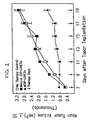
- Figure 1 is a graph illustrating a comparison of the effects of mCRP (in LUVETs) treatment, native CRP (in LUVETs), and no therapy on murine EMT6 breast adenocarcinoma primary tumor volume during Days 7-19 of the study.
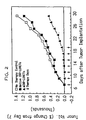
- Figure 2 is a graph illustrating a comparison of the effects of mCRP (in LUVETs) treatment, native CRP (in LUVETs) treatment, and no therapy on murine EMT6 breast adenocarcinoma primary tumor percent changes of volume for Days 7-29 of the study.
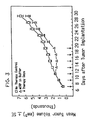
- Figure 3 is a graph illustrating a comparison of the effects of mCRP and no therapy on the EMT6 breast adenocarcinoma primary tumor volume for Days 7-29 of the study.
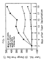
- Figure 4 is a graph illustrating a comparison of the effects of mCRP, mCRP in LUVETS, and native CRP in LUVETs, on the percent changes of tumor volumes of the murine EMT6 breast adenocarcinoma primary tumor.
- Figure 5 shows a comparison of the effects of 5-Fluorouracil and mCRP treatment on body weight and life span of P388 tumor bearing animals.
- the present invention provides methods for treating cancer using modified C-reactive protein ("mCRP").
- the mCRP useful in the practice of the present invention may be from any species. There is substantial homology between the amino acid sequences of C-reactive protein (“CRP") from different species. For instance, there is from about 50% to about 80% sequence homology between CRP from various mammalian species. Hu et al., Biochem. : 25 :7834-39 (1986); Whitehead et al., Biochem. J. , 266 :283-90 (1990); Kilpatrick et al., Immunol. Res. : 10 :43-53 (1991). It is, therefore, expected that mCRP from any species will be effective in treating cancer.
- a mammal having cancer may be treated with mCRP from a different species (e.g. , mice can be treated with human mCRP).
- the mammal is treated with homologous mCRP (e.g. , humans are treated with human mCRP) to avoid immune reactions to the mCRP.
- mCRP is preferably made using native CRP as a starting material.
- Methods of isolating CRP from natural sources are known in the art and are described, for example, by Volanakis et al., J. Immunol. : 113 :9-17 (1978); de Beer et al., J. Immunol . Meth. , 50 :17-31 (1982); Potempa et al., Mol. Immunol. , 24 :531-541 (1987).
- CRP is preferably isolated from pleural or ascites fluid by calcium-dependent affinity chromatography using phosphorylcholine-substituted BioGel A 0.5m (an agarose-based resin obtained from BioRad Laboratories, Richmond, CA) [ See , Potempa et al., Mol. Immunol. , 24 :531-541 (1987)].
- This CRP isolation method is further described in Example 1 below. Using this isolation method, CRP can be obtained which is about 99% pure.
- Genomic and cDNA clones coding for human, mouse, and rabbit CAP have been isolated. Lei et al., J. Biol. Chew. , 260 :13377-83 (1985); Woo et al., J. Biol. Chem. , 260 :13384-88 (1985); Hu et al., Biochem. , 25 :7834-39 (1986); Hu et al., J. Biol. Chem. , 263 : 1500-1504 (1988); Whitehead et al., Biochem. J. , 266 :283-90 (1990).
- probes can readily be prepared so that genomic and cDNA clones can be isolated which code for CRP from other species.
- Methods of preparing such probes and isolating genomic and cDNA clones are well known in the art. See , e.g. , Lei et al., J. Biol. Chem. , 260 :13377-83 (1985); Woo et al., J. Biol. Chem. , 260 :13384-88 (1985); Hu et al., Biochem. , 25 :7834-39 (1986); Hu et al., J. Biol. Chem.
- CRP can be prepared using conventional and well known recombinant DNA techniques and cell culture and fermentation conditions. See , e.g. , Hu et al., J. Biol. Chem. , 263 :1500-1504 (1988). However, to obtain pentameric native CRP, eukaryotic host cells, preferably mammalian host cells, should be used. See Samols et al., Protides Biol. Fluids , 34 :263-66 (1986); Hu et al., J. Biol. Chem. , 263 :1500-1504 (1988).
- mCRP can be prepared by denaturing CRP.
- CRP can be denatured by treatment with an effective amount of urea (preferably 8M) in the presence of a conventional chelator (preferably ethylenediamine tetraacetic acid (EDTA) or citric acid).
- a conventional chelator preferably ethylenediamine tetraacetic acid (EDTA) or citric acid.
- CRP can be treated to produce mCRP by adjusting the pH of the protein to below about 3 or above about 11-12.
- mCRP can be produced by heating CRP above 50°C for a time sufficient to cause denaturation (preferably at 63°C for 2 minutes) in the absence of calcium or in the presence of a chelator such as those listed above.
- mCRP can also be prepared using recombinant DNA techniques.
- the primary translation product of the CRP gene preCRP
- preCRP The primary translation product of the CRP gene
- mCRP can be prepared by selecting conditions so that the CRP subunits are not assembled into pentameric native CRP in the host cell. This can be accomplished by expressing the desired genomic or cDNA clone in a prokaryotic host. See Samols et al., Prot. Biol. Fluids , 34 :263-66 (1986).
- the mCRP produced in this manner appears to consist of aggregates of CRP subunits and/or preCRP and perhaps other CRP peptides. See Id .
- This form of mCRP is insoluble, and further purification is problematical. However, it should be possible to inject this insoluble material into mammals as a suspension without further processing, since an aggregated form of serum-purified mCRP has been shown to be effective, as described further in Example 4 below.
- mCRP may be distinguished from native CRP by several criteria. As noted in the Background section, modified CRP expresses neo-CRP antigenicity, whereas native CRP does not. Neo-CRP antigenicity can be detected using polyclonal antisera specific for neo-CRP [ See , Potempa et al., Mol. Immunol. , 24 :531-541 (1987)]. Preferably, however, mCRP is distinguished from native CRP using monoclonal antibodies like those described in co-pending application Serial Number 07/374,166, the disclosure of which is incorporated herein by reference.
- Hybridomas secreting the monoclonal antibodies disclosed in the co-pending application are deposited with the American Type Culture Collection, Rockville, Maryland, and are registered as HB10175 (mAb 15.2C10), HB10176 (mAb 26.8C10), HB10177 (mAb 13.3H12), and HB10178 (mAb 15.1D6). These monoclonal antibodies are also described in Ying et al., J. Immunol. , 143 :221-28 (1989).
- the antisera and antibodies can be used, for example, in ELISA assays, and preferably in Dot-ELIFA assays described in Example 1, part C, below, to distinguish mCRP from native CRP.
- mCRP binds immune complexes and aggregated immunoglobulin
- native CRP does not as described in co-pending application Serial Number 07/582,884 and published PCT application WO 89/09628.
- mCRP may be distinguished from native CRP on the basis of charge, solubility, binding characteristics and biological activity as referenced in the Background section.
- mCRP is formed by the dissociation of the five CRP subunits, each of which then undergoes a spontaneous conformational change to form mCRP.
- CRP subunits may have the same activities described herein for mCRP, and the use of such fragments is considered to come within the scope of the present invention.
- proteins substantially homologous to CRP will have the activities described herein for mCRP, and such proteins are also considered to come within the scope of the present invention.
- CRP subunits having certain amino acids added, deleted or substituted by, e.g. , site-directed mutagenesis of the CRP gene would likely be effective in the treatment of cancer and could be substituted for mCRP.
- mCRP is defined herein to include the primary translation product of the CRP gene.
- an effective amount of mCRP is administered to the mammal.
- cancer in the present invention is used in a broad sense and refers to the physiological condition in mammals that is usually characterized by unregulated cell growth. It is contemplated that mCRP may be used to treat a variety of cancers, including but not limited to, adenocarcinoma, lymphoma, fibrosarcoma, and leukemia.
- the mCRP may be administered to the mammal when the presence of cancer cells in the mammal is first detected or before the cancer becomes too serious. Alternatively, the mCRP may be administered after the cancer progresses so as to reduce metastasis of cancer cells, to reduce metastatic tumors, or to reduce primary tumor burden.
- mCRP may be administered to the mammal in a pharmaceutically-acceptable carrier.
- Pharmaceutically-acceptable carriers are well known to persons skilled in the art.
- suitable carriers for administering mCRP include fluids such as water, saline, and buffer. More preferably, a Tris or phosphate buffered saline is used as the carrier. It will be apparent to those persons skilled in the art that certain carriers may be more preferable depending upon, for instance, the route of administration and concentration of protein being administered.
- liposome in the present invention relates to any sac-like or hollow vesicle-like structure which is capable of encapsulating mCRP, and includes but is not limited to, multilamellar vesicles, unilamellar vesicles, and red blood cell ghosts. Methods of preparing liposomes and encapsulating molecules in liposomes are well known in the art.
- the liposomes containing mCRP are unilamellar vesicles formed by extrusion, referred to herein as "LUVETs.”
- LUVETs unilamellar vesicles formed by extrusion
- LUVETs are particularly useful for administering mCRP because LUVETs do not contain traces of organic solvents and detergents commonly used in preparing liposomes. Also, LUVETs are defined by a single lipid bilayer, contain relatively large internal volumes, and typically have greater encapsulation efficiency as compared to multilamellar vesicles. Although the exact mechanism(s) by which liposomes containing mCRP function is not fully understood, it is believed that the liposomes may act as a vehicle for efficient delivery of mCRP to the cancer cells.
- the mCRP is preferably administered to the mammal by injection (e.g. , intravenous, intraperitoneal, subcutaneous, intramuscular). Effective dosages and schedules for administering mCRP may be determined empirically, and making such determinations is within the skill of the art. Applicants have found that a dose of from about 0.10 mg to about 20 mg of mCRP per kilogram of body weight of the mammal, and preferably from about 2 mg to about 10 mg per kilogram, is effective for treating cancer, whether the mCRP is administered in a pharmaceutically-acceptable carrier or contained in liposomes.
- the dose of mCRP that must be administered will vary depending on, for example, the mammal which will receive the mCRP, the type of cancer, the extent of cancer cell growth or metastasis, the biological site or body compartment of the tumor(s), the route of administration, and the identity of any other drugs or treatment being administered to the mammal, such as radiation or surgical treatment. It is also understood that it may be necessary to give more than one dose of mCRP. Generally, multiple doses of mCRP must be given to the mammal. The interval between doses is preferably from about 1 day to about 3 days. Administration of mCRP should be continued until health has been restored to the mammal.
- the invention also relates to a method of identifying cancer cells in a mammal using mCRP as an imaging agent.
- mCRP is to be administered to the mammal by injection (e.g. intravenous, intraperitoneal, subcutaneous, intramuscular).
- injection e.g. intravenous, intraperitoneal, subcutaneous, intramuscular.
- mCRP then localizes to the cancer cell sites and binds to the cancer cells, as described further in Examples 2 and 3 below.
- labeled mCRP may be administered to the mammal.
- Labels useful in the method are well known in the art and include but are not limited to, enzymes, fluorophores, radioisotopes, and biotin-avidin. The labels can then be readily detected by techniques known in the art.
- a labeled component that binds to the mCRP may be administered to the mammal.
- a labeled component that binds to the mCRP may be administered to the mammal.
- enzyme-labeled or fluorescein-labeled antibodies having specific reactivity for mCRP epitopes may be administered to the mammal to identify and detect the presence and location of cancer cells to which the mCRP has bound.
- CRP-containing fractions having absorbance at 280 nm ⁇ 0.25, were then eluted by Tris-citrate chelation buffer (75 mM Tris-HCl, 7.5 mM citrate, 150 mM NaCl, pH 7.3).
- CRP fractions were pooled and diluted 3-fold with deionized distilled water and adjusted to pH 7.3. The diluted fractions were then applied to a DE-52 anion exchange column (Whatman Biochemicals, Kent, England) at a ratio of 50 g resin for every 500 ml of diluted CRP. Next, CRP-containing fractions were eluted with a linear 0.05-0.5 M NaCl gradient in 10mM Tris-HCl, pH 7.3. To remove residual SAP, the fractions were applied to an unsubstituted Bio Gel A 0.5m column (BioRad) and eluted with the equilibration buffer above. The CRP was tested and confirmed negative for IgG and SAP by radial immunodiffusion (RID), and confirmed negative for IgM, IgE, and complement components C1q and C3 by a double diffusion assay.
- RID radial immunodiffusion
- Protein concentration was determined by a modification of the bicinchoninic acid (BCA) protein assay (Pierce Chemical Co., Rockford, IL) as described by Sorenson et al. [ Experientia , 42 :161-162 (1986)], and by a spectrophotometric absorbance assay at 215 nm and 225 nm as described by Waddell [ J. Lab. Clin. Med. , 48 :311-314 (1956)].
- Total CRP content was calculated by using the extinction coefficient of 1.95 cm -1 (mg/ml) for CRP at 280 nm [Wood et al., J. Clin. Invest. , 30 :616-622 (1951)].
- the purity of CRP was calculated by dividing the total amount of CRP by the total amount of protein in the same sample and multiplying by 100. The protein purity of CRP was determined from these calculated values to be ⁇ 98.5%.
- the purified CRP was concentrated to 1 mg/ml by Ultrafiltration under nitrogen using an Amicon PM-30 membrane (Amicon, Danvers, MA) and dialyzed against Tris buffered saline-calcium (10 mM Tris-HCl, 150 mM NaCl, 2 mM CaCl 2 , pH 7.3).
- the CRP was then sterile filtered sequentially through a 0.45 micron and 0.2 micron Acrodisc filter assembly (Gelman Sciences, Ann Arbor, MI) and stored at 4°C in sterile glass vials.
- mCRP Modified CRP
- mCRP concentrations were then determined using the extinction coefficient of 1.95 cm -1 (mg/ml) at 280 nm and the BCA protein assay, as described above.
- the mCRP was sterile filtered as described above and stored in low ionic strength Tris buffered saline at 4°C in sterile glass vials.
- LUVETs containing either native CRP or mCRP (prepared as described above), or control buffer were prepared according to a modification of the method reported by MacDonald et al., Biochim. Biophys. Acta , 1061 :297-301 (1991). More particularly, the method employed was a combination of two previously described methods - a first method that utilized mild conditions for the entrapment of biologically active macromolecules [Kirby et al., Liposome Technology, Volume I, Preparation of Liposomes , CRC Press, pp. 19-27 (1984)] and a second method to form vesicles of well defined size and homogeneity [Olson et al., Biochim. Biophys. Acta , 557 :9-23 (1979)].
- the vesicles that spontaneously formed were subjected to nine extrusions under pressure through a single 0.1 micron polycarbonate membrane (Nucleopore, Pleasanton, CA) mounted in a hand-driven extrusion apparatus [developed in the laboratories of R.C. MacDonald and described in MacDonald et al., Biochim. Biophys. Acta , 1061 :297-301 (1991)] fitted with two 0.5 ml Hamilton syringes (Hamilton, Reno, NV). An odd number of extrusion passages were performed to avoid possible contamination during hydration of the sample and from large vesicles which might not have passed through the filter on the first pass.
- the LUVETs from the extrusion process had a homogeneous and well defined size distribution of approximately 0.1 micron. The LUVETs were stored at 4°C for a period of one to two hours.
- the amount of protein, either native CRP or mCRP, encapsulated in the LUVETs was determined by an adaptation of the procedure described by Kaplan et al., Meth. Enzymol. , 172 :393-399 (1989).
- 20 ⁇ l of each LUVET sample was diluted with 2.0 ml of distilled deionized water.
- Each sample was filtered onto a Millipore HAWP 090 00 filter (Millipore Corporation, Bedford, MA) and washed with 2.0 ml of 6% (w/v) TCA. After washing, the filter was placed in 200 ml of Amido Black staining solution [0.1% (w/v) Amido Black 10B (Bio-Rad) dissolved in methanol/glacial acetic acid/water at a ratio of 45:10:45] for 3 minutes on a shaker table. The filter was removed and washed with approximately 200 ml of distilled deionized water for 2 minutes on a shaker table.
- the filter was then blotted and minced with a clean razor blade before it was placed in 700 ⁇ l of Amido Black elution buffer (25 mM NaOH, 0.05 mM EDTA, 50% Ethanol).
- Amido Black elution buffer 25 mM NaOH, 0.05 mM EDTA, 50% Ethanol.
- the dye was eluted from the filter with occasional vortexing during thirty minutes of incubation at room temperature, and the eluate absorbance was measured at 630 nm. Absorbance values were used to calculate the protein concentration from linear standard curves generated with samples containing 2-24 ⁇ g. BSA.
- LUVET encapsulation efficiency was determined by the calcein volume phase assay (to determine the internalized volume) and Amido Black protein assay (to determine the quantity of protein). Encapsulation efficiency was also determined using a fluorescent aqueous phase volume marker as described by Oku et al., Biochim. Biophys. Acta , 691 :332-340 (1982). The amount of native CRP measured, expressed as a lipid ratio, was 2.0 ⁇ g CRP/ ⁇ mol of lipid. The vesicle internalization volume, also expressed as a lipid ratio, was 2.02 ⁇ l/ ⁇ mol lipid. The calculated LUVET encapsulation efficiency was 990 ⁇ g CRP/ml ⁇ 20 ⁇ g/ml internal volume.
- the LUVETs were also evaluated for antigenic determinants characteristic for native CRP and mCRP to determine whether the LUVETs contained homogeneous native CRP or mCRP preparations or whether there was interconversion of these CRP forms.
- LUVETs containing either native CRP, mCRP, or control buffer were analyzed by a Dot-enzyme-linked immunofiltration assay ("Dot-ELIFA").
- Dot-ELIFA was developed by an adaptation of previously described protocols [Hawkes et al., Analyt. Biochem.
- This assay utilizes nitrocellulose membranes instead of plastic microtiter plates, and thus is particularly useful with proteins like CRP which are known to undergo conformational changes when absorbed to plastic surfaces [ See , Potempa et al., Mol. Immunol. , 24 :531-541 (1987)].
- mAbs monoclonal antibodies
- 15.1D6 reactivity specific for native CRP
- 13.3H12, 26.7A8, and 26.8C10 reactivity specific for mCRP
- 15.2C10 reactivity for native CRP and mCRP
- the membrane was incubated with goat or horse anti-mouse-IgG horseradish peroxidase (HRP) conjugate. The membrane was then washed to remove unbound secondary antibody, and developed with a HRP substrate.
- HRP horseradish peroxidase
- results of Dot-ELIFA testing confirmed that mAb 13.3H12 is specific for mCRP epitopes and mAb 15.1D6 is specific for native CRP epitopes.
- the results also showed that ⁇ 1% of the native CRP encapsulated in LUVETs displayed mCRP epitopes recognized by mAb 13.3H12. Accordingly, ⁇ 1% of the native CRP had been converted to mCRP via the LUVET extrusion procedure.
- LUVETs containing mCRP were examined for reactivities with 13.3H12 and 15.1D6 antibodies. The results showed that essentially no native CRP epitopes were expressed for the LUVETs containing mCRP.
- MLVs multilamellar vesicles
- native CRP For comparison, multilamellar vesicles (“MLVs”) containing native CRP were prepared according to the methods described by Deodhar et al., J. Biol. Resp. Modifiers , 1 :27-34 (1982). The MLVs containing native CRP were then examined for CRP and mCRP epitopes using Dot-ELIFA analysis, as described above.
- the MLV preparations displayed antigenic determinants recognized by both mAb 13.3H12 and mAb 15.1D6.
- the quantity of 15.1D6 detected was greater than 13.3H12 at all titration points examined up to an MLV aliquot containing 2048 ng of native CRP.
- the data indicated that about 40% of the native CRP encapsulated in MLVs by the methods reported by Deodhar et al. was converted to mCRP.
- the encapsulation efficiency of these MLVs was also evaluated using the calcein volume phase assay and the Amido Black protein assay.
- the amount of CRP reported as a ratio to lipid, was 0.28 ⁇ g CRP/ ⁇ mol of lipid.
- the MLV internalized volume reported as a volume to lipid ratio, was 0.82 ⁇ l/ ⁇ mol of lipid.
- the MLV encapsulation efficiency was 341.5 ⁇ g CRP/ml ⁇ 53 ⁇ g/ml internal volume.
- MLVs Multilamellar Vesicles
- LUVETs Large Unilamellar Vesicles
- LUVETs yielded greater amounts of protein encapsulated per lipid amount, as well as greater internalization volume and encapsulation efficiency. Also, native CRP encapsulation in LUVETs resulted in no detectable conversion of native CRP to mCRP. Thus, it was concluded that LUVETs were superior to MLVs.
- mice A total of 80 female BALB/c mice (obtained from Harlan Laboratories, Madison, WI), 10-12 weeks of age, were randomly divided into control and experimental groups of five mice each. Experimental groups were injected with mCRP dosages ranging from 1 ⁇ g/mouse to 100 ⁇ g/mouse while control groups were injected with phosphate-buffered saline. Injections were either intravenous, intraperitoneal, or subcutaneous and were administered on Days 4, 11, and 18.
- Urinalysis testing revealed minimal variation for the measured parameters for all groups tested. At no time was glucose or blood detected in the urine. The protein content of the urine for all animals was consistently measured at 30 ⁇ g/ml without exception. The urine pH values varied minimally within normal limits and never exceeded more than a single pH unit in variation for any individual animal.
- EMT6 breast adenocarcinoma tumors The murine breast adenocarcinoma cell line, EMT6, was obtained from Dr. John Wilson, Medical College of Virginia, Richmond, Virginia, and cultivated in RPMI-1640 medium supplemented with 10% heat-inactivated fetal calf serum, 11.25 ⁇ g/ml L-glutamine, 100 U/ml penicillin, 100 ⁇ g/ml streptomycin, and 2.5 ⁇ g/ml amphotericin B. The cell line was tested and determined to be free of mycoplasm and other microbial contamination.
- Treatments described in Table 2 for each group were administered by intravenous injection every second day for 13 days, beginning on Day 7.
- the LUVETs containing native CRP, mCRP, or control buffer were subjected to separation chromatography on a disposable 2.5 ml Bio-Gel A 0.5 m column (approximately 1-2 ml bed volume/mg protein) to remove non-LUVET associated native CRP or mCRP.
- the LUVETs passed through the column with the void volume and was turbid in appearance. Protein that was not encapsulated was measured as a slow moving fraction with absorbance at 280 nm eluting from the column at a 2-3 column volume interval from the LUVET fraction.
- tumor growth rate in the various groups varied from 459 mm 3 to 863 mm 3 .
- Tumor growth rate is plotted as mean tumor volume in cubic millimeters, ⁇ standard error of the mean.
- Tumors in animals receiving native CRP in LUVETs continued to grow at a rapid rate, paralleling and not significantly different from that rate observed with animals in the no therapy control group.
- tumor growth rate in animals receiving mCRP in LUVETs was minimal throughout this time period.
- Student's t-test tumor growth in mCRP in LUVETs treated animals was significantly different from tumor growth in the no therapy control animals to p ⁇ 0.025 on Day 9, and p ⁇ 0.001 on Days 11-19.
- tumor growth in the native CRP in LUVETs treated animals was unaffected, continuing at a rapid rate.
- tumors in both the no therapy control group and the native CRP in LUVETs group were greater than 10-times larger (1000%) than those tumors measured on Day 7.
- Tumor growth in the mCRP in LUVETS group increased after therapy was stopped, growing at a rate which was comparable to that rate observed with tumors of a similar size in the no therapy control group.
- the no therapy control group animal tumor growth rate was 203.5 ⁇ 12.7 mm 3 /day during Days 7-19 and 366.9 ⁇ 34.1 mm 3 /day during Days 19-29.
- Native CRP-LUVETs group animal tumor growth rate was 186.8 ⁇ 24.4 mm 3 /day during therapy and 367.0 ⁇ 33.7 mm 3 /day on Days 19-29.
- the mCRP in LUVETs group animals had negligible average tumor growth of 44.7 ⁇ 12.0 mm 3 /day during therapy days, Days 7-19, and the growth rate increased to 327.6 ⁇ 18.1 for Days 19-29.
- CHANGE IN TUMOR VOLUME (mm 3 /day) Therapy Days 7-19 Days 11-19 Days 19-29 No therapy control 203.5 ⁇ 12.7 -- 366.9 ⁇ 34.1
- Native CRP in LUVETs
- the mCRP treated group (Group 3, receiving mCRP in buffer) is compared to the no therapy group (Group 1) with regard to tumor growth (mean tumor volume) over the 29 day course of the study.
- the results show that mCRP was also effective in preventing growth of the murine breast adenocarcinoma tumors.
- mCRP did not show an immediate protective effect commencing with the first therapeutic dose, as was observed with the mCRP in LUVETs therapy (Group 5).
- measured mean tumor volume in the mCRP therapy group increased in parallel with the no therapy control group (and the native CRP in LUVETs group) animals.
- tumor growth increased at a rate comparable to that measured in groups not showing effective anti-tumor activity.
- animals receiving mCRP therapy did show a slight retardation of tumor growth during the first three days of therapy compared to native CRP in LUVETs therapy (and no therapy control). From Days 11 to 19, essentially no mean tumor growth occurred.
- mCRP therapy was significantly different from the no therapy control or native CRP in LUVETs therapy to a p value of ⁇ 0.025.
- measured differences continued to be significant to p ⁇ 0.001 compared to both groups.
- mCRP not native CRP, is an effective therapy in preventing the growth of murine breast adenocarcinoma. Furthermore, mCRP is effective whether injected in liposomes or alone in buffer.
- necrotic lesions were defined as a blackening of the skin surface with possible involution of tissue.
- No animal in any control group (30 total animals - Groups 1, 2A, 2B, and 2C) showed signs of necrosis.
- 12 (possibly 14) of 16 animals treated with mCRP in LUVETs, and 7 (possibly 8) of 15 animals treated with mCRP showed necrosis, while 1 (possibly 2) of 15 animals receiving native CRP in LUVETs therapy showed necrosis.
- necrotic sites Six of 15 animals receiving mCRP in LUVETs therapy developed necrotic sites on Day 9, 48 hours after initial treatment. By Day 19, some animals in this therapy group showed two or three separate lesions. The necrotic lesions were soft and pliable to palpation and were well-defined subdermal marks covering up to one-third of the whole tumor surface. A fine needle aspiration biopsy was performed on one necrotic site which verified dead tumor cells and a preponderance of polymorphonuclear leukocytes and macrophages at the site. Also, analysis of the biopsy indicated the observed necrosis was not the result of an infectious process.
- necrotic lesions were of variable size and shape and did not exceed 15 mm at the largest measured dimension. While the initially recognized lesions were malleable by palpation, with time they became nonpliable to touch and hirsute with retraction, with slightly sunken borders. When tumors were resected at necropsy, additional discrete focal necrotic lesions were discovered within tumor mass not visible by surface examination. No other organs or tissues were found to be abnormally affected by mCRP therapy (mCRP in buffer or mCRP in LUVETs), indicating mCRP has a direct anti-tumor effect while being non-toxic.
- mCRP therapy mCRP in buffer or mCRP in LUVETs
- the growth characteristics of the tumors in individual animals also exhibited important differences between the various groups listed in Table 2. For example, six out of the 15 animals in Group 5 were documented to have reductions in tumor size up to a 60% decrease when compared to tumor volume prior to therapy on Day 7 (greater than one half decline in tumor size). Furthermore, tumor growth in 44% of animals (7/16) in Group 5 showed a fluctuating progression in tumor growth.
- control Groups 1 and 2 had a high incidence of metastases -- 20 out of 30 animals (67%) comprising Groups 1 and 2 had metastatic tumor growth in the lungs. More specifically, of the 15 animals comprising Groups 2A, 2B, and 2C, 12 animals (80%) had observable metastatic lung tumors. Group 4 animals (receiving native CRP in LUVETs) also showed a relatively high incidence of metastasis. In Group 4, 10 out of 15 (67%) animals had lung metastases.
- cytospin slides were prepared using a cytocentrifuge.
- Cultured EMT6 cells, described in Example 1, part E, were washed and resuspended in phosphate buffered saline (PBS) to approximately 1-2 x 10 6 /ml.
- PBS phosphate buffered saline
- Glass microscope slides with card covers were mounted in a cytocentrifuge and 0.2 - 0.3 ml of the EMT6 cell suspension placed in the reservoir. The cells were then affixed to the slides by centrifugation for 15 minutes. The resulting cell monolayer was air dried for 30 minutes and stored at room temperature.
- the slides were then overlaid with 200 ⁇ l of native CRP or mCRP in buffer, described in Example 1, parts A and B, diluted to 100 ⁇ g/ml, and incubated for 30 minutes in a humidified chamber at 37°C.
- the slides were rinsed in two changes of PBS, and preincubated with blocking solution [1% bovine serum albumin (BSA), 0.5% normal horse serum in PBS] for 15 minutes.
- BSA bovine serum albumin
- biotinylated primary antibody solution mAb15.1D6 (described in Example 1) at 4 ⁇ g/ml or biotinylated polyclonal antibody LP5 [obtained from Dr.
- the slides were washed twice with PBS and streptavidin-horseradish peroxidase secondary reagents were applied.
- diaminobenzidine substrate buffer [6 mg of DAB tetrahydrochloride (Sigma) in 10 ml of PBS, plus 0.1 ml of 3% H 2 O 2 ] was prepared and filtered to remove precipitate. Slides were then immersed in the buffer for 15 minutes. The reaction was stopped by washing in running water for 10 minutes. After washing, the slides were counterstained by immersion in Mayer's Hematoxylin (Sigma) for 3 minutes and rinsed in water to remove excess staining solution before addition of 30 mM NH 4 OH for 20 seconds. The slides were again washed in water and mounted with a glass coverslip using Permount.
- EMT6 cytospin slides with either native CRP or mCRP showed direct binding of both native CRP and mCRP to EMT6 cells.
- the EMT6 cells bound 15.1D6 mAb (5.5 ⁇ g/ml) preferentially to 13.3H12 mAb (5.5 ⁇ g/ml).
- the staining pattern for bound 15.1D6 appeared to be membrane associated, particularly the cell membrane and to a less extent, the nuclear membrane. This staining pattern was negated by competitive inhibition studies with native CRP at 10 ⁇ g/ml concentrations and seemingly unaffected by mCRP at 10 ⁇ g/ml.
- the staining pattern with 13.3H12 was membrane associated and appeared to display some diffuse nuclear specificity.
- Example 3 In Vivo Localization of Native CRP and mCRP To Human Lung Adenocarcinoma Tumor Sites
- the human lung adenocarcinoma cell line, A549 (obtained from the American Type Culture Collection, Rockville, Maryland), was cultured in RPMI-1640 medium supplemented with 10% heat-inactivated fetal calf serum, 11.25 ⁇ g/ml L-glutamine, 100 U/ml penicillin, 100 ⁇ g/ml streptomycin, and 2.5 ⁇ g/ml amphotericin B. After culturing, the cells were washed twice in RPMI-1640 and resuspended at a concentration of 3 x 10 6 viable tumor cells/0.10 ml.
- mice obtained from Harlan-Sprague-Dawley Laboratories, Madison, WI subcutaneously proximal to the foot pad of the right hind limb.
- Treatment Group Day 7 1 100 ⁇ l sterile saline ----- 2 500 ⁇ g mAb 443A6 ----- 3 500 ⁇ g mAb 443A6 100 ⁇ g native CRP 4 500 ⁇ g mAb 443A6 100 ⁇ g mCRP
- mAb 443A6 which specifically binds A549 lung adenocarcinoma cells, was obtained from Dr. James A. Radosevich, Northwestern University Medical Center, Chicago, IL. The antibody is also commercially available from Affinity BioReagents, Inc., Neshanic Station, N.J. Preparation of mAb 443A6 and its activity is further described in U.S. Patent No. 4,816,402 and Radosevich et al., Cancer Research , 46 :5808-5812 (1985). For Groups 2, 3 and 4 receiving antibody treatment, 500 ⁇ g of mAb 443A6 was diluted in 1.0 ml ascites fluid and 100 ⁇ l was injected intravenously while 900 ⁇ l was injected intraperitoneally.
- Frozen tissue sections were prepared by dissecting a small sample of tissue, about 0.5 cm 3 , and immediately submerging it in a gelatin mixture of polyvinyl alcohol, polyethylene glycol, and dimethyl benzyl ammonium chloride pooled on a square of aluminum foil. The capsule thus formed was frozen gradually on dry ice before the foil was wrapped and stored at -80°C.
- the glass slides with the affixed tissue sections were allowed to warm to room temperature and then submersed in tap water for 2 minutes. To mask endogenous peroxidase activity, the tap water incubation was followed by a 0.03% H 2 O 2 -PBS solution incubation for 15 minutes. The tissue sections were then reacted with biotinylated mAbs specific to native CRP or biotinylated polyclonal antibodies specific for mCRP, blocked, and stained, as described in Example 2 above.
- the staining pattern showed that mAb specific for native CRP was reactive with excised tumor tissue sections from A549 tumor-bearing BALB/c nude mice which received native CRP injections.
- polyclonal antibody specific for mCRP was reactive with the tumor tissue excised from the mice that received a mCRP injection.
- Tumor tissue from the mice receiving native CRP did not demonstrate any mCRP reactivity when probed with the mCRP specific polyclonal antibody.
- native CRP reactivity was not detected in the tumor tissue from the mice receiving mCRP.
- the biotinylated antibodies were not reactive with other excised tissues from the host animals (lung, spleen, skeletal muscle) except for liver tissue.
- A549 tissue culture cells were evaluated by preparing cytospin slides as described in Example 2. Incubation of the A549 cytospin slides with either native CRP or mCRP, each conjugated with fluorescein isothiocyanate, showed direct binding of both native CRP and mCRP to the cells. Furthermore, A549 cells incubated with either native CRP or mCRP displayed antibody reactivity with mAb 15.1D6 and mAb 13.3H12, respectively.
- mice Female CD2-F 1 mice (a BALB/c x DBA/2 F 1 hybrid obtained from Taconic Farms, Germantown, NY), were housed under standard laboratory conditions and fed commercial laboratory rodent chow (Purina). Housing was maintained at 64 to 72°F with a 12 hour light/dark cycle. All other conditions were maintained as described in the Guide for the Care and Use of Laboratory Animals (NIH Publication No. 86-23, Revised 1985). For purposes of the study, the animals were divided into 10 groups, as shown in Table 5 below.
- mCRP Carrier 1 20 - - - 2 6 - - Low ionic strength PBS 3 6 - - Standard PBS 4 10 5-FU (20 mg/kg) - - 5 6 - 50 ⁇ g Low ionic strength PBS 6 6 - 200 ⁇ g Standard PBS 7 6 5-FU (20 mg/kg) 50 ⁇ g Low ionic strength PBS 8 6 5-FU (20 mg/kg) 200 ⁇ g Standard PBS 9 6 5-FU (10 mg/kg) 50 ⁇ g Low ionic strength PBS 10 6 5-FU (10 mg/kg) 200 ⁇ g Standard PBS
- P388 leukemia cells obtained from the American Type Culture Collection, Rockville, Maryland were passaged twice as an intraperitoneal implant of 1 x 10 6 cells in DBA/2 mice. After the second passage, the cells were concentrated to 1 x 10 6 /0.1 ml of diluted ascitic fluid and on Day 1 of the study, were injected intraperitoneally to the CD2-F 1 mice.
- 5-Fluorouracil (Sigma) were prepared by dissolving 5-FU in sterile distilled water to a concentration of 1.0 mg/ml and 2.0 mg/ml. As shown in Table 5, either 10 mg or 20 mg 5-FU/kg body weight was injected intraperitoneally on Days 2-6 in the morning. The volume of 5-FU injected for each animal was based upon their respective Day 2 body weight.
- mCRP was injected intravenously into Group 5-10 animals, as shown in Table 5.
- mCRP was obtained from Dr. Lawrence Potempa, Immtech International, Inc., Evanston, IL, and prepared as follows.
- CRP was prepared as described in Example 1, part A.
- the CRP at 1 mg/ml was incubated in 8 M ultra-pure urea (Schwartz-Mann, Spring Valley, NY) in the presence of 10 mM EDTA for one hour at 37°C.
- the urea was removed by dialysis into 10 mM sodium phosphate buffer (pH 7.4) containing 0.015 M sodium chloride.
- the mCRP was sterile filtered through a 0.20 micron filter (Gelman). The concentration was then adjusted to 0.5 mg/ml with 10 mM sodium phosphate buffer, pH 7.4, containing 0.015 sodium chloride to produce a solution of mCRP for the low dose (50 ⁇ g) mCRP administration in a 0.1 ml volume.
- sterile filtered soluble mCRP was adjusted to physiological ionic strength by adding sodium chloride to give a final concentration of 0.15 M NaCl and then incubated in an ice bath for 15 minutes.
- the majority of this mCRP preparation self-aggregated to form an opalescent solution which was centrifuged to about 5000 x g for 10 minutes to sediment the protein.
- the sedimented protein was resuspended in sterile 10 mM sodium phosphate buffer, pH 7.4, containing 0.15 M NaCl to give a final concentration of 2 mg/ml.
- This mCRP was administered as the high dose (200 ⁇ g) mCRP treatment in a 0.1 ml volume.
- Body weights of all animals were recorded on Day 2 and Day 6, using an AND electronic balance (AND Company, Ltd., Tokyo, Japan) which was calibrated prior to use. All animals weighed between 12.1 gm and 19.0 gm at Day 2 of the study.
- the mean body weight for Group 1, receiving no therapy was 16.8 g at Day 6 of the study.
- the mean body weight of Group 2 (15.2 g) and Group 3 (16.6 g) animals receiving low dose and high dose buffer was not considered to be different from untreated control animals.
- Administration of 50 ⁇ g of mCRP (Group 5) did not alter the mean body weight of animals at Day 6.
- administration of 200 ⁇ g of mCRP (Group 6) did not exhibit toxicity as determined by change of body weight on Day 6.
- the mean life span of control Group 1 animals was 20 days.
- the life span of animals administered low dose (Group 2) or high dose (Group 3) buffer was not different from Group 1 control tumor bearing mice.
- Administration of 5-FU at 20 mg/kg body weight (Group 4) did not dramatically alter the life span of the tumor bearing animals.
- Administration of mCRP at either 50 ⁇ g (Group 5) or 200 ⁇ g (Group 6) also did not alter the life span of tumor bearing animals.
- Combination therapy of 20 mg/kg 5-FU with 50 ⁇ g of mCRP decreased the life span of animals by 40%. In contrast, the combination with 200 ⁇ g of mCRP (Group 8) did not alter the life span of animals relative to the control groups. However, combination therapy of 10 mg of 5-FU/kg body weight with 50 ⁇ g or 200 ⁇ g of mCRP (Groups 9 and 10), increased life span by 128% and 133%, respectively.
- peritoneal lavage cell counts and cytospin evaluations are shown below in Table 6. Examination of the peritoneal cells from animals that survived up to Day 31 indicated that the surviving animal in Group 4 was a "no take.” This was based on the absence of leukemia cells in the peritoneal cavity. Leukemia cells were still present in the peritoneal cavity of the surviving animals from Groups 8, 9, and 10. The number of leukemia cells, however, indicated that administration of mCRP in combination with 5-FU inhibited tumor cell proliferation. Additionally, there was a normal complement of endogenous nucleated cells indicating that the mCRP did not alter the types of cells normally found in the peritoneum.
Landscapes
- Health & Medical Sciences (AREA)
- Chemical & Material Sciences (AREA)
- Life Sciences & Earth Sciences (AREA)
- General Health & Medical Sciences (AREA)
- Medicinal Chemistry (AREA)
- Animal Behavior & Ethology (AREA)
- Organic Chemistry (AREA)
- Public Health (AREA)
- Veterinary Medicine (AREA)
- Epidemiology (AREA)
- Gastroenterology & Hepatology (AREA)
- Toxicology (AREA)
- Pharmacology & Pharmacy (AREA)
- Biochemistry (AREA)
- Biomedical Technology (AREA)
- Urology & Nephrology (AREA)
- Zoology (AREA)
- Rheumatology (AREA)
- Dispersion Chemistry (AREA)
- Biophysics (AREA)
- Genetics & Genomics (AREA)
- Molecular Biology (AREA)
- Proteomics, Peptides & Aminoacids (AREA)
- Engineering & Computer Science (AREA)
- Pathology (AREA)
- Diabetes (AREA)
- Endocrinology (AREA)
- Nuclear Medicine, Radiotherapy & Molecular Imaging (AREA)
- Chemical Kinetics & Catalysis (AREA)
- General Chemical & Material Sciences (AREA)
- Medicines That Contain Protein Lipid Enzymes And Other Medicines (AREA)
- Medicinal Preparation (AREA)
- Peptides Or Proteins (AREA)
- Medicines Containing Antibodies Or Antigens For Use As Internal Diagnostic Agents (AREA)
- Investigating Or Analysing Biological Materials (AREA)
- Preparation Of Compounds By Using Micro-Organisms (AREA)
Claims (7)
- Utilisation d'une CRP modifiée comme seul ingrédient actif pour la production d'un médicament destiné au traitement du cancer, ladite CRP modifiée exprimant une antigénicité de néo-CRP.
- Utilisation suivant la revendication 1, dans laquelle le médicament comprend un support pharmaceutiquement acceptable pour la CRP modifiée.
- Utilisation suivant la revendication 1 ou la revendication 2, dans laquelle la CRP modifiée est présente collectivement dans une pluralité de liposomes.
- Utilisation suivant la revendication 3, dans laquelle les liposomes sont des vésicules unilamellaires.
- Utilisation suivant la revendication 4, dans laquelle les vésicules unilamellaires sont des LUVET.
- Utilisation d'une CRP modifiée comme seul ingrédient actif pour la production d'une composition de diagnostic pour la détection de cellules cancéreuses, ladite CRP modifiée exprimant une antigénicité de néo-CRP.
- Utilisation suivant la revendication 6, dans laquelle la CRP modifiée est marquée pour permettre sa détection lors de sa liaison à des cellules cancéreuses.
Applications Claiming Priority (3)
| Application Number | Priority Date | Filing Date | Title |
|---|---|---|---|
| US874263 | 1986-06-13 | ||
| US07/874,263 US5283238A (en) | 1992-04-24 | 1992-04-24 | Methods of treating cancer using modified C-reactive protein |
| PCT/US1993/003769 WO1993021944A1 (fr) | 1992-04-24 | 1993-04-22 | Procedes de traitement du cancer au moyen de la proteine c-reactive |
Publications (3)
| Publication Number | Publication Date |
|---|---|
| EP0637246A1 EP0637246A1 (fr) | 1995-02-08 |
| EP0637246A4 EP0637246A4 (fr) | 1996-03-27 |
| EP0637246B1 true EP0637246B1 (fr) | 2003-02-12 |
Family
ID=25363358
Family Applications (1)
| Application Number | Title | Priority Date | Filing Date |
|---|---|---|---|
| EP93910710A Expired - Lifetime EP0637246B1 (fr) | 1992-04-24 | 1993-04-22 | Utilisation de la proteine c-reactive pour la preparation d'un medicament pour le traitement du cancer |
Country Status (8)
| Country | Link |
|---|---|
| US (2) | US5283238A (fr) |
| EP (1) | EP0637246B1 (fr) |
| JP (2) | JPH07505905A (fr) |
| AT (1) | ATE232394T1 (fr) |
| AU (1) | AU668168B2 (fr) |
| CA (1) | CA2132001C (fr) |
| DE (1) | DE69332688T2 (fr) |
| WO (1) | WO1993021944A1 (fr) |
Families Citing this family (23)
| Publication number | Priority date | Publication date | Assignee | Title |
|---|---|---|---|---|
| US5593897A (en) * | 1988-04-04 | 1997-01-14 | Northwestern University | Binding of immune complexes by modified forms of C-reactive protein |
| ATE189394T1 (de) * | 1991-11-27 | 2000-02-15 | Immtech Int Inc | Methode zur behandlung von viralen infektionen |
| US5405832A (en) * | 1991-11-27 | 1995-04-11 | Immtech International Inc. | Method of treating non-streptococcal bacterial infections |
| US5874238A (en) * | 1993-02-26 | 1999-02-23 | Immtech International Inc. | Mutant protein and methods and materials for making and using it |
| US6703219B1 (en) | 1993-02-26 | 2004-03-09 | Immtech International Inc. | Mutant protein and methods and materials for making and using it |
| US5547931A (en) * | 1994-02-23 | 1996-08-20 | Immtech International Inc. | Methods of stimulatory thrombocytopoiesis using modified C-reactive protein |
| EP1105518A4 (fr) * | 1998-08-19 | 2001-08-22 | Immtech Internat Inc | UTILISATION DE mCRP POUR L'APPORT DE SUBSTANCES DANS DES CELLULES |
| US6331403B1 (en) | 1998-08-19 | 2001-12-18 | Lawrence A. Potempa | Use of mCRP to slow cell growth and to promote maturation of cells |
| US6190670B1 (en) | 1999-04-12 | 2001-02-20 | Immtech International Inc. | Use of mCRP to enhance immune responses |
| DK1231910T3 (da) * | 1999-11-16 | 2009-08-31 | Oncozyme Pharma Inc | Pentamidin til behandling af cancer |
| US7115665B1 (en) | 2000-11-16 | 2006-10-03 | Onocozyme Pharma, Inc. | Inhibitors of endo-exonuclease activity for treating cancer |
| DE10226011A1 (de) * | 2002-06-12 | 2004-01-08 | Aventis Pharma Deutschland Gmbh | HTS-fähiges Verfahren und Testsystem zur Ermittlung der Wechselwirkung zwischen c-reaktivem Protein und an C-reaktives Protein bindenden Komponenten |
| US20030232394A1 (en) * | 2002-06-12 | 2003-12-18 | Aventis Pharma Deutschland Gmbh | High throughput screening method and assay system for determining the interaction between C-reactive protein and components binding to it |
| ITMI20022307A1 (it) * | 2002-10-30 | 2004-04-30 | Zetesis Spa | Associazioni anti-tumorali comprendenti proteine e chemioterapici. |
| ES2678143T3 (es) | 2010-01-19 | 2018-08-09 | President And Fellows Of Harvard College | Opsonina obtenida por ingeniería genética para la detección y el tratamiento de microorganismos patógenos |
| EP2734843A2 (fr) | 2011-07-18 | 2014-05-28 | President and Fellows of Harvard College | Molécules manipulées ciblant un microbe et leurs utilisations |
| WO2014119556A1 (fr) * | 2013-01-30 | 2014-08-07 | 国立大学法人秋田大学 | Inhibiteur de la métastase dans les ganglions lymphatiques d'un cancer |
| EP2976642A4 (fr) | 2013-03-15 | 2016-09-21 | Harvard College | Procédés et compositions pour améliorer la détection et/ou la capture d'une entité cible |
| US9988617B2 (en) | 2013-05-21 | 2018-06-05 | President And Fellows Of Harvard College | Engineered heme-binding compositions and uses thereof |
| EP3546474B1 (fr) | 2013-12-18 | 2021-07-07 | President and Fellows of Harvard College | Capture/détection de bactéries gram positif à l'aide de crp |
| EP3020726A1 (fr) * | 2014-11-12 | 2016-05-18 | Pentracor GmbH | Utilisation d'une solution de citrate destiné au nettoyage chromatographique d'affinés de CRP au moyen de phosphocholine et leurs dérivés |
| CN108289928B (zh) | 2015-08-06 | 2024-09-17 | 哈佛大学校长及研究员协会 | 改进的微生物-结合分子和其用途 |
| JP7222565B2 (ja) | 2020-03-17 | 2023-02-15 | 紀和化学工業株式会社 | 染料組成物 |
Family Cites Families (4)
| Publication number | Priority date | Publication date | Assignee | Title |
|---|---|---|---|---|
| US4444744A (en) * | 1980-03-03 | 1984-04-24 | Goldenberg Milton David | Tumor localization and therapy with labeled antibodies to cell surface antigens |
| US4816402A (en) * | 1986-01-07 | 1989-03-28 | Northwestern University | Murine hybridoma and diagnostic antibody produced thereby |
| US4857314A (en) * | 1986-07-18 | 1989-08-15 | Health Research , Inc. | C-reactive proteins in treatment of animal and human cancers |
| WO1989009628A1 (fr) * | 1988-04-04 | 1989-10-19 | Potempa Lawrence A | Liaison de complexes immuns a l'aide de formes modifiees de proteine c-reactive |
-
1992
- 1992-04-24 US US07/874,263 patent/US5283238A/en not_active Expired - Fee Related
-
1993
- 1993-04-22 AU AU41109/93A patent/AU668168B2/en not_active Ceased
- 1993-04-22 WO PCT/US1993/003769 patent/WO1993021944A1/fr not_active Ceased
- 1993-04-22 JP JP5519361A patent/JPH07505905A/ja active Pending
- 1993-04-22 CA CA002132001A patent/CA2132001C/fr not_active Expired - Fee Related
- 1993-04-22 AT AT93910710T patent/ATE232394T1/de not_active IP Right Cessation
- 1993-04-22 DE DE69332688T patent/DE69332688T2/de not_active Expired - Fee Related
- 1993-04-22 EP EP93910710A patent/EP0637246B1/fr not_active Expired - Lifetime
- 1993-11-09 US US08/149,663 patent/US5474904A/en not_active Expired - Fee Related
-
2003
- 2003-01-06 JP JP2003000624A patent/JP3810741B2/ja not_active Expired - Fee Related
Also Published As
| Publication number | Publication date |
|---|---|
| US5283238A (en) | 1994-02-01 |
| WO1993021944A1 (fr) | 1993-11-11 |
| EP0637246A1 (fr) | 1995-02-08 |
| AU4110993A (en) | 1993-11-29 |
| AU668168B2 (en) | 1996-04-26 |
| US5474904A (en) | 1995-12-12 |
| JP2003267894A (ja) | 2003-09-25 |
| JP3810741B2 (ja) | 2006-08-16 |
| DE69332688T2 (de) | 2003-10-16 |
| CA2132001A1 (fr) | 1993-11-11 |
| CA2132001C (fr) | 2000-02-15 |
| JPH07505905A (ja) | 1995-06-29 |
| DE69332688D1 (de) | 2003-03-20 |
| ATE232394T1 (de) | 2003-02-15 |
| EP0637246A4 (fr) | 1996-03-27 |
Similar Documents
| Publication | Publication Date | Title |
|---|---|---|
| US5474904A (en) | Methods of imaging cancer cells using modified C-reactive protein | |
| Wong et al. | Eosinophils from patients with blood eosinophilia express transforming growth factor beta 1 | |
| Lotze et al. | High-dose recombinant interleukin 2 in the treatment of patients with disseminated cancer: responses, treatment-related morbidity, and histologic findings | |
| Murray et al. | Phase I trial of liposomal muramyl tripeptide phosphatidylethanolamine in cancer patients. | |
| Gammon et al. | An in vitro model of immune complex-mediated basement membrane zone separation caused by pemphigoid antibodies, leukocytes, and complement | |
| Spitler et al. | Therapy of patients with malignant melanoma using a monoclonal antimelanoma antibody-ricin A chain immunotoxin | |
| Ohsaka et al. | In vivo activation of human neutrophil functions by administration of recombinant human granulocyte colony-stimulating factor in patients with malignant lymphoma | |
| US20020102733A1 (en) | Therapeutic liposome composition and method | |
| O'Shaughnessy et al. | Retroviral Mediated Transfer of the Human Multidrug Resistance Gene (MDR-1) into Hematopoietic Stem Cells During Autologous Transplantation after Intensive Chemotherapy for Metastatic Breast Cancer. National Institutes of Health, Bethesda, Maryland | |
| Thombre et al. | Inhibition of liver metastases in murine colon adenocarcinoma by liposomes containing human C-reactive protein or crude lymphokine | |
| US5500215A (en) | Methods for the production of antibodies and induction of immune responses to tumor-associated gangliosides by immunization with ganglioside lactones | |
| Fritsche et al. | Identification of a new oncofoetal antigen associated with several types of human carcinomas | |
| US5189014A (en) | Method of treating cellular Fc receptor mediated hypersensitivity immune disorders | |
| Bajorin et al. | Treatment with high dose mouse monoclonal (anti-GD3) antibody R24 in patients with metastatic melanoma | |
| Rowinsky et al. | A phase I and pharmacological study of topotecan infused over 30 minutes for five days in patients with refractory acute leukemia. | |
| US5672688A (en) | Immunoglobulin Fc fragment bound to an alkylating, antibiotic, or antimetabolic antitum or substance | |
| Gokhale et al. | Pharmacokinetics and tolerance of liposomal amphotericin B in patients | |
| Granier et al. | Localized cutaneous cryptococcosis successfully treated with ketoconazole | |
| Levitt | Chlorpropamide-induced pure white cell aplasia | |
| Berg et al. | Phase II trial of irofulven (6-hydroxymethylacylfulvene) for patients with advanced renal cell carcinoma | |
| JP2784401B2 (ja) | 慢性骨髄性白血病の治療方法 | |
| Koyama et al. | Effect of chemical cationization of antigen on glomerular localization of immune complexes in active models of serum sickness nephritis in rabbits | |
| van Scoy‐Mosher et al. | A clinicopathologic analysis of chronic lymphocytic leukemia | |
| Ohsaka et al. | Effects of granulocyte colony‐stimulating factor and granulocyte‐macrophage colony‐stimulating factor on respiratory burst activity of neutrophiis in patients with myelodysplastic syndromes | |
| Lawton et al. | The effect of transfer factor on neutrophil function in chronic mucocutaneous candidiasis |
Legal Events
| Date | Code | Title | Description |
|---|---|---|---|
| PUAI | Public reference made under article 153(3) epc to a published international application that has entered the european phase |
Free format text: ORIGINAL CODE: 0009012 |
|
| 17P | Request for examination filed |
Effective date: 19940928 |
|
| AK | Designated contracting states |
Kind code of ref document: A1 Designated state(s): AT BE CH DE DK ES FR GB IT LI SE |
|
| A4 | Supplementary search report drawn up and despatched |
Effective date: 19960208 |
|
| AK | Designated contracting states |
Kind code of ref document: A4 Designated state(s): AT BE CH DE DK ES FR GB IT LI SE |
|
| 17Q | First examination report despatched |
Effective date: 19971009 |
|
| GRAG | Despatch of communication of intention to grant |
Free format text: ORIGINAL CODE: EPIDOS AGRA |
|
| RIC1 | Information provided on ipc code assigned before grant |
Free format text: 7A 61K 38/16 A, 7A 61P 35/00 B |
|
| RTI1 | Title (correction) |
Free format text: USE OF MODIFIED C-REACTIVE PROTEIN FOR THE PREPARATION OF A MEDICAMENT FOR THE TREATMENT OF CANCER |
|
| GRAG | Despatch of communication of intention to grant |
Free format text: ORIGINAL CODE: EPIDOS AGRA |
|
| GRAH | Despatch of communication of intention to grant a patent |
Free format text: ORIGINAL CODE: EPIDOS IGRA |
|
| GRAH | Despatch of communication of intention to grant a patent |
Free format text: ORIGINAL CODE: EPIDOS IGRA |
|
| GRAA | (expected) grant |
Free format text: ORIGINAL CODE: 0009210 |
|
| AK | Designated contracting states |
Designated state(s): AT BE CH DE DK ES FR GB IT LI SE |
|
| REG | Reference to a national code |
Ref country code: GB Ref legal event code: FG4D |
|
| REG | Reference to a national code |
Ref country code: CH Ref legal event code: EP |
|
| REF | Corresponds to: |
Ref document number: 69332688 Country of ref document: DE Date of ref document: 20030320 Kind code of ref document: P |
|
| PG25 | Lapsed in a contracting state [announced via postgrant information from national office to epo] |
Ref country code: AT Free format text: LAPSE BECAUSE OF NON-PAYMENT OF DUE FEES Effective date: 20030422 |
|
| PG25 | Lapsed in a contracting state [announced via postgrant information from national office to epo] |
Ref country code: LI Free format text: LAPSE BECAUSE OF NON-PAYMENT OF DUE FEES Effective date: 20030430 Ref country code: CH Free format text: LAPSE BECAUSE OF NON-PAYMENT OF DUE FEES Effective date: 20030430 Ref country code: BE Free format text: LAPSE BECAUSE OF NON-PAYMENT OF DUE FEES Effective date: 20030430 |
|
| PG25 | Lapsed in a contracting state [announced via postgrant information from national office to epo] |
Ref country code: SE Free format text: LAPSE BECAUSE OF FAILURE TO SUBMIT A TRANSLATION OF THE DESCRIPTION OR TO PAY THE FEE WITHIN THE PRESCRIBED TIME-LIMIT Effective date: 20030512 Ref country code: GB Free format text: LAPSE BECAUSE OF NON-PAYMENT OF DUE FEES Effective date: 20030512 Ref country code: DK Free format text: LAPSE BECAUSE OF FAILURE TO SUBMIT A TRANSLATION OF THE DESCRIPTION OR TO PAY THE FEE WITHIN THE PRESCRIBED TIME-LIMIT Effective date: 20030512 |
|
| ET | Fr: translation filed | ||
| BERE | Be: lapsed |
Owner name: *NORTHWESTERN UNIVERSITY Effective date: 20030430 Owner name: *IMMTECH INTERNATIONAL INC. Effective date: 20030430 |
|
| PG25 | Lapsed in a contracting state [announced via postgrant information from national office to epo] |
Ref country code: DE Free format text: LAPSE BECAUSE OF NON-PAYMENT OF DUE FEES Effective date: 20031101 |
|
| REG | Reference to a national code |
Ref country code: CH Ref legal event code: PL |
|
| PLBE | No opposition filed within time limit |
Free format text: ORIGINAL CODE: 0009261 |
|
| STAA | Information on the status of an ep patent application or granted ep patent |
Free format text: STATUS: NO OPPOSITION FILED WITHIN TIME LIMIT |
|
| GBPC | Gb: european patent ceased through non-payment of renewal fee |
Effective date: 20030512 |
|
| PG25 | Lapsed in a contracting state [announced via postgrant information from national office to epo] |
Ref country code: FR Free format text: LAPSE BECAUSE OF NON-PAYMENT OF DUE FEES Effective date: 20031231 |
|
| REG | Reference to a national code |
Ref country code: FR Ref legal event code: ST |
|
| 26N | No opposition filed |
Effective date: 20031113 |
|
| PG25 | Lapsed in a contracting state [announced via postgrant information from national office to epo] |
Ref country code: IT Free format text: LAPSE BECAUSE OF NON-PAYMENT OF DUE FEES Effective date: 20050422 |
|
| PG25 | Lapsed in a contracting state [announced via postgrant information from national office to epo] |
Ref country code: ES Free format text: LAPSE BECAUSE OF NON-PAYMENT OF DUE FEES Effective date: 20030430 |