EP0646027B1 - Gaine pour la fermeture d'une blessure provoquee par un instrument tubulaire - Google Patents
Gaine pour la fermeture d'une blessure provoquee par un instrument tubulaire Download PDFInfo
- Publication number
- EP0646027B1 EP0646027B1 EP93915208A EP93915208A EP0646027B1 EP 0646027 B1 EP0646027 B1 EP 0646027B1 EP 93915208 A EP93915208 A EP 93915208A EP 93915208 A EP93915208 A EP 93915208A EP 0646027 B1 EP0646027 B1 EP 0646027B1
- Authority
- EP
- European Patent Office
- Prior art keywords
- sheath
- inner core
- outer shell
- bioabsorbable
- tissue
- Prior art date
- Legal status (The legal status is an assumption and is not a legal conclusion. Google has not performed a legal analysis and makes no representation as to the accuracy of the status listed.)
- Expired - Lifetime
Links
Images
Classifications
-
- A—HUMAN NECESSITIES
- A61—MEDICAL OR VETERINARY SCIENCE; HYGIENE
- A61B—DIAGNOSIS; SURGERY; IDENTIFICATION
- A61B10/00—Instruments for taking body samples for diagnostic purposes; Other methods or instruments for diagnosis, e.g. for vaccination diagnosis, sex determination or ovulation-period determination; Throat striking implements
- A61B10/02—Instruments for taking cell samples or for biopsy
- A61B10/0233—Pointed or sharp biopsy instruments
-
- A—HUMAN NECESSITIES
- A61—MEDICAL OR VETERINARY SCIENCE; HYGIENE
- A61B—DIAGNOSIS; SURGERY; IDENTIFICATION
- A61B17/00—Surgical instruments, devices or methods
- A61B17/0057—Implements for plugging an opening in the wall of a hollow or tubular organ, e.g. for sealing a vessel puncture or closing a cardiac septal defect
-
- A—HUMAN NECESSITIES
- A61—MEDICAL OR VETERINARY SCIENCE; HYGIENE
- A61B—DIAGNOSIS; SURGERY; IDENTIFICATION
- A61B17/00—Surgical instruments, devices or methods
- A61B2017/00004—(bio)absorbable, (bio)resorbable or resorptive
-
- A—HUMAN NECESSITIES
- A61—MEDICAL OR VETERINARY SCIENCE; HYGIENE
- A61B—DIAGNOSIS; SURGERY; IDENTIFICATION
- A61B17/00—Surgical instruments, devices or methods
- A61B17/12—Surgical instruments, devices or methods for ligaturing or otherwise compressing tubular parts of the body, e.g. blood vessels or umbilical cord
- A61B2017/12004—Surgical instruments, devices or methods for ligaturing or otherwise compressing tubular parts of the body, e.g. blood vessels or umbilical cord for haemostasis, for prevention of bleeding
Definitions
- This invention relates generally to a device for use with or in combination with a wide range of medical instruments which device is left in situ within the patient, after and/or during use of the medical instrument, for purposes of avoiding or preventing hemorrhagic complications arising from the wound caused by the instrument.
- the invention is particularly applicable to a cardiovascular device and process in the general form of a hollow tubular sheath deposited in the patient's tissue area to inhibit bleeding and seal or plug the access site and will be described with particular reference thereto.
- a cardiovascular device and process in the general form of a hollow tubular sheath deposited in the patient's tissue area to inhibit bleeding and seal or plug the access site and will be described with particular reference thereto.
- the invention has broader medical applications and is not to be limited to its use as a cardiovascular plug.
- a medical device generally tubular, such as a laparoscope, trocar, electrode, probe, introducer, or the like is inserted into the abdomen, chest, or body parts of a patient for performing some procedure or inspection after which the device is withdrawn.
- a wound results and hemorrhagic complications arise during the insertion and/or removal of the device.
- my invention there was no known device which addressed the hemorrhagic complications arising from the wound. If complications arose, the wound was treated in accordance with sound medical practice employing conventional techniques.
- gelatin as a substance for making medication capsules for internal use and adapted to be absorbed by enzyme action or other physiological processes within the body is well known.
- gelatin material for various surgical techniques has been well documented.
- the use of a gelatin material in a "sponge" form or as a foam is commercially available from the Upjohn Company under the trademark “Gelfoam”.
- Gelfoam with and without thrombin a protein which is active at the last stage of clot formation and functions to change fibrinogen to fibrin, has been used in surgery in virtually all organ systems including prostrate, brain, musculoskeletal, vascular graphs and other areas without adverse complications.
- the use of gelatin as a coating for a fabric, blood-vessel graft is disclosed in U.S. Patent no. 3,106,483 to Kline et al.
- my '280 patent I disclosed a hemostatic sheath made of a bioabsorbable material, such as gelatin, which was slidably disposed over a biopsy needle. The needle caused a wound and the gelatin sheath plugged the wound after the needle was removed to enhance blood clotting and avoid hemorrhagic complications.
- the gelatin being bioabsorbable, eventually dissolves, thus eliminating any need for post surgical treatment of the wound.
- my '835 patent I disclosed several variations and extensions of my sheath.
- the sheath was coated with thrombin, a substance recognized for its blood clotting abilities.
- the gelatin sheath could also be sized to achieve compression of the tissue wound and that gelatin, in its initial hardened state, could be easily inserted into the site.
- the non-bioabsorbable sheath embodiment permits access to the puncture site for subsequent insertion of medical devices through the sheath.
- a tubular medical device such as a medical instrument, which device is inserted through the skin of the patient and at least into the patient's subcutaneous tissue, and to which a hollow tubular sheath is slidably disposed on said tubular device.
- a means like a pusher mechanism associated with the medical device is used for depositing the sheath in situ at the wound for minimizing hemorrhagic complications arising from the wound.
- the device could be a laparascope, a trocar, an electrode, a probe, a biopsy needle, an introducer, or the like. The device need not cause the puncture in the body tissue.
- Another instrument could cause the puncture and the hemostatic sheath slidably mounted on a tubular introducer which, when removed, leaves the sheath as an in situ plug.
- the sheath could be positioned in situ either when the medical device is in use or placed in situ at the accessed tissue opening while the medical instrument or the device is removed.
- the tubular medical device is used to access an opening formed in a body part such as a blood vessel, abdomen, kidney, etc., and the sheath is positioned, in situ, at accessed opening in the body part to plug or seal the opening from body fluid leakage while also preventing bleeding from the subcutaneous tissue adjacent the body part.
- the wound thus caused in the patient is treated by positioning the sheath of the invention at the tissue of the body part where the body part has been accessed by the medical device.
- the sheath has an outer shell or casing in its initial state which becomes slippery when in contact with body fluids, such as blood, to permit the sheath to be readily inserted into the access opening.
- the tubular sheath is constructed of an absorbent material with an outer casing or alternatively, an outer coating which is relatively hard in its initial dry state whereby the absorbent material, upon exposure to body fluids, dimensionally swells to occlude the central opening of the sheath and plug the tissue access site.
- the absorbent material can be either bioabsorbable or non-bioabsorbable depending on the application.
- the outer coating or outer casing is bioabsorbable (such as hardened gelatin) to permit the sheath to be easily positioned at the access site which coating readily dissolves to permit radially-outward expansion of the absorbent material further compressing the tissue access site and minimizing bleeding.
- the thickness of the outer coating is, depending on the particular medical application, predetermined so that the absorbent material dimensionally expands radially-inwardly to occlude the opening before the outer coating is bio-absorbed and expands radially-outwardly.
- the sheath can be either bioabsorbable (for reasons discussed below) or non-absorbable and made from common materials tolerated by the tissue such as collagen, gelatin, cellulose, body tolerant polymers, and other materials.
- Still yet another embodiment of the invention is the formation of the tubular sheath with a soft, sticky distal axial end portion such as that which can be formed from soft collagen and a rigid, proximal axial end portion such as that achieved by an outer coating or an outer casing of hardened gelatin whereby the accessed opening formed in a blood vessel such as an artery is plugged by the distal end of the sheath conforming to the shape of the wall of the vessel when the sheath is pushed against the blood vessel.
- the absorbent material expands to occlude the central opening in the sheath as noted above to seal the vessel, body part, etc.
- the tubular sheath may be slit longitudinally from its distal axial end to its proximal axial end whereby the sheath can be stretched or radially expanded and applied to the tubular medical instrument or device after the instrument or device has been inserted into the patient.
- the sheath is not contaminated prior to insertion. That is, some medical treatments take several hours or more to complete. During this time, the sheath could otherwise become contaminated by body fluids from the patient escaping the skin surface area and splattering into the sheath, causing a premature expansion of the absorbable sheath material and/or dissolving of the outer coating adversely affecting placement of the sheath at the tissue access site.
- the hemostatic sheath contains a blood clotting chemical, typically the enzyme thrombin, to accelerate blood clotting about the blood vessel wall to help reduce internal bleeding and minimize complications resulting from such bleeding.
- a blood clotting chemical typically the enzyme thrombin
- One of the objects of the present invention is to provide a hemostatic sheath having a generally rigid outer casing and an absorbable inner core which can be deposited on a blood vessel wall to inhibit and prevent blood from escaping from the blood vessel.
- Still another object of the invention is to provide a hemostatic sheath which can be applied to a medical device after the medical device has been inserted into the tissue area for preventing bleeding from the wound caused by the medical device.
- Yet still a further object of an embodiment of the invention is to provide a hemostatic sheath with a sticky tip which readily attaches to the blood vessel wall or the tissue of the body part which has been accessed by a tubular medical device to prevent loss of body fluid from the accessed site.
- Another object of the invention is to provide a sheath which contains an expandable, highly absorbable inner core which permits the sheath to plug the accessed site of a body part.
- Yet another object of the present invention is to provide a sheath with a generally rigid outer core which becomes slippery when in contact with fluids to permit easy in situ application of the sheath.
- Yet still another object of the present invention is to provide a sheath with a bioabsorbable or non-bioabsorbable outer shell or coating and a bioabsorbable or non-bioabsorbable inner core to prevent hemorrhagic complications from a wound to avoid post surgical treatment of the wound.
- Another object of the invention is to provide an in situ sheath which in addition to preventing bleeding at the site also seals or plugs the tissue access opening.
- the invention may be used in any process for applying a sheath in situ to prevent hemorrhagic complications arising from a wound generally, and more specifically bleeding from the subcutaneous tissue and still more specifically bleeding from the subcutaneous tissue and loss of body fluid (including blood) arising from the opening formed in the body part.
- a still further object of the invention is to provide an in situ vascular plug to minimize bleeding from a puncture or a tissue access opening in a body part no matter how formed.
- Still yet another object of the invention is to provide a sheath in combination with a tubular pushing device to permit universal application of the sheath to the tissue of a body access site for a wide variety of medical instruments and/or introducers for preventing bleeding of the wound and thus obviate post surgical treatment of the wound.
- Still another object of an embodiment is to provide a sheath containing blood clotting enzymes to accelerate the clotting around the sheath to minimize bleeding complications.
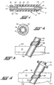
- FIGURES 1 and 2 which as noted above, correspond respectively to FIGURES 5 and 6 of my '280 patent. Because my prior patents, including the drawings, are incorporated by reference in the specifications hereof, reference numerals for FIGURES 1 and 2 are identical to and indicate the same parts, surfaces, etc., which have been described in my prior patents. Reference to the specifications of my prior patents should be had for a more detailed explanation of the functioning of the device than that disclosed herein.
- reference numeral 36 in FIGURES 1 and 2 designates a removable hemostatic sheath which has a low co-efficient of friction and may be either bioabsorbable or non-bioabsorbable in nature.
- cutting cannula 12 leaves a generally cylindrical void defined by tissue margin 51 (initially shown as the dot-dash line) and when hemostatic sheath 36 is inserted into the void, the margin is expanded to a cylindrical edge shown as 49 and the tissue surrounding the void is compressed.
- hemostatic sheath 36 compression of the tissue by means of hemostatic sheath 36 is sufficient to avoid hemorrhaging complications and that a bioabsorbable sheath, while preferred, is not necessary for all applications to prevent hemorrhaging. That is, sheath 36 can be non-bioabsorbable. Optionally, the non-bioabsorbable sheath could be coated with thrombin.
- body means and includes all ducts, organs, body parts, as well as the vasculature.
- tissue of the body means the tissue forming the outer surface of the body and can include as well, the tissue surrounding the body.
- Access is the opening, no matter how formed, in the tissue of the body and for vasculature applications includes trancutaneous access, as well as other types of access such as external access or extracorporeal access.
- sheath 36 at the site “where the body tissue has been accessed” means the use of sheath 36 at the site of the blood vessel or body organ and not just superficially at the skin level.
- tubular medical device includes not only medical instruments such as needles capable of puncture to gain access to the body, but also other devices capable of gaining access to the body such as a catheter, cannula, tube, introducer, and access devices.
- FIGURES 3, 4, and 5 show schematically, the patients skin 60, the subcutaneous tissue 61 lying beneath the skin, a membrane or body tissue 63 (such as the peritoneum) lining a body part 64 (such as the abdomen for the peritoneum) which body part 64 is to be accessed.
- a membrane or body tissue 63 such as the peritoneum
- body part 64 such as the abdomen for the peritoneum
- Access to body part 64 may be had by any tubular medical device 70 which is generally cylindrical.
- medical device could be, depending on the medical procedure performed, a laparoscope, a trocar, an electrode, a probe, an introducer, or the like.
- sheath 36 is slidably disposed on tubular medical device 70 external to the patient while medical device 70 is used to perform the desired medical procedure.
- a pusher mechanism or means i.e., plunger
- plunger 75 is simply a hand operated tubular member slidably disposed over tubular medical device 70.
- FIGURE 4 thus shows sheath 36 positioned in wound 76 by plunger 75 with plunger 75 being withdrawn from tubular medical device 70 and
- FIGURE 5 shows wound 76 with sheath 36 positioned therein at the in situ position described with tubular medical device 70 withdrawn. It should be obvious that the invention does not require precise positioning of sheath 36 by plunger 75. Because of the characteristics of sheath 36, sheath 36 can be precisely positioned in situ by the surgeon after plunger 75 and tubular medical device 70 is withdrawn. In fact, this is preferred for many procedures.
- the material for sheath 36 could be bioabsorbable or non-bioabsorbable material and common materials for sheath 36 would include collagen, gelatin, cellulose, absorbable polymers, and the like.
- sheath 36 as thus far described is that of a solid hollow cylinder.
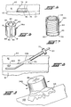
- An alternative configuration is shown for sheath 36a of FIGURE 6 and another configuration is shown for sheath 36b of FIGURE 7.
- All three sheath forms are having, in accordance with the invention, an outer hardened, rigid, or form maintaining coating or casing 83 surrounding an inner softer core 84.
- the margin for inner core 84 is diagrammatically illustrated by the broken line identified as reference numeral 85 shown for sheath 36a in FIGURE 6.
- Outer coating 83 is comprised of a substance which generally maintains its form when being inserted into the patient. Outer coating 83 may be bioabsorbable, non-bioabsorbable, or semi-bioabsorbable. Typically outer coating 83 is composed of a gelatin compound. Gelatin forms a relatively rigid outer shell structure which maintains its form during the insertion procedure. However, outer coating 83 may consist of collagen, cellulose, absorbable polymers, gelatin and combinations thereof. (Those skilled in the art will recognize that gelatin is a derivative of collagen. Thus in my prior applications, I disclosed collagen albeit in its gelatin form.
- Outer coating 83 is preferably a bioabsorbable material which dissolves within the patient's body. Outer coating 83 may also be composed of a substance that becomes slick upon contact with body fluids so as to allow sheath 36 to be easily inserted to the body tissue access site (i.e., membrane 63, blood vessel wall) without damaging the surrounding tissue during insertion. The slick outer coating 83 of sheath 36 acts as a lubricant to sheath 36 as it is being inserted.
- outer coating 83 Normally the slick surface results from the outer coating 83 dissolving in the presence of the body fluid.
- Bioabsorbable materials such as gelatin or gelatin compounds, form a very slippery surface when in contact with blood.
- outer coating 83 depending on the application, is sized to have various thickness. The thickness of outer coating 83 depends on where sheath 36 is being applied in the body of the patient and the distance the sheath has to travel within the body to reach the desired tissue access site. If outer coating 83 of sheath 36 is composed of a bioabsorbable substance such as gelatin, outer coating 83 will be absorbed into the surrounding tissue as it is being inserted through the body, causing the thickness of outer coating 36 to decrease. Therefore, outer coating 83 should be of a minimum thickness to allow sheath 36 to be properly placed at the desired tissue access site before being completely dissolved.
- the inner core 84 of sheath 36 may have the same or a different material than outer coating 83.
- Inner core 84 is made of absorbable materials.
- the material may be bioabsorbable, non-bioabsorbable or semi-bioabsorbable.
- the material of inner core 84 may be composed of collagen, gelatin, cellulose, absorbable polymers and combinations thereof.
- the material of inner core 84 Prior to being inserted into the body, the material of inner core 84 is typically compressed (i.e., by outer coating 83) to enhance the volume of body fluids which can be absorbed.
- the material of inner core 84 also has the property of expanding in volume when absorbing body fluids.
- Inner core material is preferably a less rigid material than outer coating 83 so that the inner core material can easily deform about the tissue access site during insertion.
- Collagen is a soft, highly absorbable material which is preferred for inner core 84.
- outer coating 83 permits sheath 36 to be easily moved to the tissue access site without damaging subcutaneous tissue 61 while body fluids permit collagen in inner core 84 to radially expand to occlude the longitudinally-extending central opening 89 of sheath 36 thus blocking or sealing the tissue access site at membrane 63. Further, as outer coating 83 dissolves, inner core 84 expands not only radially inwardly, but also radially outwardly to further compress subcutaneous tissue 61 and minimize bleeding.
- the thickness of coating 83 is predetermined such that occlusion of central opening 89 substantially occurs to seal the tissue access site before the radially-outwardly expansion and subsequent tissue compression occurs and the outside diameter of sheath 36 is suitably sized to achieve a sufficient level of subcutaneous tissue compression upon initial injection to minimize bleeding.
- Sheath 36 can be inserted on tubular medical device 70 prior to being inserted into the patient or prior to tubular medical device 70 being removed from the patient.
- damage to sheath 36 may occur if precautions are not taken.
- One source of damage to sheath 36 is in the form of contamination.
- In a medical environment there exists many infectious micro-organisms.
- sheath 36 is exposed to these foreign micro-organisms prior to being inserted into the patient. The longer the particular medical treatment, the more likely sheath 36 may be contaminated. Sheath 36 may also become deformed during the medical treatment.
- sheath 36 is preferably formed of a bioabsorbable material.
- gelatin has the special property of becoming slippery when body fluids such as blood contacts the gelatin surface. When the gelatin and body fluids interact, the gelatin is dissolved forming a slippery surface. During a long and/or complicated medical treatment, body fluids may come in contact with and completely dissolve the gelatin. When the time comes for in situ application of sheath 36, the slippery surface has dissolved and application becomes difficult.
- sheath 36 is comprised of absorbable materials, which absorb body fluids. As the body fluids are absorbed into sheath 36, such materials typically expand.
- sheath 36 will deform due to expansion, thus preventing sheath 36 from being inserted through skin surface 60 and/or damaging the surrounding subcutaneous tissue 61 during insertion.
- the expanded sheath 36 may also stick to tubular medical device 70 and may be damaged due to friction and/or shearing from being slid along tubular medical device 70 during insertion.
- Sheath 36 can be hermetically sealing sheath 36 with a substance, such as plastic, which protects sheath 36 from contamination and contact with body fluids.
- a substance such as plastic
- sheath 36 Prior to inserting sheath 36 into the tissue access site, the seal about sheath 36 is removed.
- sheath 36 is designed such that it can be quickly and easily inserted about tubular medical device 70 just prior to the time sheath 36 is to be inserted into the tissue access site.
- sheath 36a is generally of a cylindrical shape with a slit 90 extending, longitudinally from proximal end to distal end 81 and radially through the wall of sheath 36a from outside diameter to inside diameter.
- slit 90 Prior to sheath 36a being inserted about tubular medical device 70, slit 90 is opened and sheath 36a is inserted about the circumference of tubular medical device 70. Slit 90 is preferably spread open only enough so as to be fitted about tubular medical device 70. Otherwise, sheath 36a may be creased and/or torn on the side opposite slit 90.
- slit 90 is closed.
- the inside cylindrical surface of sheath 36a is formed such that once slit 90 is closed, the inside cylindrical surface of sheath 36a closely conforms to the outer surface of tubular medical device 70.
- At least one of the longitudinally-extruding edges 91, 92 forming slit 90 has a sticky surface maintaining slit 90 closed after sheath 36a is applied to tubular medical device 70.
- the sticky surface which could be mechanically formed such as like a "velcro" surface tends to "glue” the surfaces together.
- sheath 36a could be made of two cylindrical halves simply applied as a hollow cylinder to tubular medical device 70.
- FIGURE 7 illustrates another form of sheath 36, which is designated by reference numeral 36b and which is particularly applicable for sheath application as a vascular plug.
- Sheath 36b is essentially constructed as described above with a rigid outer coating 83 and a soft inner core 84. However, sheath 36b has a distal portion 95 extending from its distal end 80, which preferably is fretted as at 96. Distal position is composed of a soft sticky substance such as a soft collagen. A very thin bioabsorbable coating may be applied to distal position 95 just sufficient to permit sheath 36b to be inserted to the blood vessel site.
- sheath 36b as a vascular plug may be explained by reference to FIGURES 8 and 9 relative to any conventional vascular treatment such as an angiogram.
- an angiogram is a treatment for removing deposits which have accumulated along the inner wall of an artery 100 (i.e., blood vessel).
- the procedure typically employed user a conventional needle stick to locate an artery 100, the stick penetrating the subcutaneous tissue 61 and providing the tissue access opening to artery 100.
- a conventional guide wire is then fed through the needle into the artery, and the needle is removed with the guide wire left in place.
- a conventional introducer sheath 101 having a tissue dilator at its distal end with a valve and a pressure/sample port at its proximal end (not shown) is fed over the guide wire so that introducer sheath 101 extends into artery 100.
- a conventional catheter is then fed over the guide wire and the angiogram procedure performed after which the catheter and guide wire are removed. All of these steps are conventional and are not shown in the drawings.
- Sheath 36b is then placed on introducer 101 and, depending on the configuration of introducer 101, sheath 36b may or may not have slit 90. Also, positioned on introducer 101 is pusher mechanism 75.
- Pusher mechanism 75 is used to push sheath 36b against the blood vessel wall 103 (i.e., artery) and because distal portion 96 is soft, it conforms to the shape of vessel wall 103 as best shown in FIGURE 9. Sheath 36b is held in place by pusher 75 as the introducer sheath 101 is withdrawn and while sheath 36b is being held in this position, the collagen swells and expands radially-inwardly as discussed above to occlude central passage 89 and thus plug the tissue access opening (i.e., the opening caused in artery 100) to perform the angiogram.
- the tissue access opening i.e., the opening caused in artery 100
- Sheath 36b also expands radially-outwardly as discussed above, wedging itself further into subcutaneous tissue 61 to firmly seal or maintain sheath 36b, now a plug, in its in situ place or position while also minimizing bleeding from subcutaneous tissue 61.
- the soft distal portion 95 of sheath 36b spreads about the access site to seal seepage from the site while the radially-inward expansion of the non-bioabsorbable collagen plugs central opening 89 preventing escape of body fluids and the radially-outward expansion of the collagen further wedges sheath 36b into subcutaneous tissue 61 to maintain sheath 36b in its in situ position to prevent loss of body fluid thus forming an effective vascular plug.
Landscapes
- Health & Medical Sciences (AREA)
- Life Sciences & Earth Sciences (AREA)
- Surgery (AREA)
- Medical Informatics (AREA)
- Animal Behavior & Ethology (AREA)
- Engineering & Computer Science (AREA)
- Biomedical Technology (AREA)
- Heart & Thoracic Surgery (AREA)
- Veterinary Medicine (AREA)
- Molecular Biology (AREA)
- Public Health (AREA)
- General Health & Medical Sciences (AREA)
- Nuclear Medicine, Radiotherapy & Molecular Imaging (AREA)
- Cardiology (AREA)
- Pathology (AREA)
- Materials For Medical Uses (AREA)
- Surgical Instruments (AREA)
- Infusion, Injection, And Reservoir Apparatuses (AREA)
Abstract
Claims (18)
- Dispositif médical prévu pour être utilisé chez l'homme pour réduire les saignements, comprenant un élément tubulaire (14) s'étendant le long d'une trajectoire vers un site dans ledit patient, où les saignements sont susceptibles de se produire; une gaine hémostatique (36) comportant un passage central (89), s'étendant dans le sens longitudinal, et recevant, de manière généralement concentrique, ledit élément tubulaire (14) et disposé en vue de son coulissement autour dudit élément tubulaire (14) et des moyens (75) pour déposer ladite gaine (36) au niveau dudit site et pour provoquer le mouvement de coulissement relatif entre ladite gaine (36) et ledit élément tubulaire (14), caractérisé en ce que la gaine (36) comporte une enveloppe extérieure (83) et une partie centrale intérieure (84), l'enveloppe extérieure (83) étant un revêtement ou un enrobage extérieur relativement rigide dans son état initial et devenant glissant au contact des fluides corporels, la partie centrale intérieure (84 se composant d'un matériau absorbant, pouvant se dilater et est moins rigide que l'enveloppe extérieure (83), les fluides corporels au niveau du site provoquant ainsi le gonflement dimensionnel de ladite partie centrale intérieure (84) de ladite gaine hémostatique (36) de telle sorte que ledit corps est obturé au niveau du site.
- Dispositif selon la revendication 1, caractérisé en ce que la partie centrale intérieure (84) et/ou l'enveloppe extérieure (83) de la gaine (36) se compose d'un matériau défini dans une classe comprenant le collagène, la gélatine, des polymères résorbables et des combinaisons de ces derniers.
- Dispositif selon la revendication 2, caractérisé en ce que la partie centrale intérieure (84) de la gaine est biorésorbable.
- Dispositif selon la revendication 2, caractérisé en ce que la partie centrale intérieure (84) de la gaine est non résorbable.
- Dispositif selon la revendication 4, caractérisé en ce que le matériau de la partie centrale intérieure (84) de la gaine est du collagène.
- Dispositif selon la revendication 2, caractérisé en ce que l'enveloppe extérieure (83) de la gaine (36) est biorésorbable.
- Dispositif selon la revendication 6, caractérisé en ce que l'enveloppe extérieure (83) se compose de gélatine.
- Dispositif selon la revendication 1, caractérisé en ce que l'enveloppe extérieure (83) est non résorbable.
- Dispositif selon la revendication 1, caractérisé en ce que l'enveloppe extérieure (83) est partiellement biorésorbable.
- Dispositif selon la revendication 1, caractérisé en ce que l'enveloppe extérieure (83) est dimensionné selon une taille donnée en fonction de l'emplacement où la gaine (36) est appliquée dans le corps.
- Dispositif selon la revendication 1, caractérisé en ce que la partie centrale intérieure (84) de la gaine (36) est comprimée par l'enveloppe extérieure (83).
- Dispositif selon la revendication 1, caractérisé en ce que la gaine (36) comprend de la thrombine.
- Dispositif selon la revendication 1, caractérisé en ce que la gaine (36) comporte une fente (90) s'étendant dans le sens longitudinal, sur sa longueur.
- Dispositif selon l'une quelconque des revendications 1 à 13, caractérisé en ce que la gaine (36b) présente une partie d'extrémité proximale et une partie d'extrémité distale (95), la partie d'extrémité distale (95) pouvant se déformer dans le sens radial.
- Dispositif selon la revendication 14, caractérisé en ce que la partie d'extrémité distale (95) pouvant être déformée s'étend au-delà de l'enveloppe extérieure (83).
- Dispositif selon la revendication 15, caractérisé en ce que la partie d'extrémité distale (95) pouvant être déformée se compose d'une substance molle collante, et est revêtue d'une enveloppe biorésorbable très mince.
- Dispositif selon la revendication 1, dont la gaine (36) présente une partie distale s'étendant dans le sens longitudinal et une partie proximale s'étendant dans le sens longitudinal, et caractérisé en ce que la partie distale est plus souple que la partie proximale.
- Dispositif selon l'une quelconque des revendications 1 à 17, caractérisé en ce que ledit moyen de dépôt est un mécanisme de poussoir associé au dispositif ou un dispositif de poussée.
Applications Claiming Priority (3)
| Application Number | Priority Date | Filing Date | Title |
|---|---|---|---|
| US07/896,588 US5254105A (en) | 1988-05-26 | 1992-06-10 | Sheath for wound closure caused by a medical tubular device |
| PCT/US1993/005340 WO1993025255A2 (fr) | 1992-06-10 | 1993-06-04 | Gaine pour la fermeture d'une blessure provoquee par un instrument tubulaire |
| US896588 | 1997-07-17 |
Publications (3)
| Publication Number | Publication Date |
|---|---|
| EP0646027A1 EP0646027A1 (fr) | 1995-04-05 |
| EP0646027A4 EP0646027A4 (fr) | 1995-09-27 |
| EP0646027B1 true EP0646027B1 (fr) | 1999-08-25 |
Family
ID=25406459
Family Applications (1)
| Application Number | Title | Priority Date | Filing Date |
|---|---|---|---|
| EP93915208A Expired - Lifetime EP0646027B1 (fr) | 1992-06-10 | 1993-06-04 | Gaine pour la fermeture d'une blessure provoquee par un instrument tubulaire |
Country Status (6)
| Country | Link |
|---|---|
| US (1) | US5254105A (fr) |
| EP (1) | EP0646027B1 (fr) |
| JP (1) | JP2649286B2 (fr) |
| CA (1) | CA2136442C (fr) |
| DE (1) | DE69326146T2 (fr) |
| WO (1) | WO1993025255A2 (fr) |
Families Citing this family (191)
| Publication number | Priority date | Publication date | Assignee | Title |
|---|---|---|---|---|
| US5108421A (en) * | 1990-10-01 | 1992-04-28 | Quinton Instrument Company | Insertion assembly and method of inserting a vessel plug into the body of a patient |
| CZ281454B6 (cs) * | 1992-11-23 | 1996-10-16 | Milan Mudr. Csc. Krajíček | Pomůcka pro nechirurgické uzavírání otvoru ve stěně cévy |
| JPH0795982A (ja) * | 1993-04-19 | 1995-04-11 | Olympus Optical Co Ltd | ディスポーザブル医療用具 |
| US5431639A (en) | 1993-08-12 | 1995-07-11 | Boston Scientific Corporation | Treating wounds caused by medical procedures |
| US5439469A (en) * | 1993-11-05 | 1995-08-08 | Advanced Surgical, Inc. | Wound closure device |
| US6129685A (en) * | 1994-02-09 | 2000-10-10 | The University Of Iowa Research Foundation | Stereotactic hypothalamic obesity probe |
| US5697975A (en) | 1994-02-09 | 1997-12-16 | The University Of Iowa Research Foundation | Human cerebral cortex neural prosthetic for tinnitus |
| US7077822B1 (en) | 1994-02-09 | 2006-07-18 | The University Of Iowa Research Foundation | Stereotactic hypothalamic obesity probe |
| US5800535A (en) * | 1994-02-09 | 1998-09-01 | The University Of Iowa Research Foundation | Wireless prosthetic electrode for the brain |
| US5477862A (en) * | 1994-03-14 | 1995-12-26 | Haaga; John R. | Cutting tip for biopsy needle |
| WO1995026683A1 (fr) * | 1994-03-31 | 1995-10-12 | Boston Scientific Corporation | Obturateur vasculaire a localisateur de vaisseaux |
| US5645565A (en) * | 1995-06-13 | 1997-07-08 | Ethicon Endo-Surgery, Inc. | Surgical plug |
| US5810846A (en) * | 1995-08-03 | 1998-09-22 | United States Surgical Corporation | Vascular hole closure |
| US6183497B1 (en) | 1998-05-01 | 2001-02-06 | Sub-Q, Inc. | Absorbable sponge with contrasting agent |
| US6071300A (en) | 1995-09-15 | 2000-06-06 | Sub-Q Inc. | Apparatus and method for percutaneous sealing of blood vessel punctures |
| US6162192A (en) | 1998-05-01 | 2000-12-19 | Sub Q, Inc. | System and method for facilitating hemostasis of blood vessel punctures with absorbable sponge |
| KR19990064208A (ko) * | 1995-10-13 | 1999-07-26 | 트랜스바스큘라, 인코포레이티드 | 동맥 폐색부를 우회하고/거나 그 밖의 혈관 횡단 과정을 수행하기 위한 방법 및 장치 |
| US5674231A (en) * | 1995-10-20 | 1997-10-07 | United States Surgical Corporation | Apparatus and method for vascular hole closure |
| CA2244066A1 (fr) | 1996-02-02 | 1997-08-07 | Transvascular, Inc. | Dispositif, systeme et procede d'intervention transvasculaire interstitielle |
| US5797920A (en) * | 1996-06-14 | 1998-08-25 | Beth Israel Deaconess Medical Center | Catheter apparatus and method using a shape-memory alloy cuff for creating a bypass graft in-vivo |
| AU740072B2 (en) | 1996-08-06 | 2001-10-25 | Tyco Group S.A.R.L. | Insertion assembly and method of inserting a hemostatic closure device into an incision |
| US6142955A (en) | 1997-09-19 | 2000-11-07 | United States Surgical Corporation | Biopsy apparatus and method |
| US6019733A (en) * | 1997-09-19 | 2000-02-01 | United States Surgical Corporation | Biopsy apparatus and method |
| US6050955A (en) | 1997-09-19 | 2000-04-18 | United States Surgical Corporation | Biopsy apparatus and method |
| US6193673B1 (en) | 1998-02-20 | 2001-02-27 | United States Surgical Corporation | Biopsy instrument driver apparatus |
| US7625352B1 (en) | 1998-05-01 | 2009-12-01 | Sub-Q, Inc. | Depth and puncture control for system for hemostasis of blood vessel |
| US20010045575A1 (en) | 1998-05-01 | 2001-11-29 | Mark Ashby | Device and method for facilitating hemostasis of a biopsy tract |
| US6315753B1 (en) | 1998-05-01 | 2001-11-13 | Sub-Q, Inc. | System and method for facilitating hemostasis of blood vessel punctures with absorbable sponge |
| US7004962B2 (en) * | 1998-07-27 | 2006-02-28 | Schneider (Usa), Inc. | Neuroaneurysm occlusion and delivery device and method of using same |
| US6136014A (en) * | 1998-09-01 | 2000-10-24 | Vivant Medical, Inc. | Percutaneous tissue removal device |
| US6036698A (en) | 1998-10-30 | 2000-03-14 | Vivant Medical, Inc. | Expandable ring percutaneous tissue removal device |
| CA2317661C (fr) | 1998-11-20 | 2008-04-15 | Medical Industries Corp. | Dispositif d'insertion d'un agent hemostatique |
| CA2701691C (fr) | 1998-11-25 | 2011-09-13 | United States Surgical Corporation | Systeme de biopsie |
| US6162203A (en) * | 1999-01-11 | 2000-12-19 | Haaga; John R. | Cargo delivery needle |
| US7001400B1 (en) | 1999-03-04 | 2006-02-21 | Abbott Laboratories | Articulating suturing device and method |
| US6964668B2 (en) | 1999-03-04 | 2005-11-15 | Abbott Laboratories | Articulating suturing device and method |
| US7842048B2 (en) | 2006-08-18 | 2010-11-30 | Abbott Laboratories | Articulating suture device and method |
| US20040092964A1 (en) | 1999-03-04 | 2004-05-13 | Modesitt D. Bruce | Articulating suturing device and method |
| US7235087B2 (en) | 1999-03-04 | 2007-06-26 | Abbott Park | Articulating suturing device and method |
| US8137364B2 (en) | 2003-09-11 | 2012-03-20 | Abbott Laboratories | Articulating suturing device and method |
| US6984219B2 (en) | 1999-09-23 | 2006-01-10 | Mark Ashby | Depth and puncture control for blood vessel hemostasis system |
| US7842068B2 (en) | 2000-12-07 | 2010-11-30 | Integrated Vascular Systems, Inc. | Apparatus and methods for providing tactile feedback while delivering a closure device |
| US8758400B2 (en) | 2000-01-05 | 2014-06-24 | Integrated Vascular Systems, Inc. | Closure system and methods of use |
| US6461364B1 (en) | 2000-01-05 | 2002-10-08 | Integrated Vascular Systems, Inc. | Vascular sheath with bioabsorbable puncture site closure apparatus and methods of use |
| US6391048B1 (en) | 2000-01-05 | 2002-05-21 | Integrated Vascular Systems, Inc. | Integrated vascular device with puncture site closure component and sealant and methods of use |
| US9579091B2 (en) | 2000-01-05 | 2017-02-28 | Integrated Vascular Systems, Inc. | Closure system and methods of use |
| US6540735B1 (en) | 2000-05-12 | 2003-04-01 | Sub-Q, Inc. | System and method for facilitating hemostasis of blood vessel punctures with absorbable sponge |
| JP2004508092A (ja) | 2000-09-08 | 2004-03-18 | コールマン ジェイムス イー | 外科用ステープラ |
| US6712773B1 (en) | 2000-09-11 | 2004-03-30 | Tyco Healthcare Group Lp | Biopsy system |
| CA2423061A1 (fr) * | 2000-09-25 | 2002-03-28 | Cohesion Technologies, Inc. | Stents et obturateurs anastomotiques resorbables et leur utilisation chez des patients |
| US6626918B1 (en) | 2000-10-06 | 2003-09-30 | Medical Technology Group | Apparatus and methods for positioning a vascular sheath |
| US20030083539A1 (en) * | 2000-11-17 | 2003-05-01 | Boris Leschinsky | Intra-aortic balloon catheter for long-term implantation |
| WO2002041787A2 (fr) | 2000-11-27 | 2002-05-30 | Tyco Healthcare Group Lp | Appareil et procede d'echantillonnage et d'elimination de tissus |
| US7211101B2 (en) | 2000-12-07 | 2007-05-01 | Abbott Vascular Devices | Methods for manufacturing a clip and clip |
| US6623510B2 (en) | 2000-12-07 | 2003-09-23 | Integrated Vascular Systems, Inc. | Closure device and methods for making and using them |
| US8690910B2 (en) | 2000-12-07 | 2014-04-08 | Integrated Vascular Systems, Inc. | Closure device and methods for making and using them |
| US7806904B2 (en) | 2000-12-07 | 2010-10-05 | Integrated Vascular Systems, Inc. | Closure device |
| US7905900B2 (en) | 2003-01-30 | 2011-03-15 | Integrated Vascular Systems, Inc. | Clip applier and methods of use |
| WO2002087636A1 (fr) | 2001-03-12 | 2002-11-07 | Sub-Q, Inc. | Procedes permettant de steriliser des compositions de gelatine reticulee |
| US8187625B2 (en) | 2001-03-12 | 2012-05-29 | Boston Scientific Scimed, Inc. | Cross-linked gelatin composition comprising a wetting agent |
| US7008440B2 (en) * | 2001-11-08 | 2006-03-07 | Sub-Q, Inc. | System and method for delivering hemostasis promoting material to a blood vessel puncture site by fluid pressure |
| US6863680B2 (en) * | 2001-11-08 | 2005-03-08 | Sub-Q, Inc. | System and method for delivering hemostasis promoting material to a blood vessel puncture site by fluid pressure |
| IES20010547A2 (en) | 2001-06-07 | 2002-12-11 | Christy Cummins | Surgical Staple |
| US7993365B2 (en) * | 2001-06-08 | 2011-08-09 | Morris Innovative, Inc. | Method and apparatus for sealing access |
| US20060004408A1 (en) * | 2001-06-08 | 2006-01-05 | Morris Edward J | Method and apparatus for sealing access |
| US7192436B2 (en) | 2001-11-08 | 2007-03-20 | Sub-Q, Inc. | Pledget-handling system and method for delivering hemostasis promoting material to a blood vessel puncture site by fluid pressure |
| US7037323B2 (en) | 2001-11-08 | 2006-05-02 | Sub-Q, Inc. | Pledget-handling system and method for delivering hemostasis promoting material to a blood vessel puncture site by fluid pressure |
| US7025748B2 (en) | 2001-11-08 | 2006-04-11 | Boston Scientific Scimed, Inc. | Sheath based blood vessel puncture locator and depth indicator |
| US6749621B2 (en) | 2002-02-21 | 2004-06-15 | Integrated Vascular Systems, Inc. | Sheath apparatus and methods for delivering a closure device |
| CA2488688A1 (fr) | 2002-06-04 | 2003-12-11 | Christy Cummins | Clip de fermeture pour vaisseau sanguin et dispositif d'apport |
| US7955353B1 (en) * | 2002-11-04 | 2011-06-07 | Sub-Q, Inc. | Dissolvable closure device |
| US7455680B1 (en) * | 2002-11-04 | 2008-11-25 | Boston Scientific Scimed, Inc. | Apparatus and method for inhibiting blood loss |
| WO2004054156A2 (fr) * | 2002-12-06 | 2004-06-24 | Fast Country, Inc. | Systemes et procedes d'apport de ressources pour hotes interactives |
| US20040122349A1 (en) * | 2002-12-20 | 2004-06-24 | Lafontaine Daniel M. | Closure device with textured surface |
| US8709038B2 (en) * | 2002-12-20 | 2014-04-29 | Boston Scientific Scimed, Inc. | Puncture hole sealing device |
| US7160309B2 (en) | 2002-12-31 | 2007-01-09 | Laveille Kao Voss | Systems for anchoring a medical device in a body lumen |
| US7857828B2 (en) | 2003-01-30 | 2010-12-28 | Integrated Vascular Systems, Inc. | Clip applier and methods of use |
| US8758398B2 (en) | 2006-09-08 | 2014-06-24 | Integrated Vascular Systems, Inc. | Apparatus and method for delivering a closure element |
| US8821534B2 (en) | 2010-12-06 | 2014-09-02 | Integrated Vascular Systems, Inc. | Clip applier having improved hemostasis and methods of use |
| US8202293B2 (en) | 2003-01-30 | 2012-06-19 | Integrated Vascular Systems, Inc. | Clip applier and methods of use |
| US8398656B2 (en) | 2003-01-30 | 2013-03-19 | Integrated Vascular Systems, Inc. | Clip applier and methods of use |
| US8905937B2 (en) | 2009-02-26 | 2014-12-09 | Integrated Vascular Systems, Inc. | Methods and apparatus for locating a surface of a body lumen |
| US7942897B2 (en) * | 2003-07-10 | 2011-05-17 | Boston Scientific Scimed, Inc. | System for closing an opening in a body cavity |
| US20080264102A1 (en) | 2004-02-23 | 2008-10-30 | Bolton Medical, Inc. | Sheath Capture Device for Stent Graft Delivery System and Method for Operating Same |
| US8500792B2 (en) | 2003-09-03 | 2013-08-06 | Bolton Medical, Inc. | Dual capture device for stent graft delivery system and method for capturing a stent graft |
| US20070198078A1 (en) | 2003-09-03 | 2007-08-23 | Bolton Medical, Inc. | Delivery system and method for self-centering a Proximal end of a stent graft |
| US11259945B2 (en) | 2003-09-03 | 2022-03-01 | Bolton Medical, Inc. | Dual capture device for stent graft delivery system and method for capturing a stent graft |
| US7763063B2 (en) | 2003-09-03 | 2010-07-27 | Bolton Medical, Inc. | Self-aligning stent graft delivery system, kit, and method |
| US8292943B2 (en) | 2003-09-03 | 2012-10-23 | Bolton Medical, Inc. | Stent graft with longitudinal support member |
| US9198786B2 (en) | 2003-09-03 | 2015-12-01 | Bolton Medical, Inc. | Lumen repair device with capture structure |
| US11596537B2 (en) | 2003-09-03 | 2023-03-07 | Bolton Medical, Inc. | Delivery system and method for self-centering a proximal end of a stent graft |
| US7462188B2 (en) | 2003-09-26 | 2008-12-09 | Abbott Laboratories | Device and method for suturing intracardiac defects |
| US20050107867A1 (en) * | 2003-11-17 | 2005-05-19 | Taheri Syde A. | Temporary absorbable venous occlusive stent and superficial vein treatment method |
| JP4805162B2 (ja) * | 2003-11-25 | 2011-11-02 | ボストン サイエンティフィック リミテッド | 止血器具 |
| US7875043B1 (en) | 2003-12-09 | 2011-01-25 | Sub-Q, Inc. | Cinching loop |
| US7449024B2 (en) | 2003-12-23 | 2008-11-11 | Abbott Laboratories | Suturing device with split arm and method of suturing tissue |
| IES20040368A2 (en) | 2004-05-25 | 2005-11-30 | James E Coleman | Surgical stapler |
| US7402320B2 (en) * | 2004-08-31 | 2008-07-22 | Vnus Medical Technologies, Inc. | Apparatus, material compositions, and methods for permanent occlusion of a hollow anatomical structure |
| US9801982B2 (en) | 2004-09-28 | 2017-10-31 | Atrium Medical Corporation | Implantable barrier device |
| WO2006037049A2 (fr) | 2004-09-28 | 2006-04-06 | Atrium Medical Corporation | Application d'un revetement sur un dispositif medical |
| US8367099B2 (en) | 2004-09-28 | 2013-02-05 | Atrium Medical Corporation | Perforated fatty acid films |
| WO2007011385A2 (fr) | 2004-09-28 | 2007-01-25 | Atrium Medical Corporation | Gel thermodurcissable et son procede de fabrication |
| US9012506B2 (en) | 2004-09-28 | 2015-04-21 | Atrium Medical Corporation | Cross-linked fatty acid-based biomaterials |
| US8312836B2 (en) | 2004-09-28 | 2012-11-20 | Atrium Medical Corporation | Method and apparatus for application of a fresh coating on a medical device |
| US9000040B2 (en) | 2004-09-28 | 2015-04-07 | Atrium Medical Corporation | Cross-linked fatty acid-based biomaterials |
| US9592324B2 (en) | 2006-11-06 | 2017-03-14 | Atrium Medical Corporation | Tissue separating device with reinforced support for anchoring mechanisms |
| US8124127B2 (en) | 2005-10-15 | 2012-02-28 | Atrium Medical Corporation | Hydrophobic cross-linked gels for bioabsorbable drug carrier coatings |
| JP4959583B2 (ja) | 2005-01-25 | 2012-06-27 | タイコ ヘルスケア グループ リミテッド パートナーシップ | 中空解剖学的構造の永続的閉塞のための構造 |
| US8926633B2 (en) | 2005-06-24 | 2015-01-06 | Abbott Laboratories | Apparatus and method for delivering a closure element |
| US8313497B2 (en) | 2005-07-01 | 2012-11-20 | Abbott Laboratories | Clip applier and methods of use |
| US8267947B2 (en) | 2005-08-08 | 2012-09-18 | Abbott Laboratories | Vascular suturing device |
| US7883517B2 (en) | 2005-08-08 | 2011-02-08 | Abbott Laboratories | Vascular suturing device |
| US8083754B2 (en) | 2005-08-08 | 2011-12-27 | Abbott Laboratories | Vascular suturing device with needle capture |
| US9456811B2 (en) | 2005-08-24 | 2016-10-04 | Abbott Vascular Inc. | Vascular closure methods and apparatuses |
| US8758397B2 (en) | 2005-08-24 | 2014-06-24 | Abbott Vascular Inc. | Vascular closure methods and apparatuses |
| US20070060895A1 (en) | 2005-08-24 | 2007-03-15 | Sibbitt Wilmer L Jr | Vascular closure methods and apparatuses |
| US8920442B2 (en) | 2005-08-24 | 2014-12-30 | Abbott Vascular Inc. | Vascular opening edge eversion methods and apparatuses |
| US9427423B2 (en) | 2009-03-10 | 2016-08-30 | Atrium Medical Corporation | Fatty-acid based particles |
| US9278161B2 (en) | 2005-09-28 | 2016-03-08 | Atrium Medical Corporation | Tissue-separating fatty acid adhesion barrier |
| US8317822B2 (en) * | 2005-12-22 | 2012-11-27 | Ethicon, Inc. | Systems and methods for closing a vessel wound |
| US7381217B2 (en) * | 2005-12-23 | 2008-06-03 | Boston Scientific Scimed, Inc. | Serpentine stent pattern |
| US8808310B2 (en) | 2006-04-20 | 2014-08-19 | Integrated Vascular Systems, Inc. | Resettable clip applier and reset tools |
| US8556930B2 (en) | 2006-06-28 | 2013-10-15 | Abbott Laboratories | Vessel closure device |
| CA2594239A1 (fr) * | 2006-08-02 | 2008-02-02 | Tyco Healthcare Group Lp | Dispositif d'assistance de stabilisation pour trocart |
| US9492596B2 (en) | 2006-11-06 | 2016-11-15 | Atrium Medical Corporation | Barrier layer with underlying medical device and one or more reinforcing support structures |
| US8226681B2 (en) | 2007-06-25 | 2012-07-24 | Abbott Laboratories | Methods, devices, and apparatus for managing access through tissue |
| US8574244B2 (en) | 2007-06-25 | 2013-11-05 | Abbott Laboratories | System for closing a puncture in a vessel wall |
| US20090157101A1 (en) | 2007-12-17 | 2009-06-18 | Abbott Laboratories | Tissue closure system and methods of use |
| US8893947B2 (en) | 2007-12-17 | 2014-11-25 | Abbott Laboratories | Clip applier and methods of use |
| US7841502B2 (en) | 2007-12-18 | 2010-11-30 | Abbott Laboratories | Modular clip applier |
| US20090227938A1 (en) * | 2008-03-05 | 2009-09-10 | Insitu Therapeutics, Inc. | Wound Closure Devices, Methods of Use, and Kits |
| US9282965B2 (en) | 2008-05-16 | 2016-03-15 | Abbott Laboratories | Apparatus and methods for engaging tissue |
| US8118832B1 (en) | 2008-06-16 | 2012-02-21 | Morris Innovative, Inc. | Method and apparatus for sealing access |
| US20090318892A1 (en) * | 2008-06-20 | 2009-12-24 | Maria Aboytes | Removable Core Implant Delivery Catheter |
| CN102076281B (zh) | 2008-06-30 | 2014-11-05 | 波顿医疗公司 | 用于腹主动脉瘤的系统和方法 |
| GB2476772B (en) * | 2008-10-27 | 2013-09-25 | Sessions Pharmaceuticals Inc | Fluid extracting wound dressing |
| US9241696B2 (en) | 2008-10-30 | 2016-01-26 | Abbott Vascular Inc. | Closure device |
| US8323312B2 (en) | 2008-12-22 | 2012-12-04 | Abbott Laboratories | Closure device |
| US8858594B2 (en) | 2008-12-22 | 2014-10-14 | Abbott Laboratories | Curved closure device |
| US9486191B2 (en) | 2009-01-09 | 2016-11-08 | Abbott Vascular, Inc. | Closure devices |
| US20100179589A1 (en) | 2009-01-09 | 2010-07-15 | Abbott Vascular Inc. | Rapidly eroding anchor |
| US9173644B2 (en) | 2009-01-09 | 2015-11-03 | Abbott Vascular Inc. | Closure devices, systems, and methods |
| US9414820B2 (en) | 2009-01-09 | 2016-08-16 | Abbott Vascular Inc. | Closure devices, systems, and methods |
| US9089311B2 (en) | 2009-01-09 | 2015-07-28 | Abbott Vascular Inc. | Vessel closure devices and methods |
| US20100185234A1 (en) | 2009-01-16 | 2010-07-22 | Abbott Vascular Inc. | Closure devices, systems, and methods |
| USD636881S1 (en) | 2009-02-10 | 2011-04-26 | Sessions Pharmaceuticals Inc. | Wound dressing |
| US8317824B2 (en) | 2009-02-20 | 2012-11-27 | Boston Scientific Scimed, Inc. | Tissue puncture closure device |
| US8375553B2 (en) * | 2009-02-20 | 2013-02-19 | Boston Scientific Scimed, Inc. | Locking element for vascular closure device |
| US8529598B2 (en) * | 2009-02-20 | 2013-09-10 | Boston Scientific Scimed, Inc. | Tissue puncture closure device |
| US8052914B2 (en) | 2009-02-20 | 2011-11-08 | Boston Scientific Scimed, Inc. | Modified plug for arteriotomy closure |
| US9913634B2 (en) | 2009-02-20 | 2018-03-13 | Boston Scientific Scimed, Inc. | Locking element for vascular closure device |
| US8292918B2 (en) | 2009-02-20 | 2012-10-23 | Boston Scientific Scimed, Inc. | Composite plug for arteriotomy closure and method of use |
| CN106551740B (zh) | 2009-03-13 | 2020-03-27 | 波顿医疗公司 | 用于在手术部位部署腔内假体的系统和方法 |
| US20110038910A1 (en) | 2009-08-11 | 2011-02-17 | Atrium Medical Corporation | Anti-infective antimicrobial-containing biomaterials |
| US20110054492A1 (en) | 2009-08-26 | 2011-03-03 | Abbott Laboratories | Medical device for repairing a fistula |
| WO2011066340A1 (fr) | 2009-11-24 | 2011-06-03 | University Of Florida Research Foundation, Inc. | Appareil et procédés permettant de bloquer les voies d'aiguilles et de canules |
| US8858592B2 (en) * | 2009-11-24 | 2014-10-14 | Covidien Lp | Wound plugs |
| WO2011100547A2 (fr) | 2010-02-11 | 2011-08-18 | Boston Scientific Scimed, Inc. | Dispositifs et procédés automatiques de déploiement de fermeture vasculaire |
| US8303624B2 (en) | 2010-03-15 | 2012-11-06 | Abbott Cardiovascular Systems, Inc. | Bioabsorbable plug |
| EP2593141B1 (fr) | 2010-07-16 | 2018-07-04 | Atrium Medical Corporation | Composition et procédés destinés à modifier la vitesse d'hydrolyse de substances vulcanisées à base d'huile |
| US8758399B2 (en) | 2010-08-02 | 2014-06-24 | Abbott Cardiovascular Systems, Inc. | Expandable bioabsorbable plug apparatus and method |
| US8603116B2 (en) | 2010-08-04 | 2013-12-10 | Abbott Cardiovascular Systems, Inc. | Closure device with long tines |
| US9370353B2 (en) | 2010-09-01 | 2016-06-21 | Abbott Cardiovascular Systems, Inc. | Suturing devices and methods |
| US8663252B2 (en) | 2010-09-01 | 2014-03-04 | Abbott Cardiovascular Systems, Inc. | Suturing devices and methods |
| US8597340B2 (en) | 2010-09-17 | 2013-12-03 | Boston Scientific Scimed, Inc. | Torque mechanism actuated bioabsorbable vascular closure device |
| US8758402B2 (en) | 2010-12-17 | 2014-06-24 | Boston Scientific Scimed, Inc. | Tissue puncture closure device |
| US9149276B2 (en) | 2011-03-21 | 2015-10-06 | Abbott Cardiovascular Systems, Inc. | Clip and deployment apparatus for tissue closure |
| US9414822B2 (en) | 2011-05-19 | 2016-08-16 | Abbott Cardiovascular Systems, Inc. | Tissue eversion apparatus and tissue closure device and methods for use thereof |
| US9050067B2 (en) | 2011-09-26 | 2015-06-09 | Cook Medical Technologies, LLC | Percutaneous nephrostomy plug delivery device |
| US9332976B2 (en) | 2011-11-30 | 2016-05-10 | Abbott Cardiovascular Systems, Inc. | Tissue closure device |
| JPWO2013136967A1 (ja) * | 2012-03-15 | 2015-08-03 | テルモ株式会社 | 穿刺針 |
| US8864778B2 (en) | 2012-04-10 | 2014-10-21 | Abbott Cardiovascular Systems, Inc. | Apparatus and method for suturing body lumens |
| US8858573B2 (en) | 2012-04-10 | 2014-10-14 | Abbott Cardiovascular Systems, Inc. | Apparatus and method for suturing body lumens |
| EP3141223A1 (fr) | 2012-04-12 | 2017-03-15 | Bolton Medical, Inc. | Dispositif de mise en place d'une prothèse vasculaire |
| US9241707B2 (en) | 2012-05-31 | 2016-01-26 | Abbott Cardiovascular Systems, Inc. | Systems, methods, and devices for closing holes in body lumens |
| US9867880B2 (en) | 2012-06-13 | 2018-01-16 | Atrium Medical Corporation | Cured oil-hydrogel biomaterial compositions for controlled drug delivery |
| US9364209B2 (en) | 2012-12-21 | 2016-06-14 | Abbott Cardiovascular Systems, Inc. | Articulating suturing device |
| US9439751B2 (en) | 2013-03-15 | 2016-09-13 | Bolton Medical, Inc. | Hemostasis valve and delivery systems |
| US10596321B2 (en) | 2016-04-08 | 2020-03-24 | Allergan, Inc. | Aspiration and injection device |
| WO2018043427A1 (fr) * | 2016-09-01 | 2018-03-08 | テルモ株式会社 | Gaine d'introduction |
| JP6797612B2 (ja) * | 2016-09-01 | 2020-12-09 | テルモ株式会社 | イントロデューサー用シース |
| US10709444B2 (en) | 2017-01-24 | 2020-07-14 | Allergan Industrie Sas | Thread insertion devices |
| US10595977B2 (en) * | 2017-01-24 | 2020-03-24 | Allergan Industrie, Sas | Thread insertion devices |
| US10265151B2 (en) | 2017-01-24 | 2019-04-23 | Allergan Industrie Sas | Thread insertion devices |
| US10258447B2 (en) | 2017-01-24 | 2019-04-16 | Allergan Industrie Sas | Thread insertion devices |
| US10820900B2 (en) | 2017-01-24 | 2020-11-03 | Allergan Industrie Sas | Thread insertion devices |
| US10426449B2 (en) | 2017-02-16 | 2019-10-01 | Abbott Cardiovascular Systems, Inc. | Articulating suturing device with improved actuation and alignment mechanisms |
| WO2020264132A1 (fr) * | 2019-06-28 | 2020-12-30 | Kci Licensing, Inc. | Inserts de plaie pour le traitement de plaies tunnélisées |
| US20230029576A1 (en) * | 2020-01-07 | 2023-02-02 | Kci Licensing, Inc. | Means to improve usability of a wound insert for application to deep wounds |
| CN114945346A (zh) * | 2020-01-16 | 2022-08-26 | 爱德华兹生命科学公司 | 用于流体控制的系统和方法 |
| WO2021262264A1 (fr) | 2020-06-24 | 2021-12-30 | Bolton Medical, Inc. | Composant anti-retour pour dispositif de pose de prothèse vasculaire |
Family Cites Families (18)
| Publication number | Priority date | Publication date | Assignee | Title |
|---|---|---|---|---|
| US2603217A (en) * | 1952-07-15 | mcshirley | ||
| US2691373A (en) * | 1951-11-15 | 1954-10-12 | Julien A Bried | Colon flushing nozzle with dissolvable tip |
| US2814296A (en) * | 1954-04-15 | 1957-11-26 | S & R J Everett & Co Ltd | Surgical needles |
| US3106483A (en) * | 1961-07-27 | 1963-10-08 | Us Catheter & Instr Corp | Synthetic blood vessel grafts |
| US3396727A (en) * | 1964-01-06 | 1968-08-13 | Nolte Albert C Jr | Drainage tube for body fluids provided with filtering means coated with bacterial preventive material |
| US3358684A (en) * | 1965-03-12 | 1967-12-19 | Marshall Gerald | Parenteral injection devices |
| US3530860A (en) * | 1967-01-09 | 1970-09-29 | Ponce De Leon Ear | Method and apparatus for inserting a tube through the ear drum |
| US3736939A (en) * | 1972-01-07 | 1973-06-05 | Kendall & Co | Balloon catheter with soluble tip |
| US3888258A (en) * | 1972-11-07 | 1975-06-10 | Taichiro Akiyama | Drain for the eardrum and apparatus for introducing the same |
| US4564361A (en) * | 1975-11-18 | 1986-01-14 | Hiroshi Akiyama | Catheter |
| EP0019104A3 (fr) * | 1979-05-19 | 1980-12-10 | Intermedicat GmbH | Aiguille à biopsie pour recueillir des échantillons histologiques |
| US4306563A (en) * | 1979-11-28 | 1981-12-22 | Firma Pfrimmer & Co. Pharmazeutische Werke Erlangen Gmbh | Catheter for introduction into body cavities |
| US4396021A (en) * | 1980-12-15 | 1983-08-02 | Baumgartner George C | Surgical instrument and process |
| US4650488A (en) * | 1984-05-16 | 1987-03-17 | Richards Medical Company | Biodegradable prosthetic device |
| US4710181A (en) * | 1985-06-11 | 1987-12-01 | Genus Catheter Technologies, Inc. | Variable diameter catheter |
| DE8623592U1 (de) * | 1986-09-03 | 1987-10-01 | Lindenberg, Josef, 7512 Rheinstetten | Vorrichtung zur Entnahme von Flüssigkeiten, Gewebe o.dgl. |
| DE3632197A1 (de) * | 1986-09-23 | 1988-03-31 | Rainer Dr Hofmann | Stanz- oder schneidbiopsiekanuele |
| US4827940A (en) * | 1987-04-13 | 1989-05-09 | Cardiac Pacemakers, Inc. | Soluble covering for cardiac pacing electrode |
-
1992
- 1992-06-10 US US07/896,588 patent/US5254105A/en not_active Expired - Lifetime
-
1993
- 1993-06-04 JP JP6501601A patent/JP2649286B2/ja not_active Expired - Fee Related
- 1993-06-04 CA CA002136442A patent/CA2136442C/fr not_active Expired - Lifetime
- 1993-06-04 EP EP93915208A patent/EP0646027B1/fr not_active Expired - Lifetime
- 1993-06-04 DE DE69326146T patent/DE69326146T2/de not_active Expired - Lifetime
- 1993-06-04 WO PCT/US1993/005340 patent/WO1993025255A2/fr not_active Ceased
Also Published As
| Publication number | Publication date |
|---|---|
| WO1993025255A3 (fr) | 1994-02-17 |
| CA2136442A1 (fr) | 1993-12-23 |
| EP0646027A1 (fr) | 1995-04-05 |
| DE69326146D1 (de) | 1999-09-30 |
| JP2649286B2 (ja) | 1997-09-03 |
| JPH07506513A (ja) | 1995-07-20 |
| US5254105A (en) | 1993-10-19 |
| DE69326146T2 (de) | 1999-12-09 |
| EP0646027A4 (fr) | 1995-09-27 |
| WO1993025255A2 (fr) | 1993-12-23 |
| CA2136442C (fr) | 1996-12-17 |
Similar Documents
| Publication | Publication Date | Title |
|---|---|---|
| US5254105A (en) | Sheath for wound closure caused by a medical tubular device | |
| US5573518A (en) | Sheath for wound closure caused by a medical tubular device | |
| EP1572004B1 (fr) | Appareil pouvant empecher les pertes sanguines | |
| US5906631A (en) | Method and device for sealing vascular puncture wounds | |
| CA2187545C (fr) | Dispositif d'obturation d'une perforation dans un vaisseau sanguin | |
| US5643318A (en) | Vascular plug with vessel locator | |
| US5192300A (en) | Insertion assembly and method of inserting a vessel plug into the body of a patient | |
| US5478352A (en) | Insertion assembly and method of inserting a vessel plug into the body of a patient | |
| US6270484B1 (en) | Safety penetrating instrument with expandible portion and method of penetrating anatomical cavity | |
| JP2652588B2 (ja) | 止血切開封止装置 | |
| US8709039B2 (en) | Hemostatic devices and methods for use thereof | |
| JP2656689B2 (ja) | 傷凝固装置 | |
| JP2004016818A (ja) | シーリング装置 | |
| JPH0570469B2 (fr) | ||
| JPH0817777B2 (ja) | 止血装置および止血方法 | |
| JP2004521663A (ja) | 止血材を用いて血管穿刺を閉じる方法および装置 | |
| JP2005534389A (ja) | 穿刺孔の周長を減少させることにより穿刺孔をシールする装置 | |
| US20090270911A1 (en) | Vessel Sealing Device and Method of Using Same | |
| JPH10295699A (ja) | 止血用具 | |
| JPH10165510A (ja) | 医療器具導入装置 | |
| AU6552999A (en) | Assembly for sealing a puncture in a vessel |
Legal Events
| Date | Code | Title | Description |
|---|---|---|---|
| PUAI | Public reference made under article 153(3) epc to a published international application that has entered the european phase |
Free format text: ORIGINAL CODE: 0009012 |
|
| 17P | Request for examination filed |
Effective date: 19941208 |
|
| AK | Designated contracting states |
Kind code of ref document: A1 Designated state(s): BE DE DK FR GB IE IT SE |
|
| RHK1 | Main classification (correction) |
Ipc: A61B 17/00 |
|
| A4 | Supplementary search report drawn up and despatched | ||
| AK | Designated contracting states |
Kind code of ref document: A4 Designated state(s): BE DE DK FR GB IE IT SE |
|
| 17Q | First examination report despatched |
Effective date: 19970716 |
|
| APAB | Appeal dossier modified |
Free format text: ORIGINAL CODE: EPIDOS NOAPE |
|
| APBJ | Interlocutory revision of appeal recorded |
Free format text: ORIGINAL CODE: EPIDOS IRAPE |
|
| GRAG | Despatch of communication of intention to grant |
Free format text: ORIGINAL CODE: EPIDOS AGRA |
|
| GRAG | Despatch of communication of intention to grant |
Free format text: ORIGINAL CODE: EPIDOS AGRA |
|
| GRAH | Despatch of communication of intention to grant a patent |
Free format text: ORIGINAL CODE: EPIDOS IGRA |
|
| GRAH | Despatch of communication of intention to grant a patent |
Free format text: ORIGINAL CODE: EPIDOS IGRA |
|
| GRAA | (expected) grant |
Free format text: ORIGINAL CODE: 0009210 |
|
| AK | Designated contracting states |
Kind code of ref document: B1 Designated state(s): BE DE DK FR GB IE IT SE |
|
| PG25 | Lapsed in a contracting state [announced via postgrant information from national office to epo] |
Ref country code: SE Free format text: THE PATENT HAS BEEN ANNULLED BY A DECISION OF A NATIONAL AUTHORITY Effective date: 19990825 Ref country code: IT Free format text: LAPSE BECAUSE OF FAILURE TO SUBMIT A TRANSLATION OF THE DESCRIPTION OR TO PAY THE FEE WITHIN THE PRE;WARNING: LAPSES OF ITALIAN PATENTS WITH EFFECTIVE DATE BEFORE 2007 MAY HAVE OCCURRED AT ANY TIME BEFORE 2007. THE CORRECT EFFECTIVE DATE MAY BE DIFFERENT FROM THE ONE RECORDED.SCRIBED TIME-LIMIT Effective date: 19990825 Ref country code: BE Free format text: LAPSE BECAUSE OF FAILURE TO SUBMIT A TRANSLATION OF THE DESCRIPTION OR TO PAY THE FEE WITHIN THE PRESCRIBED TIME-LIMIT Effective date: 19990825 |
|
| REF | Corresponds to: |
Ref document number: 69326146 Country of ref document: DE Date of ref document: 19990930 |
|
| REG | Reference to a national code |
Ref country code: IE Ref legal event code: FG4D |
|
| PG25 | Lapsed in a contracting state [announced via postgrant information from national office to epo] |
Ref country code: DK Free format text: LAPSE BECAUSE OF FAILURE TO SUBMIT A TRANSLATION OF THE DESCRIPTION OR TO PAY THE FEE WITHIN THE PRESCRIBED TIME-LIMIT Effective date: 19991125 |
|
| ET | Fr: translation filed | ||
| PG25 | Lapsed in a contracting state [announced via postgrant information from national office to epo] |
Ref country code: GB Free format text: LAPSE BECAUSE OF NON-PAYMENT OF DUE FEES Effective date: 20000604 |
|
| PLBE | No opposition filed within time limit |
Free format text: ORIGINAL CODE: 0009261 |
|
| STAA | Information on the status of an ep patent application or granted ep patent |
Free format text: STATUS: NO OPPOSITION FILED WITHIN TIME LIMIT |
|
| 26N | No opposition filed | ||
| GBPC | Gb: european patent ceased through non-payment of renewal fee |
Effective date: 20000604 |
|
| PGFP | Annual fee paid to national office [announced via postgrant information from national office to epo] |
Ref country code: FR Payment date: 20010718 Year of fee payment: 9 |
|
| PG25 | Lapsed in a contracting state [announced via postgrant information from national office to epo] |
Ref country code: FR Free format text: LAPSE BECAUSE OF NON-PAYMENT OF DUE FEES Effective date: 20010831 |
|
| REG | Reference to a national code |
Ref country code: FR Ref legal event code: ST |
|
| PGFP | Annual fee paid to national office [announced via postgrant information from national office to epo] |
Ref country code: IE Payment date: 20020424 Year of fee payment: 10 |
|
| PG25 | Lapsed in a contracting state [announced via postgrant information from national office to epo] |
Ref country code: IE Free format text: LAPSE BECAUSE OF NON-PAYMENT OF DUE FEES Effective date: 20030604 |
|
| REG | Reference to a national code |
Ref country code: IE Ref legal event code: MM4A |
|
| PGFP | Annual fee paid to national office [announced via postgrant information from national office to epo] |
Ref country code: DE Payment date: 20120530 Year of fee payment: 20 |
|
| REG | Reference to a national code |
Ref country code: DE Ref legal event code: R071 Ref document number: 69326146 Country of ref document: DE |
|
| PG25 | Lapsed in a contracting state [announced via postgrant information from national office to epo] |
Ref country code: DE Free format text: LAPSE BECAUSE OF EXPIRATION OF PROTECTION Effective date: 20130605 |