EP0653726A1 - Technique pour trouver des régions d'histogramme intéressantes pour la reproduction améliorée d'échelles de tonalité dans des images radiographiques - Google Patents
Technique pour trouver des régions d'histogramme intéressantes pour la reproduction améliorée d'échelles de tonalité dans des images radiographiques Download PDFInfo
- Publication number
- EP0653726A1 EP0653726A1 EP94116530A EP94116530A EP0653726A1 EP 0653726 A1 EP0653726 A1 EP 0653726A1 EP 94116530 A EP94116530 A EP 94116530A EP 94116530 A EP94116530 A EP 94116530A EP 0653726 A1 EP0653726 A1 EP 0653726A1
- Authority
- EP
- European Patent Office
- Prior art keywords
- sample
- interest
- texture
- histogram
- image
- Prior art date
- Legal status (The legal status is an assumption and is not a legal conclusion. Google has not performed a legal analysis and makes no representation as to the accuracy of the status listed.)
- Granted
Links
Images






Classifications
-
- G—PHYSICS
- G06—COMPUTING OR CALCULATING; COUNTING
- G06T—IMAGE DATA PROCESSING OR GENERATION, IN GENERAL
- G06T5/00—Image enhancement or restoration
- G06T5/40—Image enhancement or restoration using histogram techniques
-
- G—PHYSICS
- G06—COMPUTING OR CALCULATING; COUNTING
- G06T—IMAGE DATA PROCESSING OR GENERATION, IN GENERAL
- G06T5/00—Image enhancement or restoration
- G06T5/90—Dynamic range modification of images or parts thereof
- G06T5/92—Dynamic range modification of images or parts thereof based on global image properties
-
- G—PHYSICS
- G06—COMPUTING OR CALCULATING; COUNTING
- G06T—IMAGE DATA PROCESSING OR GENERATION, IN GENERAL
- G06T7/00—Image analysis
- G06T7/40—Analysis of texture
- G06T7/41—Analysis of texture based on statistical description of texture
-
- G—PHYSICS
- G16—INFORMATION AND COMMUNICATION TECHNOLOGY [ICT] SPECIALLY ADAPTED FOR SPECIFIC APPLICATION FIELDS
- G16H—HEALTHCARE INFORMATICS, i.e. INFORMATION AND COMMUNICATION TECHNOLOGY [ICT] SPECIALLY ADAPTED FOR THE HANDLING OR PROCESSING OF MEDICAL OR HEALTHCARE DATA
- G16H30/00—ICT specially adapted for the handling or processing of medical images
- G16H30/40—ICT specially adapted for the handling or processing of medical images for processing medical images, e.g. editing
-
- G—PHYSICS
- G16—INFORMATION AND COMMUNICATION TECHNOLOGY [ICT] SPECIALLY ADAPTED FOR SPECIFIC APPLICATION FIELDS
- G16H—HEALTHCARE INFORMATICS, i.e. INFORMATION AND COMMUNICATION TECHNOLOGY [ICT] SPECIALLY ADAPTED FOR THE HANDLING OR PROCESSING OF MEDICAL OR HEALTHCARE DATA
- G16H50/00—ICT specially adapted for medical diagnosis, medical simulation or medical data mining; ICT specially adapted for detecting, monitoring or modelling epidemics or pandemics
- G16H50/20—ICT specially adapted for medical diagnosis, medical simulation or medical data mining; ICT specially adapted for detecting, monitoring or modelling epidemics or pandemics for computer-aided diagnosis, e.g. based on medical expert systems
-
- G—PHYSICS
- G06—COMPUTING OR CALCULATING; COUNTING
- G06T—IMAGE DATA PROCESSING OR GENERATION, IN GENERAL
- G06T2207/00—Indexing scheme for image analysis or image enhancement
- G06T2207/10—Image acquisition modality
- G06T2207/10116—X-ray image
-
- G—PHYSICS
- G06—COMPUTING OR CALCULATING; COUNTING
- G06T—IMAGE DATA PROCESSING OR GENERATION, IN GENERAL
- G06T2207/00—Indexing scheme for image analysis or image enhancement
- G06T2207/20—Special algorithmic details
- G06T2207/20021—Dividing image into blocks, subimages or windows
-
- G—PHYSICS
- G06—COMPUTING OR CALCULATING; COUNTING
- G06T—IMAGE DATA PROCESSING OR GENERATION, IN GENERAL
- G06T2207/00—Indexing scheme for image analysis or image enhancement
- G06T2207/30—Subject of image; Context of image processing
- G06T2207/30004—Biomedical image processing
-
- Y—GENERAL TAGGING OF NEW TECHNOLOGICAL DEVELOPMENTS; GENERAL TAGGING OF CROSS-SECTIONAL TECHNOLOGIES SPANNING OVER SEVERAL SECTIONS OF THE IPC; TECHNICAL SUBJECTS COVERED BY FORMER USPC CROSS-REFERENCE ART COLLECTIONS [XRACs] AND DIGESTS
- Y10—TECHNICAL SUBJECTS COVERED BY FORMER USPC
- Y10S—TECHNICAL SUBJECTS COVERED BY FORMER USPC CROSS-REFERENCE ART COLLECTIONS [XRACs] AND DIGESTS
- Y10S128/00—Surgery
- Y10S128/92—Computer assisted medical diagnostics
- Y10S128/925—Neural network
-
- Y—GENERAL TAGGING OF NEW TECHNOLOGICAL DEVELOPMENTS; GENERAL TAGGING OF CROSS-SECTIONAL TECHNOLOGIES SPANNING OVER SEVERAL SECTIONS OF THE IPC; TECHNICAL SUBJECTS COVERED BY FORMER USPC CROSS-REFERENCE ART COLLECTIONS [XRACs] AND DIGESTS
- Y10—TECHNICAL SUBJECTS COVERED BY FORMER USPC
- Y10S—TECHNICAL SUBJECTS COVERED BY FORMER USPC CROSS-REFERENCE ART COLLECTIONS [XRACs] AND DIGESTS
- Y10S378/00—X-ray or gamma ray systems or devices
- Y10S378/901—Computer tomography program or processor
-
- Y—GENERAL TAGGING OF NEW TECHNOLOGICAL DEVELOPMENTS; GENERAL TAGGING OF CROSS-SECTIONAL TECHNOLOGIES SPANNING OVER SEVERAL SECTIONS OF THE IPC; TECHNICAL SUBJECTS COVERED BY FORMER USPC CROSS-REFERENCE ART COLLECTIONS [XRACs] AND DIGESTS
- Y10—TECHNICAL SUBJECTS COVERED BY FORMER USPC
- Y10S—TECHNICAL SUBJECTS COVERED BY FORMER USPC CROSS-REFERENCE ART COLLECTIONS [XRACs] AND DIGESTS
- Y10S706/00—Data processing: artificial intelligence
- Y10S706/902—Application using ai with detail of the ai system
- Y10S706/924—Medical
Definitions
- This invention relates in general to digital image processing and more particularly to a technique for finding the histogram region of interest for improved tone scale reproduction of digital radiographic images.
- the output tonescale (that is, tone reproduction function) is generally already designed into the film by the film manufacturer, and a variety of different films are available to get different "looks" depending on exam types, exposing techniques, and observer preference.
- computed radiography (CR) (as described in US-A-Re.31,847), on the other hand, the separation of image acquisition and display stages allows an image or portion of an image to be displayed at an arbitrary output contrast depending on the particular need.
- the ability in CR systems to adjust this output tonescale in a computer before the final image is displayed is one of the powerful features of such systems.
- choosing or creating the appropriate tonescale is not always a simple operation because it depends on, among other things, the exam type, exposure conditions, imaging modality, noise sensitivity of the image acquisition device, and dynamic range of the output device.
- tone scale is to define an appropriate transformation function that maps the important image information to the maximum dynamic range of the display medium (either film or soft copy, that is, CRT display).
- Various methods have been proposed to generate tonescale curves for CR images.
- the histogram-based tonescale transformation is one of the most commonly used techniques for enhancing such images because of its efficiency.
- the following are exemplary of histogram-based imaging techniques for processing digital medical images: US-A-5,068,788; US-A-5,198,669; US-A-4,839,807; US-A-5,040,225; US-A-4,914,295; US-A-4,952,805.
- a method of finding a histogram region of interest for improved tone scale reproduction of digital radiographic images comprising the steps of: pseudo-randomly sampling a digital radiographic image with a sample having an appropriate size to delineate an region of interest; processing each sample using texture analysis techniques to extract a plurality of texture features; using the extracted texture features, classifying said sample with a previously trained neural network classifier to determine its class; and accumulating pixel values belonging to the same class to form separate histograms for each said class, wherein each said histogram can be used to optimize tone scale reproduction.
- Figure 1 is a block diagram of an imaging system in which the present invention may be used.
- Figure 2 is a block diagram of an embodiment of the present invention used in image processing of digital radiographic images.
- Figure 3 is a block diagram of a preferred image segmentation procedure.
- Figure 4 is a partially diagrammatic, partially block diagram of a preferred embodiment of the present invention.
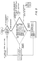
- Figure 5 is a flow chart of image samples processing routine.
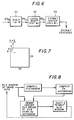
- Figure 6 is a block diagram illustrating the image sample preprocessing steps.
- Figure 7 is a diagrammatic view depicting local adaptive histogram equalization.
- Figure 8 is a flow chart useful in carrying out the preprocessing steps of Figures 6 and 7.
- Figures 9 - 14 are graphical views useful in describing the technique of the present invention.
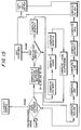
- Figure 15 if a flow chart useful in carrying out the texture feature extraction step.
- Figure 16 is a diagrammatic view illustrating a neural network classifier.
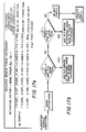
- Figures 17a and 17b are respectively an example and a flow diagram illustrating the building histogram step.
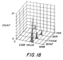
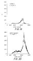
- Figures 18-23 are graphical views useful in explaining the present invention.
- FIG. 1 illustrates a typical system 10 in which the present invention can be practiced.
- the system consists of an image acquisition section (scanner) 12 in which an input image 14 is converted into a digital radiographic image.
- a digital image processing section 16 manipulates the digital image signal with image processing algorithms intended to improve the appearance, diagnostic quality, and other properties of the image.
- the digital image processing section can be a digital computer having known components, such as a digital processing computing device, memory, input and output devices, and so forth.
- the processed output image 20 is viewed on image display 18.
- the digital radiographic image signals to be processed with this invention can be acquired in a variety of different systems, for example, from storage phosphor (computed radiography) systems, film digitizer systems, image intensifier systems, and so forth.
- CT computed tomography
- MRI magnetic resonance imaging
- US ultrasound
- film writers laser printer, cathode ray tube printer
- soft display on a video monitor the most common are film writers (laser printer, cathode ray tube printer) and soft display on a video monitor.
- an image classification technique to improve the image processing algorithms described in US-A-5,164,993.
- the technique of the present invention is used after a digital radiographic image has been segmented according to the algorithm disclosed in the Jang and Schaetzing application.
- the digital radiographic image is processed with the tone scale algorithms disclosed in US-A-5,164,993. (Fig. 2, blocks 21, 22, 23 ).
- segmentation algorithm of Jang and Schaetzing is more clearly illustrated with reference to Figure 3.
- the referenced application is referred to for a more complete description of the segmentation technique disclosed.
- the segmentation method in general, detects and distinguishes in CR images both the regions of the direct x-ray exposure around the body part being imaged (background), and the regions in which beam-limiting devices have been used to restrict the size of the irradiation field (foreground).
- segmentation consists of five steps: edge detection 24, block generation 26, block classification 28, block refinement 30, and bit map generation 32.
- the edge detection step 24 can use a variety of edge detection algorithms. However, a morphological edge detector is preferred because of its low sensitivity to noise, its ability to yield an edge strength measure that is proportional to the contrast between the foreground/background and the object, and its efficient implementation.
- the output of the edge detection step is an edge strength map containing high-amplitude pixels in areas of strong edges and low-amplitude pixels in areas of little or no edge density.
- the local edge content is one indicator of the level of detail in various parts of the image, and, therefore, an indicator of whether these parts are more likely to belong to the foreground/background region or to the object.
- the image is broken into a set of nonoverlapping, contiguous blocks of pixels. Further analysis of the image and the classification into foreground, background, and object take place on a block-by-block basis.
- the block classification and block refinement steps 28, 30 are performed next. There are ten possible states each block can have, depending on whether the block is basically homogeneous (relatively low edge density) or nonhomogeneous (relatively high edge density), and what it contains (foreground pixels, background pixels, object pixels, mixtures of foreground and/or background pixels with object pixels).
- the information about edge content along with similar information from neighboring blocks is fed into a syntactic reasoning section that uses a set of clinically and empirically determined decision rules to classify each block of the image into one of two categories: foreground/background or object.
- the block refinement step reclassifies portions of the block into the latter two categories.
- the bit-map generation step 32 creates a two-valued (binary) image that functions as a template which is used in a preferred embodiment of the technique of the present invention.
- the technique of the present invention facilitates an accurate analysis of an image histogram by estimating what regions of the histogram correspond to bone, soft tissue, background and foreground based upon texture attributes. With such knowledge, it becomes possible, for example, to allow for more contrast in the bone region at the expense of contrast in the soft tissue region.
- diagram 34 depicts an original radiographic image including background, foreground, and region of interest.
- Diagram 36 depicts the binary image resulting from the segmentation processing described above.
- the delineated region of interest (the hands), are randomly sampled as illustrated in diagram 38.
- Each sample is preprocessed and texture features are extracted (box 40).
- the sample is then classified (box 42) using the features extracted by means of a neural classification technique, as to type, that is, bone, tissue, background, or foreground.
- Pixel values corresponding to the same class are accumulated (box 44) to form separate histogram estimates for bone, tissue, background, foreground. These histogram estimates can then be used (box 46) to optimize the tone scale algorithm for bone regions or tissue regions to enhance display of these regions for viewing and diagnosis.
- the random sampling step of box 38 (Fig. 4) is carried out by extracting subregions of pixels (e. g., 39x39 pixel subregions) from the segmented body part (which may, for example , have a digital value of 12 bits per pixel).
- the sampling technique preferably is a linear pixel shuffling technique as described in the article of Peter G. Anderson, Linear Pixel Shuffling for Image Processing, an Introduction, Journal of Electronic Imaging, (2), April, 1993, Pages 147-154.
- An exemplary software routine is shown in Fig. 5.
- the first preprocessing step 50 reduces the data for each sample by a factor (such as 16). As an example, each 12 bit pixel in a sample is reduced to an 8 bit pixel. A local adaptive histogram equalization process is then performed on each reduced sample.
- a histogram is computed (the frequency table of pixel code values). (Figs. 7, 9, 10) .
- an equalization histogram is computed as a cumulative frequency table based on the histogram. Then the center 3x3 pixel region is remapped based on the equalization histogram. (See Figs. 11,12). The process is repeated at successive windows after shifting by 3 pixels. (Figs. 7, 9, 10). A software routine of the process is shown in Fig. 8. Thereafter, the middle region (e. g., 32x32 pixels) of each sample is taken (Fig. 6, box 54). Figures 13 and 14 respectively illustrate a bone sample histogram before and after local adaptive histogram equalization.
- the texture of an object or an image can be described by adjectives such as random, periodic, smooth, coarse, rough, and so forth, and can be represented using statistical or structural techniques.
- Orthopedic bones have unique textural attributes because of trabecular structures in the spongy bone that form reinforcement struts that follow the lines of stress.
- the radiographic imaging process records these attributes, resulting in a unique texture compared to tissue, background, or foreground.
- two statistical methods of texture analysis are used, gray-level co-occurrence (GLC) matrix (See: Haralick, R., Shanmugam, K., Dinstein, I., (1973), Textural Features for Image Classification, IEEE Trans. Syst. Man.
- the GLC matrix is based upon the average spatial relationship chat gray-tones in an image have to one another, that is, cooccurence.
- the underlying assumption is that texture information can be adequately specified by a set of gray tone spatial-dependence matrices that are computed based various pixel differences and orientations.
- These gray-level co-occurrence matrices are estimates of the second-order joint conditional probability density functions, p(i,j,d,o) and are typically calculated at 4 orientations of 0, 45, 90, and 135 degrees.
- Each density function is the probability of going from gray level i to gray level j, given that the intersample spacing is d and the direction is given by angle o. If an image has N g gray levels, then the gray-level co-occurrence matrix is N g xN g .
- all textural features are extracted from the these matrices that were computed from 32x32 pixel samples.
- d 1-3 and the features extracted are energy, contrast, correlation, local homogeneity, and entropy.
- a limitation of the GLC matrix method is that the matrix and corresponding features must be computed 4 times, once for each orientation.
- the NGLD method overcomes this limitation by considering the relationship between a pixel and all of its neighbors at one time instead of in one direction at a time. This removes the angular dependency, facilitates invariance under linear gray-level transformations, and reduces the computational load.
- the NGLD matrix is formed from the gray-tone relationship between every element in the image and all of its neighbors at a particular distance d. All texture features are then obtained from this NGLD matrix.
- the matrix is of size N g xN n where N g is the number of possible gray levels and N n is the number of possible neighbors. It can be considered as frequency counts of grayness variation of an image and is similar in meaning as a histogram of an image. In the example given, the following features were used, small number emphasis, large number emphasis, number nonuniformity, second moment, and entropy.
- FIG. 15 there is shown a software routine for use in the feature extraction step.
- a neural network classifier Prior to classifying a sample, a neural network classifier was trained on known data (bone, soft tissue, background, foreground samples preprocessed as described above and texture features computed) which was manually hand-selected from a library of radiographic images. (See US-A-5,048,097.)
- Goal To train a classifier to recognize an unknown sample (never seen before) as one of four classes, that is, bone, tissue, background, foreground.
- Procedure Collect thousands of samples over a wide range of exams and image conditions which will serve as representative sampling of the entire population of possible samples to be seen under operating conditions. As each sample is selected, a label is affixed to it indicating the appropriate class, that is, bone, tissue, background, foreground.
- Each sample is preprocessed using the identical method to be employed under operating conditions.
- Bone would map as ⁇ 1, 0, 0, 0 ⁇ Tissue would map as ⁇ 0, 1, 0, 0 ⁇ Bgrd. would map as ⁇ 0, 0, 1, 0 ⁇ Fgrd. would map as ⁇ 0, 0, 0, 1 ⁇
- the preferred embodiment is a three-layer globally connected neural network.
- Backward error propagation is the preferred learning algorithm which utilizes gradient descent. Gradient descent is utilized to minimize the errors between the target and output vectors in an iterative fashion.
- Figure 16 shows a diagram illustrating a neural network classifier.
- the next step is to classify the samples using the previously trained neural network classifier (Box 42). If a sample is found to be of type bone, the central pixel and its surrounding neighborhood (typically 3x3) are all classified as bone and the corresponding pixel data is accumulated with other bone pixel values to build a bone histogram. (Box 44, Fig. 4). The same is done for the other three classes.
- Figures 17a and 17b are respectively an example of histogram building and a software routine for carrying out histogram building.
- Figure 18 is a three dimensional graphical view illustrating the histograms built for the four classes described above.
- Figures 19-22 are actual histograms, respectively, of bone and tissue samples.
- Figures 21 and 22 are histograms, respectively, of bone and tissue sample as classified by the technique of the present invention. The correlation between the actual histograms and the computed histograms is evident.
- Figure 23 illustrates the entire body part histogram and the tone scale curve based on it, as well as the bone only histogram and the tonescale curve based on it. Not knowing the bone region, one must make guesses about the left point and especially the right point using the tonescale algorithm of US-A-5,164,993. A wrong decision results in reduced contrast and inconsistency in image presentation. Knowing the limits of the bone and tissue regions enables accurate selection of the left and right points and results in consistency in image presentation.
- a second advantage of this system is that it facilitates customizing to the viewer. For example, if the radiologist is looking for a bone tumor, he might choose to use all of the dynamic range of the tone scale within the bone region and not use any for soft tissue or background. Or for a fracture, he may choose up to 20% of the dynamic range to examine the soft tissue for swelling and 80% for the bone in order to locate the fracture.
- Another advantage is that if the segmentation stage results in an error such that regions of background or foreground are included in the segmentation mask, the histogram analysis method fails. However, when the histogram is decomposed using texture features, this problem is overcome.
Landscapes
- Engineering & Computer Science (AREA)
- Health & Medical Sciences (AREA)
- Physics & Mathematics (AREA)
- Public Health (AREA)
- Theoretical Computer Science (AREA)
- General Physics & Mathematics (AREA)
- Medical Informatics (AREA)
- Primary Health Care (AREA)
- Biomedical Technology (AREA)
- General Health & Medical Sciences (AREA)
- Epidemiology (AREA)
- Nuclear Medicine, Radiotherapy & Molecular Imaging (AREA)
- Data Mining & Analysis (AREA)
- Databases & Information Systems (AREA)
- Pathology (AREA)
- Radiology & Medical Imaging (AREA)
- Probability & Statistics with Applications (AREA)
- Computer Vision & Pattern Recognition (AREA)
- Image Processing (AREA)
- Image Analysis (AREA)
- Apparatus For Radiation Diagnosis (AREA)
- Facsimile Image Signal Circuits (AREA)
Applications Claiming Priority (2)
| Application Number | Priority Date | Filing Date | Title |
|---|---|---|---|
| US08/152,772 US5426684A (en) | 1993-11-15 | 1993-11-15 | Technique for finding the histogram region of interest for improved tone scale reproduction of digital radiographic images |
| US152772 | 1993-11-15 |
Publications (2)
| Publication Number | Publication Date |
|---|---|
| EP0653726A1 true EP0653726A1 (fr) | 1995-05-17 |
| EP0653726B1 EP0653726B1 (fr) | 2001-06-27 |
Family
ID=22544377
Family Applications (1)
| Application Number | Title | Priority Date | Filing Date |
|---|---|---|---|
| EP94116530A Expired - Lifetime EP0653726B1 (fr) | 1993-11-15 | 1994-10-20 | Technique pour trouver des régions d'histogramme intéressantes pour la reproduction améliorée d'échelles de tonalité dans des images radiographiques |
Country Status (4)
| Country | Link |
|---|---|
| US (1) | US5426684A (fr) |
| EP (1) | EP0653726B1 (fr) |
| JP (1) | JP3619270B2 (fr) |
| DE (1) | DE69427567T2 (fr) |
Cited By (9)
| Publication number | Priority date | Publication date | Assignee | Title |
|---|---|---|---|---|
| WO1996013805A1 (fr) * | 1994-10-26 | 1996-05-09 | Minnesota Mining And Manufacturing Company | Accentuation de contraste par analyse spatiale d'histogrammes |
| WO1997004419A1 (fr) * | 1995-07-19 | 1997-02-06 | Morphometrix Technologies Inc. | Procede d'extraction de texture en fenetre pour analyse d'image |
| EP0780791A3 (fr) * | 1995-12-22 | 1998-04-29 | Eastman Kodak Company | Réglage automatique d'échelles de tonalité en utilisant une mesure de l'avtivité d'image |
| EP1126410A1 (fr) * | 2000-02-14 | 2001-08-22 | Koninklijke Philips Electronics N.V. | Amélioration d'un signal d'image |
| EP1211643A3 (fr) * | 2000-10-17 | 2003-09-17 | Eastman Kodak Company | Réglage automatique de luminosité pour radiographie numérique |
| EP1598780A1 (fr) * | 2004-05-17 | 2005-11-23 | GE Medical Systems Global Technology Company, LLC | Méthode et appareil de traitement d'images et système de tomographie par rayons X |
| CN102567736A (zh) * | 2010-12-14 | 2012-07-11 | 三星电子株式会社 | 图像识别设备及方法 |
| US20140270431A1 (en) * | 2013-03-15 | 2014-09-18 | Sony Corporation | Characterizing pathology images with statistical analysis of local neural network responses |
| US11216989B2 (en) | 2018-08-13 | 2022-01-04 | Lg Electronics Inc. | Mobile device and method for controlling same |
Families Citing this family (57)
| Publication number | Priority date | Publication date | Assignee | Title |
|---|---|---|---|---|
| FR2712415B1 (fr) * | 1993-11-09 | 1995-12-22 | Ge Medical Syst Sa | Procédé de repérage automatique de points d'intérêt lors d'un examen de stéréotaxie en mammographie. |
| US5761333A (en) * | 1995-01-31 | 1998-06-02 | General Electric Company | Contrast enhancement for CT systems |
| US5541028A (en) * | 1995-02-02 | 1996-07-30 | Eastman Kodak Company | Constructing tone scale curves |
| US5995651A (en) * | 1996-07-11 | 1999-11-30 | Duke University | Image content classification methods, systems and computer programs using texture patterns |
| US7650015B2 (en) | 1997-07-22 | 2010-01-19 | Image Processing Technologies. LLC | Image processing method |
| US6154560A (en) * | 1996-08-30 | 2000-11-28 | The Cleveland Clinic Foundation | System and method for staging regional lymph nodes using quantitative analysis of endoscopic ultrasound images |
| IL119283A0 (en) * | 1996-09-19 | 1996-12-05 | Elscint Ltd | Adaptive filtering |
| US5918196A (en) * | 1996-11-29 | 1999-06-29 | Cognex Corporation | Vision system for analyzing solid-of-revolution radius profile |
| GB9626676D0 (en) * | 1996-12-23 | 1997-02-12 | Smith & Nephew Res | Imaging method |
| US6092059A (en) * | 1996-12-27 | 2000-07-18 | Cognex Corporation | Automatic classifier for real time inspection and classification |
| US6901158B2 (en) * | 1997-09-22 | 2005-05-31 | Canon Kabushiki Kaisha | Image discrimination apparatus and image discrimination method |
| US6018590A (en) * | 1997-10-07 | 2000-01-25 | Eastman Kodak Company | Technique for finding the histogram region of interest based on landmark detection for improved tonescale reproduction of digital radiographic images |
| US6175652B1 (en) | 1997-12-31 | 2001-01-16 | Cognex Corporation | Machine vision system for analyzing features based on multiple object images |
| US6175644B1 (en) | 1998-05-01 | 2001-01-16 | Cognex Corporation | Machine vision system for object feature analysis and validation based on multiple object images |
| AU1606400A (en) * | 1998-11-13 | 2000-06-05 | Arch Development Corporation | System for detection of malignancy in pulmonary nodules |
| US6738499B1 (en) | 1998-11-13 | 2004-05-18 | Arch Development Corporation | System for detection of malignancy in pulmonary nodules |
| AUPP898499A0 (en) * | 1999-03-02 | 1999-03-25 | University Of Queensland, The | Method for image texture analysis |
| US6211971B1 (en) | 1999-03-11 | 2001-04-03 | Lockheed Martin Missiles & Space Co. | Method and apparatus to compress multi-spectral images to a single color image for display |
| US6579238B1 (en) * | 2000-04-24 | 2003-06-17 | Acuson Corporation | Medical ultrasonic imaging system with adaptive multi-dimensional back-end mapping |
| US6804384B2 (en) | 2001-06-12 | 2004-10-12 | Mclean Hospital Corporation | Color magnetic resonance imaging |
| US7088872B1 (en) * | 2002-02-14 | 2006-08-08 | Cogent Systems, Inc. | Method and apparatus for two dimensional image processing |
| DE10213284B4 (de) * | 2002-03-25 | 2007-11-08 | Siemens Ag | Verfahren zur Bilddarstellung in der medizinischen Bildgebung |
| US7006600B1 (en) * | 2004-01-15 | 2006-02-28 | Progeny, Inc. | Integrated digital dental x-ray system |
| CN1930587B (zh) * | 2004-03-15 | 2011-05-25 | 皇家飞利浦电子股份有限公司 | 图像可视化 |
| US8774355B2 (en) * | 2004-06-30 | 2014-07-08 | General Electric Company | Method and apparatus for direct reconstruction in tomosynthesis imaging |
| DE102005006658B4 (de) * | 2005-02-14 | 2011-12-15 | Siemens Ag | Verfahren bzw. Röntgensystem zur Aufnahme von Röntgenabbildungen eines auf einem digitalen Röntgendetektor abgebildeten Untersuchungsobjektes |
| DE102005010076A1 (de) * | 2005-03-04 | 2006-09-07 | Siemens Ag | Bildbearbeitungsverfahren für ein digitales medizinisches Untersuchungsbild und zugehörige Untersuchungseinrichtung |
| US8131477B2 (en) * | 2005-11-16 | 2012-03-06 | 3M Cogent, Inc. | Method and device for image-based biological data quantification |
| US7783121B1 (en) * | 2005-11-29 | 2010-08-24 | Adobe Systems Incorporated | Method and apparatus for applying a filter to an image with a large range of pixel values |
| US8594445B2 (en) * | 2005-11-29 | 2013-11-26 | Adobe Systems Incorporated | Fast bilateral filtering using rectangular regions |
| EP1840831A1 (fr) * | 2006-03-31 | 2007-10-03 | Sony Deutschland Gmbh | Égalisation d'histogramme adaptative pour des images présentant un fort contraste local |
| JP5068036B2 (ja) * | 2006-05-19 | 2012-11-07 | 富士フイルム株式会社 | 分光画像処理装置 |
| JP4232804B2 (ja) * | 2006-08-25 | 2009-03-04 | 日本電気株式会社 | 画像濃度変換方法、画像強調処理装置、およびそのプログラム |
| US8275179B2 (en) * | 2007-05-01 | 2012-09-25 | 3M Cogent, Inc. | Apparatus for capturing a high quality image of a moist finger |
| US8355595B2 (en) * | 2007-05-15 | 2013-01-15 | Xerox Corporation | Contrast enhancement methods and apparatuses |
| US7675524B1 (en) | 2007-05-17 | 2010-03-09 | Adobe Systems, Incorporated | Image processing using enclosed block convolution |
| US7920741B2 (en) | 2007-05-31 | 2011-04-05 | Adobe Systems Incorporated | System and method for image processing using integral histogram convolution |
| US7889923B1 (en) | 2007-05-31 | 2011-02-15 | Adobe Systems Incorporated | System and method for sparse histogram merging |
| US8411916B2 (en) * | 2007-06-11 | 2013-04-02 | 3M Cogent, Inc. | Bio-reader device with ticket identification |
| US7925653B2 (en) * | 2008-02-27 | 2011-04-12 | General Electric Company | Method and system for accessing a group of objects in an electronic document |
| US8655097B2 (en) | 2008-08-22 | 2014-02-18 | Adobe Systems Incorporated | Adaptive bilateral blur brush tool |
| US8315473B1 (en) | 2008-08-22 | 2012-11-20 | Adobe Systems Incorporated | Variably fast and continuous bilateral approximation filtering using histogram manipulations |
| US8666148B2 (en) | 2010-06-03 | 2014-03-04 | Adobe Systems Incorporated | Image adjustment |
| JP4691732B1 (ja) * | 2010-07-30 | 2011-06-01 | 国立大学法人 岡山大学 | 組織抽出システム |
| US9008415B2 (en) * | 2011-09-02 | 2015-04-14 | Adobe Systems Incorporated | Automatic image adjustment parameter correction |
| US8903169B1 (en) | 2011-09-02 | 2014-12-02 | Adobe Systems Incorporated | Automatic adaptation to image processing pipeline |
| US8897554B2 (en) | 2011-12-13 | 2014-11-25 | The Nielsen Company (Us), Llc | Video comparison using color histograms |
| US8750613B2 (en) | 2011-12-13 | 2014-06-10 | The Nielsen Company (Us), Llc | Detecting objects in images using color histograms |
| US8897553B2 (en) | 2011-12-13 | 2014-11-25 | The Nielsen Company (Us), Llc | Image comparison using color histograms |
| RU2653108C2 (ru) * | 2012-11-20 | 2018-05-07 | Конинклейке Филипс Н.В. | Интегрированное фенотипирование с использованием признаков текстуры изображения |
| JPWO2014203940A1 (ja) * | 2013-06-18 | 2017-02-23 | キヤノン株式会社 | トモシンセシス撮影の制御装置、撮影装置、撮影システム、制御方法および当該制御方法をコンピュータに実行させるためのプログラム |
| JP6252750B2 (ja) * | 2013-12-03 | 2017-12-27 | 株式会社島津製作所 | 骨梁解析装置 |
| US11157765B2 (en) | 2018-11-21 | 2021-10-26 | Samsung Electronics Co., Ltd. | Method and system for determining physical characteristics of objects |
| WO2020109519A1 (fr) * | 2018-11-30 | 2020-06-04 | Koninklijke Philips N.V. | Appareil et procédé de détection de fracture osseuse |
| CN109685801B (zh) * | 2018-12-10 | 2021-03-26 | 杭州帝视科技有限公司 | 结合纹理特征和深度神经网络信息的皮肤镜图像处理方法 |
| KR102575890B1 (ko) * | 2020-11-16 | 2023-09-08 | 본와이즈 주식회사 | 인공지능 시스템을 훈련시키기 위한 이미지 데이터를 처리하도록 구성되는 전자 장치 |
| CN120859558B (zh) * | 2025-09-26 | 2025-12-23 | 中国人民解放军西部战区总医院 | 一种急诊骨折自动识别与诊断的超声辅助系统 |
Citations (4)
| Publication number | Priority date | Publication date | Assignee | Title |
|---|---|---|---|---|
| JPS61278290A (ja) * | 1985-06-03 | 1986-12-09 | Matsushita Electric Ind Co Ltd | 複合カラ−映像信号のデイジタル記録再生装置 |
| US4897881A (en) * | 1988-03-23 | 1990-01-30 | Centre De Recherche Industrielle Du Quebec | Optimum fast textural feature extractor |
| EP0396171A1 (fr) * | 1989-04-26 | 1990-11-07 | Laboratoires D'electronique Philips | Procédé pour l'analyse de textures et analyseur |
| US5164993A (en) * | 1991-11-25 | 1992-11-17 | Eastman Kodak Company | Method and apparatus for automatic tonescale generation in digital radiographic images |
Family Cites Families (12)
| Publication number | Priority date | Publication date | Assignee | Title |
|---|---|---|---|---|
| US31847A (en) * | 1861-03-26 | Jesse young | ||
| US4914295A (en) * | 1985-06-25 | 1990-04-03 | Fuji Photo Film Co., Ltd. | Radiation image read-out and image signal storing apparatus |
| US4839807A (en) * | 1987-08-03 | 1989-06-13 | University Of Chicago | Method and system for automated classification of distinction between normal lungs and abnormal lungs with interstitial disease in digital chest radiographs |
| US4952805A (en) * | 1987-08-20 | 1990-08-28 | Fuji Photo Film Co., Ltd. | Method of judging the presence or absence of a limited irradiation field, method of selecting a correct irradiation field, and method of judging correctness or incorrectness of an irradiation field |
| US5040225A (en) * | 1987-12-07 | 1991-08-13 | Gdp, Inc. | Image analysis method |
| US4914589A (en) * | 1988-10-24 | 1990-04-03 | General Electric Company | Three-dimensional images obtained from tomographic data using a variable threshold |
| US5068788A (en) * | 1988-11-29 | 1991-11-26 | Columbia Scientific Inc. | Quantitative computed tomography system |
| US5198669A (en) * | 1989-09-20 | 1993-03-30 | Fujitsu Limited | Digital X-ray image processing apparatus |
| US5153926A (en) * | 1989-12-05 | 1992-10-06 | E. I. Du Pont De Nemours And Company | Parallel processing network that corrects for light scattering in image scanners |
| US5048097A (en) * | 1990-02-02 | 1991-09-10 | Eastman Kodak Company | Optical character recognition neural network system for machine-printed characters |
| US5107529A (en) * | 1990-10-03 | 1992-04-21 | Thomas Jefferson University | Radiographic equalization apparatus and method |
| US5331550A (en) * | 1991-03-05 | 1994-07-19 | E. I. Du Pont De Nemours And Company | Application of neural networks as an aid in medical diagnosis and general anomaly detection |
-
1993
- 1993-11-15 US US08/152,772 patent/US5426684A/en not_active Expired - Lifetime
-
1994
- 1994-10-20 DE DE69427567T patent/DE69427567T2/de not_active Expired - Fee Related
- 1994-10-20 EP EP94116530A patent/EP0653726B1/fr not_active Expired - Lifetime
- 1994-11-15 JP JP28043594A patent/JP3619270B2/ja not_active Expired - Fee Related
Patent Citations (4)
| Publication number | Priority date | Publication date | Assignee | Title |
|---|---|---|---|---|
| JPS61278290A (ja) * | 1985-06-03 | 1986-12-09 | Matsushita Electric Ind Co Ltd | 複合カラ−映像信号のデイジタル記録再生装置 |
| US4897881A (en) * | 1988-03-23 | 1990-01-30 | Centre De Recherche Industrielle Du Quebec | Optimum fast textural feature extractor |
| EP0396171A1 (fr) * | 1989-04-26 | 1990-11-07 | Laboratoires D'electronique Philips | Procédé pour l'analyse de textures et analyseur |
| US5164993A (en) * | 1991-11-25 | 1992-11-17 | Eastman Kodak Company | Method and apparatus for automatic tonescale generation in digital radiographic images |
Non-Patent Citations (3)
| Title |
|---|
| NEUBAUER C: "Segmentation of Defects in Textile Fabric.", 11TH IAPR INTERNATIONAL CONFERENCE ON PATTERN RECOGNITION. VOL. 1. CONFERENCE A: COMPUTER VISION AND APPLICATIONS, 30 August 1992 (1992-08-30), THE HAGUE, NL, pages 688 - 691, XP010029916, DOI: doi:10.1109/ICPR.1992.201654 * |
| OJA E ET AL.: "Regularity Measurement, Classification and Segmentation of Textures.", FROM PIXELS TO FEATURES. PROCEEDINGS OF A WORKSHOP, 22 August 1988 (1988-08-22), BONAS, FR, pages 207 - 218 * |
| PATENT ABSTRACTS OF JAPAN vol. 11, no. 138 (E - 503) 2 May 1987 (1987-05-02) * |
Cited By (15)
| Publication number | Priority date | Publication date | Assignee | Title |
|---|---|---|---|---|
| WO1996013805A1 (fr) * | 1994-10-26 | 1996-05-09 | Minnesota Mining And Manufacturing Company | Accentuation de contraste par analyse spatiale d'histogrammes |
| US5588071A (en) * | 1994-10-26 | 1996-12-24 | Minnesota Mining And Manufacturing Company | Identifying an area of interest using histogram data arranged in predetermined sequence |
| WO1997004419A1 (fr) * | 1995-07-19 | 1997-02-06 | Morphometrix Technologies Inc. | Procede d'extraction de texture en fenetre pour analyse d'image |
| US5949907A (en) * | 1995-07-19 | 1999-09-07 | Morphometrix Technologies Inc. | Window texture extraction for image analysis |
| AU723079B2 (en) * | 1995-07-19 | 2000-08-17 | Veracel Inc. | Window texture extraction for image analysis |
| EP0780791A3 (fr) * | 1995-12-22 | 1998-04-29 | Eastman Kodak Company | Réglage automatique d'échelles de tonalité en utilisant une mesure de l'avtivité d'image |
| EP1126410A1 (fr) * | 2000-02-14 | 2001-08-22 | Koninklijke Philips Electronics N.V. | Amélioration d'un signal d'image |
| WO2001059183A3 (fr) * | 2000-02-14 | 2001-12-20 | Koninkl Philips Electronics Nv | Mise en valeur d'un signal d'image |
| KR100790327B1 (ko) * | 2000-02-14 | 2008-01-02 | 코닌클리케 필립스 일렉트로닉스 엔.브이. | 화상 신호 개선 |
| EP1211643A3 (fr) * | 2000-10-17 | 2003-09-17 | Eastman Kodak Company | Réglage automatique de luminosité pour radiographie numérique |
| EP1598780A1 (fr) * | 2004-05-17 | 2005-11-23 | GE Medical Systems Global Technology Company, LLC | Méthode et appareil de traitement d'images et système de tomographie par rayons X |
| CN102567736A (zh) * | 2010-12-14 | 2012-07-11 | 三星电子株式会社 | 图像识别设备及方法 |
| US20140270431A1 (en) * | 2013-03-15 | 2014-09-18 | Sony Corporation | Characterizing pathology images with statistical analysis of local neural network responses |
| US9710695B2 (en) * | 2013-03-15 | 2017-07-18 | Sony Corporation | Characterizing pathology images with statistical analysis of local neural network responses |
| US11216989B2 (en) | 2018-08-13 | 2022-01-04 | Lg Electronics Inc. | Mobile device and method for controlling same |
Also Published As
| Publication number | Publication date |
|---|---|
| JPH07192119A (ja) | 1995-07-28 |
| US5426684A (en) | 1995-06-20 |
| DE69427567D1 (de) | 2001-08-02 |
| DE69427567T2 (de) | 2002-05-16 |
| JP3619270B2 (ja) | 2005-02-09 |
| EP0653726B1 (fr) | 2001-06-27 |
Similar Documents
| Publication | Publication Date | Title |
|---|---|---|
| EP0653726B1 (fr) | Technique pour trouver des régions d'histogramme intéressantes pour la reproduction améliorée d'échelles de tonalité dans des images radiographiques | |
| EP0576961B1 (fr) | Détection automatique du fond et du premier plan dans des images radiographiques numériques | |
| US5757953A (en) | Automated method and system for region decomposition in digital radiographic images | |
| US5825909A (en) | Automated method and system for image segmentation in digital radiographic images | |
| EP0301028B1 (fr) | Procede pour detecter automatiquement des cretes dans l'histogramme du niveau de gris d'une image numerique | |
| US7221787B2 (en) | Method for automated analysis of digital chest radiographs | |
| US6125194A (en) | Method and system for re-screening nodules in radiological images using multi-resolution processing, neural network, and image processing | |
| US5901240A (en) | Method for detecting the collimation field in a digital radiography | |
| US20090252429A1 (en) | System and method for displaying results of an image processing system that has multiple results to allow selection for subsequent image processing | |
| US6356652B1 (en) | Visualization of diagnostically irrelevant zones in a radiographic image | |
| EP1225542B1 (fr) | Segmentation en tomographie | |
| US6353674B1 (en) | Method of segmenting a radiation image into direct exposure area and diagnostically relevant area | |
| US6212291B1 (en) | Method for recognizing multiple irradiation fields in digital radiography | |
| US6608915B2 (en) | Image processing method and apparatus | |
| US5283736A (en) | Radiographic image processing apparatus | |
| Thakur et al. | A local statistics based region growing segmentation method for ultrasound medical images | |
| Schilham et al. | Multi-scale nodule detection in chest radiographs | |
| US5889882A (en) | Detection of skin-line transition in digital medical imaging | |
| EP0887769B1 (fr) | Methode de segmentation d'une image de radiation en surface directement exposee et surface pertinente pour le diagnostique | |
| TSAI | Automatic segmentation of liver structure in CT images using a neural network | |
| Digalakis et al. | Automatic adaptive contrast enhancement for radiological imaging | |
| Fathi et al. | A new fuzzy based flood-fill algorithm for 3D NMR brain segmentation | |
| Hariharan et al. | An algorithm for the enhancement of chest x-ray images of tuberculosis patients | |
| Jang et al. | Image enhancement for computed radiographic images | |
| EP0654762B1 (fr) | Visualisation de zones diagnostiquement non-pertinentes dans une image de radiographie |
Legal Events
| Date | Code | Title | Description |
|---|---|---|---|
| PUAI | Public reference made under article 153(3) epc to a published international application that has entered the european phase |
Free format text: ORIGINAL CODE: 0009012 |
|
| AK | Designated contracting states |
Kind code of ref document: A1 Designated state(s): DE FR GB |
|
| 17P | Request for examination filed |
Effective date: 19951012 |
|
| 17Q | First examination report despatched |
Effective date: 19980528 |
|
| GRAG | Despatch of communication of intention to grant |
Free format text: ORIGINAL CODE: EPIDOS AGRA |
|
| GRAG | Despatch of communication of intention to grant |
Free format text: ORIGINAL CODE: EPIDOS AGRA |
|
| GRAH | Despatch of communication of intention to grant a patent |
Free format text: ORIGINAL CODE: EPIDOS IGRA |
|
| GRAH | Despatch of communication of intention to grant a patent |
Free format text: ORIGINAL CODE: EPIDOS IGRA |
|
| GRAA | (expected) grant |
Free format text: ORIGINAL CODE: 0009210 |
|
| AK | Designated contracting states |
Kind code of ref document: B1 Designated state(s): DE FR GB |
|
| REF | Corresponds to: |
Ref document number: 69427567 Country of ref document: DE Date of ref document: 20010802 |
|
| ET | Fr: translation filed | ||
| REG | Reference to a national code |
Ref country code: GB Ref legal event code: IF02 |
|
| PLBE | No opposition filed within time limit |
Free format text: ORIGINAL CODE: 0009261 |
|
| STAA | Information on the status of an ep patent application or granted ep patent |
Free format text: STATUS: NO OPPOSITION FILED WITHIN TIME LIMIT |
|
| 26N | No opposition filed | ||
| PGFP | Annual fee paid to national office [announced via postgrant information from national office to epo] |
Ref country code: GB Payment date: 20050914 Year of fee payment: 12 |
|
| PGFP | Annual fee paid to national office [announced via postgrant information from national office to epo] |
Ref country code: FR Payment date: 20051006 Year of fee payment: 12 |
|
| PGFP | Annual fee paid to national office [announced via postgrant information from national office to epo] |
Ref country code: DE Payment date: 20051031 Year of fee payment: 12 |
|
| PG25 | Lapsed in a contracting state [announced via postgrant information from national office to epo] |
Ref country code: DE Free format text: LAPSE BECAUSE OF NON-PAYMENT OF DUE FEES Effective date: 20070501 |
|
| GBPC | Gb: european patent ceased through non-payment of renewal fee |
Effective date: 20061020 |
|
| REG | Reference to a national code |
Ref country code: FR Ref legal event code: ST Effective date: 20070629 |
|
| PG25 | Lapsed in a contracting state [announced via postgrant information from national office to epo] |
Ref country code: GB Free format text: LAPSE BECAUSE OF NON-PAYMENT OF DUE FEES Effective date: 20061020 |
|
| PG25 | Lapsed in a contracting state [announced via postgrant information from national office to epo] |
Ref country code: FR Free format text: LAPSE BECAUSE OF NON-PAYMENT OF DUE FEES Effective date: 20061031 |