EP0703754B1 - Test des maladies vasculaires peripheriques - Google Patents
Test des maladies vasculaires peripheriques Download PDFInfo
- Publication number
- EP0703754B1 EP0703754B1 EP94916353A EP94916353A EP0703754B1 EP 0703754 B1 EP0703754 B1 EP 0703754B1 EP 94916353 A EP94916353 A EP 94916353A EP 94916353 A EP94916353 A EP 94916353A EP 0703754 B1 EP0703754 B1 EP 0703754B1
- Authority
- EP
- European Patent Office
- Prior art keywords
- exercise
- pressure
- spring
- transducer
- plate
- Prior art date
- Legal status (The legal status is an assumption and is not a legal conclusion. Google has not performed a legal analysis and makes no representation as to the accuracy of the status listed.)
- Expired - Lifetime
Links
- 238000012360 testing method Methods 0.000 title claims description 23
- 208000018262 Peripheral vascular disease Diseases 0.000 title description 4
- 230000017531 blood circulation Effects 0.000 claims abstract description 35
- 230000036772 blood pressure Effects 0.000 claims abstract description 30
- 210000003205 muscle Anatomy 0.000 claims abstract description 24
- 230000000284 resting effect Effects 0.000 claims abstract description 20
- 238000000034 method Methods 0.000 claims abstract description 14
- QVGXLLKOCUKJST-UHFFFAOYSA-N atomic oxygen Chemical compound [O] QVGXLLKOCUKJST-UHFFFAOYSA-N 0.000 claims abstract description 10
- 229910052760 oxygen Inorganic materials 0.000 claims abstract description 10
- 239000001301 oxygen Substances 0.000 claims abstract description 10
- 230000001926 lymphatic effect Effects 0.000 claims abstract description 4
- 210000003414 extremity Anatomy 0.000 claims description 35
- 238000005259 measurement Methods 0.000 claims description 28
- 230000002792 vascular Effects 0.000 claims description 23
- 210000003423 ankle Anatomy 0.000 claims description 22
- 206010022562 Intermittent claudication Diseases 0.000 claims description 12
- 208000024980 claudication Diseases 0.000 claims description 11
- 238000003745 diagnosis Methods 0.000 claims description 10
- 230000033001 locomotion Effects 0.000 claims description 10
- 230000005355 Hall effect Effects 0.000 claims description 6
- 238000011065 in-situ storage Methods 0.000 claims description 4
- 230000002093 peripheral effect Effects 0.000 claims description 3
- 238000002496 oximetry Methods 0.000 claims description 2
- 230000000737 periodic effect Effects 0.000 claims description 2
- 230000004044 response Effects 0.000 claims description 2
- 230000007246 mechanism Effects 0.000 abstract description 2
- 230000008512 biological response Effects 0.000 abstract 2
- 239000000523 sample Substances 0.000 description 20
- WWGNGKGHDNYPPS-UHFFFAOYSA-N B.P.I Chemical compound B.P.I WWGNGKGHDNYPPS-UHFFFAOYSA-N 0.000 description 19
- 210000002414 leg Anatomy 0.000 description 14
- 210000002683 foot Anatomy 0.000 description 13
- 210000001367 artery Anatomy 0.000 description 10
- 210000003141 lower extremity Anatomy 0.000 description 8
- 208000037265 diseases, disorders, signs and symptoms Diseases 0.000 description 7
- 238000011835 investigation Methods 0.000 description 7
- 238000000692 Student's t-test Methods 0.000 description 6
- 244000309466 calf Species 0.000 description 6
- 201000010099 disease Diseases 0.000 description 6
- 238000011084 recovery Methods 0.000 description 6
- 208000002193 Pain Diseases 0.000 description 5
- 230000008859 change Effects 0.000 description 5
- 208000019553 vascular disease Diseases 0.000 description 5
- 208000025865 Ulcer Diseases 0.000 description 4
- 230000004872 arterial blood pressure Effects 0.000 description 4
- 230000036269 ulceration Effects 0.000 description 4
- 206010002383 Angina Pectoris Diseases 0.000 description 3
- 200000000007 Arterial disease Diseases 0.000 description 3
- 208000031481 Pathologic Constriction Diseases 0.000 description 3
- 239000000853 adhesive Substances 0.000 description 3
- 230000001070 adhesive effect Effects 0.000 description 3
- 238000004458 analytical method Methods 0.000 description 3
- 230000000747 cardiac effect Effects 0.000 description 3
- 206010012601 diabetes mellitus Diseases 0.000 description 3
- 239000003550 marker Substances 0.000 description 3
- 230000000926 neurological effect Effects 0.000 description 3
- 230000010355 oscillation Effects 0.000 description 3
- 230000002441 reversible effect Effects 0.000 description 3
- 208000024891 symptom Diseases 0.000 description 3
- 230000035488 systolic blood pressure Effects 0.000 description 3
- 238000002604 ultrasonography Methods 0.000 description 3
- 208000031104 Arterial Occlusive disease Diseases 0.000 description 2
- 206010033425 Pain in extremity Diseases 0.000 description 2
- 206010034576 Peripheral ischaemia Diseases 0.000 description 2
- 230000004075 alteration Effects 0.000 description 2
- 238000002399 angioplasty Methods 0.000 description 2
- 208000021328 arterial occlusion Diseases 0.000 description 2
- 230000000712 assembly Effects 0.000 description 2
- 238000000429 assembly Methods 0.000 description 2
- 230000036770 blood supply Effects 0.000 description 2
- 238000004364 calculation method Methods 0.000 description 2
- 230000006835 compression Effects 0.000 description 2
- 238000007906 compression Methods 0.000 description 2
- 230000008602 contraction Effects 0.000 description 2
- 238000005516 engineering process Methods 0.000 description 2
- 210000001105 femoral artery Anatomy 0.000 description 2
- 230000006872 improvement Effects 0.000 description 2
- 238000012806 monitoring device Methods 0.000 description 2
- 235000015097 nutrients Nutrition 0.000 description 2
- 239000011347 resin Substances 0.000 description 2
- 229920005989 resin Polymers 0.000 description 2
- 208000037804 stenosis Diseases 0.000 description 2
- 230000036262 stenosis Effects 0.000 description 2
- 238000001356 surgical procedure Methods 0.000 description 2
- 210000003813 thumb Anatomy 0.000 description 2
- 210000001364 upper extremity Anatomy 0.000 description 2
- 206010002388 Angina unstable Diseases 0.000 description 1
- 208000020446 Cardiac disease Diseases 0.000 description 1
- 208000000059 Dyspnea Diseases 0.000 description 1
- 206010013975 Dyspnoeas Diseases 0.000 description 1
- 208000007353 Hip Osteoarthritis Diseases 0.000 description 1
- 206010020565 Hyperaemia Diseases 0.000 description 1
- 206010020772 Hypertension Diseases 0.000 description 1
- 206010030124 Oedema peripheral Diseases 0.000 description 1
- 208000005764 Peripheral Arterial Disease Diseases 0.000 description 1
- 208000030831 Peripheral arterial occlusive disease Diseases 0.000 description 1
- 206010034568 Peripheral coldness Diseases 0.000 description 1
- 208000007814 Unstable Angina Diseases 0.000 description 1
- 208000005475 Vascular calcification Diseases 0.000 description 1
- 230000001464 adherent effect Effects 0.000 description 1
- 239000012790 adhesive layer Substances 0.000 description 1
- 238000002266 amputation Methods 0.000 description 1
- 230000008321 arterial blood flow Effects 0.000 description 1
- 238000013528 artificial neural network Methods 0.000 description 1
- 230000009286 beneficial effect Effects 0.000 description 1
- 230000002457 bidirectional effect Effects 0.000 description 1
- 210000004204 blood vessel Anatomy 0.000 description 1
- 230000004087 circulation Effects 0.000 description 1
- 238000012790 confirmation Methods 0.000 description 1
- 230000001186 cumulative effect Effects 0.000 description 1
- 230000007423 decrease Effects 0.000 description 1
- 230000000994 depressogenic effect Effects 0.000 description 1
- 239000007933 dermal patch Substances 0.000 description 1
- 238000013461 design Methods 0.000 description 1
- 238000002405 diagnostic procedure Methods 0.000 description 1
- 230000008034 disappearance Effects 0.000 description 1
- 208000035475 disorder Diseases 0.000 description 1
- 239000003814 drug Substances 0.000 description 1
- 238000002651 drug therapy Methods 0.000 description 1
- 230000000694 effects Effects 0.000 description 1
- 210000005224 forefinger Anatomy 0.000 description 1
- 208000019622 heart disease Diseases 0.000 description 1
- 230000003516 hyperlipidaemic effect Effects 0.000 description 1
- 230000001631 hypertensive effect Effects 0.000 description 1
- 230000001771 impaired effect Effects 0.000 description 1
- 201000004332 intermediate coronary syndrome Diseases 0.000 description 1
- 208000021156 intermittent vascular claudication Diseases 0.000 description 1
- 208000028867 ischemia Diseases 0.000 description 1
- 230000000302 ischemic effect Effects 0.000 description 1
- 238000002955 isolation Methods 0.000 description 1
- 210000000629 knee joint Anatomy 0.000 description 1
- 239000010410 layer Substances 0.000 description 1
- 238000007726 management method Methods 0.000 description 1
- 238000012544 monitoring process Methods 0.000 description 1
- 230000003387 muscular Effects 0.000 description 1
- 230000002981 neuropathic effect Effects 0.000 description 1
- 239000002773 nucleotide Substances 0.000 description 1
- 125000003729 nucleotide group Chemical group 0.000 description 1
- 230000036961 partial effect Effects 0.000 description 1
- 238000003909 pattern recognition Methods 0.000 description 1
- 230000002980 postoperative effect Effects 0.000 description 1
- 230000008569 process Effects 0.000 description 1
- 230000035485 pulse pressure Effects 0.000 description 1
- 230000002829 reductive effect Effects 0.000 description 1
- 230000000717 retained effect Effects 0.000 description 1
- 238000012216 screening Methods 0.000 description 1
- 208000013220 shortness of breath Diseases 0.000 description 1
- 239000000725 suspension Substances 0.000 description 1
- 238000002560 therapeutic procedure Methods 0.000 description 1
- 230000008320 venous blood flow Effects 0.000 description 1
- 230000000007 visual effect Effects 0.000 description 1
- 210000000707 wrist Anatomy 0.000 description 1
Images


Classifications
-
- A—HUMAN NECESSITIES
- A61—MEDICAL OR VETERINARY SCIENCE; HYGIENE
- A61B—DIAGNOSIS; SURGERY; IDENTIFICATION
- A61B8/00—Diagnosis using ultrasonic, sonic or infrasonic waves
- A61B8/42—Details of probe positioning or probe attachment to the patient
- A61B8/4272—Details of probe positioning or probe attachment to the patient involving the acoustic interface between the transducer and the tissue
- A61B8/4281—Details of probe positioning or probe attachment to the patient involving the acoustic interface between the transducer and the tissue characterised by sound-transmitting media or devices for coupling the transducer to the tissue
-
- A—HUMAN NECESSITIES
- A61—MEDICAL OR VETERINARY SCIENCE; HYGIENE
- A61B—DIAGNOSIS; SURGERY; IDENTIFICATION
- A61B5/00—Measuring for diagnostic purposes; Identification of persons
- A61B5/02—Detecting, measuring or recording for evaluating the cardiovascular system, e.g. pulse, heart rate, blood pressure or blood flow
- A61B5/02007—Evaluating blood vessel condition, e.g. elasticity, compliance
-
- A—HUMAN NECESSITIES
- A61—MEDICAL OR VETERINARY SCIENCE; HYGIENE
- A61B—DIAGNOSIS; SURGERY; IDENTIFICATION
- A61B5/00—Measuring for diagnostic purposes; Identification of persons
- A61B5/22—Ergometry; Measuring muscular strength or the force of a muscular blow
- A61B5/221—Ergometry, e.g. by using bicycle type apparatus
- A61B5/222—Ergometry, e.g. by using bicycle type apparatus combined with detection or measurement of physiological parameters, e.g. heart rate
-
- A—HUMAN NECESSITIES
- A61—MEDICAL OR VETERINARY SCIENCE; HYGIENE
- A61B—DIAGNOSIS; SURGERY; IDENTIFICATION
- A61B8/00—Diagnosis using ultrasonic, sonic or infrasonic waves
- A61B8/42—Details of probe positioning or probe attachment to the patient
- A61B8/4209—Details of probe positioning or probe attachment to the patient by using holders, e.g. positioning frames
-
- A—HUMAN NECESSITIES
- A61—MEDICAL OR VETERINARY SCIENCE; HYGIENE
- A61B—DIAGNOSIS; SURGERY; IDENTIFICATION
- A61B8/00—Diagnosis using ultrasonic, sonic or infrasonic waves
- A61B8/42—Details of probe positioning or probe attachment to the patient
- A61B8/4209—Details of probe positioning or probe attachment to the patient by using holders, e.g. positioning frames
- A61B8/4236—Details of probe positioning or probe attachment to the patient by using holders, e.g. positioning frames characterised by adhesive patches
-
- A—HUMAN NECESSITIES
- A63—SPORTS; GAMES; AMUSEMENTS
- A63B—APPARATUS FOR PHYSICAL TRAINING, GYMNASTICS, SWIMMING, CLIMBING, OR FENCING; BALL GAMES; TRAINING EQUIPMENT
- A63B23/00—Exercising apparatus specially adapted for particular parts of the body
- A63B23/035—Exercising apparatus specially adapted for particular parts of the body for limbs, i.e. upper or lower limbs, e.g. simultaneously
- A63B23/04—Exercising apparatus specially adapted for particular parts of the body for limbs, i.e. upper or lower limbs, e.g. simultaneously for lower limbs
- A63B23/08—Exercising apparatus specially adapted for particular parts of the body for limbs, i.e. upper or lower limbs, e.g. simultaneously for lower limbs for ankle joints
- A63B23/085—Exercising apparatus specially adapted for particular parts of the body for limbs, i.e. upper or lower limbs, e.g. simultaneously for lower limbs for ankle joints by rotational movement of the joint in a plane substantially parallel to the body-symmetrical-plane
-
- A—HUMAN NECESSITIES
- A61—MEDICAL OR VETERINARY SCIENCE; HYGIENE
- A61B—DIAGNOSIS; SURGERY; IDENTIFICATION
- A61B5/00—Measuring for diagnostic purposes; Identification of persons
- A61B5/45—For evaluating or diagnosing the musculoskeletal system or teeth
- A61B5/4519—Muscles
Definitions
- the present invention relates to peripheral vascular disease testing and particularly to an apparatus and method for (1) screening patients for further study, and (2) investigating in greater detail, and more accurately, patients who are known to have peripheral vascular disease and may be either under observation or have had angioplasty or an operation.
- the treadmill even if available, is of limited use in diagnosis in these cases.
- the blood flow rate measurable in the distal circulation remains normal or elevates after exercise in a limb with a normal blood flow.
- blood pressure falls, or even reaches zero, in a limb with abnormally impaired blood flow.
- the overall heart rate is not much affected (up to about 20%) and hence not only can the diagnostic method be carried out upon the frail and elderly more easily, but the results obtained relate more directly to the limb under test.
- measurement can be made in the non-exercised limbs.
- the applicant has devised a simple non-invasive technique to assist not only the vascular specialist in more detailed analysis of the disease before and after treatment, but also to assist the General Practitioner in diagnosis. Further, with instruction a patient can perform self-assessment at home as now happens with blood pressure monitoring.
- the invention as set out in claim 1 provides in a general sense a diagnostic apparatus comprising a standardizable resistance means to resist a force applied thereto by a group of limb muscles during exercise, and test means for physiologically measuring a biological component resultant from said limb exercise.
- the biological component may be selected from one or more of blood flow, blood pressure, transcutaneous oxygen and lymphatic clearance rate. Where the resting blood flow is measured before and after exercise with the standardizable resistance means, if there is a vascular obstruction in the limb the blood flow will markedly fall or even reach zero. Similarly transcutaneous oxygen values will fall.
- the device may also be used to test lymphatic clearance of an injected labelled nucleotide before and after limb exercise.
- Measurements that can be made before and after exercise, in addition to blood flow by Doppler include for example transcutaneous oxygen measurements, compartmental pressures, leg volumes and other measurements that can be readily monitored.
- vascular disease tends to show up at or towards the ankle and lower calf muscles because these are furthest from the heart.
- asymptomatic vascular disease can also be present in the other limbs and an estimation of the disease in these limbs allows a more accurate diagnosis as to whether, for example, by-pass surgery is likely to be more effective than drug therapy.
- the resistance means is preferably such as to resist multiple motions to induce fatigue by using up oxygen and available nutrients in the limb that has an inadequate blood supply.
- EP-A-0430067 discloses a device for exercising the leg muscles against a known force.
- the apparatus is complex and would be difficult to utilize with the upper limb.
- WO 88/08276 reveals a muscle testing apparatus for testing the strength of muscles or groups of muscles. Again data is taken direct from the apparatus itself, and although it exercises groups of muscles the data taken cannot be directed specifically to the presence or absence of vascular disease.
- EP-A-5090421 reveals an apparatus for testing muscular strength utilising a pressure plate and a pressure transducer.
- US-A-4732038 reveals the interposing of a force sensing device within a range of motion of a limb or other to sense the flexion of a selected group of muscles. Again, in each case data from said flexion is collected from the device itself rather than, as in the instance of the present invention, the device being utilized essentially to induce fatigue on a controlled basis.
- US-A-4280486 reveals a motor driven foot exercising device comprising a treadle associated with a non-invasive blood flow detector; the detector is utilized to sound an alarm or restart a motor if blood pressure falls too low.
- EP-A-0330463 relates to a blood flow resistance measuring device comprising a wrist band pressure pad.
- US-A-4454885 provides diagnostic apparatus comprising a standardizable resistance means to resist a force applied thereto by a selected group of limb muscles during exercise, and test means for physiologically measuring a biological component resultant from said limb exercise, the standardizable resistance means comprising a pressure plate pivoted about an axle, spring means operatively connected to said plate to bias the plate to an at rest position, and adjustment means for preloading the spring means to a desired value, whereby in use the plate is reciprocatable by a limb against the spring means bias to induce fatigue or claudication.
- the present invention is characterized by means indicative of the load applied by the spring means, the spring means being associated with a signal generating means indicative of each completed movement thereby to generate an output signal for each completed stroke, and further characterized by the longitudinal axis of the patient's leg being arranged, in use, to pass close to or through the axle.
- the device may comprise a base supporting the axle and enclosing or supporting the adjustment means.
- the adjustment means may be a screw thread and the spring may be a tension spring.
- the adjustment means may include a screw threaded block connecting the screw thread to the tension spring, and the load indicator means may indicate a range of spring tension values, for example high, medium or low, or delineated in Kg, for example. With small design adjustments a compression spring may be similarly utilized.
- the patient may, in use, press the plate to the end of its travel a set number of times, or may continue working on the pressure plate until fatigue or claudication sets in, before blood flow is remeasured.
- the means for physiologically testing blood flow is preferably a Doppler Ultrasound blood flow rate monitoring device, for example that sold under the Registered Trade Mark "Dopplex". It is known that resting arterial and venous blood flow rate measurements provide characteristic doppler wave forms when measured at various sites with a hand held probe containing a terminal transducer. For example, the normal blood flow rate in the common femoral artery is tri-phasic in that it has a positive wave form in the systolic flow phase followed by a negative flow rate in the reverse flow phase followed by a third phase of positive flow before the next systole.
- Partial stenosis of the common femoral artery will reduce the readings for the first systolic flow phase and the reverse phase and may result in the disappearance of the third phase altogether. Severe stenosis results in the reverse phase disappearing as well. Similar wave forms for other areas in the limb, and for the venous system, are also known in the art to have characteristic signatures.
- a patient can be made to exercise against a gradient which is beneficial to diabetic patients with rigid, non-collapsible vessels.
- the apparatus of the present invention may also be arranged such that the arc of travel of the pressure plate is adjustable.
- the spring means is operatively associated with a signal generating means indicative of each completed movement whereby work done can be readily calculated.
- the spring means may be associated with a permanent magnet and a hall effect sensor thereby to generate an output signal for each completed stroke of the pressure plate.
- a second hall effect device can be used to define the position of the remote end of the spring means so that spring loading can be readily measured by electronic means. This enables the audible or visual signal to be produced when a patient has completed a given quantity of work. This enables ready comparison with other patients and normal volunteers.
- a further improvement of the present invention additionally lies in the problem that after exercise has been taken, a certain period lapses before a hand held Doppler probe, for example, can be correctly positioned to measure pressure and flow rate in a artery. This is particularly so where the pressure and flow rate are low as is likely to be the case after claudication.
- a specially adapted Doppler transducer assembly may be used which can be secured to the skin in a correct position and used prior to, during, and after exercise.
- the Doppler transducer assembly In order to use the Doppler transducer assembly during exercise it is usually necessary to briefly suspend the exercise so that readings may be taken, alternatively readings may be taken continuously and a computer utilized to discount traces within certain limits so that only readings taken during said suspension are recorded and displayed.
- the applicants have found the key to successfully positioning a Doppler transducer is to ensure that it is correctly positioned over the desired artery. In the first place it is desirable therefore to mark, with a standard Doppler probe, the position of the artery on the skin with a marker pen. This mark may also include the precise direction of the artery.
- a housing secured over the mark with the marker pen should be hollow so that the mark can be seen clearly through the housing.
- Two transducers are preferably utilised one to monitor flow and the other to monitor pressure. It is important that these are correctly positioned relative to the artery so that the flow transducer i.e. the 60° transducer, is directed along the axis of the artery. For this reason the transducer should be rotatable in the housing as well as being secured therein. This can be achieved by providing a flow line on the transducer housing so that it may be rotated into accord with the line applied earlier, and for it to be either an interference fit in the housing or to be screwed thereinto so that relative rotation can occur.
- US-A-5058592 relates to a similar acoustic pad but wherein the transducer is steerable. This is effected by providing an annular body with an adhesive foot to be secured to the skin and a moveable Doppler probe secured within the annulus and steered by means of being rocked into the correct position and held in position by means of a "VELCRO" tape.
- This arrangement tends to overcome the problem of faulty positioning because it is possible to see where the housing is going prior to securing the probe within the housing.
- the device only measures blood pressure and provides no means whereby blood flow may be assessed or means for aligning the two so that the two components can be measured from the same position.
- EP-A-467853 relates to a complex device comprising, inter alia, a sensor held in position by a cuff which extends over an extensive portion of the arm.
- This comprises a blood pressure and separately a blood flow monitoring device but these are not combined, they are not in a single skin patch and relative positioning clearly presents problems.
- WO92/07508 similarly relates to a non-invasive arterial pressure monitor which comprises a pair of spaced Doppler transducers which both measure blood flow to achieve bidirectional flow determinations. There are no means for combining these two together or for measuring blood pressure discussed.
- the test means may be an in-situ Doppler device adapted to measure both blood flow and blood pressure.
- the Doppler transducer means may thus comprise at least two fixed transducers, first of said transducers being positioned substantially perpendicular to the skin contact surface of the housing and the second of said transducers been fixed at a predetermined angle relative thereto.
- the predetermined angle of said transducer may be at or about 60° to the skin contact surface of the transducer.
- the Doppler transducer means may comprise a transducer rotatable about its horizontal axis or oscillatable through a predetermined angle, said transducer being adapted to output a first signal indicative of pressure and a second signal indicative of flow.
- the housing may comprise means to drive the transducer such as an electric motor or oscillator means for example, an electromagnet.
- the housing may be square, rectangular, circular or ovoid in transverse cross-section and may be so arranged that the transducer assembly heads are recessed from the plane of the skin contact surface of the housing thereby to accommodate a Doppler wave transmitting gel.
- the housing may be releasably retained on the skin either because the skin contact face of the housing is provided with an adhesive surface, or by the provision of a high friction surface associated with the resilient means for positioning about a body part to retain the housing in its operative position.
- the Doppler transducer assemblies as just described may be utilized in conjunction with an automatic blood pressure cuff so that at periodic intervals during diagnosis said cuff is inflated, preferably in response to a predetermined cuff control signal, to allow blood pressure and flow rate to be automatically sampled.
- the assemblies are particularly suited to utilization with a dedicated microprocessor, the output signals obtained from the varied sources are at best collated and displayed on a computer monitor screen.
- a method for the non-invasive measurement of peripheral vascular supply to a limb using apparatus comprises establishing a resting pre-exercise vascular pressure or flow rate, utilizing a limb to repeatedly move the plate of the apparatus over a predetermined distance for a measured time, and establishing a post exercise pressure or flow rate.
- the the pre- and post-exercise vascular pressures may be denoted by an ankle pressure index as herein set forth.
- the vascular flow and/or pressure may be monitored during timed intervals during an exercise routine, and when a cuff is secured about the ankle it may be inflated, likewise at timed intervals during exercise, thereby to allow vascular flow and pressure values to be monitored during short resting periods during the exercise. Oximetry determinations may also be utilized in the arrangement in accordance with the present invention.
- Signals from all the foregoing may be input to a neural network or other pattern recognition programme so that simple diagnoses can be computer generated.
- the main components of the standardizable resistance means are a pressure plate (1) supported on a base frame (2) which also accommodates a tension spring (3).
- the pressure plate (1) is generally planar and extends upwardly from the base frame (2) and is provided at its lower end with an axle (12) journalled for rotation in an axle stand (13) forming part of the base frame (2).
- a heel plate (11) may optionally be provided, which extends normal to the plane of the pressure plate (1) at or adjacent the axle (12).
- the tension spring (3) extends longitudinally of the base frame (2) towards the rear (14) thereof.
- a tension controller (4) which comprises a screw threaded member terminating at its remote outer end in an adjuster wheel (5).
- a tension block (6) provided with an attachment for the spring (3) whereby a hook at the rear end of the spring (3) contacts the tension block (6) and is secured thereto by means of pin (7).
- the tension block is associated to the exterior of the device with a range of values, for example "high”, “medium” and “low”, which allows the paramedic to set the pressure required to actuate the plate.
- an extremity of an upper or lower limb is placed upon the pressure plate (1) and the indicator (8) is set to an appropriate value for the patient concerned.
- the patient is then invited either to depress the plate a set number of times, or to depress the plate an indeterminate number of times for a certain period or until the muscle groups under test become significantly fatigued.
- the flow rate in the most appropriate artery is recorded on a resting basis and is recorded again when the group of muscles have been satisfactorily fatigued. If the peripheral flow rate remains much the same then the patient's pain is more likely to be orthopaedic in origin, whereas If the blood flow is significantly reduced the problem is most likely to be vascular in origin and require further investigation.
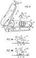
- Figure 2 shows a standardizable resistance means (20) of the present invention.
- the standardizable resistance means (20) comprises a base frame (22) supporting a cylinder portion (22).
- the frame (22') terminates at its forward end (25) in an axle stand portion (33) provided at its upper most portion with an axle (32).
- Axle (32) supports pressure plate (21) provided at its lower end with a heel plate (31).
- the heel plate (31) is moveable relative to the axle (32) and is positioned so that with the patient's foot on pressure plate (21) and the patient's heel resting on the heel plate (31), the longitudinal axis of the patient's leg passes through the axle (32).
- the pressure plate (21) is also provided with an extension handle (27) which is extendable from the position shown in Figure 2 to an extended position at a variable height where it may be secured by means of a thumb screw or other convenient means (not shown).
- a saddle (28) is optionally provided for use with the resistance means (20) such that it clips on to the cylinder portion (21) prime and is provided with an upstanding elbow rest (29). It will be observed that with the handle (27) extended and with the saddle (28) correctly positioned on the cylinder (22') an arm can be exercised.
- the cylinder (21') is provided at a remote end (24) with tension controller (26).
- the tension controller (26) is arranged such that it provides a reasonably accurate reflection of the tension applied to spring (23).
- Spring (23) extends from the rear end of the cylinder (24) to the forward end of the cylinder (25) and is secured at both ends by means of generally annular spring retainers. Either or both of the spring retainers are provided with a permanent magnet for cooperation with a hall effect device (not shown), positioned suitably on the internal face of the cylinder (22').
- the hall effect device is connected to outputs (30) by means of this device and an output signal is thus generated each time the spring is elongated to a predetermined degree. This provides a means of counting, and because the extension of the spring by a set amount is known, work done can be readily calculated.
- Figures 1 and 2 can be utilized by for example a general practitioner to ascertain whether or not a problem of leg pain experienced by a patient is vascular in origin merely by taking a first reading with a Doppler probe and repeating the reading after an exercise period. As explained before the absence of blood flow pressure will indicate a vascular problem and hence indicate the need for specialist care.
- Figure 2 particularly provides also a simple and effective mechanism whereby all non-invasive investigations related to the diagnosis and surveillance of patients with peripheral vascular disease can be computer linked and automated.
- Blood flow rate determinations utilizing the standardizable resistance means of the present invention can be made with a Dopplex Vascular Blood Flow Monitor in accordance with manufacturer's directions.
- Calculations can also be effected of the work done to reach ischaemic pain and this can be equated to the distance that the patient is actually able to walk, which is more accurate than the values presently obtainable.
- Tables can be prepared for the normal and the diseased, equating work done on the device in accordance with the present invention with distance actually walked and hence values can be given in mobility ranges if desired.
- Measurements can also be made of the time to recover after exercise-induced ischaemia.
- utilizing the device of the present invention to make more than one measurement after exercise, whereas with prior art treadmill arrangements only one measurement is actually made because of the time delay in getting the patient to the couch.
- the resistance means is preferably such as to resist multiple motions to induce fatigue by using up oxygen and available nutrients in the limb that has an inadequate blood supply.
- Figure 3A shows a temporarily adherent probe (40) which is adapted, by means of skin contact member (41) for location at a precisely defined spot on the skin surface.
- the skin contact member (41) may be held in position by means of an adhesive or by means of a resilient member about the limb, for example.
- the underside of the skin contact member may be provided with a high friction surface or with a weakly adhesive layer if necessary.
- the probe (40) is provided with an upstanding housing (43) which surrounds a removable transducer assembly (49) provided with an input and output connector portion (42).
- the transducer assembly (49) includes a pair of transducers; embedded in resin; transducer (44) being angled at 60° to the perpendicular and transducer (45) being perpendicular to the skin surface.
- the tranzducers (44) and (45) and the resin in which they are embedded are spaced inwardly of the skin contact surface level so as to provide a space for a gel layer (48) to provide an intimate contact between the transducer assembly (49) and skin.
- transducer (44) being angled at 60° is adapted to measure flow rate through a target vessel, whereas transducer (45) is arranged to measure pressure therein.
- transducer (46) is adapted for rotation or oscillation about a central point (47).
- housing is similar to that of Figure 3A but includes actuation means (not shown) such as a small electric motor or an electromagnetic oscillator.
- actuation means such as a small electric motor or an electromagnetic oscillator.
- an oscillator is utilized the oscillation will be over the 60° between perpendicular and an angle of 60° to the perpendicular, and that means are provided to output a signal at each change of direction of the oscillation thereby providing flow and pressure outputs. It is envisaged that a cycle of about one second will be necessary for this to occur.
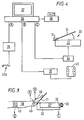
- Figure 4 the arrangements of Figures 2, 3A and 3B can be utilized together to provide an automated read out from the standardized resistance means (20). The operation of this is best shown in Figure 5.
- the Doppler probe of Figures 3A or 3B are positioned on a patient's leg as shown, for example, in Figure 5.
- an automatically inflatable cuff connected to port B of a computer (36) is disposed about the patient's leg (50).
- the outputs from probe (40) are connected to a port A of the computer (36) while recording means (34) associated with the standardized resistant means (20) is connected to port C of the computer (36).
- a sphygmananometer cuff is disposed about an arm and inflated.
- a Dopplex probe held between the forefinger and thumb at 45° angle is disposed over the brachial pulse.
- the cuff is inflated until doppler sound disappears and slowly reflated until the sound returns. This gives a brachial systolic pressure (BSP).
- BSP brachial systolic pressure
- Ankle systolic pressure is then found by utilizing a Doppler probe to locate the dorsalis pedis or posterior tibial pulse.
- the cuff is disposed about the leg just above the ankle and inflated.
- the pulse is located with the Doppler probe and the cuff is inflated until the Doppler sound disappears and slowly deflated until the sound returns. This gives a pressure reading.
- These two readings may be utilized to provide an ankle pressure index by dividing the ankle pressure reading by the branchial pressure reading.
- a normal ankle pressure index is equal to or greater than one whereupon it is safe to apply compression therapy.
- the patient is required to sequentially depress the pressure plate (21) shown in Figure 5 either by a set member of times, or usually over a set period of time or until claudication occurs.
- the tension controller (26) is adjusted to a value suited to the patient and the value read from the controller (26) may be input into the computer to give an indication of work done.
- the operation of the pressure plate (21) causes the permanent magnet associated with the spring (20) to operate the hall effect device each time the pressure plate (21) is fully depressed, thereby giving a count of a number of depressions achieved. Work done therefore by each patient may be readily measured and recorded for future comparison.
- the patient may be requested to cease movement of the leg so that intermediate determinations of the vascular flow may be rapidly made and input into the computer via port A.
- the present invention therefore provides a portable space saving device.
- the arrangement is suitable for use by leg muscles.
- the angle of the foot plate may be adjustable, as may be the arc travelled by the foot plate.
- the heel plate may be adjustable to ensure that the foot is, as far as is possible, in a standardised position.
- the measurement of exercise is totally variable depending upon the abilities of the patient and allows for patients to provide accurate data according to severity of symptoms without loss of information.
- An LED display can be provided to indicate Kilogram loading and cumulative total number of flexions. These outputs can be input into a suitably programmed computer.
- the branchial blood pressure and pulse rate do not significantly change with exercise.
- the treadmill there is an increase in blood pressure, pulse rate and cardiac output with exercise, for example:- RESTING PRESSURE POST-EXERCISE TREADMILL POST EXERCISE STRESST'ER Brachial 150 180 150 Dorsial Pedis 100 40 40 Pressure Index 0.66 0.22 0.27
- a post exercise index is valuable in management scoring systems. 40% to 45% of patients cannot use the treadmill at all or can only do so inadequately. Added to this there is an inaccuracy of 5% to 20% in the measurement which means post exercise data is of limited use when obtained from the treadmill in 50% to 70% of cases. Inadequate data using the present invention only occurs in 2% to 5% of cases.
- the remaining 29 claudicant patients were tested using both the treadmill and the selective exercise device of the invention.
- Each patient underwent resting Doppler arterial assessment of each lower limb, including measurement of pedal arterial occlusion pressures in each foot.
- A.B.P.I. measurements were performed with the handheld "Dopplex" Doppler ultrasound (Huntleigh Technology Plc). using the "Dopplex reporter" computer programme.
- the brachial arterial pressure was also measured, and A.B.P. I.s were calculated.
- the patient was then asked to stop exercising, and the A.B.P.I.'s, the systolic blood pressure and the pulse rate were measured immediately, and at 30sec, 1,3,5,8 and 10 minutes post exercise, and thereafter at 5 minute intervals until the A.B.P.I.'s had reached resting levels.
- the pedal arterial occlusion pressures were measured using standard blood pressure cuffs, and sphygmomanometer. The occlusion pressure being deemed to have been reached when the Doppler signal from the artery in question reappeared after reducing the cuff pressure around the ankle.
- the total exercise times on the treadmill and on the selective exercise device, were also recorded for each patient.
- the resistance of the selective exercise device could be varied between 10 and 20Kg. In this study the resistance was adjusted to the individual patient so that the selective exercise device could be operated without undue effort.
- the mean resting brachial blood pressure for the claudicant patients was 142mmHg (S.D. 13mmHg, range 15-175mmHg), and the mean pulse rate was 72 beats per minute (S.D.9.1, range 55-98 beats per minute).
- the mean brachial blood pressure 1 minute following exercise on the treadmill was 177mmHg (S.D.11mmHg), range 120-215mmHg.
- the mean pulse rate was 141 beats per minute (S.D. 13.6, range 125-170 beats per minute).
- the mean brachial blood pressure immediately following selective exercise was 146mmHg (S.D.15mmHg, range 120-180mmHg).
- the mean pulse rate was 76 beats per minute (S.D. 11.7, range 74-106 beats per minute).
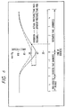
- the change in A.B.P.I. is plotted against time after exercise, on the treadmill (Fig.8) and on the selective exercise device (Fig. 9).
- Fig. 2 demonstrates the mean change in A.B.P.I. at 0 secs., 30 secs., 1 min., 3 min., and 5 mins., post-exercise for these five patients.
- the demonstration of a fall in the A.B.P.I. following exercise has been used in the diagnosis and surveillance of arterial disease of the lower limb for nearly 20 years.
- the treadmill until now has been the only standardized method of exercise in routine use. Attempts at exercising the calf muscles without any resistance, to induce claudication have been shown to produce submaximal decreases in ankle Doppler pressures, as have procedures that cause postocclusive reactive hyperaemia.
- the selective exercise device exercises the calf muscles against a known resistance and depresses the ankle Doppler pressures to a similar degree as the treadmill and is the first effective alternative in this respect.
- the selective exercise device allows immediate measurement of the A.B.P.I.
- the selective exercise device may be used to evaluate asymmetric vascular occlusive disease, which may be difficult using the treadmill because the worst leg will prevent maximal stress to the less affected leg. This may be of practical importance when planning intervention by percutaneous angioplasty, stenting, atherectomy, or by operation.
Landscapes
- Health & Medical Sciences (AREA)
- Life Sciences & Earth Sciences (AREA)
- Physics & Mathematics (AREA)
- General Health & Medical Sciences (AREA)
- Engineering & Computer Science (AREA)
- Public Health (AREA)
- Veterinary Medicine (AREA)
- Pathology (AREA)
- Biomedical Technology (AREA)
- Heart & Thoracic Surgery (AREA)
- Medical Informatics (AREA)
- Molecular Biology (AREA)
- Surgery (AREA)
- Animal Behavior & Ethology (AREA)
- Biophysics (AREA)
- Nuclear Medicine, Radiotherapy & Molecular Imaging (AREA)
- Radiology & Medical Imaging (AREA)
- Orthopedic Medicine & Surgery (AREA)
- Cardiology (AREA)
- Physiology (AREA)
- Physical Education & Sports Medicine (AREA)
- Acoustics & Sound (AREA)
- Vascular Medicine (AREA)
- Measuring Pulse, Heart Rate, Blood Pressure Or Blood Flow (AREA)
- Pharmaceuticals Containing Other Organic And Inorganic Compounds (AREA)
- Medicines That Contain Protein Lipid Enzymes And Other Medicines (AREA)
- Medicinal Preparation (AREA)
- Investigating Or Analysing Biological Materials (AREA)
Claims (15)
- Appareil de diagnostic comprenant un dispositif de résistance standardisable (20) pour résister à une force appliquée sur celui-ci par un groupe sélectionné de muscles des membres inférieurs pendant l'exercice, et un élément de test pour mesurer physiologiquement un composant biologique résultant dudit exercice des membres, le dispositif de résistance standardisable comprenant une plaque de pression (21) montée à pivotement autour d'un axe (32), un ressort (23) raccordé de manière fonctionnelle à ladite plaque afin de solliciter la plaque vers une position de repos, et un élément de réglage (26) pour précharger le ressort à une valeur désirée, ce par quoi, lors de l'utilisation, la plaque est susceptible d'effectuer un mouvement de va-et-vient grâce à un membre, à l'encontre de la sollicitation du ressort, afin d'induire une fatigue ou une claudication, caractérisé par des moyens (26) indicatifs de la charge appliquée par le ressort, le ressort étant associé à un moyen générateur de signaux, indicatif de chaque mouvement accompli, pour générer de cette façon un signal de sortie pour chaque course accomplie, et des moyens étant prévus de telle sorte que l'axe longitudinal de la jambe des patients est disposé, lors de l'utilisation, pour passer par l'axe.
- Appareil selon la revendication 1, dans lequel ledit composant biologique est choisi parmi au moins l'un parmi le débit sanguin, la pression sanguine, l'oxygène transcutané et la vitesse de clairance lymphatique.
- Appareil selon la revendication 1, dans lequel l'élément de test est choisi parmi un dispositif Doppler (40) pour mesurer le débit sanguin, un oxymètre pour mesurer les valeurs d'oxygène transcutané, des moyens pour mesurer les pressions compartimentales et des moyens pour mesurer le volume des jambes.
- Appareil selon l'une quelconque des revendications précédentes, dans lequel la plaque de pression (21) sollicitée par ressort est dotée d'une plaque de talon (31), et dans lequel l'élément de test est un dispositif Doppler (40A) in situ, adapté pour mesurer à la fois le débit sanguin et la pression sanguine.
- Appareil selon la revendication 4, dans lequel le dispositif Doppler comprend un moyen transducteur (46) ayant un transducteur apte à tourner autour de son axe horizontal ou à osciller sur un angle prédéterminé, ledit transducteur était adapté pour émettre un premier signal indicatif de la pression sanguine et un second signal indicatif du débit sanguin.
- Appareil selon la revendication 5, dans lequel le transducteur est entraíné par un moteur électrique ou est amené à osciller par des moyens électromagnétiques.
- Appareil selon l'une quelconque des revendications précédentes, dans lequel le ressort est un ressort de rappel (23) et l'élément de réglage (26) comprend un bloc fileté connectant un filet de vis au ressort de rappel.
- Appareil selon l'une quelconque des revendications précédentes, dans lequel l'arc de déplacement de la plaque de pression (21) est ajustable.
- Appareil selon la revendication 8, dans lequel le ressort est associé à un aimant permanent et un détecteur à effet Hall, pour générer de cette façon un signal de sortie pour chaque mouvement accompli de la plaque de pression (21).
- Appareil selon l'une quelconque des revendications précédentes, dans lequel la plaque de pression (21) comprend en outre une poignée étirable, et la base comprend un moyen de retenue coudé sur une surface supérieure de celle-ci.
- Appareil selon l'une quelconque des revendications 5 à 10, comprenant un manchon (49) de sphygmomanomètre, l'appareil étant réalisé et disposé d'une façon telle qu'à des intervalles périodiques pendant le diagnostic, ledit manchon est gonflé en réponse à un signal de commande de manchon prédéterminé pour permette à la pression sanguine et au débit sanguin d'être enregistrés automatiquement.
- Procédé pour la mesure non-effractive de l'alimentation vasculaire périphérique à un membre (50), à l'aide d'un appareil tel que défini à l'une quelconque des revendications 1 à 11, lequel procédé comprend :caractérisé par le fait que la jambe est positionnée de telle sorte que l'axe longitudinal de la jambe des patients est disposé, lors de l'utilisation, pour passer par l'axe.l'établissement d'une pression ou d'un débit vasculaire de pré-exercice de repos ;l'utilisation du membre pour déplacer de façon répétée la plaque sous résistance d'une distance prédéterminée pendant un laps de temps mesuré ; etl'établissement d'une pression ou d'un débit de post-exercice,
- Procédé selon la revendication 12, dans lequel les pressions de pré- et post-exercice sont dénotées par un indice de pression de cheville.
- Procédé selon l'une ou l'autre des revendications 12 ou 13, dans lequel les valeurs de débit et/ou de pression vasculaires sont surveillées pendant des intervalles chronométrés pendant la réalisation courante de l'exercice, et dans lequel un manchon fixé autour de la cheville est gonflé à des intervalles chronométrés pendant l'exercice, permettant ainsi à des valeurs de débit et de pression vasculaires d'être surveillées pendant de courtes périodes de repos pendant l'exercice.
- Procédé selon l'une quelconque des revendications 12 à 14, comprenant en outre des déterminations par oxymétrie.
Applications Claiming Priority (5)
| Application Number | Priority Date | Filing Date | Title |
|---|---|---|---|
| GB939311816A GB9311816D0 (en) | 1993-06-08 | 1993-06-08 | Peripheral vascular disease testing |
| GB9311816 | 1993-06-08 | ||
| GB9406174A GB9406174D0 (en) | 1994-03-29 | 1994-03-29 | Peripheral vascular disease testing |
| GB9406174 | 1994-03-29 | ||
| PCT/GB1994/001217 WO1994028794A1 (fr) | 1993-06-08 | 1994-06-03 | Test des maladies vasculaires peripheriques |
Publications (2)
| Publication Number | Publication Date |
|---|---|
| EP0703754A1 EP0703754A1 (fr) | 1996-04-03 |
| EP0703754B1 true EP0703754B1 (fr) | 1998-07-15 |
Family
ID=26303023
Family Applications (1)
| Application Number | Title | Priority Date | Filing Date |
|---|---|---|---|
| EP94916353A Expired - Lifetime EP0703754B1 (fr) | 1993-06-08 | 1994-06-03 | Test des maladies vasculaires peripheriques |
Country Status (6)
| Country | Link |
|---|---|
| US (1) | US5803907A (fr) |
| EP (1) | EP0703754B1 (fr) |
| AT (1) | ATE168249T1 (fr) |
| AU (1) | AU6804494A (fr) |
| DE (1) | DE69411739T2 (fr) |
| WO (1) | WO1994028794A1 (fr) |
Cited By (1)
| Publication number | Priority date | Publication date | Assignee | Title |
|---|---|---|---|---|
| CN107930030A (zh) * | 2017-11-15 | 2018-04-20 | 闫星晨 | 多功能下肢神经康复器 |
Families Citing this family (14)
| Publication number | Priority date | Publication date | Assignee | Title |
|---|---|---|---|---|
| US5991654A (en) * | 1997-06-06 | 1999-11-23 | Kci New Technologies, Inc. | Apparatus and method for detecting deep vein thrombosis |
| USD447709S1 (en) | 2000-03-23 | 2001-09-11 | Innothera Topic International Societe Anonyme | Measuring apparatus |
| GB2378908A (en) * | 2001-08-24 | 2003-02-26 | Robert Philip Warr | Leg exerciser |
| GB0204192D0 (en) | 2002-02-22 | 2002-04-10 | Stu Ert Medical Devices Ltd | Apparatus and method for assessing peripheral vascular disease |
| US6683240B1 (en) * | 2002-09-10 | 2004-01-27 | Mark Cubranich | Drummers foot exerciser and bass drum practice pedal |
| US20060111636A1 (en) * | 2004-11-23 | 2006-05-25 | Jacober Jeffrey M | Wrist-mount blood pressure monitor |
| US20060111637A1 (en) * | 2004-11-23 | 2006-05-25 | Jacober Jeffrey M | Wrist-mount blood pressure monitor with auditory feature |
| GB2423484B (en) * | 2005-02-26 | 2009-02-25 | John Patrick Lambert | A device for stimulating blood circulation |
| US20090048525A1 (en) * | 2007-08-14 | 2009-02-19 | Biomedix, Inc. | Venous refill testing system and method |
| US20100292591A1 (en) * | 2009-05-14 | 2010-11-18 | Rooke Thom W | Venous measurement system |
| US20100324431A1 (en) * | 2009-06-18 | 2010-12-23 | Nellcor Puritan Bennett Ireland | Determining Disease State Using An Induced Load |
| US20110245693A1 (en) * | 2010-03-30 | 2011-10-06 | Boston Scientific Scimed, Inc. | Intravascular pressure sensing |
| CN107106056B (zh) * | 2014-11-10 | 2020-03-27 | 泰尔茂株式会社 | 皮肤灌注压的测定装置 |
| WO2016207884A1 (fr) * | 2015-06-21 | 2016-12-29 | Yissum Research Development Company Of The Hebrew University Of Jerusalem Ltd. | Détection de maladie artérielle périphérique |
Family Cites Families (17)
| Publication number | Priority date | Publication date | Assignee | Title |
|---|---|---|---|---|
| FR2024708A5 (fr) * | 1969-10-15 | 1970-08-28 | Andrier Louis | |
| DE2911258C2 (de) * | 1979-03-22 | 1982-09-23 | Chmiel, Horst, Prof. Dr.-Ing., 7250 Leonberg | Vorrichtung zum noninvasiven Messen der Blutströmungsgeschwindigkeit nach der Ultraschall-Doppler-Effekt-Methode |
| US4280486A (en) * | 1979-10-29 | 1981-07-28 | World Medical Marketing Corporation | Foot exerciser |
| US4454885A (en) * | 1981-06-22 | 1984-06-19 | Henry Ford Hospital | Assessing arterial systems |
| US4476874A (en) * | 1982-06-01 | 1984-10-16 | Sri International | Ultrasonic imaging with volume flow measuring method and apparatus |
| US4502680A (en) * | 1982-12-23 | 1985-03-05 | Blum Alvin S | Foot exerciser |
| US4556066A (en) * | 1983-11-04 | 1985-12-03 | The Kendall Company | Ultrasound acoustical coupling pad |
| US4790325A (en) * | 1985-11-25 | 1988-12-13 | Lee Arnold S | Automatic arterial blood pressure recorder |
| JPH0787834B2 (ja) * | 1985-12-11 | 1995-09-27 | マイヤ−,ニ−ルス | 筋肉収縮を検査するための装置 |
| US4805455A (en) * | 1987-04-24 | 1989-02-21 | Myo-Tech Corp. | Muscle testing apparatus and method |
| US4732038A (en) * | 1986-07-11 | 1988-03-22 | Delgiorno Daniel | Muscle testing method |
| US5090421A (en) * | 1986-12-09 | 1992-02-25 | Hoggan Health Industries, Inc. | Apparatus for testing muscle strength |
| JP2882797B2 (ja) * | 1988-02-24 | 1999-04-12 | コーリン電子株式会社 | 末梢抵抗検査出装置 |
| DE8909993U1 (de) * | 1989-08-21 | 1990-01-04 | Pützler, Manfred, Dipl.-Phys., 4400 Münster | Wadenmuskulatur-Ergometer zur NMR-Spektroskopie |
| DE59107232D1 (de) * | 1990-07-18 | 1996-02-22 | Avl Medical Instr Ag | Einrichtung und Verfahren zur Blutdruckmessung |
| US5241964A (en) * | 1990-10-31 | 1993-09-07 | Medwave, Incorporated | Noninvasive, non-occlusive method and apparatus which provides a continuous indication of arterial pressure and a beat-by-beat characterization of the arterial system |
| US5058592A (en) * | 1990-11-02 | 1991-10-22 | Whisler G Douglas | Adjustable mountable doppler ultrasound transducer device |
-
1994
- 1994-06-03 AT AT94916353T patent/ATE168249T1/de not_active IP Right Cessation
- 1994-06-03 EP EP94916353A patent/EP0703754B1/fr not_active Expired - Lifetime
- 1994-06-03 AU AU68044/94A patent/AU6804494A/en not_active Abandoned
- 1994-06-03 US US08/557,021 patent/US5803907A/en not_active Expired - Fee Related
- 1994-06-03 DE DE69411739T patent/DE69411739T2/de not_active Expired - Fee Related
- 1994-06-03 WO PCT/GB1994/001217 patent/WO1994028794A1/fr not_active Ceased
Cited By (1)
| Publication number | Priority date | Publication date | Assignee | Title |
|---|---|---|---|---|
| CN107930030A (zh) * | 2017-11-15 | 2018-04-20 | 闫星晨 | 多功能下肢神经康复器 |
Also Published As
| Publication number | Publication date |
|---|---|
| AU6804494A (en) | 1995-01-03 |
| ATE168249T1 (de) | 1998-08-15 |
| DE69411739T2 (de) | 1998-11-12 |
| US5803907A (en) | 1998-09-08 |
| WO1994028794A1 (fr) | 1994-12-22 |
| DE69411739D1 (de) | 1998-08-20 |
| EP0703754A1 (fr) | 1996-04-03 |
Similar Documents
| Publication | Publication Date | Title |
|---|---|---|
| EP0703754B1 (fr) | Test des maladies vasculaires peripheriques | |
| Ramsey III | Noninvasive automatic determination of mean arterial pressure | |
| Clapper et al. | Comparison of the reliability of the Orthoranger and the standard goniometer for assessing active lower extremity range of motion | |
| Hummel et al. | Reactive hyperemia vs treadmill exercise testing in arterial disease | |
| US4569355A (en) | Method and apparatus for monitoring and diagnosing peripheral blood flow | |
| McMullin et al. | An evaluation of Doppler ultrasound and photoplethysmography in the investigation of venous insufficiency | |
| US5365924A (en) | Method and apparatus for non-invasive cardiovascular diagnosis | |
| Begg et al. | Instrumentation used in clinical gait studies: a review | |
| McMahon et al. | Effects of muscle group and placement site on reliability of hand-held dynamometry strength measurements | |
| JP3712418B2 (ja) | 誘発された摂動を測定してヒト動脈系の物理的状態を決定するための装置および方法 | |
| JP2586910B2 (ja) | 足関節痙縮度測定装置 | |
| Weaver et al. | Design and validation of an instrument package designed to increase the reliability of ankle range of motion measurements | |
| Kelechi et al. | Measuring venous insufficiency objectively in the clinical setting | |
| Wong et al. | Investigating the influence of limb blood flow on contraction-induced muscle growth and the impact of that growth on changes in maximal strength | |
| Forconi et al. | Strain gauge plethysmography in the study of circulation of the limbs | |
| Eagle | Doppler ultrasound-basics revisited | |
| Brahim et al. | Ultrasound measurement of the anterior leg compartment | |
| Barnes | Noninvasive evaluation of peripheral arterial disease | |
| Fronek et al. | Non-invasively determined ambulatory venous pressure | |
| Harwood et al. | Exercise testing in peripheral arterial disease | |
| Wassermann et al. | Role of ultrasound as a non-invasive method of diagnosis of chronic exertional compartment syndrome | |
| Malatino et al. | Comparison of a new portable electronic sphygmomanometer (Copal UA251) with the Hawksley random zero machine | |
| Schäffler et al. | Neuropathy Tests | |
| Faghri et al. | Prediction of calf volume during muscle contraction | |
| Ramsey III | Automatic oscillometric noninvasive blood pressure: Theory and practice |
Legal Events
| Date | Code | Title | Description |
|---|---|---|---|
| PUAI | Public reference made under article 153(3) epc to a published international application that has entered the european phase |
Free format text: ORIGINAL CODE: 0009012 |
|
| 17P | Request for examination filed |
Effective date: 19960105 |
|
| AK | Designated contracting states |
Kind code of ref document: A1 Designated state(s): AT BE CH DE DK ES FR GB GR IE IT LI LU MC NL PT SE |
|
| 17Q | First examination report despatched |
Effective date: 19960611 |
|
| GRAG | Despatch of communication of intention to grant |
Free format text: ORIGINAL CODE: EPIDOS AGRA |
|
| GRAG | Despatch of communication of intention to grant |
Free format text: ORIGINAL CODE: EPIDOS AGRA |
|
| GRAH | Despatch of communication of intention to grant a patent |
Free format text: ORIGINAL CODE: EPIDOS IGRA |
|
| GRAH | Despatch of communication of intention to grant a patent |
Free format text: ORIGINAL CODE: EPIDOS IGRA |
|
| GRAA | (expected) grant |
Free format text: ORIGINAL CODE: 0009210 |
|
| AK | Designated contracting states |
Kind code of ref document: B1 Designated state(s): AT BE CH DE DK ES FR GB GR IE IT LI LU MC NL PT SE |
|
| PG25 | Lapsed in a contracting state [announced via postgrant information from national office to epo] |
Ref country code: LI Free format text: LAPSE BECAUSE OF FAILURE TO SUBMIT A TRANSLATION OF THE DESCRIPTION OR TO PAY THE FEE WITHIN THE PRESCRIBED TIME-LIMIT Effective date: 19980715 Ref country code: GR Free format text: LAPSE BECAUSE OF NON-PAYMENT OF DUE FEES Effective date: 19980715 Ref country code: ES Free format text: THE PATENT HAS BEEN ANNULLED BY A DECISION OF A NATIONAL AUTHORITY Effective date: 19980715 Ref country code: CH Free format text: LAPSE BECAUSE OF FAILURE TO SUBMIT A TRANSLATION OF THE DESCRIPTION OR TO PAY THE FEE WITHIN THE PRESCRIBED TIME-LIMIT Effective date: 19980715 Ref country code: BE Free format text: LAPSE BECAUSE OF FAILURE TO SUBMIT A TRANSLATION OF THE DESCRIPTION OR TO PAY THE FEE WITHIN THE PRESCRIBED TIME-LIMIT Effective date: 19980715 Ref country code: AT Free format text: LAPSE BECAUSE OF FAILURE TO SUBMIT A TRANSLATION OF THE DESCRIPTION OR TO PAY THE FEE WITHIN THE PRESCRIBED TIME-LIMIT Effective date: 19980715 |
|
| REF | Corresponds to: |
Ref document number: 168249 Country of ref document: AT Date of ref document: 19980815 Kind code of ref document: T |
|
| REG | Reference to a national code |
Ref country code: CH Ref legal event code: EP |
|
| REF | Corresponds to: |
Ref document number: 69411739 Country of ref document: DE Date of ref document: 19980820 |
|
| ITF | It: translation for a ep patent filed | ||
| ET | Fr: translation filed | ||
| RAP4 | Party data changed (patent owner data changed or rights of a patent transferred) |
Owner name: STU-ERT MEDICAL DEVICES LIMITED |
|
| REG | Reference to a national code |
Ref country code: IE Ref legal event code: FG4D |
|
| PG25 | Lapsed in a contracting state [announced via postgrant information from national office to epo] |
Ref country code: SE Free format text: LAPSE BECAUSE OF FAILURE TO SUBMIT A TRANSLATION OF THE DESCRIPTION OR TO PAY THE FEE WITHIN THE PRESCRIBED TIME-LIMIT Effective date: 19981015 Ref country code: PT Free format text: LAPSE BECAUSE OF FAILURE TO SUBMIT A TRANSLATION OF THE DESCRIPTION OR TO PAY THE FEE WITHIN THE PRESCRIBED TIME-LIMIT Effective date: 19981015 Ref country code: DK Free format text: LAPSE BECAUSE OF FAILURE TO SUBMIT A TRANSLATION OF THE DESCRIPTION OR TO PAY THE FEE WITHIN THE PRESCRIBED TIME-LIMIT Effective date: 19981015 |
|
| REG | Reference to a national code |
Ref country code: CH Ref legal event code: PL |
|
| PLBE | No opposition filed within time limit |
Free format text: ORIGINAL CODE: 0009261 |
|
| STAA | Information on the status of an ep patent application or granted ep patent |
Free format text: STATUS: NO OPPOSITION FILED WITHIN TIME LIMIT |
|
| PG25 | Lapsed in a contracting state [announced via postgrant information from national office to epo] |
Ref country code: LU Free format text: LAPSE BECAUSE OF NON-PAYMENT OF DUE FEES Effective date: 19990603 Ref country code: IE Free format text: LAPSE BECAUSE OF NON-PAYMENT OF DUE FEES Effective date: 19990603 |
|
| 26N | No opposition filed | ||
| PG25 | Lapsed in a contracting state [announced via postgrant information from national office to epo] |
Ref country code: MC Free format text: LAPSE BECAUSE OF NON-PAYMENT OF DUE FEES Effective date: 19991231 |
|
| REG | Reference to a national code |
Ref country code: IE Ref legal event code: MM4A |
|
| REG | Reference to a national code |
Ref country code: GB Ref legal event code: IF02 |
|
| PGFP | Annual fee paid to national office [announced via postgrant information from national office to epo] |
Ref country code: NL Payment date: 20031224 Year of fee payment: 10 |
|
| PGFP | Annual fee paid to national office [announced via postgrant information from national office to epo] |
Ref country code: DE Payment date: 20041203 Year of fee payment: 11 |
|
| PGFP | Annual fee paid to national office [announced via postgrant information from national office to epo] |
Ref country code: FR Payment date: 20041222 Year of fee payment: 11 |
|
| PG25 | Lapsed in a contracting state [announced via postgrant information from national office to epo] |
Ref country code: NL Free format text: LAPSE BECAUSE OF NON-PAYMENT OF DUE FEES Effective date: 20050101 |
|
| NLV4 | Nl: lapsed or anulled due to non-payment of the annual fee |
Effective date: 20050101 |
|
| PG25 | Lapsed in a contracting state [announced via postgrant information from national office to epo] |
Ref country code: IT Free format text: LAPSE BECAUSE OF NON-PAYMENT OF DUE FEES Effective date: 20050603 |
|
| PGFP | Annual fee paid to national office [announced via postgrant information from national office to epo] |
Ref country code: GB Payment date: 20050630 Year of fee payment: 12 |
|
| PG25 | Lapsed in a contracting state [announced via postgrant information from national office to epo] |
Ref country code: DE Free format text: LAPSE BECAUSE OF NON-PAYMENT OF DUE FEES Effective date: 20060103 |
|
| PG25 | Lapsed in a contracting state [announced via postgrant information from national office to epo] |
Ref country code: FR Free format text: LAPSE BECAUSE OF NON-PAYMENT OF DUE FEES Effective date: 20060228 |
|
| REG | Reference to a national code |
Ref country code: FR Ref legal event code: ST Effective date: 20060228 |
|
| PG25 | Lapsed in a contracting state [announced via postgrant information from national office to epo] |
Ref country code: GB Free format text: LAPSE BECAUSE OF NON-PAYMENT OF DUE FEES Effective date: 20060603 |
|
| GBPC | Gb: european patent ceased through non-payment of renewal fee |
Effective date: 20060603 |