EP0703785B1 - Methoden zur behandlung von muskelerkrankungen und muskelstörungen - Google Patents
Methoden zur behandlung von muskelerkrankungen und muskelstörungen Download PDFInfo
- Publication number
- EP0703785B1 EP0703785B1 EP94916690A EP94916690A EP0703785B1 EP 0703785 B1 EP0703785 B1 EP 0703785B1 EP 94916690 A EP94916690 A EP 94916690A EP 94916690 A EP94916690 A EP 94916690A EP 0703785 B1 EP0703785 B1 EP 0703785B1
- Authority
- EP
- European Patent Office
- Prior art keywords
- polypeptide
- seq
- sequence
- muscle
- information
- Prior art date
- Legal status (The legal status is an assumption and is not a legal conclusion. Google has not performed a legal analysis and makes no representation as to the accuracy of the status listed.)
- Expired - Lifetime
Links
Images























Classifications
-
- C—CHEMISTRY; METALLURGY
- C07—ORGANIC CHEMISTRY
- C07K—PEPTIDES
- C07K14/00—Peptides having more than 20 amino acids; Gastrins; Somatostatins; Melanotropins; Derivatives thereof
- C07K14/82—Translation products from oncogenes
-
- A—HUMAN NECESSITIES
- A61—MEDICAL OR VETERINARY SCIENCE; HYGIENE
- A61K—PREPARATIONS FOR MEDICAL, DENTAL OR TOILETRY PURPOSES
- A61K38/00—Medicinal preparations containing peptides
- A61K38/16—Peptides having more than 20 amino acids; Gastrins; Somatostatins; Melanotropins; Derivatives thereof
-
- A—HUMAN NECESSITIES
- A61—MEDICAL OR VETERINARY SCIENCE; HYGIENE
- A61K—PREPARATIONS FOR MEDICAL, DENTAL OR TOILETRY PURPOSES
- A61K38/00—Medicinal preparations containing peptides
- A61K38/16—Peptides having more than 20 amino acids; Gastrins; Somatostatins; Melanotropins; Derivatives thereof
- A61K38/17—Peptides having more than 20 amino acids; Gastrins; Somatostatins; Melanotropins; Derivatives thereof from animals; from humans
- A61K38/22—Hormones
- A61K38/27—Growth hormone [GH], i.e. somatotropin
-
- A—HUMAN NECESSITIES
- A61—MEDICAL OR VETERINARY SCIENCE; HYGIENE
- A61P—SPECIFIC THERAPEUTIC ACTIVITY OF CHEMICAL COMPOUNDS OR MEDICINAL PREPARATIONS
- A61P19/00—Drugs for skeletal disorders
-
- A—HUMAN NECESSITIES
- A61—MEDICAL OR VETERINARY SCIENCE; HYGIENE
- A61P—SPECIFIC THERAPEUTIC ACTIVITY OF CHEMICAL COMPOUNDS OR MEDICINAL PREPARATIONS
- A61P21/00—Drugs for disorders of the muscular or neuromuscular system
-
- A—HUMAN NECESSITIES
- A61—MEDICAL OR VETERINARY SCIENCE; HYGIENE
- A61P—SPECIFIC THERAPEUTIC ACTIVITY OF CHEMICAL COMPOUNDS OR MEDICINAL PREPARATIONS
- A61P21/00—Drugs for disorders of the muscular or neuromuscular system
- A61P21/04—Drugs for disorders of the muscular or neuromuscular system for myasthenia gravis
-
- A—HUMAN NECESSITIES
- A61—MEDICAL OR VETERINARY SCIENCE; HYGIENE
- A61P—SPECIFIC THERAPEUTIC ACTIVITY OF CHEMICAL COMPOUNDS OR MEDICINAL PREPARATIONS
- A61P25/00—Drugs for disorders of the nervous system
-
- A—HUMAN NECESSITIES
- A61—MEDICAL OR VETERINARY SCIENCE; HYGIENE
- A61P—SPECIFIC THERAPEUTIC ACTIVITY OF CHEMICAL COMPOUNDS OR MEDICINAL PREPARATIONS
- A61P35/00—Antineoplastic agents
-
- A—HUMAN NECESSITIES
- A61—MEDICAL OR VETERINARY SCIENCE; HYGIENE
- A61P—SPECIFIC THERAPEUTIC ACTIVITY OF CHEMICAL COMPOUNDS OR MEDICINAL PREPARATIONS
- A61P43/00—Drugs for specific purposes, not provided for in groups A61P1/00-A61P41/00
-
- A—HUMAN NECESSITIES
- A61—MEDICAL OR VETERINARY SCIENCE; HYGIENE
- A61P—SPECIFIC THERAPEUTIC ACTIVITY OF CHEMICAL COMPOUNDS OR MEDICINAL PREPARATIONS
- A61P9/00—Drugs for disorders of the cardiovascular system
-
- A—HUMAN NECESSITIES
- A61—MEDICAL OR VETERINARY SCIENCE; HYGIENE
- A61P—SPECIFIC THERAPEUTIC ACTIVITY OF CHEMICAL COMPOUNDS OR MEDICINAL PREPARATIONS
- A61P9/00—Drugs for disorders of the cardiovascular system
- A61P9/10—Drugs for disorders of the cardiovascular system for treating ischaemic or atherosclerotic diseases, e.g. antianginal drugs, coronary vasodilators, drugs for myocardial infarction, retinopathy, cerebrovascula insufficiency, renal arteriosclerosis
-
- C—CHEMISTRY; METALLURGY
- C07—ORGANIC CHEMISTRY
- C07K—PEPTIDES
- C07K14/00—Peptides having more than 20 amino acids; Gastrins; Somatostatins; Melanotropins; Derivatives thereof
- C07K14/435—Peptides having more than 20 amino acids; Gastrins; Somatostatins; Melanotropins; Derivatives thereof from animals; from humans
- C07K14/475—Growth factors; Growth regulators
- C07K14/4756—Neuregulins, i.e. p185erbB2 ligands, glial growth factor, heregulin, ARIA, neu differentiation factor
-
- A—HUMAN NECESSITIES
- A61—MEDICAL OR VETERINARY SCIENCE; HYGIENE
- A61K—PREPARATIONS FOR MEDICAL, DENTAL OR TOILETRY PURPOSES
- A61K38/00—Medicinal preparations containing peptides
Definitions
- the invention relates to prophylactic or affirmative treatment of diseases and disorders of the musculature by administering polypeptides found in vertebrate species, which polypeptides are growth, differentiation and survival factors for muscle cells.
- Satellite cells are distributed throughout muscle tissue and are mitotically quiescent in the absence of injury or disease. Following muscle injury or during recovery from disease, satellite cells will reenter the cell cycle, proliferate and 1) enter existing muscle fibers or 2) undergo differentiation into multinucleate myotubes which form new muscle fiber. The myoblasts ultimately yield replacement muscle fibers or fuse into existing muscle fibers, thereby increasing fiber girth by the synthesis of contractile apparatus components. This process is illustrated, for example, by the nearly complete regeneration which occurs in mammals following induced muscle fiber degeneration; the muscle progenitor cells proliferate and fuse together regenerating muscle fibers.
- Fibroblast growth factor FLGF
- TGF ⁇ Transforming growth factor ⁇
- IGFs Insulin-like growth factors
- PDGF Platelet derived growth factor
- both muscle tissue and neurons are potential sources of factors which stimulate myoblast proliferation and differentiation.
- diseases affecting the neuromuscular system which are neural in origin i.e., neurogenic
- the muscle tissue innervated by the affected nerve becomes paralyzed and wastes progressively.
- neurons may provide a source of growth factors which elicit the muscle regeneration described above and provide a mechanism for muscle recovery from wasting and atrophy.
- neuregulins A recently described family of growth factors, the neuregulins, are synthesized by motor neurons (Marchioni et al. Nature 362:313, 1993) and inflammatory cells (Tarakhovsky et al., Oncogene 6:2187-2196 (1991)).
- the neuregulins and related p185 erbB2 binding factors have been purified, cloned and expressed (Benveniste et al., PNAS 82 :3930-3934, 1985; Kimura et al., Nature 348 :257-260, 1990; Davis and Stroobant, J. Cell. Biol.
- Recombinant neuregulins have been shown to be mitogenic for peripheral glia (Marchionni et al., Nature 362:313, 1993) and have been shown to influence the formation of the neuromuscular junction (Falls et al., Cell 72:801, 1993).
- the regenerating neuron and the inflammatory cells associated with the recovery from neurogenic disease and nerve injury provide a source of factors which coordinate the remyelination of motor neurons and their ability to form the appropriate connection with their target. After muscle has been reinnervated the motor neuron may provide factors to muscle, stimulating muscle growth and survival.
- glial growth factors proteins heretofore described as glial growth factors, acetylcholine receptor inducing activity (ARIA), heregulins, neu differentiation factor, and, more generally, neuregulins.
- ARIA acetylcholine receptor inducing activity
- these compounds are capable of inducing both the proliferation of muscle cells and the differentiation and survival of myotubes. These phenomena may occur in cardiac and smooth muscle tissues in addition to skeletal muscle tissues.
- the above compounds, regulatory compounds which induce synthesis of these compounds, and small molecules which mimic these compounds by binding to the receptors on muscle or by stimulating through other means the second messenger systems activated by the ligand-receptor complex are all extremely useful as prophylactic and affirmative therapies for muscle diseases.
- a novel aspect of the invention involves the use of the above named proteins as growth factors for the manufacture of a medicament for the treating or prophylaxis of a disease or disorder of the musculature by increasing the mitogenesis, survival, growth and differentiation of muscle cells. Treating of the muscle cells to achieve these effects may be achieved by contacting muscle cells with a polypeptide described herein. The use may be provided to slow or halt net muscle loss or to increase the amount or quality of muscle present in the vertebrate.
- a vertebrate preferably a mammal, more preferably a human
- Neuregulin effects on muscle may occur, for example, by causing an increase in muscle performance by inducing the synthesis of particular isoforms of the contractile apparatus such as the myosin heavy chain slow and fast isoforms; by promoting muscle fiber survival via the induction of synthesis of protective molecules such as, but not limited to, dystrophin; and/or by increasing muscle innervation by, for example, increasing acetylcholine receptor molecules at the neuromuscular junction.
- muscle cell refers to any cell which contributes to muscle tissue.
- Myoblasts, satellite cells, myotubes, and myofibril tissues are all included in the term “muscle cells” and may all be treated using the methods of the invention. Muscle cell effects may be induced within skeletal, cardiac and smooth muscles.
- Mitogenesis may be induced in muscle cells, including myoblasts or satellite cells, of skeletal muscle, smooth muscle or cardiac muscle. Mitogenesis as used herein refers to any cell division which results in the production of new muscle cells in the patient. More specifically, mitogenesis in vitro is defined as an increase in mitotic index relative to untreated cells of 50%, more preferably 100%, and most preferably 300%, when the cells are exposed to labelling agent for a time equivalent to two doubling times.
- the mitotic index is the fraction of cells in the culture which have labelled nuclei when grown in the presence of a tracer which only incorporates during S phase (i.e., BrdU) and the doubling time is defined as the average time required for the number of cells in the culture to increase by a factor of two).
- An effect on mitogenesis in vivo is defined as an increase in satellite cell activation as measured by the appearance of labelled satellite cells in the muscle tissue of a mammal exposed to a tracer which only incorporates during S phase (i.e., BrdU).
- a useful therapeutic is defined as a compound when administered in-vivo increases satellite cell activation relative to a control mammal by at least 10%, more preferably by at least 50%, and most preferably by more than 200% when the mammal is exposed to labelling agent for a period of greater than 15 minutes and tissues are assayed between 10 hours and 24 hours after administration of the mitogen at the therapeutic dose.
- satellite cell activation in vivo may be detected by monitoring the appearance of the intermediate filament vimentin by immunological or RNA analysis methods.
- the useful mitogen is defined as one which causes expression of detectable levels of vimentin in the muscle tissue when the therapeutically useful dosage is provided.
- Myogenesis refers to any fusion of myoblasts to yield myotubes. Most preferably, an effect on myogenesis is defined as an increase in the fusion of myoblasts and the enablement of the muscle differentiation program.
- the useful myogenic therapeutic is defined as a polypeptide which confers any increase in the fusion index in vitro. More preferably, the polypeptide confers at least a 2.0-fold increase and, most preferably, the compound confers a 3-fold or greater increase in the fusion index relative to the control.
- the fusion index is defined as the fraction of nuclei present in multinucleated cells in the culture relative to the total number of nuclei present in the culture.
- the percentages provided above are for cells assayed after 6 days of exposure to the myogenic polypeptide and are relative to an untreated control.
- Myogenesis may also be determined by assaying the number of nuclei per area in myotubes or by measurement of the levels of muscle specific protein by Western analysis.
- the polypeptide confers at least a 2.0-fold increase in the density of myotubes using the assay provided, for example, herein, and, most preferably, the polypeptide confers a 3-fold or greater increase.
- the growth of muscle may occur by the increase in the fiber size and/or by increasing the number of fibers.
- the growth of muscle as used herein may be measured by A) an increase in wet weight, B) an increase in protein content, C) an increase in the number of muscle fibers, or D) an increase in muscle fiber diameter.
- An increase in growth of a muscle fiber can be defined as an increase in the diameter where the diameter is defined as the minor axis of ellipsis of the cross section.
- the useful therapeutic is one which increases the wet weight, protein content and/or diameter by 10% or more, more preferably by more than 50% and most preferably by more than 100% in an animal whose muscles have been previously degenerated by at least 10% and relative to a similarly treated control animal (i.e., an animal with degenerated muscle tissue which is not treated with the muscle growth compound).
- a compound which increases growth by increasing the number of muscle fibers is useful as a therapeutic when it increases the number of fibers in the diseased tissue by at least 1%, more preferably at least 20%, and most preferably, by at least 50%. These percentages are determined relative to the basal level in a comparable untreated undiseased mammal or in the contralateral undiseased muscle when the polypeptide is administered and acts locally.
- the survival of muscle fibers as used herein refers to the prevention of loss of muscle fibers as evidenced by necrosis or apoptosis or the prevention of other mechanisms of muscle fiber loss. Survival as used herein indicates an decrease in the rate of cell death of at least 10%, more preferably by at least 50%, and most preferably by at least 300% relative to an untreated control. The rate of survival may be measured by counting cells stainable with a dye specific for dead cells (such as propidium iodide) in culture when the cells are 8 days post-differentiation (i.e., 8 days after the media is changed from 20% to 0.5% serum).
- a dye specific for dead cells such as propidium iodide
- Muscle regeneration refers to the process by which new muscle fibers form from muscle progenitor cells.
- the useful therapeutic for regeneration confers an increase in the number of new fibers by at least 1%, more preferably by at least 20%, and most preferably by at least 50%, as defined above.
- the differentiation of muscle cells as used herein refers to the induction of a muscle developmental program which specifies the components of the muscle fiber such as the contractile apparatus (the myofibril).
- the therapeutic useful for differentiation increases the quantity of any component of the muscle fiber in the diseased tissue by at least 10% or more, more preferably by 50% or more, and most preferably by more than 100% relative to the equivalent tissue in a similarly treated control animal.
- Atrophy of muscle refers to a significant loss in muscle fiber girth.
- significant atrophy is meant a reduction of muscle fiber diameter in diseased, injured or unused muscle tissue of at least 10% relative to undiseased, uninjured, or normally utilized tissue.
- muscular disorders which may be treated include skeletal muscle diseases and disorders such as myopathies, dystrophies, myoneural conductive diseases, traumatic muscle injury, and nerve injury.
- Cardiac muscle pathologies such as cardiomyopathies, ischemic damage, congenital disease, and traumatic injury may also be treated using our methods, as may smooth muscle diseases and disorders such as arterial sclerosis, vascular lesions, and congenital vascular diseases.
- Duchennes muscular dystrophy, Beckkers' dystrophy, and Myasthenia gravis are but three of the diseases which may be treated using the methods of the invention.
- tumour of muscle cell origin such as rhabdomyosarcoma.
- This may include administration of an effective amount of a substance which inhibits the binding of one or more of the polypeptides described herein and inhibiting the proliferation of the cells which contribute to the tumour.
- neuregulin proteins are encoded by the same gene.
- a variety of messenger RNA splicing variants (and their resultant proteins) are derived from this gene and many of these products show binding to p185 ergB2 and activation of the same. Products of this gene have been used by us to show muscle cell mitogenic activity (see Examples 1 and 2, below), differentiation (Examples 3 and 6), and survival (Examples 4 and 5).
- We provide a use for products of the neuregulin gene (described herein and in the references listed above) which have the stated activities as muscle cell mitogens, differentiation factors, and survival factors.
- rhGGF2 recombinant human GGF2
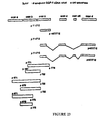
- Fig. 29 shows the known patterns of splicing. These patterns are derived from polymerase chain reaction experiments (on reverse transcribed RNA), analysis of cDNA clones (as presented within), and analysis of published sequences encoding neuregulins (Peles et al., Cell 69 :205 (1992) and Wen et al., Cell 69 :559 (1992)). These patterns, as well as additional patterns disclosed herein, represent probable splicing variants which exist. The splicing variants are fully described in Goodearl et al., USSN 08/036,555, filed March 24, 1993, incorporated herein by reference.
- cell division, survival, differentiation and growth of muscle cells may be achieved by contacting muscle cells with a polypeptide defined by the formula WBAZCX wherein WBAZCX is composed of the polypeptide segments shown in Fig. 30 (SEQ ID Nos.
- W comprises the polypeptide segment F, or is absent; wherein Z comprises the polypeptide segment G or is absent; and wherein X comprises the polypeptide segment C/D HKL, C/D H, C/D HL, C/D D, C/D' HL, C/D' HKL, C/D' H, C/D' D, C/D C/D' HKL, C/D C/D' H, C/D C/D' HL, C/D C/D' D, C/D D' H, C/D D' HL, C/D D' HKL, C/D' D' H, C/D' D' HL, C/D' D' HKL, C/D' D' H, C/D' D' HKL, C/D C/D' D' H, C/D C/D' D' HL, or C/D C/D' D' HKL and/or by contacting muscle cells with a polypeptide defined by the formula YBAZ
- Y comprises the polypeptide segment E, or is absent; wherein Z comprises the polypeptide segment G or is absent; and wherein X comprises the polypeptide segment C/D HKL, C/D H, C/D HL, C/D D, C/D' HL, C/D' HKL, C/D' H, C/D' D, C/D C/D' HKL, C/D C/D' H, C/D C/D' HL, C/D C/D' D, C/D D' H, C/D D' HL, C/D D' HKL, C/D' D' H, C/D' D' HL, C/O' D' HKL, C/D C/D' D' H, C/D C/D' D' HL, or C/D C/D' D' HKL.
- the N-terminus of the above-described polypeptides begins with either the F or E polypeptide segments.
- F polypeptide When the F polypeptide is present it may be cleaved upon maturation of the protein to yield the mature polypeptide.
- E sequence When the E sequence is present the first 50 amino acids which represent the N-terminal signal sequence may be absent from the polypeptides.
- polypeptide for the manufacture of a medicament for the treating or prophylaxis of a disease or disorder of the musculature by increasing the mitogenesis, growth, differentiation or survival of a muscle cell, wherein said polypeptide binds receptors and activates second messenger systems of said muscle cell wherein the polypeptide is selected from;
- GGF2 polypeptide whose sequence is shown in Fig. 44 for the treatment of muscle cells.
- DNA encoding the polypeptides in the manufacture of a medicament for muscle cell treatments may also be achieved by the use of DNA encoding the polypeptide compounds described above in an expressible genetic construction.
- DNA encoding the polypeptide may be administered to the patient using techniques known in the art for delivering DNA to the cells. For example, retroviral vectors, electroporation or liposomes may be used to deliver DNA.
- a candidate polypeptide can be routinely screened for p185 erbB2 binding, and, if it binds, can then be screened for glial cell mitogenic activity using the methods described herein.
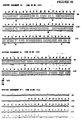
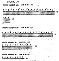
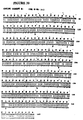
- the human peptide sequences described above and presented in Figs. 30, 31, 32 and 33, SEQ ID Nos. 132-146, respectively, represent a series of splicing variants which can be isolated as full length complementary DNAs (cDNAS) from natural sources (cDNA libraries prepared from the appropriate tissues) or can be assembled as DNA constructs with individual exons (e.g., derived as separate exons) by someone skilled in the art.
- cDNAS full length complementary DNAs
- the medicament of the present invention is made by administering the polypeptide with a pharmaceutically effective carrier optionally together with an acceptable diluent, carrier or excipient and/or in unit dosage form.
- a pharmaceutically effective carrier optionally together with an acceptable diluent, carrier or excipient and/or in unit dosage form.
- conventional pharmaceutical or veterinary practice may be employed to provide suitable formulations or compositions.
- the formulations to be used can be applied to parenteral administration, for example, intravenous, subcutaneous, intramuscular, intraorbital, ophthalmic, intraventricular, intracranial, intracapsular, intraspinal, intracisternal, intraperitoneal, topical, intranasal, aerosol, scarification, and also oral, buccal, rectal or vaginal administration.
- parenteral administration for example, intravenous, subcutaneous, intramuscular, intraorbital, ophthalmic, intraventricular, intracranial, intracapsular, intraspinal, intracisternal, intraperitoneal, topical, intranasal, aerosol, scarification, and also oral, buccal, rectal or vaginal administration.
- formulations of this invention may also be administered by the transplantation into the patient of host cells expressing the DNA encoding polypeptides which are effective for the purpose or by the use of surgical implants which release the formulations of the invention.
- Parenteral formulations may be in the form of liquid solutions or suspensions; for oral administration, formulations may be in the form of tablets or capsules; and for intranasal formulations, in the form of powders, nasal drops, or aerosols.
- Formulations for parenteral administration may, for example, contain as excipients sterile water or saline, polyalkylene glycols such as polyethylene glycol, oils of vegetable origin, or hydrogenated naphthalenes, biocompatible, biodegradable lactide polymer, or polyoxyethylene-polyoxypropylene copolymers may be used to control the release of the present factors.
- polyalkylene glycols such as polyethylene glycol, oils of vegetable origin, or hydrogenated naphthalenes
- biocompatible, biodegradable lactide polymer or polyoxyethylene-polyoxypropylene copolymers
- Other potentially useful parenteral delivery systems for the factors include ethylene-vinyl acetate copolymer particles, osmotic pumps, implantable infusion systems, and liposomes.
- Formulations for inhalation may contain as excipients, for example, lactose, or may be aqueous solutions containing, for example, polyoxyethylene-9-lauryl ether, glycocholate and deoxycholate, or may be oily solutions for administration in the form of nasal drops, or as a gel to be applied intranasally.
- Formulations for parenteral administration may also include glycocholate for buccal administration, methoxysalicylate for rectal administration, or citric acid for vaginal administration.
- the present factors can be used as the sole active agents, or can be used in combination with other active ingredients, e.g., other growth factors which could facilitate neuronal survival in neurological diseases, or peptidase or protease inhibitors.
- concentration of the present factors in the formulations of the invention will vary depending upon a number of issues, including the dosage to be administered, and the route of administration.
- the factors may be provided in an aqueous physiological buffer solution containing about 0.1 to 10% w/v compound for parenteral administration.
- General dose ranges are from about 1 mg/kg to about 1 g/kg of body weight per day; a preferred dose range is from about 0.01 mg/kg to 100 mg/kg of body weight per day.
- the preferred dosage to be administered is likely to depend upon the type and extent of progression of the pathophysiological condition being addressed, the overall health of the patient, the make up of the formulation, and the route of administration.
- polypeptide factors utilized in the methods of the invention can also be used as immunogens for making antibodies, such as monoclonal antibodies, following standard techniques. These antibodies can, in turn, be used for therapeutic or diagnostic purposes. Thus, conditions perhaps associated with muscle diseases resulting from abnormal levels of the factor may be tracked by using such antibodies.
- In vitro techniques can be used, employing assays on isolated samples using standard methods. Imaging methods in which the antibodies are, for example, tagged with radioactive isotopes which can be imaged outside the body using techniques for the art of tumour imaging may also be employed.
- GGF2 The "GGF2" designation is used for all clones which were previously isolated with peptide sequence data derived from GGF-II protein (i.e., GGF2HBS5, GGF2BPP3) and, when present alone (i.e., GGF2 or rhGGF2), to indicate recombinant human protein encoded by plasmids isolated with peptide sequence data derived from the GGF-II protein (i.e., as produced in insect cells from the plasmid HBSS).
- Recombinant human GGF from the GGFHBS5 clone is called GGF2, rhGGF2 and GGF2HBS5 polypeptide.
- Treating as used herein means any administration of the compounds described herein for the purpose of increasing muscle cell mitogenesis, survival, and/or differentiation, and/or decreasing muscle atrophy and degeneration. Most preferably, the treating is for the purpose of reducing or diminishing the symptoms or progression of a disease or disorder of the muscle cells. Treating as used herein also means the administration of the compounds for increasing or altering the muscle cells in healthy individuals. The treating may be brought about by the contacing of the muscle cells which are sensitive or responsive to the compounds described herein with an effective amount of the compound, as described above. Inhibitors of the compounds described herein may also be used to halt or slow diseases of muscle cell proliferation.
- GGF/p185 erbB2 binding neuregulin proteins produces a number of variably-sized, differentially-spliced RNA transcripts that give rise to a series of proteins. These proteins are of different lengths and contain some common peptide sequences and some unique peptide sequences. The conclusion that these factors are encoded by a single gene is supported by the differentially-spliced RNA sequences which are recoverable from bovine posterior pituitary and human breast cancer cells (MDA-MB-231)). Further support for this conclusion derives from the size range of proteins which act as both mitogens for muscle tissue (as disclosed herein) and as ligands for the p185 erbB2 receptor (see below).
- p185 erbB2 receptor binding proteins stimulate muscle cell mitogenesis and hence, stimulates myotube formation (myogenesis). This stimulation results in increased formation of myoblasts and increased formation of myotubes (myogenesis).
- the polypeptides described herein also stimulate increased muscle growth, differentiation, and survival of muscle cells.
- These ligands include, but are not limited to the GGF's, the neuregulins, the heregulins, NDF, and ARIA.
- these proteins, DNA encoding these proteins, and related polypeptides may be administered to patients suffering from traumatic damage or diseases of the muscle tissue. It is understood that all methods provided for the purpose of mitogenesis are useful for the purpose of myogenesis. While not forming part of the present invention inhibitors of these ligands (such as antibodies or peptide fragments) may be administered for the treatment of muscle.derived tumors.
- polypeptides may be obtained using the protocols described herein (Examples 9-17) and in Holmes et al., science 256 : 1205 (1992); Peles et al., Cell 69 :205 (1992); Wen et al., Cell 69 :559 (1992); Lupu et al., Proc. Natl. Acad. Sci. USA 89:2287 (1992); Yarden and Peles, Biochemistry 30:3543 (1991); Lupu et al., Science 249:1552 (1990); Dobashi et al., Biochem. Biophys. Res. Comm. 179:1536 (1991); Huang et al., J. Biol. Chem.
- Polypeptides may be assayed for their usefulness in vitro using the methods provided in the examples below. In vivo testing may be performed as described in Example 1 and in Sklar et al., In Vitro Cellular and Developmental Biology 27A:433-434, 1991.
- the invention includes methods for the use as defined previously of any protein which is substantially homologous to the coding segments in Fig. 30 (SEQ ID Nos. 132-143, 156, 1576-147) for the purpose of inducing muscle mitogenesis. Also included are the use of: allelic variations; natural mutants; induced mutants; proteins encoded by DNA that hybridizes under high or low stringency conditions to a nucleic acid naturally occurring (for definitions of high and low stringency see Current Protocols in Molecular Biology, John Wiley & Sons, New York, 1989, 6.3.1 - 6.3.6, hereby incorporated by reference); and the use of polypeptides or proteins specifically bound by antisera to GGF polypeptides. The term also includes the use of chimeric polypeptides that include the GGF polypeptides comprising sequences from Fig. 28 for the induction of muscle mitogenesis.
- rhGGF2 recombinant human GGF2
- the examples illustrate our discovery that recombinant human GGF2 (rhGGF2) confers several effects on primary human muscle culture.
- rhGGF2 has significant effects in three independent biological activity assays on muscle cultures.
- the polypeptide increased mitogenesis as measured by proliferation of subconfluent quiescent myoblasts, increased differentiation by confluent myoblasts in the presence of growth factor, and increased survival of differentiated myotubes as measured by loss of dye exclusion and increased acetylcholine receptor synthesis. These activities indicate efficacy of GGF2 and other neuregulins in inducing muscle repair, regeneration, and prophylactic effects on muscle degeneration.
- Clone GGF2HBS5 was expressed in recombinant Baculovirus infected insect cells as described in Example 14, infra , and the resultant recombinant human GGF2 was added to myoblasts in culture (conditioned medium added at 40 ⁇ l/ml).
- Myoblasts (057A cells) were grown to preconfluence in a 24 well dish.
- Medium was removed and replaced with DMEM containing 0.5% fetal calf serum with or without GGF2 conditioned medium at a concentration of 40 ⁇ l/ml.
- Medium was changed after 2 days and cells were fixed and stained after 5 days. Total nuclei were counted as were the number of nuclei in myoblasts (Table 1).
- the mitogenic activity of rhGGF2 may be measured in vivo by giving a continuous supply of GGF2 and [ 3 H]thymidine to rat muscle via an osmotic mini pump. The muscle bulk is determined by wet weight after one and two weeks of treatment. DNA replication is measured by counting labeled nuclei in sections after coating for autoradiography (Sklar et al., In Vitro Cellular and Developmental Biology 27A:433-434, 1991) in sham and rhGGF2-treated muscle. Denervated muscle is also examined in this rat animal model via these methods and this method allows the assessment of the role of rhGGF2 in muscle atrophy and repair. Mean fiber diameter can also be used for assessing effects of FGF on prevention of atrophy.
- Quiescent primary clonal human myoblasts were prepared as previously described (Sklar, R., Hudson, A., Brown, R., In vitro Cellular and Developmental Biology 1991; 27A:433-434).
- the quiescent cells were treated with the indicated agents (rhGGF2 conditioned media, PDGF with and without methylprednisolone, and control media) in the presence of 10 ⁇ M BrdU, 0.5% FCS in DMEM. After two days the cells were fixed in 4% paraformaldehyde in PBS for 30 minutes, and washed with 70% ethanol. The cells were then incubated with an anti-BrdU antibody, washed, and antibody binding was visualized with a peroxidase reaction.
- rhGGF2 purified to homogeneity is also mitogenic for human myoblasts (Fig. 1).
- Recombinant human GGF2 also causes mitogenesis of primary human myoblasts (see Table 2 and Fig. 1).
- the mitogenesis assay is performed as described above.
- the mitotic index is then calculated by dividing the number of BrdU positive cells by the total number of cells.
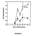
- rhGGF2 The effects of purified rhGGF2 (95% pure) on muscle culture differentiation were examined (Fig. 2).
- Confluent myoblast cultures were induced to differentiate by lowering the serum content of the culture medium from 20% to 0.5%.
- the test cultures were treated with the indicated concentration of rhGGF2 for six days, refreshing the culture medium every 2 days.
- the cultures were then fixed, stained, and the number of nuclei counted per millimeter.
- the data in Fig. 2 demonstrate a large increase in the number of nuclei in myotubes when rhGGF2 is present, relative to controls.
- the survival assay was also performed with other growth factors which have known effects on muscle culture.
- the rhGGF2 effect was unique among the growth factors tested (Fig. 4).
- cultures were treated in parallel with the rhGGF2 treated plates with the indicated concentrations of the various growth factors. Survival of myotubes was measured as above at 8 days of differentiation of 057A myoblast cells.
- rhGGF2 100ng/ml
- human platelet derived growth factor 20ng/ml
- human basic fibroblast growth factor 25ng/ml
- human epidermal growth factor 30ng/ml
- human leucocyte inhibitory factor 10ng/ml
- human insulin like growth factor I 30ng/ml
- human insulin like growth factor II 25ng/ml.
- rhGGF2 has promise as a therapy for intervention of muscle degeneration characterized by numerous muscle diseases.
- agents which increase the extramuscular concentration of neuregulins may have a prophylactic effect or slow the progress of muscle-wasting disorders and increase rates of muscle differentiation, repair, conditioning, and regeneration.
- rhGGF2 Promotes Survival of Differentiated Myotubes with a Genetic Defect at the Duchenne Muscular Dystrophy Locus
- rhGGF2 can also promote survival of differentiated Duchenne myotubes and provides strong evidence that rhGGF2 may slow or prevent the course of muscle degeneration and wasting in mammals.
- the effects of purified rhGGF2 on muscle culture differentiation was also examined by Western analysis of culture lysates.
- the levels of muscle specific proteins were determined in triplicate treated and untreated cultures. These cultures were prepared and treated as above except that the plate size was increased to 150 mm and the muscle culture layer was scraped off for Western analysis as described in Sklar, R., and Brown, R. ( J. Neurol. Sci. 101:73-81, 1991).
- the results presented in Table A indicate that rhGGF2 treatment increases the levels of several muscle specific proteins, including dystrophin, myosin heavy chain (MHC, adult slow and fast isoforms), but does not increase the levels of HSP72 or MHC neonate isoform to a similar level per amount of protein loaded on the Western.
- the levels of muscle specific proteins induced by rhGGF2 were similar to the quantitative increases in the number of myonuclei/mm 2 (Table 3).
- the rhGGF2 dependent increase in the adult myosin heavy chain isoforms may represent a maturation of the myotubes, as the neonatal isoform was not significantly increased by rhGGF2 treatment.
- MHC isoforms switch from fetal to neonatal forms followed by a switch to mature adult slow and fast MHC isoforms (Periasamy et al. J . Biol. Chem. 259:13573-13578, 1984; Periasamy et al. J. Biol. Chem.
- Neuregulins including rhGGF2, induce the synthesis of acetylcholine receptors in muscle.
- AchR subunit proteins can be induced by exposing muscle cells to neuregulins. More specifically, we have shown that contacting muscle cells with rhGGF2 can induce the synthesis of AchR subunit proteins. This induction following rhGGF2 exposure was observed in two ways: first, we detected increased expression of human growth hormone via the product of a reporter gene construct and second we detected increased binding of alpha-bungarotoxin to cells.
- a mouse myoblast cell line C2 was used. C2 cells were transfected with a transgene that contained the 5' regulatory sequences of the AChR delta subunit gene of mouse linked to a human growth hormone full-length cDNA (Baldwin and Burden, 1988. J. Cell Biol. 107:2271-2279).
- This reporter construct allows the measurement of the induction of AChR delta gene expression by assaying the quantity of growth hormone secreted into the media.
- the line can be induced to form myotubes by lowering serum concentration in the media from 20% to 0.5%.
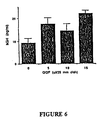
- mice C2 myoblasts transfected with an AChR-human growth hormone reporter construct and were assayed for expression of hGH following treatment with rhGGF2.
- the results of two separate experiments are summarized in Table 4 and in Figures 6 (hGH expression) and 7 (hGH expression and alpha-bungarotoxin binding). Shown are the dose response curves for secreted human growth hormone and for bungarotoxin binding from muscle cultures treated with rhGGF2.
- C2 myotubes were treated with cold ⁇ -BTX (20 nM) for 1 hr. at 37°C, washed with culture medium twice and then treated with GGF2.
- Culture medium was adjusted with bovine serum albumin at the concentration of 1 mg/ml. 24 hours later, culture medium was removed and saved for hGH assay.
- Muscle cultures were treated with 125 I- ⁇ -BTX (20 nM) for 1 hour at 37°C, washed and scraped in PBS containing 1% SDS. Non-specific binding was determined in the presence of cold ⁇ -BTX (40 nM). The cell homogenate was counted for radioactivity and assayed for total protein amount.
- rhGGF2 led to a greater than 2-fold increase in hGH gene expression, thereby indicating that rhGGF2 induced the synthesis of the delta subunit of the acetylcholine receptor. Furthermore, increased bungarotoxin binding is consistant with assembly of these subunit proteins into functional acetylcholine receptors. To strenthen the interpretation of these data the analysis was repeated on cultures that had the hGH reporter linked to a metallothiene promoter, which should not be responsive to rhGGF2. The results of that control experiment showed that the hGH response was mediated through transcriptional activation of the AchR delta subunit gene control elements.
- rhGGF2 could be useful in replenishing AchRs as part of the therapy for the autoimmune disease Myasthenia gravis. This activity may also be beneficial in treatment of peripheral nerve regeneration and neuropathy by stimulating a key step in re-innervation of muscle.
- the mitogenic activity of a highly purified sample containing both GGFs I and II was studied using a quantitative method, which allows a single microculture to be examined for DNA synthesis, cell morphology, cell number and expression of cell antigens.
- This technique has been modified from a method previously reported by Muir et al., Analytical Biochemistry 185, 377-382, 1990. The main modifications are: 1) the use of uncoated microtiter plates, 2) the cell number per well, 3) the use of 5% Foetal Bovine Plasma (FBP) instead of 10% Foetal Calf Serum (FCS), and 4) the time of incubation in presence of mitogens and bromodeoxyuridine (BrdU), added simultaneously to the cultures.
- FBP Foetal Bovine Plasma
- FCS Foetal Calf Serum
- the assay optimized for rat sciatic nerve Schwann cells, has also been used for several cell lines, after appropriate modifications to the cell culture conditions.
- Unbound antibodies were removed by three washes in PBS containing 0.1% Triton X-100 and peroxidase-conjugated goat anti-mouse IgG antibody (Dako Corp., Santa Barbara, CA) (50 ⁇ l/well, 2 ⁇ g/ml diluted in blocking buffer) was added and incubated for one hour at 37°C. After three washes in PBS/Triton and a final rinse in PBS, wells received 100 ⁇ l/well of 50 mM phosphate/citrate buffer, pH 5.0, containing 0.05% of the soluble chromogen o-phenylenediamine (OPD) and 0.02% H 2 O 2 .
- PBS/Triton 50 mM phosphate/citrate buffer, pH 5.0, containing 0.05% of the soluble chromogen o-phenylenediamine (OPD) and 0.02% H 2 O 2 .
- the reaction was terminated after 5-20 min at room temperature, by pipetting 80 ⁇ l from each well to a clean plate containing 40 ⁇ l/well of 2N sulfuric acid. The absorbance was recorded at 490nm using a plate reader (Dynatech Labs).
- the assay plates containing the cell monolayers were washed twice with PBS and immunocytochemically stained for BrdU-DNA by adding 100 ⁇ l/well of the substrate diaminobenzidine (DAB) and 0.02% H 2 O 2 to generate an insoluble product. After 10-20 min the staining reaction was stopped by washing with water, and BrdU-positive nuclei observed and counted using an inverted microscope. occasionally, negative nuclei were counterstained with 0.001% Toluidine blue and counted as before.
- DAB diaminobenzidine
- Swiss 3T3 Fibroblasts Cells, from Flow Labs, were maintained in DMEM supplemented with 10% FCS, penicillin and streptomycin, at 37°C in a humidified atmosphere of 10% CO 2 in air. Cells were fed or subcultured every two days. For mitogenic assay, cells were plated at a density of 5,000 cells/well in complete medium and incubated for a week until cells were confluent and quiescent. The serum containing medium was removed and the cell monolayer washed twice with serum free-medium. 100 ⁇ l of serum free medium containing mitogens and 10 ⁇ M of BrdU were added to each well and incubated for 48 hours. Dose responses to GGFs and serum or PDGF (as a positive control) were performed.
- BHK (Baby Hamster Kidney) 21 C13 Fibroblasts Cells from European Collection of Animal Cell Cultures (ECACC), were maintained in Glasgow Modified Eagle Medium (GMEM) supplemented with 5% tryptose phosphate broth, 5% FCS, penicillin and streptomycin, at 37°C in a humidified atmosphere of 5% CO 2 in air. Cells were fed or subcultured every two to three days. For mitogenic assay, cells were plated at a density of 2,000 cell/well in complete medium for 24 hours. The serum containing medium was then removed and after washing with serum free medium, replaced with 100 ⁇ l of 0.1% FCS containing GMEM or GMEM alone. GGFs and FCS or bFGF as positive controls were added, coincident with 10 ⁇ M BrdU, and incubated for 48 hours. Cell cultures were then processed as described for Schwann cells.
- GMEM Glasgow Modified Eagle Medium
- FCS penicillin and streptomycin
- C6 Rat Glioma Cell Line Cells, obtained at passage 39, were maintained in DMEM containing 5% FCS, 5% Horse serum (HS), penicillin and streptomycin, at 37°C in a humidified atmosphere of 10% CO 2 in air. Cells were fed or subcultured every three days. For mitogenic assay, cells were plated at a density of 2,000 cells/well in complete medium and incubated for 24 hours. Then medium was replaced with a mixture of 1:1 DMEM and F12 medium containing 0.1% FCS, after washing in serum free medium. Dose responses to GGFs, FCS and ⁇ FGF were then performed and cells were processed through the ELISA as previously described for the other cell types.
- HS Horse serum
- PC12 Rat Adrenal Pheochromocytoma Cells
- ECACC ECACC
- RPMI 1640 RPMI 1640 supplemented with 10% HS, 5% FCS, penicillin and streptomycin, in collagen coated flasks, at 37°C in a humidified atmosphere of 5% CO 2 in air. Cells were fed every three days by replacing 80% of the medium.
- cells were plated at a density of 3,000 cells/well in complete medium, on collagen coated plates (50 ⁇ l/well collagen, Vitrogen Collagen Corp., diluted 1 : 50, 30 min at 37°C) and incubated for 24 hours. The medium was then placed with fresh RPMI either alone or containing 1 mM insulin or 1% FCS. Dose responses to FCS/HS (1:2) as positive control and to GGFs were performed as before. After 48 hours cells were fixed and the ELISA performed as previously described.
- Fig. 12 shows the comparison of data obtained with the two assays, performed in the same cell culture conditions (5,000 cells/well, in 5% FBP/DMEM, incubated in presence of GGFs for 48 hrs). As clearly shown, the results are comparable, but BrdU incorporation assay appears to be slightly more sensitive, as suggested by the shift of the curve to the left of the graph, i.e. to lower concentrations of GGFS.
- the original assay plates containing cell monolayers can undergo the second reaction resulting in the insoluble DAB product, which stains the BrdU positive nuclei.
- the microcultures can then be examined under an inverted microscope, and cell morphology and the numbers of BrdU-positive and negative nuclei can be observed.
- Fig. 13A and Fig. 13B the BrdU-DNA immunoreactivity, evaluated by reading absorbance at 490 nm, is compared to the number of BrdU-positive nuclei and to the percentage of BrdU-positive nuclei on the total number of cells per well, counted in the same cultures. Standard deviations were less than 10%. The two evaluation methods show a very good correlation and the discrepancy between the values at the highest dose of GGFs can be explained by the different extent of DNA synthesis in cells detected as BrdU-positive.
- the BrdU incorporation assay can therefore provide additional useful information about the biological activity of polypeptides on Schwann cells when compared to the (125) 1-UdR incorporation assay.
- the data reported in Fig. 15 show that GGFs can act on Schwann cells to induce DNA synthesis, but at lower doses to increase the number of negative cells present in the microculture after 48 hours.
- the assay has then been used on several cell lines of different origin.
- Fig. 15 the mitogenic responses of Schwann cells and Swiss 3T3 fibroblasts to GGFs are compared; despite the weak response obtained in 3T3 fibroblasts, some clearly BrdU-positive nuclei were detected in these cultures.
- Control cultures were run in parallel in presence of several doses of FCS or human recombinant PDGF, showing that the cells could respond to appropriate stimuli (not shown).
- fibroblasts The ability of fibroblasts to respond to GGFs was further investigated using the BHK 21 C13 cell line. These fibroblasts, derived from kidney, do not exhibit contact inhibition or reach a quiescent state when confluent. Therefore the experimental conditions were designed to have a very low background proliferation without compromising the cell viability.
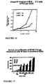
- GGFs have a significant mitogenic activity on BHK21 C13 cells as shown by Fig. 16 and Fig. 17.
- Fig. 16 shows the Brdu incorporation into DNA by BHK 21 C13 cells stimulated by GGFS in the presence of 0.1% FCS. The good mitogenic response to FCS indicates that cell culture conditions were not limiting.
- Fig. 16 shows the Brdu incorporation into DNA by BHK 21 C13 cells stimulated by GGFS in the presence of 0.1% FCS. The good mitogenic response to FCS indicates that cell culture conditions were not limiting.
- the mitogenic effect of GGFs is expressed as the number of BrdU-positive and BrdU-negative cells and as the total number of cells counted per well. Data are representative of two experiments run in duplicates; at least three fields per well were counted. As observed for Schwann cells in addition to a proliferative effect at low doses, GGFs also increase the numbers of nonresponding cells surviving. The percentage of BrdU positive cells is proportional to the increasing amounts of GGFs added to the cultures. The total number of cells after 48 hours in presence of higher doses of GGFs is at least doubled, confirming that GGFs induce DNA synthesis and proliferation in BHK21 C13 cells. Under the same conditions, cells maintained for 48 hours in the presence of 2% FCS showed an increase of about six fold (not shown).
- C6 glioma cells have provided a useful model to study glial cell properties.
- the phenotype expressed seems to be dependent on the cell passage, the cells more closely resembling an astrocyte phenotype at an early stage, and an oligodendrocyte phenotype at later stages (beyond passage 70).
- C6 cells used in these experiments were from passage 39 to passage 52.
- C6 cells are a highly proliferating population, therefore the experimental conditions were optimized to have a very low background of BrdU incorporation.
- the presence of 0.1% serum was necessary to maintain cell viability without significantly affecting the mitogenic responses, as shown by the dose response to FCS (Fig. 18).
- aFGF acidic Fibroblast growth factor
- GGFs are expressed as the percentages of maximal BrdU incorporation obtained in the presence of FCS (8%). Values are averages of two experiments, run in duplicates. The effect of GGFs was comparable to that of a pure preparation of aFGF.
- aFGF has been described as a specific growth factor for C6 cells (Lim R. et al., Cell Regulation 1 :741-746, 1990) and for that reason it was used as a positive control. The direct counting of BrdU positive and negative cells was not possible because of the high cell density in the microcultures.
- PC12 cells did not show any evident responsiveness to GGFS, when treated under culture conditions in which PC12 could respond to sera (mixture of FCS and HS as used routinely for cell maintenance). Nevertheless the number of cells plated per well seems to affect the behavior of PC12 cells, and therefore further experiments are required.
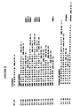
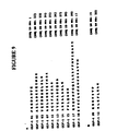
- a total of 21 peptide sequences (see Fig. 8, SEQ ID Nos. 1-20, 165) were obtained for GGF-I, of which 12 peptides (see Fig. 9, SEQ ID Nos. 1, 22-29, 17, 19, and 32) are not present in current protein databases and therefore represent unique sequences.
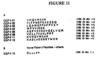
- a total of 12 peptide sequences (see Fig. 10, SEQ ID Nos. 42-50 and 161-163) were obtained for GGF-II, of which 10 peptides (see Fig. 11, SEQ ID Nos. 42-50) are not present in current protein databases and therefore represent unique sequences (an exception is peptide GGF-II 06 which shows identical sequences in many proteins which are probably of no significance given the small number of residues).
- sequence X S S is consistent with the presence of an N linked carbohydrate moiety on an asparagine at the position denoted by X.
- X represents an unknown residue denoting a sequencing cycle where a single position could not be called with certainty either because there was more than one signal of equal size in the cycle or because no signal was present.
- asterisk denotes those peptides where the last amino acid called corresponds to the last amino acid present in that peptide. In the remaining peptides, the signal strength after the last amino acid called was insufficient to continue sequence calling to the end of that peptide.
- the right hand column indicates the results of a computer database search using the GCG package FASTA and TFASTA programs to analyze the NBRF and EMBL sequence databases.
- HMG-1 High Mobility Group protein-1 HMG-2 High Mobility Group protein-2 LH-alpha Luteinizing hormone alpha subunit LH-beta Luteinizing hormone beta subunit
- DNA sequence and polypeptide sequence should be obtainable by this means as with GGF-II, and also DNA constructs and expression vectors incorporating such DNA sequence, host cells genetically altered by incorporating such constructs/vectors, and protein obtainable by cultivating such host cells.
- the invention envisages such subject matter.
- DNA oligomer probes were designed by backtranslating the amino acid sequences (derived from the peptides generated from purified GGF protein) into nucleotide sequences. Oligomers represented either the coding strand or the non-coding strand of the DNA sequence.
- oligomers represented either the coding strand or the non-coding strand of the DNA sequence.
- serine, arginine or leucine were included in the oligomer design, then two separate syntheses were prepared to avoid ambiguities. For example, serine was encoded by either TCN or AGY as in 537 and 538 or 609 and 610. Similar codon splitting was done for arginine or leucine (e.g. 544, 545).
- DNA oligomers were synthesized on a Biosearch 8750 4-column DNA synthesizer using ⁇ -cyanoethyl chemistry operated at 0.2 micromole scale synthesis. Oligomers were cleaved off the column (500 angstrom CpG resins) and deprotected in concentrated ammonium hydroxide for 6-24 hours at 55-60°C. Deprotected oligomers were dried under vacuum (Speedvac) and purified by electrophoresis in gels of 15% acrylamide (20 mono : 1 bis), 50 mM Tris-borate-EDTA buffer containing 7M urea.
- PCR primers were prepared by essentially the same procedures that were used for probes with the following modifications. Linkers of thirteen nucleotides containing restriction sites were included at the 5' ends of the degenerate oligomers for use in cloning into vectors. DNA synthesis was performed at 1 micromole scale using 1,000 angstrom CpG resins and inosine was used at positions where all four nucleotides were incorporated normally into degenerate probes. Purifications of PCR primers included an ethanol precipitation following the gel electrophoresis purification.
- a bovine genomic DNA library was purchased from Stratagene (Catalogue Number: 945701). The library contained 2 x 10 6 15-20kb Sau3Al partial bovine DNA fragments cloned into the vector lambda DashII.
- a bovine total brain cDNA library was purchased from Clonetech (Catalogue Number: BL 10139).
- Complementary DNA libraries were constructed (In Vitrogen; Stratagene) from mRNA prepared from bovine total brain, from bovine pituitary and from bovine posterior pituitary. In Vitrogen prepared two cDNA libraries: one library was in the vector lambda g10, the other in vector pcDNAI (a plasmid library). The Stratagene libraries were prepared in the vector lambda unizap. Collectively, the cDMA libraries contained 14 million primary recombinant phage.
- the bovine genomic library was plated on E . coli K12 host strain LE392 on 23 x 23 cm plates (Nunc) at 150,000 to 200,000 phage plaques per plate. Each plate represented approximately one bovine genome equivalent. Following an overnight incubation at 37°C, the plates were chilled and replicate filters were prepared according to procedures of Maniatis et al. (2:60-81). Four plaque lifts were prepared from each plate onto uncharged nylon membranes (Pall Biodyne A or MSI Nitropure). The DNA was immobilized onto the membranes by cross-linking under UV light for 5 minutes or, by baking at 80°C under vacuum for two hours.
- DNA probes were labelled using T4 polynucleotide kinase (New England Biolabs) with gamma 32P ATP (New England Nuclear; 6500 Ci/mmol) according to the specifications of the suppliers. Briefly, 50 pmols of degenerate DNA oligomer were incubated in the presence of 600 ⁇ Ci gamma 32 P-ATP and 5 units T4 polynucleotide kinase for 30 minutes at 37°C. Reactions were terminated, gel electrophoresis loading buffer was added and then radiolabelled probes were purified by electrophoresis. 32P labelled probes were excised from gel slices and eluted into water.
- DNA probes were labelled via PCR amplification by incorporation of ⁇ -32P-dATP or ⁇ -32P dCTP according to the protocol of Schowalter and Sommer, Anal. Biochem 177:90-94 (1989). Probes labelled in PCR reactions were purified by desalting on Sephadex G-150 columns.
- Prehybridization and hybridization were performed in GMC buffer (0.52 M NaPi, 7% SDS, 1% BSA, 1.5 mM EDTA, 0.1 M NaCl 10 mg/ml tRNA). Washing was performed in oligowash (160 ml 1 M Na 2 HPO 4 , 200 ml 20% SDS, 8.0 ml 0.5 M EDTA, 100 ml 5M NaCl, 3632 ml H2O). Typically, 20 filters (400 sq. centimeters each) representing replicate copies of ten bovine genome equivalents were incubated in 200 ml hybridization solution with 100 pmols of degenerate oligonucleotide probe (128-512 fold degenerate). Hybridization was allowed to occur overnight at 5°C below the minimum melting temperature calculated for the degenerate probe. The calculation of minimum melting temperature assumes 2°C for an AT pair and 4°C for a GC pair.
- Filters were washed in repeated changes of oligowash at the hybridization temperatures four to five hours and finally, in 3.2M tetramethylammonium chloride, 1% SDS twice for 30 min at a temperature dependent on the DNA probe length. For 20mers, the final wash temperature was 60°C. Filters were mounted, then exposed to X-ray film (Kodak XAR5) using intensifying screens (Dupont Cronex Lightening Plus). Usually, a three to five day film exposure at minus 80°C was sufficient to detect duplicate signals in these library screens. Following analysis of the results, filters could be stripped and reprobed.
- Filters were stripped by incubating through two successive cycles of fifteen minutes in a microwave oven at full power in a solution of 1% SDS containing 10mM EDTA pH8. Filters were taken through at least three to four cycles of stripping and reprobing with various probes.
- Recombinant Phage DNA samples (2 micrograms) were digested according to conditions recommended by the restriction endonuclease supplier (New England Biolabs). Following a four hour incubation at 37°C, the reactions products were precipitated in the presence of 0.1M sodium acetate and three volumes of ethanol. Precipitated DNA was collected by centrifugation, rinsed in 75% ethanol and dried. All resuspended samples were loaded onto agarose gels (typically 1% in TAE buffer; 0.04M Tris acetate, 0.002M EDTA). Gel runs were at 1 volt per centimeter from 4 to 20 hours. Markers included lambda Hind III DNA fragments and/or ⁇ X174HaeIII DNA fragments (New England Biolabs).
- the gels were stained with 0.5 micrograms/ml of ethidium bromide and photographed.
- DNA was first depurinated in the gel by treatment with 0.125 N HCl, denatured in 0.5 N NaOH and transferred in 20x SSC (3M sodium chloride, 0.03 M sodium citrate) to uncharged nylon membranes. Blotting was done for 6 hours up to 24 hours, then the filters were neutralized in 0.5 Tris HCl pH 7.5, 0.15 M sodium chloride, then rinsed briefly in 50 mM Tris-borate EDTA.
- the filters were wrapped first in transparent plastic wrap, then the DNA side exposed for five minutes to an ultraviolet light. Hybridization and washing was performed as described for library screening (see section 2 of this Example). For hybridization analysis to determine whether similar genes exist in other species slight modifications were made.
- the DNA filter was purchased from Clonetech (Catalogue Number 7753-1) and contains 5 micrograms of EcoRI digested DNA from various species per lane.
- the probe was labelled by PCR amplification reactions as described in section 2 above, and hybridizations were done in 80% buffer B(2 g polyvinylpyrrolidine, 2 g Ficoll-400, 2 g bovine serum albumin, 50 ml 1M Tris-HCl (pH 7.5) 58 g NaCl, 1 g sodium pyrophosphate, 10 g sodium dodecyl sulfate, 950ml H 2 O) containing 10% dextran sulfate.
- the probes were denatured by boiling for ten minutes then rapidly cooling in ice water. The probe was added to the hybridization buffer at 10 6 dpm 32 P per ml and incubated overnight at 60°C.
- the filters were washed at 60°C first in buffer B followed by 2X SSC, 0.1% SDS then in 1x SSC, 0.1% SDS. For high stringency, experiments, final washes were done in 0.1 x SSC, 1% SDS and the temperature raised to 65°C.
- Southern blot data were used to prepare a restriction map of the genomic clone and to indicate which subfragments hybridized to the GGF probes (candidates for subcloning).
- DNA digests e.g. 5 micrograms
- DNA was loaded onto 1% agarose gels then appropriate fragments excised from the gels following staining.
- the DNA was purified by adsorption onto glass beads followed by elution using the protocol described by the supplier (Bio 101).
- Recovered DNA fragments 100-200 ng
- pT3T7 a derivative of pUC18
- T4 ligase New England Biolabs.
- This vector carries the E. coli ⁇ lactamase gene, hence, transformants can be selected on plates containing ampicillin.
- the vector also supplies ⁇ -galactosidase complementation to the host cell, therefore non-recombinants (blue) can be detected using isopropylthiogalactoside and Bluogal (Bethesda Research Labs).
- a portion of the ligation reactions was used to transform E . coli K12 XL1 blue competent cells (Stratagene Catalogue Number: 200236) and then the transformants were selected on LB plates containing 50 micrograms per ml ampicillin. White colonies were selected and plasmid mini preps were prepared for DNA digestion and for DNA sequence analysis. Selected clones were retested to determine if their insert DNA hybridized with the GGF probes.
- Double stranded plasmid DNA templates were prepared from 5 ml cultures according to standard protocols. Sequencing was by the dideoxy chain termination method using Sequenase 2.0 and a dideoxynucleotide sequencing kit (US Biochemical) according to the manufacturers protocol (a modification of Sanger et al. PNAS; USA 74 :5463 (1977)]. Alternatively, sequencing was done in a DNA thermal cycler (Perkin Elmer, model 4800) using a cycle sequencing kit (New England Biolabs; Bethesda Research Laboratories) and was performed according to manufacturers instructions using a 5'-end labelled primer. Sequence primers were either those supplied with the sequencing kits or were synthesized according to sequence determined from the clones.
- Sequencing reactions were loaded on and resolved on 0.4mm thick sequencing gels of 6% polyacrylamide. Gels were dried and exposed to X-Ray film. Typically, 35S was incorporated when standard sequencing kits were used and a 32P end labelled primer was used for cycle sequencing reactions. Sequences were read into a DNA sequence editor from the bottom of the gel to the top (5' direction to 3') and data were analyzed using programs supplied by Genetics Computer Group (GCG, University of Wisconsin).
- Specific DNA target sequences were amplified beginning with either total RNA or polyadenylated RNA samples that had been converted to cDNA using the Perkin Elmer PCR/RNA Kit Number: N808-0017.
- First strand reverse transcription reactions used 1 ⁇ g template RNA and either primers of oligo dT with restriction enzyme recognition site linkers attached or specific antisense primers determined from cloned sequences with restriction sites attached.
- the primers either were plus strand unique sequences as used in 3' RACE reactions (Frohman et.
- the amplification profiles followed the following general scheme: 1) five minutes soak file at 95°C; 2) thermal cycle file of 1 minute, 95°C; 1 minute ramped down to an annealing temperature of 45°C, 50°C or 55°C; maintain the annealing temperature for one minute; ramp up to 72°C over one minute; extend at 72°C for one minute or for one minute plus a 10 second auto extension; 3) extension cycle at 72°C, five minutes, and; 4) soak file 4°C for infinite time.
- Thermal cycle files (#2) usually were run for 30 cycles.
- a sixteen ⁇ l sample of each 100 ⁇ l amplification reaction was analyzed by electrophoresis in 2% Nusieve 1% agarose gels run in TAE buffer at 4 volts per centimeter for three hours. The gels were stained, then blotted to uncharged nylon membranes which were probed with labelled DNA probes that were internal to the primers.
- DNA amplification products could be identified in the blotting experiments and their positions used as a guide to purification and reamplification.
- the remaining portions of selected samples were loaded onto preparative gels, then following electrophoresis four to five slices of 0.5 mm thickness (bracketing the expected position of the specific product) were taken from the gel.
- the agarose was crushed, then soaked in 0.5 ml of electrophoresis buffer from 2-16 hours at 40°C. The crushed agarose was centrifuged for two minutes and the aqueous phase was transferred to fresh tubes.
- Reamplification was done on five microliters (roughly 1% of the product) of the eluted material using the same sets of primers and the reaction profiles as in the original reactions.
- samples were extracted with chloroform and transferred to fresh tubes.
- Concentrated restriction enzyme buffers and enzymes were added to the reactions in order to cleave at the restriction sites present in the linkers.
- the digested PCR products were purified by gel electrophoresis, then subcloned into vectors as described in the subcloning section above. DNA sequencing was done described as above.
- DNA sequences were assembled using a fragment assembly program and the amino acid sequences deduced by the GCG programs GelAssemble, Map and Translate.
- the deduced protein sequences were used as a query sequence to search protein sequence databases using WordSearch. Analysis was done on a VAX Station 3100 workstation operating under VMS 5.1. The database search was done on SwissProt release number 21 using GCG Version 7.0.
- GGF-II 12 (SEQ ID No. 44), a peptide generated via lysyl endopeptidase digestion of a purified GGF-II preparation (see Figs. 16 and 12) showed strong amino acid sequence homology with GGF-I 07 (SEQ ID No. 39), a tryptic peptide generated from a purified GGF-I preparation.
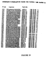
- GGF-II 12 was thus used to create ten degenerate oligonucleotide probes (see oligos 609, 610 and 649 to 656 in Fig.
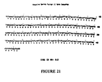
- Fig. 21 shows the nucleotide sequence, SEQ ID No. 89) and the deduced amino acid sequence of the initial DNA sequence readings that included the hybridization sites of probes 609 and 650, and confirmed that a portion of this bovine genomic DNA encoded peptide 12 (KASLADSGEYM).
- GGF-II 12 resided on a 66 amino acid open reading frame (see below) which has become the starting point for the isolation of overlapping sequences representing a putative bovine GGF-II gene and a cDNA.
- RNA and oligo dT-selected (poly A containing) RNA samples were prepared from bovine total pituitary, anterior pituitary, posterior pituitary, and hypothalamus. Using primers from the list shown in Fig. 22, SEQ ID Nos. 109-119, one-sided PCR reactions (RACE) were used to amplify cDNA ends in both the 3' and 5' directions, and anchored PCR reactions were performed with degenerate oligonucleotide primers representing additional GGF-II peptides.
- Fig. 29 summarizes the contiguous DNA structures and sequences obtained in those experiments.
- the cloned gene was characterized first by constructing a physical map of GGF2BG1 that allowed us to position the coding sequences as they were found (see below, Fig. 30). DNA probes from the coding sequences described above have been used to identify further DNA fragments containing the exons on this phage clone and to identify clones that overlap in both directions.
- the putative bovine GGF-II gene is divided into at least 5 coding segments. Coding segments are defined as discrete lengths of DNA sequence which can be translated into polypeptide sequences using the universal genetic code.
- the coding segments described in Fig. 36 and referred to in the present application are: 1) particular exons present within the GGF gene (e.g.
- coding segment a or 2) derived from sets of two or more exons that appear in specific sub-groups of mRNAs, where each set can be translated into the specific polypeptide segments as in the gene products shown.
- the polypeptide segments referred to in the claims are the translation products of the analogous DNA coding segments. Only coding segments A and B have been defined as exons and sequenced and mapped thus far. The summary of the contiguous coding sequences identified is shown in Fig. 31. The exons are listed (alphabetically) in the order of their discovery. It is apparent from the intron/exon boundaries that exon B may be included in cDNAs that connect coding segment E and coding segment A.
- the three deduced structures encode proteins of lengths 206, 281 and 257 amino acids.
- the first 183 residues of the deduced protein sequence are identical in all three gene products.
- the clones differ significantly.
- a codon for glycine GGT in GGF2BPP1 also serves as a splice donor for GGF2BPP2 and GGF2BPP3, which alternatively add on exons C, C/D, C/D' and D or C, C/D and D, respectively, and shown in Fig. 32, SEQ ID No. 145).
- GGFIIBPP1 is a truncated gene product which is generated by reading past the coding segment A splice junction into the following intervening sequence (intron).
- a probe encompassing the B and A exons was labelled via PCR amplification and used to screen a cDNA library made from RNA isolated from bovine posterior pituitary.
- One clone (GGF2BPP5) showed the pattern indicated in Fig. 29 and contained an additional DNA coding segment (G) between coding segments A and C.
- the entire nucleic acid sequence is shown in Fig. 31 (SEQ ID No. 144).
- the predicted translation product from the longest open reading frame is 241 amino acids.
- a portion of a second cDNA (GGF2BPP4) was also isolated from the bovine posterior pituitary library using the probe described above. This clone showed the pattern indicated in Fig. 29.
- BPP4 also displays a novel 3' end with regions H, K and L beyond region C/D.
- the sequence of BPP4 is shown in Fig. 33 (SEQ ID No. 146).
- the GGF proteins are the members of a new superfamily of proteins.
- DNA probes from this bovine recombinant molecule can readily detect specific sequences in a variety of samples tested.
- a highly homologous sequence is also detected in human genomic DNA.
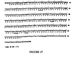
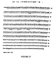
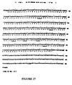
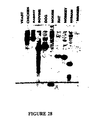
- the autoradiogram is shown in Fig. 28.
- the signals in the lanes containing rat and human DNA represent the rat and human equivalents of the GGF gene, the sequences of several cDNA's encoded by this gene have been recently reported by Holmes et al. (Science 256 : 1205 (1992)) and Wen et al. (Cell 69 : 559 (1992)).
- a probe derived from coding segment A (see Fig. 30), which was produced by labeling a polymerase chain reaction (PCR) product from segment A, was also used to screen the primary library.
- PCR polymerase chain reaction
- Several clones that hybridized with both A and E derived probes were selected and one particular clone, GGF2HBS5, was selected for further analysis.
- This clone is represented by the pattern of coding segments (EBACC/D'D as shown in Fig. 30).
- the E segment in this clone is the human equivalent of the truncated bovine version of E shown in Fig. 30.
- GGF2HBS5 is the most likely candidate to encode GGF-II of all the "putative" GGF-II candidates described.
- the length of coding sequence segment E is 786 nucleotides plus 264 bases of untranslated sequence.
- the predicted size of the protein encoded by GGF2HBS5 is approximately 423 amino acids (approximately 45 kilodaltons, see Fig. 44, SEQ ID NO: 21), which is similar to the size of the deglycosylated form of GGF-II (see Example 20). Additionally, seven of the GGF-II peptides listed in Fig. 26 have equivalent sequences which fall within the protein sequence predicted from region E.
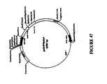
- RNA encoding the GGF2HBS5 protein was produced in an in vitro transcription system driven by the bacteriophage T7 promoter resident in the vector (Bluescript SK [Stratagene Inc.] see Fig. 47) containing the GGF2HBS5 insert. This RNA was translated in a cell free (rabbit reticulocyte) translation system and the size of the protein product was 45 Kd. Additionally, the cell-free product has been assayed in a Schwann cell mitogenic assay to confirm biological activity. Schwann cells treated with conditioned medium show both increased proliferation as measured by incorporation of 125 I-Uridine and phosphorylation on tyrosine of a protein in the 185 kilodalton range.
- GGF2HBS5 encodes the human equivalent of bovine GGF2.
- the GGF2HBS5 cDNA clone encoding human GGF2 was cloned into vector pcDL-SR ⁇ 296 and COS-7 cells were transfected in 100 mm dishes by the DEAE-dextran method. Cell lysates or conditioned media from transiently expressing COS cells were harvested at 3 or 4 days post-transfection. To prepare lysates, cell monolayers were washed with PBS, scraped from the dishes lysed by three freeze/thaw cycles in 150 ⁇ m of 0.25 M Tris-HCl, pH8. Cell debris was pelleted and the supernatant recovered.
- Conditioned media samples (7 mls.) were collected, then concentrated and buffer exchanged with 10 mm Tris, pH 7.4 using centiprep-10 and Centricon-10 units as described by the manufactures (Amicon, Beverly, MA). Rat nerve Schwann cells were assayed for incorporation of DNA synthesis precursors, as described. Conditioned media or cell lysate samples were tested in the Schwann cell proliferation assay as described in Marchionni et al., Nature 362:313 (1993). The cDNA, GGF2HBS5, encoding GGF2 directed the secretion of the protein product to the medium. Minimal activity was detectable inside the cells as determined by assays using cell lysates. GGF2HFB1 and GGFBPP5 cDNA's failed to direct the secretion of the product to the extracellular medium. GGF activity from these clones was detectable only in cell lysates.
- Recombinant GGF2 was also expressed in CHO cells.
- the GGF2HBS5 cDNA encoding GGF2 was cloned into the EcoRI site of vector pcdhfrpolyA and transfected into the DHFR negative CHO cell line (GG44) by the calcium phosphate coprecipitation method.
- Clones were selected in nucleotide and nucleoside free a medium (Gibco) in 96-well plates. After 3 weeks, conditioned media samples from individual clones were screened for expression of GGF by the Schwann cell proliferation assay as described in Marchionni et al., Nature 362:313 (1993). Stable clones which secreted significant levels of GGF activity into the medium were identified.
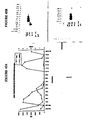
- Fig. 46 Schwann cell proliferation activity data from different volume aliquots of CHO cell conditioned medium were used to produce the dose response curve shown in Fig. 46 (Graham and Van Der Eb, Virology 52:456, 1973). This material was analyzed on a Western blot probed with polyclonal antisera raised against a GGF2 specific peptide. A band of approximately 65 Kd (the expected size of GGF2 extracted from pituitary) is specifically labeled (Fig. 48, lane 12).
- Recombinant GGF2 was also expressed in insect cells using the Baculovirus expression.
- Sf9 insect cells were infected with baculovirus containing the GGF2HBS5 cDNA clone at a multiplicity of 3-5 (10 6 cells/ml) and cultured in Sf900-II medium.
- Schwann cell mitogenic activity was secreted into the extracellular medium.
- Different volumes of insect cell conditioned medium were tested in the Schwann cell proliferation assay in the absence of forskolin and the data used to produce a dose response curve.
- Schwann cell mitogenic activity of recombinant human and bovine glial growth factors was determined as follows: Mitogenic responses of cultured Schwann cells were measured in the presence of 5 ⁇ M forskolin using crude recombinant GGF preparations obtained from transient mammalian expression experiments. Incorporation of [ 125 I]-Urd was determined following an 18-24 hour exposure to materials obtained from transfected or mock transfected cos cells as described in the Methods. The mean and standard deviation of four sets of data are shown. The mitogenic response to partially purified native bovine pituitary GGF (carboxymethyl cellulose fraction; Goodearl et al., submitted) is shown (GGF) as a standard of one hundred percent activity.
- GGF mitogenic response to partially purified native bovine pituitary GGF (carboxymethyl cellulose fraction; Goodearl et al., submitted) is shown (GGF) as a standard of one hundred percent activity.
- cDNAs (Fig. 46, SEQ ID NOs. 166-168) were cloned into pcDL-SR ⁇ 296 (Takebe et al., Mol. Cell Biol. 8:466-472 (1988)), and COS-7 cells were transfected in 100 mm dishes by the DEAE-dextran method (Sambrook et al., In Molecular Cloning. A Laboratory Manual, 2nd. ed. (Cold Spring Harbor Laboratory Press, Cold Spring Harbor, NY, 1989)). Cell lysates or conditioned media were harvested at 3 or 4 days post-transfection.
- the deduced structures of the family of GGF sequences indicate that the longest forms (as represented by GGF2BPP4) encode transmembrane proteins where the extracellular part contains a domain which resembles epidermal growth factor (see carpenter and Wahl in Peptide Growth Factors and Their Receptors I pp. 69-133, Springer-Verlag, NY 1991).
- the positions of the cysteine residues in coding segments C and C/D or C/D' peptide sequence are conserved with respect to the analogous residues in the epidermal growth factor (EGF) peptide sequence (see Fig. 32, SEQ ID Nos. 147-149). This suggests that the extracellular domain functions as receptor recognition and biological activation sites.
- GGF DNA sequences encoding polypeptides which encompass the EGF-like domain can have full biological activity for stimulating glial cell mitogenic activity.
- Membrane bound versions of this protein may induce Schwann cell proliferation if expressed on the surface of neurons during embryogenesis or during nerve regeneration (where the surfaces of neurons are intimately associated with the surfaces of proliferating Schwann cells).
- GGFs may act as classically diffusible factors which can interact with Schwann cells at some distance from their point of secretion. Other forms may be released from intracells by sources via tissue injury and cell disruption.
- An example of a secreted GGF is the protein encoded by GGF2HBS5; this is the only GGF known which has been found to be directed to the exterior of the cell. Secretion is probably mediated via an N-terminal hydrophobic sequence found only in region E, which is the N-terminal domain contained within recombinant GGF2 encoded by GGF2HBSS.
- GGF's appear to be non-secreted. These GGFs may be injury response forms which are released as a consequence of tissue damage.
- GGF2HBS5 Other regions of the predicted protein structure of GGF2 (encoded by GGF2HBS5) and other proteins containing regions B and A exhibit similarities to the human basement membrane heparan sulfate proteoglycan core protein.
- the peptide ADSGEY which is located next to the second cysteine of the C2 immunoglobulin fold in these GGF's, occurs in nine of twenty-two C-2 repeats found in that basal lamina protein. This evidence strongly suggests that these proteins may associate with matrix proteins such as those associated with neurons and glia, and may suggest a method for sequestration of glial growth factors at target sites.
- the proteins can be overproduced using cloned DNA.
- a recombinant E . coli cell containing the sequences described above can be constructed.
- Expression systems such as pNH8a or pHH16a (Stratagene, Inc.) can be used for this purpose by following manufacturers procedures.
- these sequences can be inserted in a mammalian expression vector and an overproducing cell line can be constructed.
- DNA encoding a GGF clone GGF2BPP5 has been expressed in COS cells and can be expressed in Chinese hamster ovary cells using the pMSXND expression vector (Lee and Nathans, J. Biol. Chem. 263, 3521-3527, (1981)).
- This vector containing GGF DNA sequences can be transfected into host cells using established procedures.
- Transient expression can be examined or G418-resistant clones can be grown in the presence of methotrexate to select for cells that amplify the dhfr gene (contained on the pMSXND vector) and, in the process, co-amplify the adjacent GGF protein encoding sequence. Because CHO cells can be maintained in a totally protein-free medium (Hamilton and Ham, In Vitro 13 , 537-547 (1977)), the desired protein can be purified from the medium. Western analysis using the antisera produced in Example 17 can be used to detect the presence of the desired protein in the conditioned medium of the overproducing cells.
- the desired protein was purified from the medium conditioned by transiently expressing cos cells as follows.
- rGGF II was harvested from the conditioned medium and partially purified using Cation Exchange Chromatography (POROS-HS).
- the column was equilibrated with 33.3 mM MES pH 6.0.
- Conditioned media was loaded at flow rate of 10 ml/min.
- the peak containing Schwann cell proliferation activity and immunoreactive (using the polyclonal antisera was against a GGF2 peptide described above) was eluted with 50 mM Tris, 1M NaCl pH 8.0.
- rhGGF2 is also expressed using a stable Chinese ovary Hamster cell line.
- rGGF2 from the harvested conditioned media was partially purified using Cation Exchange Chromatograph (POROS-HS). The column was equilibrated with PBS pH 7.4. Conditioned media was loaded at 10 ml/min. The peak containing the Schwann Cell Proliferative activity and immunoreactivity (using GGF2 polyclonal antisera) was eluted with 50 mM Hepes, 500 mM NaCl pH 8.0. An additional peak was observed at 50 mM Hepes, 1M NaCl pH 8.0 with both proliferation as well as immunoreactivity (Fig. 45).
- rhGGF2 can be further purified using Hydrophobic Interaction Chromatography as a high resolution step; Cation exchange/Reserve phase Chromatography (if needed as second high resolution step); A viral inactivation step and a DNA removal step such as Anion exchange chromatography.
- Schwann Cell Proliferation Activity of recombinant GGF2 peak eluted from the Cation Exchange column was determined as follows: Mitogenic responses of the cultured Schwann cells were measured in the presence of 5 M Forskolin using the peak eluted by 50 mM Tris 1 M NaCl pH 8.0. The peak was added at 20 1, 10 1 (1:10) 10 1 and (1:100) 10 1. Incorporation of 125 I-Uridine was determined and expressed as (CPM) following an 18-24 hour exposure.
- GGF2 purification on cation exchange columns was performed as follows: CHO cell conditioned media expressing rGGFII was loaded on the cation exchange column at 10 ml/min. The column was equilibrated with PBS pH 7.4. The elution was achieved with 50 mM Hepes 500 mM NaCl pH 8.0 and 50 mM Hepes 1M NaCl pH 8.0 respectively. All fractions were analyzed using the Schwann cell proliferation assay (CPM) described herein. The protein concentration (mg/ml) was determined by the Bradford assay using BSA as the standard.
- CPM Schwann cell proliferation assay
- the protein may be assayed at various points in the procedure using a Western blot assay.
- the Schwann cell mitogenic assay described herein may be used to assay the expressed product of the full length clone or any biologically active portions thereof.
- the full length clone GGF2BPP5 has been expressed transiently in COS cells. Intracellular extracts of transfected COS cells show biological activity when assayed in the Schwann cell proliferation assay described in Example 8.
- the full length close encoding GGF2HBS5 has been expressed transiently in COS cells. In this case both cell extract and conditioned media show biological activity in the Schwann cell proliferation assay described in Example 8.
- Any member of the family of splicing variant complementary DNA's derived from the GGF gene can be expressed in this manner and assayed in the Schwann cell proliferation assay by one skilled in the art.
- recombinant material may be isolated from other variants according to Wen et al. (Cell 69 :559 (1992)) who expressed the splicing variant Neu differentiation factor (NDF) in COS-7 cells.
- cDNA clones inserted in the pJT-2 eukaryotic plasmid vector are under the control of the SV40 early promoter, and are 3'-flanked with the SV40 termination and polyadenylation signals.
- COS-7 cells were transfected with the pJT-2 plasmid DNA by electroporation as follows: 6 x 10 6 cells (in 0.8 ml of DMEM and 10% FEBS) were transferred to a 0.4 cm cuvette and mixed with 20 ⁇ g of plasmid DNA in 10 ⁇ l of TE solution (10 mM Tris-HCl (pH 8.0), 1 mM EDTA). Electroporation was performed at room temperature at 1600 V and 25 ⁇ F using a Bio-Rad Gene Pulser apparatus with the pulse controller unit set at 200 ohms. The cells were then diluted into 20 ml of DMEM, 10% FBS and transferred into a T75 flask (Falcon). After 14 hr.
- Conditioned medium containing recombinant protein which was harvested from the cells demonstrated biological activity in a cell line expressing the receptor for this protein.
- This cell line (cultured human breast carcinoma cell line AU 565) was treated with recombinant material.
- the treated cells exhibited a morphology change which is characteristic of the activation of the erbB2 receptor.
- Conditioned medium of this type also can be tested in the Schwann cell proliferation assay.
- hGGF2 The cDNA encoding hGGF2 was cloned into the amplifiable vector pcdhfrpolyA and transfected into CHO-DG44 cells for stable expression.
- rhGGF2 is secreted into the conditioned media.
- the ability of the recombinant GGF2 to be secreted is presumably mediated through the N-terminal hydrophobic stretch (signal sequence).
- a signal sequence once having initiated export of a growing protein chain across the rough endoplasmic reticulum, is cleaved from the mature protein at a specific site. N-terminal sequence analysis of the expressed and purified rhGGF2 indicates the site of cleavage as shown below.
- N-terminal sequence analysis of rhGGF2 Cycle # Primary Sequence pMoles 1 Gly (G) 210.6 2 Asn (N) 163 3 GLU (E) 149 4 Ala (A) 220 5 Ala (A) 180 6 Pro (P) 173 7 Ala (A) 177 8 Gly (G) 154.9 9 Ala (A) 162.4 10 Ser (S) 65.4 11 Val (V) 132.7 12 Val (V) (Cys) 11.7 13 Tyr (Y) 112.7 14 Ser (S) 47.6 15 Ser (S) 27.1
- the following sequence represents the amino acid sequence of hGGF2.
- the shaded area indicates the cleaved signal sequence.
- the shaded area represents experimentally determined 15 amino acid residues at the N-terminal of the rhGGF2, indicating A 50 -G 51 bond to be the cleavage site for the signal sequence.
- the cDNA clone was obtained as follows (via the purification and sequencing of a p185 erbB2 binding protein from a transformed rat fibroblast cell line).
- a p185 erbB2 binding protein was purified from conditioned medium as follows. Pooled conditioned medium from three harvests of 500 roller bottles (120 liters total) was cleared by filtration through 0.2 ⁇ filters and concentrated 31-fold with a Pelicon ultrafiltration system using membranes with a 20kd molecular size cutoff. All the purification steps were performed by using a Pharmacia fast protein liquid chromatography system.
- the concentrated material was directly loaded on a column of heparin-Sepharose (150 ml, preequilibrated with phosphate-buffered saline (PBS)).
- PBS phosphate-buffered saline
- the column was washed with PBS containing 0.2 M NaCl until no absorbance at 280 nm wavelength could be detected.
- Bound proteins were then eluted with a continuous gradient (250 ml) of NaCl (from 0.2 M to 1.0 M), and 5 ml fractions were collected. Samples (0.01 ml of the collected fractions were used for the quantitative assay of the kinase stimulatory activity.
- the column was developed with a 45 ml gradient of (NH 4 ) 2 SO 4 (from 1.7 M to no salt) in 0.1 M Na 2 PO 4 (pH 7.4), and 2 ml fractions were collected and assayed (0.002 ml per sample) for kinase stimulation (as described in Example 19).
- the major peak of activity was pooled and dialyzed against 50 mM sodium phosphate buffer (pH 7.3).
- a Mono-S cation-exchange column HR5/5, Pharmacia was preequilibrated with 50 mM sodium phosphate. After loading the active material (0.884 mg of protein; 35 ml), the column was washed with the starting buffer and then developed at a rate of 1 ml/min.
- Samples from various steps of purification were analyzed by gel electrophoresis followed by silver staining using a kit from ICN (Costa Mesa, CA), and their protein contents were determined with a Coomassie blue dye binding assay using a kit from Bio-Rad (Richmond, CA).
- the p44 protein (10 ⁇ g) was reconstituted in 200 ⁇ l of 0.1 M ammonium bicarbonate buffer (pH 7.8). Digestion was conducted with L-1-tosyl-amide 2-phenylethyl chloromethyl ketone-treated trypsin (Serva) at 37°C for 18 hr. at an enzyme-to-substrate ratio of 1:10. The resulting peptide mixture was separated by reverse-phase HPLC and monitored at 215 nm using a Vydac C4 micro column (2.1 mm i.d. x 15 cm, 300 ⁇ ) and an HP 1090 liquid chromatographic system equipped with a diode-array detector and a workstation.
- Serva L-1-tosyl-amide 2-phenylethyl chloromethyl ketone-treated trypsin
- the column was equilibrated with 0.1% trifluoroacetic acid (mobile phase A), and elution was effected with a linear gradient from 0%-55% mobile phase B (90% acetonitrile in 0.1% trifluoroacetic acid) over 70 min.
- the flow rate was 0.2 ml/min. and the column temperature was controlled at 25°C.
- One-third aliquots of the peptide peaks collected manually from the HPLC system were characterized by N-terminal sequence analysis by Edman degradation. The fraction eluted after 27.7 min.
- T27.7 contained mixed amino acid sequences and was further rechromatographed after reduction as follows: A 70% aliquot of the peptide fraction was dried in vacuo and reconstituted in 100 ⁇ l of 0.2 M ammonium bicarbonate buffer (pH 7.8). DTT (final concentration 2 mM) was added to the solution, which was then incubated at 37°C for 30 min. The reduced peptide mixture was then separated by reverse-phase HPLC using a Vydac column (2.1 mm i.d. x 15 cm). Elution conditions and flow rat were identical to those described above.
- Amino acid sequence analysis of the peptide was performed with a Model 477 protein sequencer (Applied Biosystems, Inc., Foster City, CA) equipped with an on-line phenylthiohydantoin (PTH) amino acid analyzer and a Model 900 data analysis system (Hunkapiller et al. (1986) In Methods of Protein Microcharacterization , J.E. Shively, ed. (Clifton, New Jersey: Humana Press p. 223-247). The protein was loaded onto a trifluoroacetic acid-treated glass fiber disc precycled with polybrene and NaCl.
- the synthetic oligonucleotides were end-labeled with [ ⁇ - 32 P]ATP with T4 polynucleotide kinase and used to screen replicate sets of nitrocellulose filters.
- the hybridization solution contained 6 x SSC, 50 mM sodium phosphate (pH 6.8), 0.1% sodium pyrophosphate, 2 x Denhardt's solution, 50 ⁇ g/ml salmon sperm DNA, and 20% formamide (for probe 1) or no formamide (for probe 2).
- the filters were washed at either 50°C with 0.5 x SSC, 0.2% SDS, 2 mM EDTA (for probe 1) or at 37°C with 2 x SSC, 0.2% SDS, 2 mM EDTA (for probe 2). Autoradiography of the filters gave ten clones that hybridized with both probes. These clones were purified by replating and probe hybridization as described above.
- the cDNA clones were sequenced using an Applied Biosystems 373A automated DNA sequencer and Applied Biosystems Taq DyeDeoxyTM Terminator cycle sequencing kits following the manufacture's instructions. In some instances, sequences were obtained using [ 35 S]dATP (Amersham) and SequenaseTM kits from U.S.
- Lupu et al. (Proc. Natl. Acad. Sci. 89 , 2287 (1992)) purified another protein which binds to the p185 erbB2 receptor.
- This particular protein, p75 was purified from conditioned medium used for the growth of SKBr-3 (a human breast cancer cell line) propagated in improved Eagle's medium (IMEM: GIBCO) supplemented with 10% fetal bovine serum (GIBCO).
- ARIA a 42 kD protein which stimulates acetylcholine receptor synthesis, has been isolated in the laboratory of Gerald Fischbach (Falls et al., (1993) Cell 72:801-815).
- ARIA induces tyrosine phosphorylation of a 185 Kda muscle transmembrane protein which resembles p185 erbB2 , and stimulates acetylcholine receptor synthesis in cultured embryonic myotubes.
- ARIA is most likely a member of the GGF/erbB2 ligand group of proteins, and this is potentially useful in the glial cell mitogenesis stimulation and other applications of, e.g., GGF2 described herein.
- Rat Schwann cells following treatment with sufficient levels of Glial Growth Factor to induce proliferation, show stimulation of protein tyrosine phosphorylation.
- Varying amounts of partially purified GGF were applied to a primary culture of rat Schwann cells according to the procedure outlined in Example 9.
- Schwann cells were grown in DMEM/10% fetal calf serum/5 ⁇ M forskolin/0.5 ⁇ g per mL GGF-CM (0.5mL per well) in poly D-lysine coated 24 well plates. When confluent, the cells were fed with DMEM/10% fetal calf serum at 0.5mL per well and left in the incubator overnight to quiesce.
- the cells were fed with 0.2mL of DMEM/10% fetal calf serum and left in the incubator for 1 hour. Test samples were then added directly to the medium at different concentrations and for different lengths of time as required. The cells were then lysed in boiling lysis buffer (sodium phosphate, 5mM, pH 6.8; SDS, 2%, ⁇ -mercapteothanol, 5%; dithiothreitol, 0.1M; glycerol, 10%; Bromophenol Blue, 0.4%; sodium vanadate, 10mM), incubated in a boiling water bath for 10 minutes and then either analyzed directly or frozen at -70°C.
- boiling lysis buffer sodium phosphate, 5mM, pH 6.8; SDS, 2%, ⁇ -mercapteothanol, 5%; dithiothreitol, 0.1M; glycerol, 10%; Bromophenol Blue, 0.4%; sodium vanadate, 10mM
- the dose responses of protein phosphorylation and Schwann cell proliferation are very similar (Fig. 33).
- the molecular weight of the phosphorylated band is very close to the molecular weight of p185 erbB2 .
- Similar results were obtained when Schwann cells were treated with conditioned media prepared from COS cells translates with the GGF2HBS5 clone. These results correlate well with the expected interaction of the GGFs with and activation of 185 erbB2 .
- Conditioned medium derived from a CHO cell line stably transformed with the GGF2 clone stimulates protein tyrosine phosphorylation using the assay described above. Mock transfected CHO cells fail to stimulate this activity.
- the protein sequence predicted from the cDNA sequence of GGF-II candidate clones GGF2BPP1,2 and 3 contains a number of consensus N-glycosylation motifs.
- a gap in the GGFII02 peptide sequence coincides with the asparagine residue in one of these motifs, indicating that carbohydrate is probably bound at this site.
- N-glycosylation of the GGFs was studied by observing mobility changes on SDS-PAGE after incubation with N-glycanase, an enzyme that cleaves the covalent linkages between carbohydrate and aspargine residues in proteins.
- N-Glycanase treatment of GGF-II yielded a major band of MW 40-42 kDa and a minor band at 45-48 kDa.
- Activity single active deglycosylated species at ca 45-50 kDa.
Landscapes
- Health & Medical Sciences (AREA)
- Chemical & Material Sciences (AREA)
- Life Sciences & Earth Sciences (AREA)
- Organic Chemistry (AREA)
- General Health & Medical Sciences (AREA)
- Medicinal Chemistry (AREA)
- Engineering & Computer Science (AREA)
- Bioinformatics & Cheminformatics (AREA)
- Veterinary Medicine (AREA)
- Public Health (AREA)
- Animal Behavior & Ethology (AREA)
- Pharmacology & Pharmacy (AREA)
- Chemical Kinetics & Catalysis (AREA)
- Nuclear Medicine, Radiotherapy & Molecular Imaging (AREA)
- General Chemical & Material Sciences (AREA)
- Proteomics, Peptides & Aminoacids (AREA)
- Gastroenterology & Hepatology (AREA)
- Molecular Biology (AREA)
- Genetics & Genomics (AREA)
- Biophysics (AREA)
- Biochemistry (AREA)
- Zoology (AREA)
- Neurology (AREA)
- Physical Education & Sports Medicine (AREA)
- Orthopedic Medicine & Surgery (AREA)
- Immunology (AREA)
- Heart & Thoracic Surgery (AREA)
- Endocrinology (AREA)
- Epidemiology (AREA)
- Oncology (AREA)
- Cardiology (AREA)
- Toxicology (AREA)
- Vascular Medicine (AREA)
- Urology & Nephrology (AREA)
- Biomedical Technology (AREA)
- Neurosurgery (AREA)
- Medicines That Contain Protein Lipid Enzymes And Other Medicines (AREA)
- Peptides Or Proteins (AREA)
- Pharmaceuticals Containing Other Organic And Inorganic Compounds (AREA)
- Acyclic And Carbocyclic Compounds In Medicinal Compositions (AREA)
Claims (31)
- Verwendung eines Polypeptids für die Herstellung eines Medikaments zur Behandlung oder Prophylaxe einer Erkrankung oder Fehlfunktion der Muskulatur durch Erhöhen von Mitogenese, Wachstum, Differenzierung oder Überleben einer Muskelzelle, wobei das genannte Polypeptid oder die genannte Verbindung Rezeptoren bindet oder zweite Messenger-Systeme der genannten Muskelzelle aktiviert und wobei das genannte Polypeptid oder die genannte Verbindung ausgewählt ist aus der Gruppe bestehend aus:a) einem Polypeptid, umfassend die E-Sequenz, die durch die Nukleinsäuresequenz gemäß SEQ ID Nr: 133 oder 159 codiert ist;b) einem Polypeptid, umfassend die F-Sequenz, die durch die Nukleinsäuresequenz gemäß SEQ ID Nr. 132 codiert wird;c) einem Polypeptid, das durch die Formel YBAZCX definiert wird,
wobei YBAZCX aus den in Fig. 30 gezeigten Polypeptidsegmenten besteht (SEQ ID Nrn. 132-143, 156, 157, 159); wobei Y das Polypeptidsegment E umfasst oder fehlt; wobei Z das Polypeptidsegment G umfasst oder fehlt; und wobei X die Polypeptidsegmente C/D HKL, C/D H, C/D HL, C/D D, C/D' HL, C/D' HKL, C/D' H, C/D' D, C/D C/D' HKL, C/D C/D' H, C/D C/D' HL, C/D C/D' D, C/D D' H, C/D D' HL, C/D D' HKL, C/D' D' H, C/D' D' HL, C/D' D' HKL, C/D C/D' D' H, C/D C/D' D' HL oder C/D C/D' D' HKL umfasst;d) einem Polypeptid, das durch die Formel WBAZCX definiert wird,
wobei WBAZCX aus den in Fig. 30 gezeigten Polypeptidsegmenten besteht (SEQ ID Nrn. 132-143, 156, 157, 159); wobei W das Polypeptidsegment F umfasst oder fehlt; wobei Z das Polypeptidsegment G umfasst oder fehlt; und wobei X die Polypeptidsegmente C/D HKL, C/D H, C/D HL, C/D D, C/D' HL, C/D' HKL, C/D' H, C/D' D, C/D C/D' HKL, C/D C/D' H, C/D C/D' HL, C/D C/D' D, C/D D' H, C/D D' HL, C/D D' HKL, C/D' D' H, C/D' D' HL, C/D' D' HKL, C/D C/D' D' H, C/D C/D' D' HL oder C/D C/D' D' HKL umfasst;e) einem Polypeptidsegment, das spezifisch den p185erbB2 Rezeptor von Muskelzellen bindet und zweite Messenger-Systeme der genannten Muskelzellen aktiviert;f) einem Polypeptid, umfassend eine epidermale Wachstumsfaktor-ähnliche (EGF-ähnliche) Domäne, wobei die genannte epidermale Wachstumsfaktordomäne eine Aminosäuresequenz umfasst, die durch die Nukleinsäuresequenz codiert wird, die ausgewählt wird aus der Gruppe bestehend aus:i) SEQ ID Nr: 150 (EGFL1);ii) SEQ ID Nr: 151 (EGFL2);iii) SEQ ID Nr: 152 (EGFL3);iv) SEQ ID Nr: 153 (EGFL4);v) SEQ ID Nr: 154 (EGFL5); undvi) SEQ ID Nr: 155 (EGFL6);g) einem Polypeptid, ausgewählt aus der Gruppe bestehend aus:i) einem 35 kD Polypeptidfaktor, der von der transformierten I-EJ-Ratten-Fibroblast-Zelllinie isoliert wurde;ii) einem 75 kD Polypeptidfaktor, der von der humanen SKBR-3 Brustzelllinie isoliert wurde;iii) einem 44 kD Polypeptidfaktor, der von der transformierten 1-EJ-Ratten-Fibroblast-Zelllinie isoliert wurde;iv) einem 45 kD Polypeptidfaktor, der von der humanen MDA - MB 231 Brustzelllinie isoliert wurde;v) einem 7 bis 14 kD Polypeptidfaktor, der von der humanen ATL-2 T-Zelllinie isoliert wurde;vi) einem 25 kD Polypeptidfaktor, der von aktivierten peritonealen Mausmakrophagen isoliert wurde;vii) einem 25 kD Polypeptidfaktor, der von Rinderniere isoliert wurde;viii) ARIA-Polypeptid; undix) einem 46 bis 47 kD Polypeptidfaktor, der 0-2A Progenitor-Gliazellen stimuliert; undh) einem Polypeptid, das GGFIII umfasst. - Verwendung nach Anspruch 1, wobei das genannte Polypeptid die E-Sequenz umfasst, die durch die Nukleinsäuresequenz gemäß SEQ ID Nr: 133 oder 159 codiert wird.
- Verwendung nach Anspruch 1, wobei das genannte Polypeptid die F-Sequenz umfasst, die durch die Nukleinsäuresequenz gemäß SEQ ID Nr: 132 codiert wird.
- Verwendung nach Anspruch 1, wobei das genannte Polypeptid ein Polypeptid umfasst, das durch die Formel XBAZCX definiert wird,
wobei YBAZCX aus den in Fig. 30 gezeigten Polypeptidsegmenten besteht (SEQ ID Nrn. 132-143, 156, 157, 159); wobei Y das Polypeptidsegment E umfasst oder fehlt; wobei Z das Polypeptidsegment G umfasst oder fehlt; und wobei X die Polypeptidsegmente C/D HKL, C/D H, C/D HL, C/D D, C/D' HL, C/D' HKL, C/D' H, C/D' D, C/D C/D' HKL, C/D C/D' H, C/D C/D' HL, C/D C/D' D, C/D D' H, C/D D' HL, C/D D' HKL, C/D' D' H, C/D' D' HL, C/D' D' HKL, C/D C/D' D' H, C/D C/D' D' HL oder C/D C/D' D' HKL umfasst. - Verwendung nach Anspruch 1, wobei das genannte Polypeptid ein Polypeptid umfasst, das durch die Formel WBAZCX definiert wird,
wobei WBAZCX aus den in Fig. 30 gezeigten Polypeptidsegmenten besteht (SEQ ID Nrn. 132-143, 156, 157, 159); wobei W das Polypeptidsegment F umfasst oder fehlt; wobei Z das Polypeptidsegment G umfasst oder fehlt und wobei X die Polypeptidsegmente C/D HKL, C/D H, C/D HL, C/D D, C/D' HL, C/D' HKL, C/D' H, C/D' D, C/D C/D' HKL, C/D C/D' H, C/D C/D' HL, C/D C/D' D, C/D D' H, C/D D' HL, C/D D' HKL, C/D' D' H, C/D' D' HL, C/D' D' HKL, C/D C/D' D' H, C/D C/D' D' HL oder C/D C/D' D' HKL umfasst. - Verwendung nach Anspruch 1, wobei das genannte Polypeptid ein Polypeptid umfasst, das spezifisch den p185erbB2 Rezeptor von Muskelzellen bindet und zweite Messenger-Systeme von Muskelzellen aktiviert.
- Verwendung nach Anspruch 1, wobei das genannte Polypeptid eine epidermale Wachstumsfaktor-ähnliche (EGF-ähnliche) Domäne umfasst, wobei die genannte epidermale Wachstumsfaktordomäne eine Aminosäuresequenz umfasst, die durch die Nukleinsäuresequenz codiert wird, die ausgewählt ist aus der Gruppe bestehend aus:i) SEQ ID Nr: 150 (EGFL1);ii) SEQ ID Nr: 151 (EGFL2);iii) SEQ ID Nr: 152 (EGFL3);iv) SEQ ID Nr: 153 (EGFL4);v) SEQ ID Nr: 154 (EGFL5); undvi) SEQ ID Nr: 155 (EGFL6).
- Verwendung nach Anspruch 1, wobei das genannte Polypeptid ein Polypeptid umfasst, das ausgewählt ist aus der Gruppe bestehend aus:i) einem 35 kD Polypeptidfaktor, der von der transformierten 1-EJ-Ratten-Fibroblast-Zelllinie isoliert wurde;ii) einem 75 kD Polypeptidfaktor, der von der humanen SKBR-3 Brustzelllinie isoliert wurde;iii) einem 44 kD Polypeptidfaktor, der von der transformierten I-EJ-Ratten-Fibroblast-Zelllinie isoliert wurde;iv) einem 45 kD Polypeptidfaktor, der von der humanen MDA - MB 231 Brustzelllinie isoliert wurde;v) einem 7 bis 14 kD Polypeptidfaktor, der von der humanen ATL-2 T-Zelllinie isoliert wurde;vi) einem 25 kD Polypeptidfaktor, der von aktivierten peritonealen Mausmakrophagen isoliert wurde;vii) einem 25 kD Polypeptidfaktor, der von Rinderniere isoliert wurde;viii) ARIA-Polypeptid; undix) einem 46 bis 47 kD Polypeptidfaktor, der 0-2A Progenitor-Gliazellen stimuliert.
- Verwendung nach Anspruch 1, wobei das genannte Polypeptid GGFIII umfasst.
- Verwendung nach Anspruch 1, wobei das genannte Polypeptid die E-Sequenz umfasst, die SEQ ID Nr: 185 umfasst.
- Verwendung nach Anspruch 1, wobei das genannte, die E-Sequenz umfassende Polypeptid auch wenigstens einen Teil des Peptids umfasst, das durch die DNA-Sequenzen 5' und 3' zur E-codierenden Sequenz auf Klon pGGF2HBS5 codiert wird, hinterlegt bei der A.T.C.C. am 6. November 1992 (A.T.C.C.-Hinterlegungsnummer 75347).
- Verwendung des genannten Polypeptids, umfassend die E-Sequenz nach Anspruch 1 oder 2, wobei 50 N-terminale Aminosäuren von dem genannten Peptid abgespalten werden, das die genannte E-Sequenz umfasst.
- Verwendung nach Anspruch 1, wobei das genannte, die F-Sequenz umfassende Polypeptid die Polypeptidsegmente FBA mit den in Fig. 30 gezeigten Aminosäuresequenzen (SEQ ID Nrn. 132, 134, 135), die Polypeptidsegmente FBA' mit den in Fig. 30 gezeigten Aminosäuresequenzen (SEQ ID Nrn. 132, 134, 136), die Polypeptidsegmente FEBA mit den in Fig. 30 gezeigten Aminosäuresequenzen (SEQ ID Nrn. 132, 135, 159) oder die Polypeptidsegmente FEBA' mit den Aminosäuresequenzen umfasst, die den in Fig. 30 gezeigten Polypeptidsegmenten entsprechen (SEQ ID Nrn. 132-134, 136, 159).
- Verwendung nach Anspruch 2, wobei das genannte Polypeptid rekombinantes humanes GGF2 ist.
- Verwendung nach einem der Ansprüche 1 bis 5 oder 7 bis 14, wobei das genannte Polypeptid durch eine DNA-Sequenz in einem exprimierbaren genetischen Konstrukt codiert wird.
- Verwendung nach einem der Ansprüche 1 bis 15, wobei die genannte Behandlung oder Prophylaxe die eines pathophysiologischen Zustands der Muskulatur in einem Säugetier ist, in dem der genannte Zustand einen Muskelzellentyp involviert, der für das genannte Polypeptid sensitiv oder responsiv ist.
- Verwendung nach einem der Ansprüche 1 bis 15, wobei die genannte Behandlung oder Prophylaxe die eines Zustands ist, der Muskelschäden in einem Säugetier involviert.
- Verwendung nach einem der Ansprüche 1 bis 15, wobei die genannte Behandlung oder Prophylaxe die Minderung von Atrophie der genannten Muskelzellen ist.
- Verwendung nach einem der Ansprüche 1 bis 15, wobei die genannte Behandlung oder Prophylaxe die Erhöhung der Zahl der in dem genannten Säugetier vorhandenen Muskelfasern ist.
- Verwendung nach einem der Ansprüche 1 bis 15, wobei die Behandlung oder Prophylaxe die Steigerung der Muskelregeneration in dem genannten Säugetier ist.
- Verwendung nach Anspruch 1, wobei das genannte Polypeptid, das spezifisch den p185erbB2 Rezeptor von Muskelzellen bindet und zweite Messenger-Systeme der genannten Muskelzellen aktiviert, ein gliärer Wachstumsfaktor ist.
- Verwendung nach einem der Ansprüche 1 bis 15, wobei die genannte Muskelzelle ein Myoblast, eine Satellitenzelle, eine Muskelzelle im Skelettmuskel, eine Muskelzelle im Herzmuskel oder eine Muskelzelle in einem glatten Muskel ist.
- Verwendung nach einem der Ansprüche 1 bis 15, wobei die genannte Erkrankung oder Fehlfunktion eine Skelettmuskelerkrankung oder -fehlfunktion ist.
- Verwendung nach Anspruch 23, wobei die genannte Skelettmuskelerkrankung oder -fehlfunktion eine Myopathie oder eine Dystrophie ist.
- Verwendung nach Anspruch 24, wobei die genannte Dystrophie eine Duchennes-Muskeldystrophie oder eine Becker-Dystrophie ist.
- Verwendung nach einem der Ansprüche 1 bis 15, wobei die genannte Erkrankung oder Fehlfunktion eine Herzmuskelfehlfunktion ist.
- Verwendung nach Anspruch 26, wobei die genannte Herzmuskelfehlfunktion Kardiomyopathie, ein Ischämieschaden, eine kongenitale Muskelerkrankung oder ein Herztrauma ist.
- Verwendung nach einem der Ansprüche 1 bis 15, wobei die genannte Erkrankung oder Fehlfunktion eine Glattmuskelfehlfunktion ist.
- Verwendung nach Anspruch 28, wobei die genannte Glattmuskelfehlfunktion arterielle Sklerose, eine Gefäßverletzung oder eine kongenitale Gefäßerkrankung oder -fehlfunktion ist.
- Verwendung nach einem der Ansprüche 1 bis 15, wobei die genannte Muskelzelle unzureichende funktionelle Acetylcholinrezeptoren hat und eine Muskelzelle in einem Patienten mit Myasthenia gravis oder in einem Patienten mit einem Zustand ist, der Muskelschäden involviert.
- Verwendung nach einem der vorherigen Ansprüche, bei dem die Behandlung oder Prophylaxe bei einem Menschen ist.
Applications Claiming Priority (5)
| Application Number | Priority Date | Filing Date | Title |
|---|---|---|---|
| US5902293A | 1993-05-06 | 1993-05-06 | |
| US59022 | 1993-05-06 | ||
| US08/209,204 US7115554B1 (en) | 1993-05-06 | 1994-03-08 | Methods of increasing myotube formation or survival or muscle cell mitogenesis differentiation or survival using neuregulin GGF III |
| US209204 | 1994-03-08 | ||
| PCT/US1994/005083 WO1994026298A1 (en) | 1993-05-06 | 1994-05-06 | Methods for treating muscle diseases and disorders |
Publications (4)
| Publication Number | Publication Date |
|---|---|
| EP0703785A1 EP0703785A1 (de) | 1996-04-03 |
| EP0703785A4 EP0703785A4 (de) | 1998-12-30 |
| EP0703785B1 true EP0703785B1 (de) | 2005-07-20 |
| EP0703785B8 EP0703785B8 (de) | 2005-09-14 |
Family
ID=26738273
Family Applications (1)
| Application Number | Title | Priority Date | Filing Date |
|---|---|---|---|
| EP94916690A Expired - Lifetime EP0703785B8 (de) | 1993-05-06 | 1994-05-06 | Methoden zur behandlung von muskelerkrankungen und muskelstörungen |
Country Status (10)
| Country | Link |
|---|---|
| US (5) | US7115554B1 (de) |
| EP (1) | EP0703785B8 (de) |
| JP (3) | JP4035159B2 (de) |
| KR (2) | KR100284909B1 (de) |
| AT (1) | ATE299710T1 (de) |
| AU (1) | AU691810B2 (de) |
| CA (1) | CA2162262C (de) |
| DE (1) | DE69434431T2 (de) |
| ES (1) | ES2243928T3 (de) |
| WO (1) | WO1994026298A1 (de) |
Families Citing this family (40)
| Publication number | Priority date | Publication date | Assignee | Title |
|---|---|---|---|---|
| US7115554B1 (en) * | 1993-05-06 | 2006-10-03 | Acorda Therapeutics, Inc. | Methods of increasing myotube formation or survival or muscle cell mitogenesis differentiation or survival using neuregulin GGF III |
| US7037888B1 (en) * | 1992-04-03 | 2006-05-02 | Acorda Therapeutics, Inc. | Methods for treating muscle diseases and disorders |
| ES2293643T5 (es) | 1994-10-20 | 2012-02-02 | Institut Pasteur | Secuencias nucleotídicas de antígenos retrovíricos vih-1 del grupo (o subgrupo) o. |
| FR2731225B1 (fr) * | 1995-03-03 | 2003-10-31 | Pasteur Institut | Peptides de glycoproteine transmembranaire d'enveloppe et de proteine de capside du retrovirus humain du type hiv-1 et peptides presentant avec eux une parente immunologique |
| AU727606B2 (en) * | 1996-07-12 | 2000-12-14 | Genentech Inc. | Gamma-heregulin |
| DE69840699D1 (de) | 1997-02-10 | 2009-05-14 | Genentech Inc | Heregulin varianten |
| US6136558A (en) * | 1997-02-10 | 2000-10-24 | Genentech, Inc. | Heregulin variants |
| US20010023241A1 (en) | 1998-02-04 | 2001-09-20 | Sliwkowski Mark X. | Use of heregulin as a growth factor |
| WO2000035474A1 (en) * | 1998-12-11 | 2000-06-22 | Khurana Tejvir S | Neurite derived growth factors for use in the treatment of muscular dystrophy |
| AUPP785098A0 (en) | 1998-12-21 | 1999-01-21 | Victor Chang Cardiac Research Institute, The | Treatment of heart disease |
| US6967102B1 (en) * | 1999-03-11 | 2005-11-22 | University Of Manitoba | Nitric oxide manipulation of muscle satellite cell activation |
| US6635249B1 (en) | 1999-04-23 | 2003-10-21 | Cenes Pharmaceuticals, Inc. | Methods for treating congestive heart failure |
| US7495147B2 (en) | 2000-02-28 | 2009-02-24 | Decode Genetics Ehf. | Neuregulin-1 transgenic mouse and methods of use |
| US20040005578A1 (en) * | 2000-04-27 | 2004-01-08 | Yoji Yamada | Myocardial cell proliferation-associated genes |
| ES2610353T3 (es) * | 2000-05-23 | 2017-04-27 | Cenes Pharmaceuticals, Inc. | Moléculas de ácido nucleico NRG-2, polipéptidos y métodos diagnósticos y terapéuticos |
| AU2002304965A1 (en) | 2002-05-24 | 2003-12-12 | Zensun (Shanghai) Sci-Tech.Ltd | Neuregulin based methods and compositions for treating viral myocarditis and dilated cardiomyopathy |
| AU2003272805A1 (en) * | 2002-10-01 | 2004-04-23 | Peter K. Law | Bioactive implants |
| US20070213264A1 (en) | 2005-12-02 | 2007-09-13 | Mingdong Zhou | Neuregulin variants and methods of screening and using thereof |
| JP5738516B2 (ja) | 2005-12-30 | 2015-06-24 | ゼンサン (シャンハイ) サイエンス アンド テクノロジー リミテッド | 心機能改善のためのニューレグリンの徐放 |
| US8936806B2 (en) * | 2007-01-22 | 2015-01-20 | Children's Medical Center Corporation | Periostin induces proliferation of cardiomyocytes and promotes cardiac regeneration |
| RU2498309C2 (ru) * | 2007-05-10 | 2013-11-10 | Акорда Терапьютикс, Инк. | Способы выявления сердечных нарушений |
| JP5539221B2 (ja) | 2007-12-21 | 2014-07-02 | リガンド・ファーマシューティカルズ・インコーポレイテッド | 選択的アンドロゲン受容体モジュレーター(sarm)およびその使用 |
| US20110166068A1 (en) * | 2008-07-17 | 2011-07-07 | Acorda Therapeutics, Inc. | Therapeutic Dosing of a Neuregulin or a Subsequence Thereof for Treatment or Pro-phylaxis of Heart Failure |
| EP2328606B1 (de) | 2008-08-15 | 2016-10-05 | Acorda Therapeutics, Inc. | Zusammensetzungen und verfahren zur behandlung in nicht-akuten phasen nach einer neurologischen cns-verletzung |
| EP2808339B1 (de) | 2008-11-28 | 2017-02-15 | Zensun (Shanghai) Science & Technology, Co., Ltd. | Neuregulin-Peptide und deren Verwendung |
| WO2011011388A2 (en) * | 2009-07-22 | 2011-01-27 | Children's Medical Center Corporation | Neuregulin induced regeneraton of heart muscle muscle |
| WO2011147981A2 (en) * | 2010-05-28 | 2011-12-01 | Mind-Nrg Sa | Neuregulin isoforms, neuregulin polypeptides and uses thereof |
| WO2013053076A1 (en) | 2011-10-10 | 2013-04-18 | Zensun (Shanghai)Science & Technology Limited | Compositions and methods for treating heart failure |
| EP2999499B1 (de) | 2013-05-22 | 2019-07-17 | Zensun (Shanghai) Science & Technology, Co., Ltd. | Verlängerte freisetzung von neuregulin zur behandlung von herzinsuffizienz |
| KR102051433B1 (ko) * | 2013-08-27 | 2020-01-08 | (주)네오팜 | 근육 분화 촉진 및 근육 강화용 조성물 및 외용제 |
| CN104758922A (zh) | 2014-01-03 | 2015-07-08 | 上海泽生科技开发有限公司 | 纽兰格林制剂的配方 |
| WO2015108988A2 (en) | 2014-01-17 | 2015-07-23 | Ligand Pharmaceuticals, Inc. | Methods and compositions for modulating hormone levels |
| CN105497876B (zh) | 2014-09-24 | 2021-01-15 | 上海泽生科技开发股份有限公司 | 神经调节蛋白用于预防、治疗或延迟心脏室性心律失常的方法和组合物 |
| CN111407882A (zh) | 2014-10-17 | 2020-07-14 | 上海泽生科技开发股份有限公司 | 神经调节蛋白用于预防、治疗或延迟射血分数保留的心力衰竭的方法和组合物 |
| AR121035A1 (es) | 2019-04-01 | 2022-04-13 | Lilly Co Eli | Compuestos de neuregulina-4 y métodos de uso |
| KR20220003554A (ko) | 2019-04-19 | 2022-01-10 | 리간드 파마슈티칼스 인코포레이티드 | 화합물의 결정질 형태 및 결정질 형태를 제조하는 방법 |
| US20230057622A1 (en) | 2019-09-16 | 2023-02-23 | Zensun (Shanghai) Science & Technology, Co., Ltd. | Recombinant Human Neuregulin Derivatives and Use Thereof |
| CN113289002A (zh) | 2020-02-24 | 2021-08-24 | 上海泽生科技开发股份有限公司 | 神经调节蛋白用于预防、治疗或延缓心力衰竭的方法和组合物 |
| EP4351722A1 (de) | 2021-06-10 | 2024-04-17 | Amgen Inc. | Manipulierte nrg-1-varianten mit verbesserter selektivität gegenüber erbb4 aber nicht gegen erbb3 |
| CN118879713B (zh) * | 2024-07-17 | 2025-09-26 | 天津医科大学 | 一种人工设计的新型nrg-1在疾病治疗中的应用 |
Family Cites Families (14)
| Publication number | Priority date | Publication date | Assignee | Title |
|---|---|---|---|---|
| US4935341A (en) | 1986-06-04 | 1990-06-19 | Whitehead Institute For Biomedical Research | Detection of point mutations in neu genes |
| US4968603A (en) | 1986-12-31 | 1990-11-06 | The Regents Of The University Of California | Determination of status in neoplastic disease |
| WO1989006692A1 (en) | 1988-01-12 | 1989-07-27 | Genentech, Inc. | Method of treating tumor cells by inhibiting growth factor receptor function |
| US5143842A (en) * | 1988-11-01 | 1992-09-01 | The University Of Colorado Foundation, Inc. | Media for normal human muscle satellite cells |
| DK0474727T3 (da) | 1989-05-19 | 1998-01-12 | Genentech Inc | HER2 ekstracellulært domæne |
| DE69126430T2 (de) | 1990-04-06 | 1997-09-18 | Univ Pennsylvania | Ligand für das neu-gen-produkt |
| EP0597845A1 (de) | 1990-05-25 | 1994-05-25 | Georgetown University | Wachstumsfaktor der den wachstum von zellen, die das onkogen erbb-2 aus dem menschen überexpremieren, inhibiert |
| ES2157889T3 (es) | 1990-06-27 | 2001-09-01 | Biogen Inc | Linfotoxina complejada de superficie. |
| ATE182152T1 (de) | 1991-01-14 | 1999-07-15 | Univ Georgetown | Wachstumsfaktor-ligand, der an den erbb-2- rezeptor bindet und die zellulären reaktionen induziert |
| US7115554B1 (en) * | 1993-05-06 | 2006-10-03 | Acorda Therapeutics, Inc. | Methods of increasing myotube formation or survival or muscle cell mitogenesis differentiation or survival using neuregulin GGF III |
| US5530109A (en) * | 1991-04-10 | 1996-06-25 | Ludwig Institute For Cancer Research | DNA encoding glial mitogenic factors |
| US5367060A (en) | 1991-05-24 | 1994-11-22 | Genentech, Inc. | Structure, production and use of heregulin |
| GB9217316D0 (en) * | 1992-08-14 | 1992-09-30 | Ludwig Inst Cancer Res | Schwann cell mitogenic factor,its preparation and use |
| US6635249B1 (en) * | 1999-04-23 | 2003-10-21 | Cenes Pharmaceuticals, Inc. | Methods for treating congestive heart failure |
-
1994
- 1994-03-08 US US08/209,204 patent/US7115554B1/en not_active Expired - Lifetime
- 1994-05-06 KR KR1019950704903A patent/KR100284909B1/ko not_active Expired - Fee Related
- 1994-05-06 ES ES94916690T patent/ES2243928T3/es not_active Expired - Lifetime
- 1994-05-06 JP JP52559394A patent/JP4035159B2/ja not_active Expired - Lifetime
- 1994-05-06 EP EP94916690A patent/EP0703785B8/de not_active Expired - Lifetime
- 1994-05-06 WO PCT/US1994/005083 patent/WO1994026298A1/en not_active Ceased
- 1994-05-06 CA CA2162262A patent/CA2162262C/en not_active Expired - Lifetime
- 1994-05-06 AU AU68278/94A patent/AU691810B2/en not_active Expired
- 1994-05-06 AT AT94916690T patent/ATE299710T1/de not_active IP Right Cessation
- 1994-05-06 DE DE69434431T patent/DE69434431T2/de not_active Expired - Lifetime
-
1995
- 1995-06-06 US US08/467,602 patent/US6444642B1/en not_active Expired - Lifetime
- 1995-06-06 US US08/468,731 patent/US7384756B1/en not_active Expired - Lifetime
-
2000
- 2000-09-14 KR KR1020000054080A patent/KR100306244B1/ko not_active Expired - Fee Related
-
2003
- 2003-04-30 JP JP2003125885A patent/JP4018028B2/ja not_active Expired - Lifetime
-
2007
- 2007-08-03 JP JP2007203508A patent/JP2007332153A/ja active Pending
-
2008
- 2008-06-10 US US12/157,614 patent/US7718606B2/en not_active Expired - Fee Related
-
2010
- 2010-05-17 US US12/781,633 patent/US8026213B2/en not_active Expired - Fee Related
Also Published As
| Publication number | Publication date |
|---|---|
| AU6827894A (en) | 1994-12-12 |
| US6444642B1 (en) | 2002-09-03 |
| US20110144015A1 (en) | 2011-06-16 |
| US20090018073A1 (en) | 2009-01-15 |
| EP0703785A1 (de) | 1996-04-03 |
| EP0703785B8 (de) | 2005-09-14 |
| CA2162262C (en) | 2011-07-12 |
| KR100284909B1 (ko) | 2001-03-15 |
| CA2162262A1 (en) | 1994-11-24 |
| WO1994026298A1 (en) | 1994-11-24 |
| JP2004043437A (ja) | 2004-02-12 |
| US7115554B1 (en) | 2006-10-03 |
| DE69434431T2 (de) | 2006-05-24 |
| JP4035159B2 (ja) | 2008-01-16 |
| JP2007332153A (ja) | 2007-12-27 |
| KR100306244B1 (ko) | 2001-09-24 |
| US8026213B2 (en) | 2011-09-27 |
| ATE299710T1 (de) | 2005-08-15 |
| US7718606B2 (en) | 2010-05-18 |
| EP0703785A4 (de) | 1998-12-30 |
| JP4018028B2 (ja) | 2007-12-05 |
| JPH11507201A (ja) | 1999-06-29 |
| AU691810B2 (en) | 1998-05-28 |
| US7384756B1 (en) | 2008-06-10 |
| DE69434431D1 (de) | 2005-08-25 |
| KR960702318A (ko) | 1996-04-27 |
| ES2243928T3 (es) | 2005-12-01 |
Similar Documents
| Publication | Publication Date | Title |
|---|---|---|
| US7115554B1 (en) | Methods of increasing myotube formation or survival or muscle cell mitogenesis differentiation or survival using neuregulin GGF III | |
| AU709968B2 (en) | Glial mitogenic factors, their preparation and use | |
| EP0653940B1 (de) | Glialmitogene faktoren, ihre herstellung und verwendung | |
| WO1994026298A9 (en) | Methods for treating muscle diseases and disorders | |
| US7037888B1 (en) | Methods for treating muscle diseases and disorders | |
| AU693767B2 (en) | Inhibitors of cell proliferation, their preparation and use | |
| US7968678B2 (en) | Glial mitogenic factors, their preparation and use | |
| US7541338B2 (en) | Glial mitogenic factors, their preparation and use | |
| US6972280B2 (en) | Glial mitogenic factors, their preparation and use | |
| US7094749B1 (en) | Glial mitogenic factors, their preparation and use | |
| US20050106666A1 (en) | Glial mitogenic factors, their preparation and use |
Legal Events
| Date | Code | Title | Description |
|---|---|---|---|
| PUAI | Public reference made under article 153(3) epc to a published international application that has entered the european phase |
Free format text: ORIGINAL CODE: 0009012 |
|
| 17P | Request for examination filed |
Effective date: 19951205 |
|
| AK | Designated contracting states |
Kind code of ref document: A1 Designated state(s): AT BE CH DE DK ES FR GB GR IE IT LI LU MC NL PT SE |
|
| A4 | Supplementary search report drawn up and despatched |
Effective date: 19981113 |
|
| AK | Designated contracting states |
Kind code of ref document: A4 Designated state(s): AT BE CH DE DK ES FR GB GR IE IT LI LU MC NL PT SE |
|
| 17Q | First examination report despatched |
Effective date: 20000403 |
|
| GRAP | Despatch of communication of intention to grant a patent |
Free format text: ORIGINAL CODE: EPIDOSNIGR1 |
|
| RIC1 | Information provided on ipc code assigned before grant |
Ipc: 7A 61P 21/00 B Ipc: 7A 61K 38/16 A |
|
| GRAS | Grant fee paid |
Free format text: ORIGINAL CODE: EPIDOSNIGR3 |
|
| GRAA | (expected) grant |
Free format text: ORIGINAL CODE: 0009210 |
|
| AK | Designated contracting states |
Kind code of ref document: B1 Designated state(s): AT BE CH DE DK ES FR GB GR IE IT LI LU MC NL PT SE |
|
| PG25 | Lapsed in a contracting state [announced via postgrant information from national office to epo] |
Ref country code: NL Free format text: LAPSE BECAUSE OF FAILURE TO SUBMIT A TRANSLATION OF THE DESCRIPTION OR TO PAY THE FEE WITHIN THE PRESCRIBED TIME-LIMIT Effective date: 20050720 Ref country code: LI Free format text: LAPSE BECAUSE OF FAILURE TO SUBMIT A TRANSLATION OF THE DESCRIPTION OR TO PAY THE FEE WITHIN THE PRESCRIBED TIME-LIMIT Effective date: 20050720 Ref country code: CH Free format text: LAPSE BECAUSE OF FAILURE TO SUBMIT A TRANSLATION OF THE DESCRIPTION OR TO PAY THE FEE WITHIN THE PRESCRIBED TIME-LIMIT Effective date: 20050720 Ref country code: BE Free format text: LAPSE BECAUSE OF FAILURE TO SUBMIT A TRANSLATION OF THE DESCRIPTION OR TO PAY THE FEE WITHIN THE PRESCRIBED TIME-LIMIT Effective date: 20050720 Ref country code: AT Free format text: LAPSE BECAUSE OF FAILURE TO SUBMIT A TRANSLATION OF THE DESCRIPTION OR TO PAY THE FEE WITHIN THE PRESCRIBED TIME-LIMIT Effective date: 20050720 |
|
| REG | Reference to a national code |
Ref country code: GB Ref legal event code: FG4D |
|
| REG | Reference to a national code |
Ref country code: CH Ref legal event code: EP |
|
| RAP2 | Party data changed (patent owner data changed or rights of a patent transferred) |
Owner name: ACORDA THERAPEUTICS, INC. |
|
| REG | Reference to a national code |
Ref country code: IE Ref legal event code: FG4D |
|
| REF | Corresponds to: |
Ref document number: 69434431 Country of ref document: DE Date of ref document: 20050825 Kind code of ref document: P |
|
| NLT2 | Nl: modifications (of names), taken from the european patent patent bulletin |
Owner name: ACORDA THERAPEUTICS, INC. Effective date: 20050803 |
|
| PG25 | Lapsed in a contracting state [announced via postgrant information from national office to epo] |
Ref country code: SE Free format text: LAPSE BECAUSE OF FAILURE TO SUBMIT A TRANSLATION OF THE DESCRIPTION OR TO PAY THE FEE WITHIN THE PRESCRIBED TIME-LIMIT Effective date: 20051020 Ref country code: GR Free format text: LAPSE BECAUSE OF FAILURE TO SUBMIT A TRANSLATION OF THE DESCRIPTION OR TO PAY THE FEE WITHIN THE PRESCRIBED TIME-LIMIT Effective date: 20051020 Ref country code: DK Free format text: LAPSE BECAUSE OF FAILURE TO SUBMIT A TRANSLATION OF THE DESCRIPTION OR TO PAY THE FEE WITHIN THE PRESCRIBED TIME-LIMIT Effective date: 20051020 |
|
| REG | Reference to a national code |
Ref country code: ES Ref legal event code: FG2A Ref document number: 2243928 Country of ref document: ES Kind code of ref document: T3 |
|
| PG25 | Lapsed in a contracting state [announced via postgrant information from national office to epo] |
Ref country code: PT Free format text: LAPSE BECAUSE OF FAILURE TO SUBMIT A TRANSLATION OF THE DESCRIPTION OR TO PAY THE FEE WITHIN THE PRESCRIBED TIME-LIMIT Effective date: 20051221 |
|
| REG | Reference to a national code |
Ref country code: CH Ref legal event code: PL |
|
| NLV1 | Nl: lapsed or annulled due to failure to fulfill the requirements of art. 29p and 29m of the patents act | ||
| ET | Fr: translation filed | ||
| PG25 | Lapsed in a contracting state [announced via postgrant information from national office to epo] |
Ref country code: IE Free format text: LAPSE BECAUSE OF NON-PAYMENT OF DUE FEES Effective date: 20060508 |
|
| PLBE | No opposition filed within time limit |
Free format text: ORIGINAL CODE: 0009261 |
|
| STAA | Information on the status of an ep patent application or granted ep patent |
Free format text: STATUS: NO OPPOSITION FILED WITHIN TIME LIMIT |
|
| PG25 | Lapsed in a contracting state [announced via postgrant information from national office to epo] |
Ref country code: MC Free format text: LAPSE BECAUSE OF NON-PAYMENT OF DUE FEES Effective date: 20060531 |
|
| 26N | No opposition filed |
Effective date: 20060421 |
|
| PG25 | Lapsed in a contracting state [announced via postgrant information from national office to epo] |
Ref country code: LU Free format text: LAPSE BECAUSE OF NON-PAYMENT OF DUE FEES Effective date: 20060506 |
|
| PGFP | Annual fee paid to national office [announced via postgrant information from national office to epo] |
Ref country code: ES Payment date: 20120528 Year of fee payment: 19 |
|
| PGFP | Annual fee paid to national office [announced via postgrant information from national office to epo] |
Ref country code: GB Payment date: 20130528 Year of fee payment: 20 Ref country code: DE Payment date: 20130530 Year of fee payment: 20 |
|
| PGFP | Annual fee paid to national office [announced via postgrant information from national office to epo] |
Ref country code: FR Payment date: 20130606 Year of fee payment: 20 Ref country code: IT Payment date: 20130524 Year of fee payment: 20 |
|
| REG | Reference to a national code |
Ref country code: DE Ref legal event code: R071 Ref document number: 69434431 Country of ref document: DE |
|
| REG | Reference to a national code |
Ref country code: DE Ref legal event code: R071 Ref document number: 69434431 Country of ref document: DE |
|
| REG | Reference to a national code |
Ref country code: GB Ref legal event code: PE20 Expiry date: 20140505 |
|
| PG25 | Lapsed in a contracting state [announced via postgrant information from national office to epo] |
Ref country code: GB Free format text: LAPSE BECAUSE OF EXPIRATION OF PROTECTION Effective date: 20140505 |
|
| PG25 | Lapsed in a contracting state [announced via postgrant information from national office to epo] |
Ref country code: DE Free format text: LAPSE BECAUSE OF EXPIRATION OF PROTECTION Effective date: 20140507 |
|
| REG | Reference to a national code |
Ref country code: ES Ref legal event code: FD2A Effective date: 20140926 |
|
| PG25 | Lapsed in a contracting state [announced via postgrant information from national office to epo] |
Ref country code: ES Free format text: LAPSE BECAUSE OF EXPIRATION OF PROTECTION Effective date: 20140507 |