EP0707066A2 - Method for producing the SspI restriction endonuclease and methylase - Google Patents
Method for producing the SspI restriction endonuclease and methylase Download PDFInfo
- Publication number
- EP0707066A2 EP0707066A2 EP95307106A EP95307106A EP0707066A2 EP 0707066 A2 EP0707066 A2 EP 0707066A2 EP 95307106 A EP95307106 A EP 95307106A EP 95307106 A EP95307106 A EP 95307106A EP 0707066 A2 EP0707066 A2 EP 0707066A2
- Authority
- EP
- European Patent Office
- Prior art keywords
- dna
- ssp
- methylase
- endonuclease
- gene
- Prior art date
- Legal status (The legal status is an assumption and is not a legal conclusion. Google has not performed a legal analysis and makes no representation as to the accuracy of the status listed.)
- Granted
Links
- 108091008146 restriction endonucleases Proteins 0.000 title claims abstract description 61
- 238000004519 manufacturing process Methods 0.000 title description 4
- 238000010367 cloning Methods 0.000 claims abstract description 25
- 238000000034 method Methods 0.000 claims abstract description 18
- 239000013598 vector Substances 0.000 claims abstract description 17
- 108020004414 DNA Proteins 0.000 claims description 115
- 108010042407 Endonucleases Proteins 0.000 claims description 34
- 102000004533 Endonucleases Human genes 0.000 claims description 15
- 108020004511 Recombinant DNA Proteins 0.000 claims description 9
- 102000055027 Protein Methyltransferases Human genes 0.000 claims description 7
- 108700040121 Protein Methyltransferases Proteins 0.000 claims description 7
- 239000013599 cloning vector Substances 0.000 claims description 4
- 238000012258 culturing Methods 0.000 claims 1
- 108090000623 proteins and genes Proteins 0.000 abstract description 54
- 230000000694 effects Effects 0.000 abstract description 22
- 241001478894 Sphaerotilus Species 0.000 abstract description 20
- FAPWRFPIFSIZLT-UHFFFAOYSA-M Sodium chloride Chemical compound [Na+].[Cl-] FAPWRFPIFSIZLT-UHFFFAOYSA-M 0.000 description 70
- 210000004027 cell Anatomy 0.000 description 55
- TWRXJAOTZQYOKJ-UHFFFAOYSA-L Magnesium chloride Chemical compound [Mg+2].[Cl-].[Cl-] TWRXJAOTZQYOKJ-UHFFFAOYSA-L 0.000 description 54
- 239000000047 product Substances 0.000 description 39
- 239000011780 sodium chloride Substances 0.000 description 34
- QKNYBSVHEMOAJP-UHFFFAOYSA-N 2-amino-2-(hydroxymethyl)propane-1,3-diol;hydron;chloride Chemical compound Cl.OCC(N)(CO)CO QKNYBSVHEMOAJP-UHFFFAOYSA-N 0.000 description 31
- LFQSCWFLJHTTHZ-UHFFFAOYSA-N Ethanol Chemical compound CCO LFQSCWFLJHTTHZ-UHFFFAOYSA-N 0.000 description 28
- 239000000499 gel Substances 0.000 description 27
- 229910001629 magnesium chloride Inorganic materials 0.000 description 27
- HEDRZPFGACZZDS-UHFFFAOYSA-N Chloroform Chemical compound ClC(Cl)Cl HEDRZPFGACZZDS-UHFFFAOYSA-N 0.000 description 26
- HEMHJVSKTPXQMS-UHFFFAOYSA-M Sodium hydroxide Chemical compound [OH-].[Na+] HEMHJVSKTPXQMS-UHFFFAOYSA-M 0.000 description 24
- 239000013612 plasmid Substances 0.000 description 23
- 230000029087 digestion Effects 0.000 description 22
- 108010051943 endodeoxyribonuclease SspI Proteins 0.000 description 22
- 239000012634 fragment Substances 0.000 description 19
- KCXVZYZYPLLWCC-UHFFFAOYSA-N EDTA Chemical compound OC(=O)CN(CC(O)=O)CCN(CC(O)=O)CC(O)=O KCXVZYZYPLLWCC-UHFFFAOYSA-N 0.000 description 18
- 241000588724 Escherichia coli Species 0.000 description 18
- 238000012986 modification Methods 0.000 description 17
- 239000013615 primer Substances 0.000 description 17
- 239000000872 buffer Substances 0.000 description 16
- 239000008188 pellet Substances 0.000 description 16
- 238000007852 inverse PCR Methods 0.000 description 15
- 230000004048 modification Effects 0.000 description 14
- 102000004190 Enzymes Human genes 0.000 description 13
- 108090000790 Enzymes Proteins 0.000 description 13
- ISWSIDIOOBJBQZ-UHFFFAOYSA-N Phenol Chemical compound OC1=CC=CC=C1 ISWSIDIOOBJBQZ-UHFFFAOYSA-N 0.000 description 13
- 229960000723 ampicillin Drugs 0.000 description 13
- AVKUERGKIZMTKX-NJBDSQKTSA-N ampicillin Chemical compound C1([C@@H](N)C(=O)N[C@H]2[C@H]3SC([C@@H](N3C2=O)C(O)=O)(C)C)=CC=CC=C1 AVKUERGKIZMTKX-NJBDSQKTSA-N 0.000 description 13
- 229940088598 enzyme Drugs 0.000 description 13
- KFZMGEQAYNKOFK-UHFFFAOYSA-N Isopropanol Chemical class CC(C)O KFZMGEQAYNKOFK-UHFFFAOYSA-N 0.000 description 12
- 238000005119 centrifugation Methods 0.000 description 12
- 239000011543 agarose gel Substances 0.000 description 11
- 238000002105 Southern blotting Methods 0.000 description 10
- 239000007983 Tris buffer Substances 0.000 description 10
- 239000000203 mixture Substances 0.000 description 10
- 108020004707 nucleic acids Proteins 0.000 description 10
- 102000039446 nucleic acids Human genes 0.000 description 10
- 150000007523 nucleic acids Chemical class 0.000 description 10
- 102000004169 proteins and genes Human genes 0.000 description 10
- 239000000523 sample Substances 0.000 description 10
- 239000000243 solution Substances 0.000 description 10
- LENZDBCJOHFCAS-UHFFFAOYSA-N tris Chemical compound OCC(N)(CO)CO LENZDBCJOHFCAS-UHFFFAOYSA-N 0.000 description 10
- 108091028043 Nucleic acid sequence Proteins 0.000 description 9
- DBMJMQXJHONAFJ-UHFFFAOYSA-M Sodium laurylsulphate Chemical compound [Na+].CCCCCCCCCCCCOS([O-])(=O)=O DBMJMQXJHONAFJ-UHFFFAOYSA-M 0.000 description 9
- BPHPUYQFMNQIOC-NXRLNHOXSA-N isopropyl beta-D-thiogalactopyranoside Chemical compound CC(C)S[C@@H]1O[C@H](CO)[C@H](O)[C@H](O)[C@H]1O BPHPUYQFMNQIOC-NXRLNHOXSA-N 0.000 description 9
- 239000006228 supernatant Substances 0.000 description 9
- CSNNHWWHGAXBCP-UHFFFAOYSA-L Magnesium sulfate Chemical compound [Mg+2].[O-][S+2]([O-])([O-])[O-] CSNNHWWHGAXBCP-UHFFFAOYSA-L 0.000 description 8
- 239000000020 Nitrocellulose Substances 0.000 description 8
- AIYUHDOJVYHVIT-UHFFFAOYSA-M caesium chloride Chemical compound [Cl-].[Cs+] AIYUHDOJVYHVIT-UHFFFAOYSA-M 0.000 description 8
- 238000012217 deletion Methods 0.000 description 8
- 230000037430 deletion Effects 0.000 description 8
- 229920001220 nitrocellulos Polymers 0.000 description 8
- SCVFZCLFOSHCOH-UHFFFAOYSA-M potassium acetate Chemical compound [K+].CC([O-])=O SCVFZCLFOSHCOH-UHFFFAOYSA-M 0.000 description 8
- 241000894006 Bacteria Species 0.000 description 7
- 102000012410 DNA Ligases Human genes 0.000 description 7
- 108010061982 DNA Ligases Proteins 0.000 description 7
- 210000004899 c-terminal region Anatomy 0.000 description 7
- 238000002360 preparation method Methods 0.000 description 7
- 238000006243 chemical reaction Methods 0.000 description 6
- ZPWVASYFFYYZEW-UHFFFAOYSA-L dipotassium hydrogen phosphate Chemical compound [K+].[K+].OP([O-])([O-])=O ZPWVASYFFYYZEW-UHFFFAOYSA-L 0.000 description 6
- 229910000396 dipotassium phosphate Inorganic materials 0.000 description 6
- 235000019797 dipotassium phosphate Nutrition 0.000 description 6
- 239000000284 extract Substances 0.000 description 6
- 229920001817 Agar Polymers 0.000 description 5
- 101710110830 Beta-agarase Proteins 0.000 description 5
- 108010044289 DNA Restriction-Modification Enzymes Proteins 0.000 description 5
- 102000006465 DNA Restriction-Modification Enzymes Human genes 0.000 description 5
- 239000008272 agar Substances 0.000 description 5
- 125000003275 alpha amino acid group Chemical group 0.000 description 5
- 230000001580 bacterial effect Effects 0.000 description 5
- OPTASPLRGRRNAP-UHFFFAOYSA-N cytosine Chemical compound NC=1C=CNC(=O)N=1 OPTASPLRGRRNAP-UHFFFAOYSA-N 0.000 description 5
- 238000013507 mapping Methods 0.000 description 5
- 238000012216 screening Methods 0.000 description 5
- 229920000936 Agarose Polymers 0.000 description 4
- USFZMSVCRYTOJT-UHFFFAOYSA-N Ammonium acetate Chemical compound N.CC(O)=O USFZMSVCRYTOJT-UHFFFAOYSA-N 0.000 description 4
- 102000003960 Ligases Human genes 0.000 description 4
- 108090000364 Ligases Proteins 0.000 description 4
- 101000865057 Thermococcus litoralis DNA polymerase Proteins 0.000 description 4
- 229920004890 Triton X-100 Polymers 0.000 description 4
- 239000013504 Triton X-100 Substances 0.000 description 4
- 229960005542 ethidium bromide Drugs 0.000 description 4
- 238000011534 incubation Methods 0.000 description 4
- 229910052943 magnesium sulfate Inorganic materials 0.000 description 4
- 235000019341 magnesium sulphate Nutrition 0.000 description 4
- 235000011056 potassium acetate Nutrition 0.000 description 4
- 238000000746 purification Methods 0.000 description 4
- 238000011160 research Methods 0.000 description 4
- 241000894007 species Species 0.000 description 4
- 238000012546 transfer Methods 0.000 description 4
- OPIFSICVWOWJMJ-AEOCFKNESA-N 5-bromo-4-chloro-3-indolyl beta-D-galactoside Chemical compound O[C@@H]1[C@@H](O)[C@@H](O)[C@@H](CO)O[C@H]1OC1=CNC2=CC=C(Br)C(Cl)=C12 OPIFSICVWOWJMJ-AEOCFKNESA-N 0.000 description 3
- GFFGJBXGBJISGV-UHFFFAOYSA-N Adenine Chemical compound NC1=NC=NC2=C1N=CN2 GFFGJBXGBJISGV-UHFFFAOYSA-N 0.000 description 3
- 102000013381 DNA Modification Methylases Human genes 0.000 description 3
- 108010090738 DNA Modification Methylases Proteins 0.000 description 3
- WQZGKKKJIJFFOK-GASJEMHNSA-N Glucose Natural products OC[C@H]1OC(O)[C@H](O)[C@@H](O)[C@@H]1O WQZGKKKJIJFFOK-GASJEMHNSA-N 0.000 description 3
- 239000006142 Luria-Bertani Agar Substances 0.000 description 3
- 102000016943 Muramidase Human genes 0.000 description 3
- 108010014251 Muramidase Proteins 0.000 description 3
- 108010062010 N-Acetylmuramoyl-L-alanine Amidase Proteins 0.000 description 3
- 239000004098 Tetracycline Substances 0.000 description 3
- BFNBIHQBYMNNAN-UHFFFAOYSA-N ammonium sulfate Chemical compound N.N.OS(O)(=O)=O BFNBIHQBYMNNAN-UHFFFAOYSA-N 0.000 description 3
- 229910052921 ammonium sulfate Inorganic materials 0.000 description 3
- 235000011130 ammonium sulphate Nutrition 0.000 description 3
- 238000013459 approach Methods 0.000 description 3
- 238000004113 cell culture Methods 0.000 description 3
- SUYVUBYJARFZHO-RRKCRQDMSA-N dATP Chemical compound C1=NC=2C(N)=NC=NC=2N1[C@H]1C[C@H](O)[C@@H](COP(O)(=O)OP(O)(=O)OP(O)(O)=O)O1 SUYVUBYJARFZHO-RRKCRQDMSA-N 0.000 description 3
- SUYVUBYJARFZHO-UHFFFAOYSA-N dATP Natural products C1=NC=2C(N)=NC=NC=2N1C1CC(O)C(COP(O)(=O)OP(O)(=O)OP(O)(O)=O)O1 SUYVUBYJARFZHO-UHFFFAOYSA-N 0.000 description 3
- ZMMJGEGLRURXTF-UHFFFAOYSA-N ethidium bromide Chemical compound [Br-].C12=CC(N)=CC=C2C2=CC=C(N)C=C2[N+](CC)=C1C1=CC=CC=C1 ZMMJGEGLRURXTF-UHFFFAOYSA-N 0.000 description 3
- 239000008103 glucose Substances 0.000 description 3
- 238000009396 hybridization Methods 0.000 description 3
- 208000015181 infectious disease Diseases 0.000 description 3
- 239000004325 lysozyme Substances 0.000 description 3
- 229960000274 lysozyme Drugs 0.000 description 3
- 235000010335 lysozyme Nutrition 0.000 description 3
- 230000011987 methylation Effects 0.000 description 3
- 238000007069 methylation reaction Methods 0.000 description 3
- 239000002773 nucleotide Substances 0.000 description 3
- 125000003729 nucleotide group Chemical group 0.000 description 3
- 125000002924 primary amino group Chemical group [H]N([H])* 0.000 description 3
- 238000000734 protein sequencing Methods 0.000 description 3
- 229960002180 tetracycline Drugs 0.000 description 3
- 229930101283 tetracycline Natural products 0.000 description 3
- 235000019364 tetracycline Nutrition 0.000 description 3
- 150000003522 tetracyclines Chemical class 0.000 description 3
- PIEPQKCYPFFYMG-UHFFFAOYSA-N tris acetate Chemical compound CC(O)=O.OCC(N)(CO)CO PIEPQKCYPFFYMG-UHFFFAOYSA-N 0.000 description 3
- QTBSBXVTEAMEQO-UHFFFAOYSA-M Acetate Chemical compound CC([O-])=O QTBSBXVTEAMEQO-UHFFFAOYSA-M 0.000 description 2
- 229930024421 Adenine Natural products 0.000 description 2
- 238000001712 DNA sequencing Methods 0.000 description 2
- 102000016928 DNA-directed DNA polymerase Human genes 0.000 description 2
- 108010014303 DNA-directed DNA polymerase Proteins 0.000 description 2
- 229920001917 Ficoll Polymers 0.000 description 2
- 102000004160 Phosphoric Monoester Hydrolases Human genes 0.000 description 2
- 108090000608 Phosphoric Monoester Hydrolases Proteins 0.000 description 2
- 108010021757 Polynucleotide 5'-Hydroxyl-Kinase Proteins 0.000 description 2
- 102000008422 Polynucleotide 5'-hydroxyl-kinase Human genes 0.000 description 2
- 230000027151 SOS response Effects 0.000 description 2
- VMHLLURERBWHNL-UHFFFAOYSA-M Sodium acetate Chemical compound [Na+].CC([O-])=O VMHLLURERBWHNL-UHFFFAOYSA-M 0.000 description 2
- 229960000643 adenine Drugs 0.000 description 2
- 238000000246 agarose gel electrophoresis Methods 0.000 description 2
- 230000003321 amplification Effects 0.000 description 2
- 244000309466 calf Species 0.000 description 2
- 150000001720 carbohydrates Chemical class 0.000 description 2
- 235000014633 carbohydrates Nutrition 0.000 description 2
- 229940104302 cytosine Drugs 0.000 description 2
- HAAZLUGHYHWQIW-KVQBGUIXSA-N dGTP Chemical compound C1=NC=2C(=O)NC(N)=NC=2N1[C@H]1C[C@H](O)[C@@H](COP(O)(=O)OP(O)(=O)OP(O)(O)=O)O1 HAAZLUGHYHWQIW-KVQBGUIXSA-N 0.000 description 2
- NHVNXKFIZYSCEB-XLPZGREQSA-N dTTP Chemical compound O=C1NC(=O)C(C)=CN1[C@@H]1O[C@H](COP(O)(=O)OP(O)(=O)OP(O)(O)=O)[C@@H](O)C1 NHVNXKFIZYSCEB-XLPZGREQSA-N 0.000 description 2
- 230000006378 damage Effects 0.000 description 2
- 229960000633 dextran sulfate Drugs 0.000 description 2
- 238000000502 dialysis Methods 0.000 description 2
- 238000010353 genetic engineering Methods 0.000 description 2
- 239000011521 glass Substances 0.000 description 2
- 230000001939 inductive effect Effects 0.000 description 2
- 238000003199 nucleic acid amplification method Methods 0.000 description 2
- 229940080469 phosphocellulose Drugs 0.000 description 2
- 229920003023 plastic Polymers 0.000 description 2
- 239000001267 polyvinylpyrrolidone Substances 0.000 description 2
- 229920000036 polyvinylpyrrolidone Polymers 0.000 description 2
- 235000013855 polyvinylpyrrolidone Nutrition 0.000 description 2
- 239000011148 porous material Substances 0.000 description 2
- 239000002244 precipitate Substances 0.000 description 2
- 230000037452 priming Effects 0.000 description 2
- 230000000644 propagated effect Effects 0.000 description 2
- 230000001681 protective effect Effects 0.000 description 2
- 239000001632 sodium acetate Substances 0.000 description 2
- 235000017281 sodium acetate Nutrition 0.000 description 2
- 239000001509 sodium citrate Substances 0.000 description 2
- NLJMYIDDQXHKNR-UHFFFAOYSA-K sodium citrate Chemical compound O.O.[Na+].[Na+].[Na+].[O-]C(=O)CC(O)(CC([O-])=O)C([O-])=O NLJMYIDDQXHKNR-UHFFFAOYSA-K 0.000 description 2
- 238000009987 spinning Methods 0.000 description 2
- 241001515965 unidentified phage Species 0.000 description 2
- 241000283690 Bos taurus Species 0.000 description 1
- 108091003079 Bovine Serum Albumin Proteins 0.000 description 1
- 108010072454 CTGCAG-specific type II deoxyribonucleases Proteins 0.000 description 1
- 102000053602 DNA Human genes 0.000 description 1
- 102000004594 DNA Polymerase I Human genes 0.000 description 1
- 108010017826 DNA Polymerase I Proteins 0.000 description 1
- 230000005778 DNA damage Effects 0.000 description 1
- 231100000277 DNA damage Toxicity 0.000 description 1
- 239000003155 DNA primer Substances 0.000 description 1
- 102000007260 Deoxyribonuclease I Human genes 0.000 description 1
- 108010008532 Deoxyribonuclease I Proteins 0.000 description 1
- 102100021587 Embryonic testis differentiation protein homolog A Human genes 0.000 description 1
- 108010067770 Endopeptidase K Proteins 0.000 description 1
- 101100063439 Escherichia coli (strain K12) dinD gene Proteins 0.000 description 1
- 108060002716 Exonuclease Proteins 0.000 description 1
- 101000898120 Homo sapiens Embryonic testis differentiation protein homolog A Proteins 0.000 description 1
- 239000007836 KH2PO4 Substances 0.000 description 1
- 101710192606 Latent membrane protein 2 Proteins 0.000 description 1
- 239000006137 Luria-Bertani broth Substances 0.000 description 1
- 108091034117 Oligonucleotide Proteins 0.000 description 1
- 108020005187 Oligonucleotide Probes Proteins 0.000 description 1
- 206010034759 Petit mal epilepsy Diseases 0.000 description 1
- 102000006382 Ribonucleases Human genes 0.000 description 1
- 108010083644 Ribonucleases Proteins 0.000 description 1
- 229920002684 Sepharose Polymers 0.000 description 1
- 241000815015 Sphaerotilus sp. Species 0.000 description 1
- 101710109576 Terminal protein Proteins 0.000 description 1
- 241000700605 Viruses Species 0.000 description 1
- 230000002411 adverse Effects 0.000 description 1
- 238000001042 affinity chromatography Methods 0.000 description 1
- 238000004458 analytical method Methods 0.000 description 1
- 239000003242 anti bacterial agent Substances 0.000 description 1
- 229940088710 antibiotic agent Drugs 0.000 description 1
- 239000007864 aqueous solution Substances 0.000 description 1
- 238000003556 assay Methods 0.000 description 1
- 108010005774 beta-Galactosidase Proteins 0.000 description 1
- 230000003115 biocidal effect Effects 0.000 description 1
- 229940098773 bovine serum albumin Drugs 0.000 description 1
- 229910052792 caesium Inorganic materials 0.000 description 1
- TVFDJXOCXUVLDH-UHFFFAOYSA-N caesium atom Chemical compound [Cs] TVFDJXOCXUVLDH-UHFFFAOYSA-N 0.000 description 1
- 229940041514 candida albicans extract Drugs 0.000 description 1
- 230000015556 catabolic process Effects 0.000 description 1
- 239000006285 cell suspension Substances 0.000 description 1
- 108091092356 cellular DNA Proteins 0.000 description 1
- 230000008859 change Effects 0.000 description 1
- 229960005091 chloramphenicol Drugs 0.000 description 1
- WIIZWVCIJKGZOK-RKDXNWHRSA-N chloramphenicol Chemical compound ClC(Cl)C(=O)N[C@H](CO)[C@H](O)C1=CC=C([N+]([O-])=O)C=C1 WIIZWVCIJKGZOK-RKDXNWHRSA-N 0.000 description 1
- 230000002759 chromosomal effect Effects 0.000 description 1
- 238000003776 cleavage reaction Methods 0.000 description 1
- 238000007398 colorimetric assay Methods 0.000 description 1
- 230000000295 complement effect Effects 0.000 description 1
- 239000000470 constituent Substances 0.000 description 1
- 238000010276 construction Methods 0.000 description 1
- RGWHQCVHVJXOKC-SHYZEUOFSA-J dCTP(4-) Chemical compound O=C1N=C(N)C=CN1[C@@H]1O[C@H](COP([O-])(=O)OP([O-])(=O)OP([O-])([O-])=O)[C@@H](O)C1 RGWHQCVHVJXOKC-SHYZEUOFSA-J 0.000 description 1
- 230000002950 deficient Effects 0.000 description 1
- 238000006731 degradation reaction Methods 0.000 description 1
- 230000001419 dependent effect Effects 0.000 description 1
- 238000013461 design Methods 0.000 description 1
- 238000001514 detection method Methods 0.000 description 1
- 238000010790 dilution Methods 0.000 description 1
- 239000012895 dilution Substances 0.000 description 1
- 238000001962 electrophoresis Methods 0.000 description 1
- IDGUHHHQCWSQLU-UHFFFAOYSA-N ethanol;hydrate Chemical compound O.CCO IDGUHHHQCWSQLU-UHFFFAOYSA-N 0.000 description 1
- 102000013165 exonuclease Human genes 0.000 description 1
- 239000013604 expression vector Substances 0.000 description 1
- 239000012530 fluid Substances 0.000 description 1
- 230000004927 fusion Effects 0.000 description 1
- 238000012252 genetic analysis Methods 0.000 description 1
- 230000002068 genetic effect Effects 0.000 description 1
- 229940025294 hemin Drugs 0.000 description 1
- BTIJJDXEELBZFS-QDUVMHSLSA-K hemin Chemical compound CC1=C(CCC(O)=O)C(C=C2C(CCC(O)=O)=C(C)\C(N2[Fe](Cl)N23)=C\4)=N\C1=C/C2=C(C)C(C=C)=C3\C=C/1C(C)=C(C=C)C/4=N\1 BTIJJDXEELBZFS-QDUVMHSLSA-K 0.000 description 1
- 238000000338 in vitro Methods 0.000 description 1
- 230000006698 induction Effects 0.000 description 1
- 230000000968 intestinal effect Effects 0.000 description 1
- 238000004255 ion exchange chromatography Methods 0.000 description 1
- 229960000318 kanamycin Drugs 0.000 description 1
- 229930027917 kanamycin Natural products 0.000 description 1
- SBUJHOSQTJFQJX-NOAMYHISSA-N kanamycin Chemical compound O[C@@H]1[C@@H](O)[C@H](O)[C@@H](CN)O[C@@H]1O[C@H]1[C@H](O)[C@@H](O[C@@H]2[C@@H]([C@@H](N)[C@H](O)[C@@H](CO)O2)O)[C@H](N)C[C@@H]1N SBUJHOSQTJFQJX-NOAMYHISSA-N 0.000 description 1
- 229930182823 kanamycin A Natural products 0.000 description 1
- 101150066555 lacZ gene Proteins 0.000 description 1
- UEGPKNKPLBYCNK-UHFFFAOYSA-L magnesium acetate Chemical compound [Mg+2].CC([O-])=O.CC([O-])=O UEGPKNKPLBYCNK-UHFFFAOYSA-L 0.000 description 1
- 239000011654 magnesium acetate Substances 0.000 description 1
- 235000011285 magnesium acetate Nutrition 0.000 description 1
- 229940069446 magnesium acetate Drugs 0.000 description 1
- 239000002609 medium Substances 0.000 description 1
- 125000002496 methyl group Chemical group [H]C([H])([H])* 0.000 description 1
- 238000002156 mixing Methods 0.000 description 1
- 229910000402 monopotassium phosphate Inorganic materials 0.000 description 1
- 235000019796 monopotassium phosphate Nutrition 0.000 description 1
- 239000002751 oligonucleotide probe Substances 0.000 description 1
- 238000007747 plating Methods 0.000 description 1
- 229920002401 polyacrylamide Polymers 0.000 description 1
- GNSKLFRGEWLPPA-UHFFFAOYSA-M potassium dihydrogen phosphate Chemical compound [K+].OP(O)([O-])=O GNSKLFRGEWLPPA-UHFFFAOYSA-M 0.000 description 1
- 239000011541 reaction mixture Substances 0.000 description 1
- 239000011369 resultant mixture Substances 0.000 description 1
- 230000000717 retained effect Effects 0.000 description 1
- 238000012552 review Methods 0.000 description 1
- 239000007320 rich medium Substances 0.000 description 1
- 230000007017 scission Effects 0.000 description 1
- 239000006152 selective media Substances 0.000 description 1
- 230000035939 shock Effects 0.000 description 1
- 238000002415 sodium dodecyl sulfate polyacrylamide gel electrophoresis Methods 0.000 description 1
- 238000000527 sonication Methods 0.000 description 1
- 239000011550 stock solution Substances 0.000 description 1
- 230000004083 survival effect Effects 0.000 description 1
- 239000000725 suspension Substances 0.000 description 1
- 230000009466 transformation Effects 0.000 description 1
- 239000012137 tryptone Substances 0.000 description 1
- 239000012138 yeast extract Substances 0.000 description 1
- DGVVWUTYPXICAM-UHFFFAOYSA-N β‐Mercaptoethanol Chemical compound OCCS DGVVWUTYPXICAM-UHFFFAOYSA-N 0.000 description 1
Images







Classifications
-
- C—CHEMISTRY; METALLURGY
- C12—BIOCHEMISTRY; BEER; SPIRITS; WINE; VINEGAR; MICROBIOLOGY; ENZYMOLOGY; MUTATION OR GENETIC ENGINEERING
- C12N—MICROORGANISMS OR ENZYMES; COMPOSITIONS THEREOF; PROPAGATING, PRESERVING, OR MAINTAINING MICROORGANISMS; MUTATION OR GENETIC ENGINEERING; CULTURE MEDIA
- C12N9/00—Enzymes; Proenzymes; Compositions thereof; Processes for preparing, activating, inhibiting, separating or purifying enzymes
- C12N9/14—Hydrolases (3)
- C12N9/16—Hydrolases (3) acting on ester bonds (3.1)
- C12N9/22—Ribonucleases [RNase]; Deoxyribonucleases [DNase]
-
- C—CHEMISTRY; METALLURGY
- C12—BIOCHEMISTRY; BEER; SPIRITS; WINE; VINEGAR; MICROBIOLOGY; ENZYMOLOGY; MUTATION OR GENETIC ENGINEERING
- C12N—MICROORGANISMS OR ENZYMES; COMPOSITIONS THEREOF; PROPAGATING, PRESERVING, OR MAINTAINING MICROORGANISMS; MUTATION OR GENETIC ENGINEERING; CULTURE MEDIA
- C12N9/00—Enzymes; Proenzymes; Compositions thereof; Processes for preparing, activating, inhibiting, separating or purifying enzymes
- C12N9/10—Transferases (2.)
- C12N9/1003—Transferases (2.) transferring one-carbon groups (2.1)
- C12N9/1007—Methyltransferases (general) (2.1.1.)
Definitions
- the present invention relates to recombinant DNA which encodes the Ssp I restriction endonuclease and modification methylase, and to the production of these enzymes from the recombinant DNA.
- Restriction endonucleases are a class of enzymes that occur naturally in bacteria. When they are purified away from other contaminating bacterial components, restriction endonucleases can be used in the laboratory to break DNA molecules into precise fragments. This property enables DNA molecules to be uniquely identified and to be fractionated into their constituent genes. Restriction endonucleases have proved to be indispensable tools in modern genetic research. They are the biochemical 'scissors' by means of which genetic engineering and analysis is performed.
- Restriction endonucleases act by recognizing and binding to particular sequences of nucleotides (the 'recognition sequence') along the DNA molecule. Once bound, they cleave the molecule within, or to one side of, the sequence. Different restriction endonucleases have affinity for different recognition sequences. Over one hundred different restriction endonucleases have been identified among many hundreds of bacterial species that have been examined to date.
- Bacteria usually possess only a small number restriction endonucleases per species.
- the endonucleases are named according to the bacteria from which they are derived.
- Sphaerotilus species ATCC 13925
- Ssp I This enzyme recognizes and cleaves the sequence AAT ⁇ ATT.
- restriction endonucleases play a protective role in the welfare of the bacterial cell. They enable bacteria to resist infection by foreign DNA molecules like viruses and plasmids that would otherwise destroy or parasitize them. They impart resistance by binding to infecting DNA molecules and cleaving them each time that the recognition sequence occurs. The disintegration that results inactivates many of the infecting genes and renders the DNA susceptible to further degradation by exonucleases.
- a second component of bacterial protective systems are the modification methylases. These enzymes are complementary to restriction endonucleases and they provide the means by which bacteria are able to protect their own DNA and distinguish it from foreign, infecting DNA. Modification methylases recognize and bind to the same nucleotide recognition sequence as the corresponding restriction endonuclease, but instead of breaking the DNA, they chemically modify one or other of the nucleotides within the sequence by the addition of a methyl group. Following methylation, the recognition sequence is no longer bound or cleaved by the restriction endonuclease.
- the DNA of a bacterial cell is always fully modified, by virtue of the activity of its modification methylase and it is therefore completely insensitive to the presence of the endogenous restriction endonuclease. It is only unmodified, and therefore identifiably foreign, DNA that is sensitive to restriction endonuclease recognition and attack.
- the key to isolating clones of restriction endonuclease genes is to develop a simple and reliable method to identify such clones within complex 'libraries', i.e. populations of clones derived by 'shotgun' procedures, when they occur at frequencies as low as 10 ⁇ 3 to 10 ⁇ 4.
- the method should be selective, such that the unwanted, majority, of clones are destroyed while the desirable, rare, clones survive.
- Type II restriction-modification systems are being cloned with increasing frequency.
- the first cloned systems used bacteriophage infection as a means of identifying or selecting restriction endonuclease clones ( Hha II: Mann et al., Gene 3:97-112, (1978); Eco RII: Kosykh et al ., Molec. Gen. Genet 178:717-719, (1980); Pst I: Walder et al., Proc. Nat. Acad. Sci. USA 78:1503-1507, (1981)).
- Another cloning approach involves transferring systems initially characterized as plasmid-borne into E.coli cloning plasmids ( Eco RV: Bougueleret et al., Nucleic Acids Res. 12:3659-3676, (1984); Pae R7: Gingeras and Brooks, Proc . Natl. Acad. Sci. USA 80:402-406, (1983); Theriault and Roy, Gene 19:355-359, (1982); Pvu II: Blumenthal et al., J.Bacteriol. 164:501-509, (1985)).
- a third approach and one that is being used to clone a growing number of systems, involves selecting for an active methylase gene (see, e.g. EPO Publication No. 193, 413, published September 3, 1986 and Bsu RI: Kiss et al., Nucleic Acids Res. 13:6403-6421, (1985)). Since restriction and modification genes tend to be closely linked, clones containing both genes can often be isolated by selecting for just the one gene.
- Another method for cloning methylase and endonuclease genes is based on a colorimetric assay for DNA damage.
- the plasmid library is transformed into the host E.coli strain AP1-200.
- the expression of a methylase will induce the SOS response in an E.coli strain which is McrA+, McrBC+, or Mrr+.
- the AP1-200 strain is temperature sensitive for the Mcr and Mrr systems and includes a lac-Z gene fused to the damage inducible dinD locus of E.coli.
- the detection of recombinant plasmids encoding a methylase or endonuclease gene is based on induction at the restrictive temperature of the lacZ gene.
- Transformants encoding methylase genes are detected on LB agar plates containing X-gal as blue colonies. (Piekarowicz, et.al., Nucleic Acids Res. 19:1831-1835, (1991) and Piekarowicz, et.al. J. Bacteriology 173:150-155 (1991)).
- the E.coli strain ER1992 contains a dinD1-Lac Z fusion but is lacking the methylation dependent restriction systems McrA, McrBC and Mrr.
- the endonuclease gene can be detected in the abscence of it's cognate methylase when the endonuclease damages the host cell DNA, inducing the SOS response.
- the SOS-induced cells form deep blue colonies on LB agar plates supplemented with X-gal. (Xu et.al. Nucleic Acids Res. 22:2399-2403 (1994))
- a potential obstacle to cloning restriction-modification genes lies in trying to introduce the endonuclease gene into a host not already protected by modification. If the methylase gene and endonuclease gene are introduced together as a single clone, the methylase must protectively modify the host DNA before the endonuclease has the opportunity to cleave it. On occasion, therefore, it might only be possible to clone the genes sequentially, methylase first then endonuclease.
- Cytosine-specific or adenine-specific methylase genes cannot be cloned easily into these strains, either on their own, or together with their corresponding endonuclease genes. To avoid this problem it is necessary to use mutant strains of E.coli (McrA ⁇ and McrB ⁇ or Mrr ⁇ ) in which these systems are defective.
- the present invention relates to recombinant DNA which encodes the gene for the Ssp I restriction endonuclease and modification methylase from Sphaerotilus species (NEB strain #315, obtained from the American Type Culture Collection (ATCC) under the designation number #13925), as well as to related methods for production of these enzymes from the recombinant DNA.
- This invention also relates to a transformed host which expresses the restriction endonuclease Ssp I, an enzyme which recognizes the DNA sequence AAT ⁇ ATT and cleaves as indicated between the first 5' T and third 5' A by the arrow the disclosure of which is hereby incorporated by reference herein.
- the preferred method for cloning Ssp I comprises forming a sufficient number of libraries containing DNA express the corresponding methylase gene by incubating the library DNA with an appropriate restriction endonuclease, i.e. an enzyme that cleaves its recognition sequence when it is not methylated; and retransforming a host with recombinant DNA which has not been cleaved by being incubated with the restriction endonuclease and screening the resulting transformants for positive clones among survivors.
- an appropriate restriction endonuclease i.e. an enzyme that cleaves its recognition sequence when it is not methylated
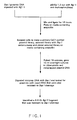
- Figure 1 illustrates the scheme for cloning the Ssp I restriction methylase.
- Figure 2 is a restriction map of the 9.6 Kb Bgl II fragment that encodes the Ssp I methylase with the deletions of the Bgl II clone.
- Figure 3 is the DNA sequence (SEQ ID NO:11 and SEQ ID NO:13) and corresponding amino acid sequence (SEQ ID NO:12) for the C-terminal portion of the endonuclease and amino acid sequence (SEQ ID NO:14) for entire methylase of the Bgl II- Xho I methylase subclone.
- FIG. 4 illustrates the scheme for producing the Ssp I restriction endonuclease.
- Figure 5 is a map of the Sphaerotilus sp. genomic DNA in the region of the methylase and endonuclease genes.
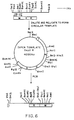
- Figure 6 is the scheme for inverse PCR on the Sac II cut and religated genomic DNA.
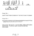
- Figure 7 is a map of where the PCR primers used to directly amplify the endonuclease gene were derived from.
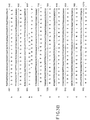
- Figure 8 is a photograph of an agarose gel demonstrating Ssp I restriction endonuclease activity in cell extracts of E.coli ER 2169 carrying the endonuclease gene in the plasmid pAII17.
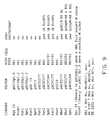
- Figure 9 is a table of different libraries prepared.
- Figure 11 is an explanation of the one letter code for amino acid sequence.
- the present invention relates to recombinant DNA which encodes the Ssp I restriction endonuclease and modification methylase, as well as to the enzymes produced from such a recombinant DNA.
- the nitrocellulose blot was pre hybridized in 15 mls of 50X Denhardt's (5 g ficoll, 5g polyvinylpyrrolidone, 5g BSA in 500 mls H2O), 20x SSC (175.3 g NaCl, 88.2 g Sodium citrate in 1L H2O), 10% SDS and 10% dextran sulfate. After prehybridizing at room temperature for one hour, the labelled probe was added and the hybridization step was carried out at 68°C overnight. The blot was washed in three changes of 2x SSC at 68°C and three changes of 2x SSC with 0.1% SDS over the period of one hour. The blot was exposed to X-ray film for 4 and 18 hours.
- 50X Denhardt's 5 g ficoll, 5g polyvinylpyrrolidone, 5g BSA in 500 mls H2O
- 20x SSC 17.5.3 g NaCl, 88.2
- pSspIM14.0-B6 was also digested with Ase I in 50 mM Tris-HCl, pH7.9, 10 mM MgCl2, 100 mM NaCl 1mm DTT then a 5 Kb digestion product was gel prepped, religated, and plated on 15 ug/ml tetracycline. Miniprepped DNA was then subjected to SspI digestion and were found to not be resistant to Ssp I.
- pSspIM14.0-B6 was also digested with Bam HI in 10 mM Tris-HCl, pH 7.9, 10 mM MgCl2, 150 mM NaCl, 1 mM DTT then a 7 Kb digestion product was gel prepped, religated, and plated on 100 ug/ml ampicillin. The miniprepped DNA was then subjected to Ssp I digestion and was found to be resistant to Ssp I.
- pSspIM14.0-B6 was also digested with Eco RV in 10 mM Tris-HCl, pH 7.9, 10 mM MgCl2, 50 mM NaCl, 1 mM DTT, then a 13 Kb digestion product was gel prepped, religated and plated on 100 ug/ml ampicillin. The miniprepped DNA was subjected to Ssp I digestion and was found to be resistant to Ssp I.
- pSspIM14.0-B6 was also digested with Xho I in 10 mM Tris-HCl, pH 7.9, 10 mM MgCl2, 50 mM NaCl, 1 mM DTT, then a 6.8 Kb digestion product was gel prepped, religated and plated on 100 ug/ml ampicillin. The miniprepped DNA was subjected to Ssp I digestion and was found to be resistant to Ssp I.
- pSspIM14.0-B6 was also double digested with Cla I and Bst BI.
- the Cla I digest was carried out first in 20 mM Tris-acetate, pH 7.9, 10 mM magnesium acetate, 50 mM potassium acetate and 1 mM DTT at 37°C. Then 5,000 units of Bst BI was added and the mixture was incubated at 65° C for one hour. An 11.4 Kb digestion product was gel prepped, religated, and plated on 100 ug/ml ampicillin. The miniprepped DNA was subjected to Ssp I digestion and was found to be not resistant to Ssp I. The deletion clones of Ssp I methylase are summarized in Figure 2.
- the DNA was recovered from the gel using ⁇ -agarase as follows: the gel slice was melted at 55°C and brought to 10 mM Tris-HCl (pH 6.5), 1 mM EDTA. Six units of ⁇ -agarase were added and the agarose was digested at 42°C for 1 hour. The undigested carbohydrates were pelleted by spinning at 15,000 x g at 4°C for 15 minutes. The DNA containing supernate was brought to 0.5M NaCl and two volumes of isopropanol were added. This was mixed and chilled at -20°C for 15 minutes before being centrifuged at 15,000 x g for 15 minutes. The DNA pellet was washed in 70% isopropanol and dried.
- the DNA was resuspended in 20 ul of TE. 10 ul of the Bgl II - Xho I methylase fragment was ligated into the Sal I - Bam HI site of pUC18 and pUC19, respectively. These two methylase subclones were subjected to DNA sequencing. The DNA sequence obtained is shown in Figure 3.
- the oligomer probes were end labelled with ⁇ -32-P in the following manner: 5 ul of the oligomer probe (250 ng) is resuspended in 20 ul of 70 mM Tris-HCl, pH7.6, 10mM MgCl2, 5 mM DTT. 5 ul of ⁇ -32-P is added followed by the addition of 1 ul T4 Polynucleotide kinase. This was incubated at 37°C for 30 minutes.
- the Southern blot was prepared as follows: 1 ug of Sphaerotilus genomic DNA was digested with Ase I, Bam HI, Bgl II, Bsm I, Bst BI, Bst EII, Eco RI, Eco RV, Pst I, Pvu II, SacII, SphI, or XhoI. The digests were run on a 0.7% agarose gel overnight. The gel was washed in two changes of 0.25 M HCl for 15 minutes, then in two changes of 0.5M NaOH, 1.5 M NaCl for 15 minutes each and finally in two changes of 1M NH4OAc, 0.02 M NaOH for 30 minutes.
- nitrocellulose To transfer the DNA to nitrocellulose, a sheet of 0.45 um pore size nitrocellulose was wet in 1M NH4OAc, 0.02 M NaOH and a piece the same size as the gel was placed on either side of the gel. This was placed on top of a two inch high stack of paper towels and another two inch stack of paper towels was placed on top. A glass plate was placed on the top of the stack and a small weight was placed on top. The DNA transfer was allowed to proceed overnight. The nitrocellulose was baked at 80°C for one hour.
- the nitrocellulose blot was pre hybridized in 15 mls of 50X Denhardt's (5 g ficoll, 5g polyvinylpyrrolidone, 5g BSA in 500 mls H2O), 20x SSC ( 175.3 g NaCl, 88.2 g Sodium citrate in 1L H2O), 10% SDS and 10% dextran sulfate. After prehybridizing at room temperature for one hour, the labelled probe was added and the hybridization step was carried out at 37°C overnight. The blot was washed in three changes of 2x SSC and three changes of 2x SSC with 0.1% SDS over the period of one hour at 37°C. The blot was exposed to X-ray film for 4 and 7 days.
- 50X Denhardt's 5 g ficoll, 5g polyvinylpyrrolidone, 5g BSA in 500 mls H2O
- 20x SSC 175.3 g NaCl, 88.2
- Figure 5 is a map of the restriction sites in the region of the Ssp I restriction/modification system in the Sphaerotilus genome.
- the 900 bp PCR product was microdialyzed against TE for 1 hour, then characterized by mapping with Eco RI, Bgl II, and Bam HI .
- the PCR product was then digested with Nde I and Bam HI as follows:70 ul of the PCR product was brought to 150 mM NaCl, 10 mM Tris-HCl (pH 7.9), 10 mM MgCl 2, 1 mM DTT; 40 units of Bam HI and 40 units of Nde I were added.
- the digest was incubated at 37°C for one hour then run on a 1% low melt agarose gel.
- the band was excised and the DNA was recovered from the gel using ⁇ -agarase as follows: the gel slice was melted at 55°C.
- the assay for Ssp I activity proceeded as follows: 1 ug of lambda DNA was diluted to 10 mM Tris-HCl, 10 mM MgCl2, 50 mM NaCl, 1 mM DTT in a total volume of 20 ul. 1 ul of the supernatant from the sonicated cells was added and incubated for 30 minutes at 37°C. The digest was run on a 0.7% agarose gel. Ssp I activity was detected in 6 out of 7 of the crude cell extracts.
- Figure 8 is a photograph of an agarose gel demonstrating Ssp I restriction endonuclease activity.
- the plasmids with Ssp I activity were grown and CsCl prepped and are referred to as p(pAII17)SspR7.2-A3, p(pAII17)SspR7.2-A9, p(pAII17)-SspR7.2-A10, p(pAII17)SspR7.2-A12, p(pAII17)SspR7.2-B1, and p(pAII17)SspR7.2-B6.
- Plasmid p(pAII17)SspR7.2-B1 was deposited with the American Type Culture Collection (ATCC) under the terms of the Budapest Treaty on October 6, 1994, and received ATCC Accession No 75909.
Landscapes
- Health & Medical Sciences (AREA)
- Life Sciences & Earth Sciences (AREA)
- Chemical & Material Sciences (AREA)
- Organic Chemistry (AREA)
- Engineering & Computer Science (AREA)
- Bioinformatics & Cheminformatics (AREA)
- Zoology (AREA)
- Wood Science & Technology (AREA)
- Genetics & Genomics (AREA)
- Molecular Biology (AREA)
- Microbiology (AREA)
- Biotechnology (AREA)
- Biomedical Technology (AREA)
- Biochemistry (AREA)
- General Engineering & Computer Science (AREA)
- General Health & Medical Sciences (AREA)
- Medicinal Chemistry (AREA)
- Enzymes And Modification Thereof (AREA)
- Micro-Organisms Or Cultivation Processes Thereof (AREA)
- Preparation Of Compounds By Using Micro-Organisms (AREA)
- Saccharide Compounds (AREA)
Abstract
Description
- The present invention relates to recombinant DNA which encodes the SspI restriction endonuclease and modification methylase, and to the production of these enzymes from the recombinant DNA.
- Restriction endonucleases are a class of enzymes that occur naturally in bacteria. When they are purified away from other contaminating bacterial components, restriction endonucleases can be used in the laboratory to break DNA molecules into precise fragments. This property enables DNA molecules to be uniquely identified and to be fractionated into their constituent genes. Restriction endonucleases have proved to be indispensable tools in modern genetic research. They are the biochemical 'scissors' by means of which genetic engineering and analysis is performed.
- Restriction endonucleases act by recognizing and binding to particular sequences of nucleotides (the 'recognition sequence') along the DNA molecule. Once bound, they cleave the molecule within, or to one side of, the sequence. Different restriction endonucleases have affinity for different recognition sequences. Over one hundred different restriction endonucleases have been identified among many hundreds of bacterial species that have been examined to date.
- Bacteria usually possess only a small number restriction endonucleases per species. The endonucleases are named according to the bacteria from which they are derived. Thus, Sphaerotilus species (ATCC 13925), synthesizes a restriction endonuclease named SspI. This enzyme recognizes and cleaves the sequence AAT▼ATT.
- While not wishing to be bound by theory, it is thought that in nature, restriction endonucleases play a protective role in the welfare of the bacterial cell. They enable bacteria to resist infection by foreign DNA molecules like viruses and plasmids that would otherwise destroy or parasitize them. They impart resistance by binding to infecting DNA molecules and cleaving them each time that the recognition sequence occurs. The disintegration that results inactivates many of the infecting genes and renders the DNA susceptible to further degradation by exonucleases.
- A second component of bacterial protective systems are the modification methylases. These enzymes are complementary to restriction endonucleases and they provide the means by which bacteria are able to protect their own DNA and distinguish it from foreign, infecting DNA. Modification methylases recognize and bind to the same nucleotide recognition sequence as the corresponding restriction endonuclease, but instead of breaking the DNA, they chemically modify one or other of the nucleotides within the sequence by the addition of a methyl group. Following methylation, the recognition sequence is no longer bound or cleaved by the restriction endonuclease. The DNA of a bacterial cell is always fully modified, by virtue of the activity of its modification methylase and it is therefore completely insensitive to the presence of the endogenous restriction endonuclease. It is only unmodified, and therefore identifiably foreign, DNA that is sensitive to restriction endonuclease recognition and attack.
- With the advent of genetic engineering technology, it is now possible to clone genes and to produce the proteins and enzymes that they encode in greater quantities than are obtainable by conventional purification techniques. The key to isolating clones of restriction endonuclease genes is to develop a simple and reliable method to identify such clones within complex 'libraries', i.e. populations of clones derived by 'shotgun' procedures, when they occur at frequencies as low as 10⁻³ to 10⁻⁴. Preferably, the method should be selective, such that the unwanted, majority, of clones are destroyed while the desirable, rare, clones survive.
- Type II restriction-modification systems are being cloned with increasing frequency. The first cloned systems used bacteriophage infection as a means of identifying or selecting restriction endonuclease clones (HhaII: Mann et al., Gene 3:97-112, (1978); EcoRII: Kosykh et al., Molec. Gen. Genet 178:717-719, (1980); PstI: Walder et al., Proc. Nat. Acad. Sci. USA 78:1503-1507, (1981)). Since the presence of restriction-modification systems in bacteria enables them to resist infection by bacteriophages, cells that carry cloned restriction-modification genes can, in principle, be selectively isolated as survivors from libraries that have been exposed to phage. This method has been found, however, to have only limited value. Specifically, it has been found that cloned restriction-modification genes do not always manifest sufficient phage resistance to confer selective survival.
- Another cloning approach involves transferring systems initially characterized as plasmid-borne into E.coli cloning plasmids (EcoRV: Bougueleret et al., Nucleic Acids Res. 12:3659-3676, (1984); PaeR7: Gingeras and Brooks, Proc. Natl. Acad. Sci. USA 80:402-406, (1983); Theriault and Roy, Gene 19:355-359, (1982); PvuII: Blumenthal et al., J.Bacteriol. 164:501-509, (1985)).
- A third approach, and one that is being used to clone a growing number of systems, involves selecting for an active methylase gene (see, e.g. EPO Publication No. 193, 413, published September 3, 1986 and BsuRI: Kiss et al., Nucleic Acids Res. 13:6403-6421, (1985)). Since restriction and modification genes tend to be closely linked, clones containing both genes can often be isolated by selecting for just the one gene. Selection for methylation activity does not always yield a complete restriction-modification system however, but instead sometimes yields only the methylase gene (BspRI: Szomolanyi et al., Gene 10:219-225, (1980); BcnI: Janulaitis et al, Gene 20:197-204 (1982); BsuRI: Kiss and Baldauf, Gene 21:111-119, (1983); and MspI: Walder et al., J. Biol. Chem. 258:1235-1241, (1983)). For an overall review of cloning restriction-modification systems, see e.g., Lunnen et al., Gene 74:25-32 (1988) and Wilson, G.G., Gene 74:281-285 (1988).
- Another method for cloning methylase and endonuclease genes is based on a colorimetric assay for DNA damage. When screening for a methylase, the plasmid library is transformed into the host E.coli strain AP1-200. The expression of a methylase will induce the SOS response in an E.coli strain which is McrA+, McrBC+, or Mrr+. The AP1-200 strain is temperature sensitive for the Mcr and Mrr systems and includes a lac-Z gene fused to the damage inducible dinD locus of E.coli. The detection of recombinant plasmids encoding a methylase or endonuclease gene is based on induction at the restrictive temperature of the lacZ gene. Transformants encoding methylase genes are detected on LB agar plates containing X-gal as blue colonies. (Piekarowicz, et.al., Nucleic Acids Res. 19:1831-1835, (1991) and Piekarowicz, et.al. J. Bacteriology 173:150-155 (1991)). Likewise, the E.coli strain ER1992 contains a dinD1-Lac Z fusion but is lacking the methylation dependent restriction systems McrA, McrBC and Mrr. In this system (called the "endo-blue" method), the endonuclease gene can be detected in the abscence of it's cognate methylase when the endonuclease damages the host cell DNA, inducing the SOS response. The SOS-induced cells form deep blue colonies on LB agar plates supplemented with X-gal. (Xu et.al. Nucleic Acids Res. 22:2399-2403 (1994))
- A potential obstacle to cloning restriction-modification genes lies in trying to introduce the endonuclease gene into a host not already protected by modification. If the methylase gene and endonuclease gene are introduced together as a single clone, the methylase must protectively modify the host DNA before the endonuclease has the opportunity to cleave it. On occasion, therefore, it might only be possible to clone the genes sequentially, methylase first then endonuclease. Another obstacle to cloning restriction-modification systems lies in the discovery that some strains of E.coli react adversely to cytosine or adenine modification; they possess systems that destroy DNA containing methylated cytosine (Raleigh and Wilson, Proc. Natl. Acad. Sci., USA 83:9070-9074, (1986)) or methylated adenine (Heitman and Model, J. Bact., 169:3243-3250, (1987); Raleigh, Trimarchi, and Revel, Genetics, 122:279-296, (1989) Waite-Rees, Keating, Moran. Slatko, Hornstra and Benner, J. Bacteriology, 173:5207-5219 (1991)). Cytosine-specific or adenine-specific methylase genes cannot be cloned easily into these strains, either on their own, or together with their corresponding endonuclease genes. To avoid this problem it is necessary to use mutant strains of E.coli (McrA⁻ and McrB⁻ or Mrr⁻) in which these systems are defective.
- Because purified restriction endonucleases, and to a lesser extent, modification methylases, are useful tools for characterizing and rearranging DNA in the laboratory, there is a commercial incentive to obtain strains of bacteria through recombinant DNA techniques that synthesize these enzymes in abundance. Such strains would be useful because they would simplify the task of purification as well as providing the means for production in commercially useful amounts.
- The present invention relates to recombinant DNA which encodes the gene for the SspI restriction endonuclease and modification methylase from Sphaerotilus species (NEB strain #315, obtained from the American Type Culture Collection (ATCC) under the designation number #13925), as well as to related methods for production of these enzymes from the recombinant DNA. This invention also relates to a transformed host which expresses the restriction endonuclease SspI, an enzyme which recognizes the DNA sequence AAT▼ATT and cleaves as indicated between the first 5' T and third 5' A by the arrow the disclosure of which is hereby incorporated by reference herein.
- The preferred method for cloning SspI comprises forming a sufficient number of libraries containing DNA express the corresponding methylase gene by incubating the library DNA with an appropriate restriction endonuclease, i.e. an enzyme that cleaves its recognition sequence when it is not methylated; and retransforming a host with recombinant DNA which has not been cleaved by being incubated with the restriction endonuclease and screening the resulting transformants for positive clones among survivors.
- After consructing several libraries of Sphaerotilus species DNA, however, we were only able to obtain the methylase or parts of the methylase gene and part of the endonuclease gene, but never the entire endonuclease gene. This led to an alternative strategy for cloning the SspI endonuclease gene. This strategy involved cloning the endonuclease gene directly under control of the T7 promotor system in the vector pAII17 with no methylase present in the host cell.
- Figure 1 illustrates the scheme for cloning the SspI restriction methylase.
- Figure 2 is a restriction map of the 9.6 Kb BglII fragment that encodes the SspI methylase with the deletions of the BglII clone.
- Figure 3 is the DNA sequence (SEQ ID NO:11 and SEQ ID NO:13) and corresponding amino acid sequence (SEQ ID NO:12) for the C-terminal portion of the endonuclease and amino acid sequence (SEQ ID NO:14) for entire methylase of the BglII-XhoI methylase subclone.
- Figure 4 illustrates the scheme for producing the SspI restriction endonuclease.
- Figure 5 is a map of the Sphaerotilus sp. genomic DNA in the region of the methylase and endonuclease genes.
- Figure 6 is the scheme for inverse PCR on the SacII cut and religated genomic DNA.
- Figure 7 is a map of where the PCR primers used to directly amplify the endonuclease gene were derived from.
- Figure 8 is a photograph of an agarose gel demonstrating SspI restriction endonuclease activity in cell extracts of E.coli ER 2169 carrying the endonuclease gene in the plasmid pAII17.
- Figure 9 is a table of different libraries prepared.
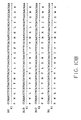
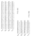
- Figure 10 is the DNA sequence and corresponding amino acid sequence (A9 = SEQ ID NO:15, SEQ ID NO:16; A10 = SEQ ID N:17, SEQ ID NO:18; A12 = SEQ ID NO:19, SEQ ID NO:20, and B1 = SEQ ID NO:21, and SEQ ID NO:22) of four different clones with SspI activity.
- Figure 11 is an explanation of the one letter code for amino acid sequence.
- The present invention relates to recombinant DNA which encodes the SspI restriction endonuclease and modification methylase, as well as to the enzymes produced from such a recombinant DNA.
- The method described herein by which the SspI restriction gene and methylase gene are preferably cloned and expressed is illustrated in Figures 1, 6 and 7 and includes the following steps:
-
- A-1. Sphaerotilus species is grown accordance with the standard protocols for growing Sphaerotilus species at New England Biolabs as described in detail in the Examples. The cells are lysed and the genomic DNA purified by the techniques described in Brooks, et al., Nucleic Acids Research, 17:979-997, (1989).
- A-2. The genomic DNA is digested fully with the following restriction endonucleases: BglII, EcoRI, PstI, SphI, and XhoI.
- A-3. These restriction enzyme fragments are ligated into a corresponding cloning site (e.g., BglII generated fragments are ligated into the BglII cloning site, and so on) of a cloning vector, ideally one that has one, or two SspI sites and the cloning site, such as pBIISpI.2, pUC19, pACYC177, or pACYC184, and the mixture is used to transform an appropriate host cell such as E. coli RR1 cells which are Mrr⁻ or any other E. coli strain which is Mrr⁻ and/or McrA⁻.
- A-4. The transformed mixture is plated onto media selective for transformed cells, such as the antibiotics ampicillin, tetracycline, kanamycin or chloramphenicol. After incubation, the transformed colonies are collected together into a single culture, the cell library.
- A-5. The recombinant plasmids are purified in toto from the cell library to make the plasmid library.
-
- B-1. The plasmid library is digested to completion in vitro with the SspI restriction endonuclease, prepared from Sphaerotilus species, by a method similar to that described in Watson et al, supra. SspI digestion differentially destroys unmodified, non-methylase-containing, clones, increasing the relative frequency of SspI methylase clones.
- B-2. The selected DNA is transformed back into an appropriate host such as E.coli RR1, and transformants are recovered by plating onto selective media. The colonies are picked and their DNA is analyzed for the presence of the SspI modification gene: the plasmids that they carry are purified and incubated with the SspI restriction endonuclease to determine whether they are resistant to digestion. Total cellular DNA (chromosomal and plasmid) is also purified and incubated with the SspI restriction endonuclease. The DNA of clones that carry the SspI modification gene should be fully modified, and both plasmid DNA and total DNA should be substantially resistant to digestion.
- B-3. The DNA libraries generated, such as EcoRI, PstI, SphI and XhoI are prepared for Southern blotting and probed with the cloned methylase gene, such as pSspIM14.0-B6.
-
- C-1. The SspI methylase clone pSspIM14.0-B6 was mapped with a number of different restriction enzymes. The restriction map appears in Figure 2.
- C-2. The SspI methylase clone pSspIM14.0-B6 was digested with the following restriction enzymes: PstI, AseI, BamHI, EcoRV, and XhoI; and religated. These deletion subclones were assayed for methylase activity by subjecting them to digestion with SspI and screening for survivors. Some of these deletion subclones had methylase activity, others had no methylase activity. This demarked an area of about 1.2 kilobases in length which was the putative methylase gene (see Figure 2).This area was between the XhoI site and the BglII site. pSspIM14.0-B6 was digested with BglII and XhoI. This fragment was subcloned into the SalI to BamHI site on pUC18 and in pUC19.
- C-3. The smallest methylase subclone, a 1.2 Kb BglII to XhoI fragment, was subjected to DNA sequencing. The DNA sequence of this region appears in figure 3.
-
- D-1. The SspI restriction endonuclease is produced from Sphaerotilus species cells carrying the SspI restriction and modification genes. The cells are propagated in a fermenter in a rich medium. The cells are harvested by centrifugation. The cells are disrupted by a gaulin mill to produce crude cell extract containing the SspI restriction endonuclease activity. The crude cell extract containing the SspI restriction endonuclease activity is purified by standard ion-exchange and affinity chromatography techniques. Figure 4 illustrates the scheme for producing the SspI restriction endonuclease.
- D-2. The endonuclease so purified is homogeneous on SDS polyacrylamide gel electrophoresis and has an apparent molecular weight of 32,000 daltons and a specific activity of approximately 140,000 units/mg (or more) of protein titered on lambda DNA.
- D-3. The amino terminal sequence of the endonuclease is obtained using an Applied Biosystems 470A Protein Sequencer (Brooks, et al., Nucleic Acids Research, 17:979-997, (1989)), and a DNA oligonucleotide probe is made based on the protein sequence.
- D-4. The probe is used to map the location of the endonuclease relative to the methylase clone in the Sphaerotilus species genome. Figure 5 illustrates the methylase and endonuclease genes in the Sphaerotilus species genome.
-
- A-1. Based on the DNA sequence of the methylase, one can design primers specifically for cloning the SspI methylase gene. Two PCR primers were designed. The SspI methylase was amplified from pSspIM14.0-B6 and subcloned into the polylinker of pUC18 and pUC19 so that the direction of the methylase gene was running with the lac promotor in the pUC18 construct (construct pSspM-B5) and against the lac promotor in the pUC19 construct (construct pSspM-A8).
- A-2. New SphI and XhoI libraries were made in pACYC184. These libraries were transformed into an McrA⁻ host (ER1797) pre-protected with the with the overexpressed methylase constructs, pSspM-B5 or pSspM-A8. These libraries were prepared for Southern blotting and were probed with an oligomer specific for the endonuclease gene. There was no detectable SspI restriction endonuclease gene in any of these libraries.
-
- B-1. A template for inverted PCR of the SspI endonuclease gene was prepared by doing a limit digest of SspI genomic DNA with SacII. The target SacII fragment is about 4 Kb in length and should encode the entire methylase and endonuclease gene. After digestion, the DNA was diluted out and religated to form a circular template.
- B-2. PCR primers were designed which flanked the AseI site in the methylase gene. Amplification was performed and the expected 4 Kb product was identified. Figure 6 illustrates the scheme for doing inverse PCR on the genomic template.
- B-3. The 4 Kb PCR product was random primed and used to probe a Southern blot of Sphaerotilus genomic DNA. The PCR product was determined to map to the location of the endonuclease.
- B-4. Attempts to clone the inverse PCR product into
competent ER 2252 cells pre-protected with the pSspM-A8 or pSspM-B5 methylase construct failed. - B-5. The inverse PCR product was cut with BglII and XhoI to isolate the N-terminal half of the endonuclease gene. This product was cloned into the BamHI to SalI site on pUC19.
- B-6. The BglII - XhoI fragment of the inverse PCR product was mapped with EcoRI, SacII, BamHI and SalI and determined to have the correct structure to be the N-terminal half of the SspI endonuclease gene. This N-terminal portion of the endonuclease gene could be added to the C-terminal portion already cloned to obtain a fully functional endonuclease.
-
- C-1. Primers for PCR were designed for the N-terminal and C-terminal ends of the SspI endonuclease. The degenerate primer for the N-terminal was based on the protein sequence obtained for the SspI endonuclease with an NdeI site engineered in. The primer for the C-terminal was based on DNA sequence of the modification methylase with a BamHI site engineered in. Figure 7 illustrates where the primers for amplifying the endonuclease gene were derived from.
- C-2. Amplification was performed on the Sphaerotilus species genomic template with the primers from C-1 using Vent DNA polymerase in the presence of dNTP's and MgSO₄.
-
- D-1. The 900 base pair PCR product was cloned into the NdeI to BamHI site on the vector pAII17. The plasmid pAII17 is a T₇ vector based on pET11c. (Kong, et al. Journal of Biological Chemistry, 268:1965-1975, (1993)) The ligation was transformed into both E. coli RR1 and ER 2169. Neither cell strain was pre-protected with the SspI methylase.
- D-2. Ninety-six colonies of ER 2169 transformants were picked and replated on L-agar ampicillin plates and L-agar, ampicillin with 10 mM IPTG plates. Colonies corresponding to those which did not grow in the presence of IPTG were grown individually and induced with 10 mM IPTG. The crude cell extracts were assayed for SspI activity on lambda DNA. Figure 8 is a photograph of SspI activity in the crude cell extracts as assayed on lambda DNA.
- Although the above-outlined steps represent the preferred mode for practicing the present invention, it will be apparent to those skilled in the art that the above-described approach can vary in accordance with techniques known in the art.
- The following Example is given to illustrate embodiments of the present invention as it is presently preferred to practice. It will be understood that this example is illustrative, and that the invention is not to be considered as restricted thereto except as indicated in the appended claims.
-
- A-1. Genomic DNA purification: Approximately five grams of Sphaerotilus species cells (ATCC #13925) were thawed and resuspended in 0.1M Tris-HCl, pH 7.1, 0.1M ETDA (25 ml) in a Corning plastic tube (50 ml). A solution of 60mg of lysozyme in 35 ml of the above buffer was divided into two 50 ml plastic tubes and equal portions (15 ml) of the cell suspension added to each. The solutions were incubated at 37°C for fifteen minutes. SDS was added from a 20% stock solution to adjust the final concentration of SDS to 1%. 200 ul of Proteinase K (20 mg/ml stock) was added and incubated for one hour at 37°C. The solution appeared stringy and diffuse at this point but was not clear. Two mls of 10% SDS/8% sarcosyl was added to the tubes (1 ml each) and heated at 55°C for two hours. The sample remained stringy but not totally cleared. The samples were dialyzed against TE (10 mM Tris-HCl, pH 7.1, 1 mM EDTA) (2 L) with a single change - total 16 hours. After dialysis the solution (98 ml) was prepared for CsCl gradients by dilution with an equal vol. of TE pH 8.0, divided into two portions and to each an addition of 98.0 g of CsCl and 1ml of a 5 mg/ml Ethidium bromide was made. The twenty tubes were spun in the Ti70 rotor for 48 hrs at 44,000 rpm. The bands were removed and extracted with CsCl-water-saturated isopropanol. The solution was dialyzed against the same buffer (4 L) as before and then phenol and chloroform extracted (one time each). This solution was dialyzed once again to remove phenol and then subjected to electrophoresis.
- A-2. Limit digestion: The purified DNA was cut with BglII, to achieve total digestion as follows: 300 ul of DNA at 100 ug/ml in 50 mM Tris pH 7.5, 10 mM MgCl₂, 100 mM NaCl, 1 mM DTT buffer was dispensed into three tubes. To the tube was added 50 units of the appropriate restriction enzyme. The tubes were incubated at 37°C for one hour, then phenol/chloroform extracted and ethanol precipitated. The pellets were redissolved in 300 ul of 10 mM Tris-HCl, 1mM EDTA, pH 8.0 and 10 ul from each analyzed by agarose gel electrophoresis.
- A-3. Ligation: The fragmented DNA was ligated to pBIISp1.2 (pBII01(ATCC #67901) cut with XcaI and a linker with an SspI site inserted) as follows: 10.0 ug of BglII digested Sphaerotilis species DNA (100 ul) was mixed with 2.0 ug BglII-cleaved and dephosphorylated pBIISp1.2 (20.0ul) and ethanol precipitated. The DNA was centrifuged at 12,000g, 4°C for 15 minutes and washed once with 100ul 70% ethanol. The DNA was resuspended in 99ul of 1X ligation buffer (50 mM Tris, pH 7.5, 10
mM MgCl₂ 10 mM DTT, .5mM ATP), 1 ul of T4 DNA ligase was added and the mixture allowed to incubate at 16°C for 16 hours. Aliquots of 3 ul were used to transform E. coli strain RR1 as follows. Each aliquot was mixed with 200 ul of ice-cold competent E. coli RR1 cells and placed on ice for thirty minutes. After a 2-minute heat shock at 42°C, the cells were diluted with one ml of Luria-broth (L-broth) and grown for one hour at 37°C. - A-4. Primary Cell Libraries: The transformed cell cultures were centrifuged, resuspended in 250 ul volumes and plated onto Luria-agar (L-agar) plates containing 100 ug/ml ampicillin. After overnight incubation at 37°C, the plates were removed and the approximately 114,000 colonies scraped-up into 25 ml of LB with antibiotic. Plasmid DNA was prepared from these cells as follows: the cells were pelleted by centrifugation and three grams of cell paste was resuspended in 14 ml of 25 mM Tris-HCl, 10 mM EDTA pH 8.0 and 50 mM glucose. The suspension was made 4.0 mg/ml in lysozyme and incubated at 25 degrees for 5 minutes. A 27 ml aliquot of 1% sodium dodecyl sulfate and 0.2 N NaOH was added followed by mixing of the solution and incubation for 5 minutes at 0 degrees. Genomic DNA was precipitated by the addition of 20 ml of ice-cold 3M potassium acetate, pH 4.8, vortexed gently for 10 seconds, left on ice for 5 minutes and centrifuged at 12,000xg for ten minutes. The supernatant was removed and extracted with an equal volume of phenol/chloroform (1:1). The layers were separated by centrifugation at 10,000xg for 5 minutes. The upper layer was removed and extracted with an equal volume of chloroform. The layers were separated by centrifugation at 10,000xg for 5 minutes. The upper layer was removed and the nucleic acids precipitated by the addition of two volumes of ethanol. The precipitate was collected by centrifugation at 12,000xg for twenty minutes. The pellet was washed with 70% ethanol once and repelleted as before. The pellet was dried under vacuum and resuspended in 8 ml of 10 mM Tris-HCl, 1mM EDTA, pH 8.0. The DNA solution was prepared for cesium chloride-ethidium bromide equilibrium density centrifugation by the addition of 8 grams of cesium chloride and 0.5 ml of a solution of ethidium bromide (5mg/ml) were added. The DNA solution was centrifuged at 44,000 rpm for 48 hours and the resulting band of plasmid DNA was removed with a syringe and 18g needle. The ethidium bromide was removed by extracting with an equal volume of CsCl-water-saturated isopropanol. The cesium chloride was removed by dialysis. The DNA was extracted with an equal volume of phenol/chloroform (1:1), and dialyzed against 10mM Tris-HCl, 1mm EDTA, pH 8.0, overnight.
-
- B-1. Primary Selection and Selected Library: 1 ug (12.0 ul) of the BglII plasmid library was diluted into 27 ul of restriction endonuclease digestion buffer (10 mM Tris pH 7.5, 10 mM MgCl₂, 1 mM DTT, 50 mM NaCl and 100ug of bovine serum albumin). 100 units (1 ul) of SspI restriction endonuclease was added and the tube was incubated at 37°C for 2 hours, at which time 7U (1 ul) of calf intenstinal phosphatase was added and the reaction was incubated for an additional 30 minutes. 5 ul aliquots of this reaction mixture were mixed with 200 ul of ice-cold competent E. coli RR1 cells and transformed, plated and grown overnight as for the primary library.
- B-2. Analysis of individuals: Colonies from the above transformation were picked and plated on LB agar plates containing ampicillin. Eighteen colonies were grown up in 10 ml cultures and the plasmids that they carried were prepared by the following miniprep purification procedure, adapted from the method of Birnboim and Doly (Nucleic Acids Res. 7:1513 (1979)).
Miniprep Procedure: Each culture was processed as follows: 1.5 mls of the overnight culture was pelleted at 6,000xg for 5 minutes. The supernatant was poured off and the cell pellet was resuspended in 150 ul of 25mM Tris, 10 mM EDTA, 50 mM glucose, pH 8.0, containing 2 mg/ml lysozyme. After five minutes at room temperature, 200 ul of 0.2M NaOH, 1% SDS was added and the tube was shaken to lyse the cells, then placed on ice. After five minutes, 150 ul of 3M sodium acetate, pH 4.8, was added and shaken and placed on ice for an additional five minutes. The precipitate that formed was spun down at 12,000xg, at 4°C for five minutes. The supernatant was removed and extracted with an equal volume of phenol/chloroform (1:1). The layers were separated by centrifugation at 10,000xg for five minutes. The supernatant was poured into a centrifuge tube containing 880 ul of ethanol and mixed. After 10 minutes at room temperature, the tube was spun at 12,000xg for 10 minutes to pellet the precipitated nucleic acids. The supernatant was discarded and the pellet was washed again with one ml of 70% ethanol-water, repelleted and dried at room temperature for 30 minutes under vacuum. Once dry, the pellet was resuspended in 50 ul of 10 mM Tris, 1mm EDTA, pH 8.0 containing 20 ug/ml RNase and incubated for 1 hour at 37°C to digest the RNA.
The plasmid minipreps were subsequently analyzed by digestion with SspI and BglII. - B-3. Methylase Gene Clones: 11% of the plasmids that were analyzed were found to be resistant to SspI and to carry a BglII fragment of approximately 9.6 Kb in length. These plasmids were subsequently shown to encode only a functional SspI modification methylase gene and not the restriction endonuclease gene. The other 89% of the plasmids looked at were not resistant to SspI and contained spurious fragments or were vector religated. No clones were found in the other four libraries, EcoRI, PstI, SphI and XhoI, that were resistant to cleavage by SspI endonuclease. These four libraries were prepared for Southern blotting as follows: 1 ug of the library DNA was digested with SspI, or the cloning enzyme (i.e., PstI for the PstI library, EcoRI for the EcoRI library, etc.) The digests were run with uncut library DNA and genomic DNA digested with the cloning enzyme on a 0.7% agarose gel overnight. The gel was washed in two changes of 0.25 M HCl for 15 minutes, then in two changes of 0.5M NaOH, 1.5 M NaCl for 15 minutes each and finally in two changes of 1M NH₄OAc, 0.02 M NaOH for 30 minutes. To transfer the DNA to nitrocellulose, a sheet of 0.45 um pore size nitrocellulose was wet in 1M NH₄OAc, 0.02 M NaOH and a piece the same size as the gel was placed on either side of the gel. This was placed on top of a two inch high stack of paper towels and another two inch stack of paper towels was placed on top. A glass plate was placed on the top of the stack and a small weight was placed on top. The DNA transfer was allowed to proceed overnight. The nitrocellulose was baked at 80° C for one hour. A ³P-labeled probe was prepared by nick translating the methylase clone pSspIM14.0-B6 as follows: 1 ug of pSspIM14.0-B6 was resuspended in 0.5M Tris-HCl, pH 7.8, 50 mM MgCl₂, 0.1M beta-mercaptoethanol, and 0.5 mg/ml BSA. 4 ul of each of a 0.1mM dCTP, dGTP and dTTP were added along with 10 ul of 650 Ci/mmol α³-P dATP. Four picograms of DNAase I were added along with 10 units of E. coli DNA Polymerase I. This mixture was incubated for three hours at 16° C.
- The nitrocellulose blot was pre hybridized in 15 mls of 50X Denhardt's (5 g ficoll, 5g polyvinylpyrrolidone, 5g BSA in 500 mls H₂O), 20x SSC (175.3 g NaCl, 88.2 g Sodium citrate in 1L H₂O), 10% SDS and 10% dextran sulfate. After prehybridizing at room temperature for one hour, the labelled probe was added and the hybridization step was carried out at 68°C overnight. The blot was washed in three changes of 2x SSC at 68°C and three changes of 2x SSC with 0.1% SDS over the period of one hour. The blot was exposed to X-ray film for 4 and 18 hours.
- Only the EcoRI library was found to contain a methylase clone which hybridized to the probe.
-
- C-1. 5 ug of pSspIM14.0-B6 were digested with PstI restriction endonuclease as follows: 50 ul of DNA at a concentration of 100 ug/lml in 50 mM Tris, pH7.9, 10 mM MgCl₂, 100 mM NaCl, 1 mM DTT was dispensed in one tube. To the tube, 100 units of PstI endonuclease was added and the reaction was incubated for 2 hours at 37°C. The whole digest was run out on a 0.7% agarose preparative gel. The fragment of choice, an approximately 7 kb PstI fragment, determined to contain the whole methylase gene, was cut out of the gel. The gel fragment was alternately extruded through a 21 gauge needle and frozen. This was repeated three times. The resultant mixture was centrifuged at 100,000xg for 1 hour at 4°C to pellet the agarose. The aqueous solution remaining was brought up to a NaCl concentration of 0.4M and precipitated with 2 volumes of isopropanol. The DNA was pelleted by centrifugation at 12,000xg for 20 minutes and washed once with cold 70% ethanol. The DNA pellet was resuspended in 2 ml TE (10 mM Tris, 1 mM EDTA, pH 8) and extracted with an equal volume of phenol. The layers were separated by centrifugation at 10,000xg for 10 minutes. The upper layer was removed and extracted with an equal volume of phenol/chloroform (1:1), and the layers were separated by centrifugation at 10,000xg for 10 minutes. The upper layer was removed and extracted with an equal volume of chloroform and centrifuged at 10,000xg to separate the layers. The aqueous layer was removed, and the DNA precipitated by the addition of 1/10 volume (.2ml) 2.75M sodium acetate and.2 volumes of cold ethanol. The DNA was pelleted by centrifuging at 12,000xg for 20 minutes and washed once with cold 70% ethanol. The DNA was resuspended in 0.5ml TE (10 mM Tris, 1mm EDTA, pH 8).
- C-2. The gel prepped DNA fragments were religated in the following manner: 5 ul of 10x ligation buffer (50 mM Tris, pH7.5, 10 mM MgCl₂, 10 mM DTT, .5mM ATP) was added to 45 ul of the gel prepped restricion digest fragment and 1 ul of T4 DNA Ligase was added and the mixture was allowed to incubate at 16°C for 16 hours. Aliquots of 1, 2, and 3ul were used to transform E. coli strain RRI as described in section I A-3. The transformed cell cultures were centrifuged, resuspended in 250 ul volumes and plated onto L-agar containing 15 ug/ml tetracycline. The cultures, now on plates, were incubated overnight at 37°C.
- C-3. Several colonies were miniprepped as described in Section I (B-2) and were found to have the correct sized fragment. The PstI deletion clone was found to be resistant to SspI digestion and thus contained the entire methylase gene.
- In the same manner described in I (C-1), pSspIM14.0-B6 was also digested with AseI in 50 mM Tris-HCl, pH7.9, 10 mM MgCl₂, 100 mM NaCl 1mm DTT then a 5 Kb digestion product was gel prepped, religated, and plated on 15 ug/ml tetracycline. Miniprepped DNA was then subjected to SspI digestion and were found to not be resistant to SspI. pSspIM14.0-B6 was also digested with BamHI in 10 mM Tris-HCl, pH 7.9, 10 mM MgCl₂, 150 mM NaCl, 1 mM DTT then a 7 Kb digestion product was gel prepped, religated, and plated on 100 ug/ml ampicillin. The miniprepped DNA was then subjected to SspI digestion and was found to be resistant to SspI. pSspIM14.0-B6 was also digested with EcoRV in 10 mM Tris-HCl, pH 7.9, 10 mM MgCl₂, 50 mM NaCl, 1 mM DTT, then a 13 Kb digestion product was gel prepped, religated and plated on 100 ug/ml ampicillin. The miniprepped DNA was subjected to SspI digestion and was found to be resistant to SspI. pSspIM14.0-B6 was also digested with XhoI in 10 mM Tris-HCl, pH 7.9, 10 mM MgCl₂, 50 mM NaCl, 1 mM DTT, then a 6.8 Kb digestion product was gel prepped, religated and plated on 100 ug/ml ampicillin. The miniprepped DNA was subjected to SspI digestion and was found to be resistant to SspI. pSspIM14.0-B6 was also double digested with ClaI and BstBI. The ClaI digest was carried out first in 20 mM Tris-acetate, pH 7.9, 10 mM magnesium acetate, 50 mM potassium acetate and 1 mM DTT at 37°C. Then 5,000 units of BstBI was added and the mixture was incubated at 65° C for one hour. An 11.4 Kb digestion product was gel prepped, religated, and plated on 100 ug/ml ampicillin. The miniprepped DNA was subjected to SspI digestion and was found to be not resistant to SspI. The deletion clones of SspI methylase are summarized in Figure 2.
- From all these deletion clones, the smallest methylase containing region of DNA was between the BglII site and the XhoI site. Thus, 5 ug of pSspIM14.0-B6 were digested with 40 units of BglII and 40 units of XhoI in 50 mM Tris-HCl, pH 7.9, 10 mM MgCl₂, 100 mM NaCl, 1 mM DTT for 1 hour. The digestion products were run out on a 0.7% low melt agarose gel. The 1.2 Kb BglII- XhoI fragment was excised from the gel. The DNA was recovered from the gel using β-agarase as follows: the gel slice was melted at 55°C and brought to 10 mM Tris-HCl (pH 6.5), 1 mM EDTA. Six units of β-agarase were added and the agarose was digested at 42°C for 1 hour. The undigested carbohydrates were pelleted by spinning at 15,000 x g at 4°C for 15 minutes. The DNA containing supernate was brought to 0.5M NaCl and two volumes of isopropanol were added. This was mixed and chilled at -20°C for 15 minutes before being centrifuged at 15,000 x g for 15 minutes. The DNA pellet was washed in 70% isopropanol and dried. The DNA was resuspended in 20 ul of TE. 10 ul of the BglII - XhoI methylase fragment was ligated into the SalI - BamHI site of pUC18 and pUC19, respectively. These two methylase subclones were subjected to DNA sequencing. The DNA sequence obtained is shown in Figure 3.
-
- D-1. SspI endonuclease from Sphaerotilus species designated NEB# 315 was propagated in a fermenter at 37°C in TRY-YE Broth medium consisting of: tryptone, 10.0 g per liter; yeast extract, 5.0 g per liter; NaCl, 2.0 g per liter; K₂HPO₄, 4.4 g per liter; glucose, 2.0 g per liter; hemin bovine, 10 mg per liter; NAD;DPN, 2.0 mg per liter. The cells are collected by centrifugation and the cell paste is used fresh or stored at -70°C. All subsequent steps are carried out at 4°C.
- D-2. The cell paste (253 grams) is thawed and the cells are resuspended in 500 ml sonication buffer (20 mM Tris-HCl, pH 7.6, 0.1 mM EDTA, 50 mM NaCl, 1 mM DTT).
- D-3. The cells are disrupted by a gaulin mill to achieve release of approximately 35 mg of soluble protein per ml of suspended cells.
- D-4. The insoluble cell debris is removed by centrifugation at 15,000 x g for 40 minutes.
- D-5. 50 g of Cell Debris Remover (Whatman) was added to the supernatant and centrifuged at 10,000 x g for 10 minutes.
- D-6. The supernatant fluid is applied to a phosphocellulose column (5 x 35 cm) (Whatman P-11) equilibrated with 20 mM K₂HPO₄, pH 6.9, 50 mM NaCl,0.1 mM EDTA, 1 mM DTT. The column is washed with two column volumes of the above buffer. The flow-though from the column is collected in a single flask. SspI endonuclease is retained by the column and elutes between 0.3 and 0.6M NaCl. The most active fractions are pooled and dialyzed overnight against 20 mM K₂HPO₄, pH7.4, 50mM NaCl, 0.1 mM EDTA, 1 mM DTT.
- D-7. The pool from the phosphocellulose column is applied to a Heparin-Sepharose CL-6B column (2.5 x 25 cm) equilibrated with 20 mM K₂HPO₄, pH 7.4, 0.05mM NaCl, 0.1 mM EDTA, 1 mM DTT, and washed with two column volumes of the same buffer. A linear gradient of NaCl from 0.05 M to 0.8 M (total volume 500 ml) is developed and applied to the column. Three ml fractions are collected. The fractions are assayed for the presence of the SspI restriction endonuclease activity on lambda DNA. The active fractions are pooled and dialysed against 100 volumes of 20 mM K₂HPO₄, pH 7.4, 0.05mM NaCl, 0.1 mM EDTA, 1 mM DTT.
- D-8. The dialyzed pool (25 ml) of SspI activity is applied to a 1 ml Mono S FPLC column (Pharmacia) and washed with 20 mM K₂HPO₄, pH 7.4, 0.05mM NaCl, 0.1 mM EDTA, 1 mM DTT and a 40 ml linear gradient from 50 mM KCl to 1.0 M KCl is developed in the same buffer and applied to the column. One ml fractions are collected and assayed for the presence of SspI restriction endonuclease activity. The four most active fractions were homogeneous and were found to have a specific activity of approximately 140,000 units/mg protein and a molecular weight on SDS-polyacrylamide gels of 32,000 Daltons.
- D-9. 4 ug of the homogeneous SspI endonuclease was subjected to amino terminal protein sequencing on an Applied Biosystems Model 470A gas phase protein sequencer (Brooks, et al., Nucleic Acids Research, 17:979-997, (1989)). The first 30 residues were degraded. The sequence of the first 25 residues obtained was the following: SKAAYQDFTKXSLLIKKXXNLITM (SEQ ID NO:1) (refer to Table 1 for explanation of 1 letter code for protein sequence).
- D-10. Based on the protein sequence, two 17-mers were made with the following sequences: 5'GCNGCNTAYC ARGACTT3' (SEQ ID NO:2) and 5'GCNGCNTAYCARGATTT3" (SEQ ID NO:3) (Y=T or C; D=A, G or T; R=A or G; N=A, C, G, or T) which were used to map the location of the amino terminal end of the endonuclease on Sphaerotilus genomic DNA.
- The oligomer probes were end labelled with γ-32-P in the following manner: 5 ul of the oligomer probe (250 ng) is resuspended in 20 ul of 70 mM Tris-HCl, pH7.6, 10mM MgCl₂, 5 mM DTT. 5 ul of γ-32-P is added followed by the addition of 1 ul T₄ Polynucleotide kinase. This was incubated at 37°C for 30 minutes.
- The Southern blot was prepared as follows: 1 ug of Sphaerotilus genomic DNA was digested with AseI, BamHI, BglII, BsmI, BstBI, BstEII, EcoRI, EcoRV, PstI, PvuII, SacII, SphI, or XhoI. The digests were run on a 0.7% agarose gel overnight. The gel was washed in two changes of 0.25 M HCl for 15 minutes, then in two changes of 0.5M NaOH, 1.5 M NaCl for 15 minutes each and finally in two changes of 1M NH₄OAc, 0.02 M NaOH for 30 minutes. To transfer the DNA to nitrocellulose, a sheet of 0.45 um pore size nitrocellulose was wet in 1M NH₄OAc, 0.02 M NaOH and a piece the same size as the gel was placed on either side of the gel. This was placed on top of a two inch high stack of paper towels and another two inch stack of paper towels was placed on top. A glass plate was placed on the top of the stack and a small weight was placed on top. The DNA transfer was allowed to proceed overnight. The nitrocellulose was baked at 80°C for one hour.
- The nitrocellulose blot was pre hybridized in 15 mls of 50X Denhardt's (5 g ficoll, 5g polyvinylpyrrolidone, 5g BSA in 500 mls H₂O), 20x SSC ( 175.3 g NaCl, 88.2 g Sodium citrate in 1L H₂O), 10% SDS and 10% dextran sulfate. After prehybridizing at room temperature for one hour, the labelled probe was added and the hybridization step was carried out at 37°C overnight. The blot was washed in three changes of 2x SSC and three changes of 2x SSC with 0.1% SDS over the period of one hour at 37°C. The blot was exposed to X-ray film for 4 and 7 days.
- From this blot, and the map of the pSspIMl4.0-B6 clone, a genomic map in the region of the restriction endonuclease was constructed. Figure 5 is a map of the restriction sites in the region of the SspI restriction/modification system in the Sphaerotilus genome.
-
- A-1. Based on the DNA sequence of the methylase, PCR primers were designed . The primer for the N-terminal is as follows 5'GCTTGAAGATCTAGAGGATTTCATA TGGGATCAATGTTTAACACCACACAA3' (SEQ ID NO:4) The sequence of the C-terminal primer is: 5'TTCTTGTTGGCGTTCGCTCGAGC ACCCAGTTAGGAA3' (SEQ ID NO:5). The SspI methylase was amplified out of pSspIMl4.0-B6 as follows: 2 ng of template DNA was diluted in 10 mMKCl, 20 mMTris-HCl (pH 8.8), 10 mM (NH₄)₂SO₄, 6 mM MgSO₄, 0.1% Triton X-100; 200 uM dNTP's, the primers, and 1 U of Vent DNA polymerase was added. Thirty five cycle of denature at 95°C for 1 minute, anneal at 72°C for 1 minute and extend at 75°C for 2 minutes were run in a thermal cycler. A 1.1 Kb PCR product was obtained. The PCR product was microdialyzed against TE for one hour, then 10 ul was digested with BglII and XhoI as follows: 10 ul of PCR product plus 2 ul of 500 mM Tris-HC, pH 7.9, 100 mM MgCl₂, 1 M NaCl, 10 mM DTT, plus 4 ul H₂O, plus 24 units of BglII and 20 units of XhoI were incubated at 37°C overnight.
The vector was prepared as follows: 5 ug of pUC18 orpUC 19 was incubated in 20 ul of 150 mM NaCl, 10 mM Tris-HCl, pH 7.9, 10 mM MgCl₂, 1 mM DTT; 20 units of BamHI and 60 units of SalI were added. The digests were incubated for 1 hour at 37°C. The DNA was then phenol/chloroform extracted and ethanol precipitated. The DNA was resuspended in 50 ul TE.
The plasmids and BglII- XhoI cut PCR product were ligated overnight at 16°C. The amplified methylase constructs were transformed into E.coli strain ER2252 and competent cells were prepared. The methylase in pUC19 (running in the opposite orientation of the lac promotor) is construct pSspM-A8. The methylase in pUC18 (running in the same orientation as the lac promotor) is construct pSspM-B5. - A-2. Based on the data obtained in I (D-10), purified Sphaerotilus species genomic DNA (prepared as in I (A-1)) was subjected to a limit digestion using SphI, or XhoI as follows: 10 ug of genomic DNA was diluted into 10 mM Tris-HCI, pH 7.9, 10 mM MgCl₂, 50 mM NaCl, 1 mMDTT, 50 units of SphI or XhoI were added to the appropriate tube. The tubes were incubated at 37°C for one hour, then phenol/chloroform extracted and ethanol precipitated. The pellets were redissolved in 50 ul of 10 mM Tris-HCl, 1mM EDTA, pH 8.0 and 1 ul from each analyzed by agarose gel electrophoresis.
The pACYC184 vectors were prepared as follows: 10 ug of pACYC184 was resuspended in 200 ul of 10 mM Tris-HCl, pH 7.9, 10 mM MgCl₂, 50 mM NaCl, 1 mMDTT. 40 units of SphI or SalI was added and the digest was incubated at 37°C for one and a half hours at whichtime 4 units of cip were added and the incubation continued for another 30 minutes. The digests were phenol/chloroform extracted and ethanol precipitated. The pellets were resuspended in 100 ul of TE.
Ligation was as follows: 10 ug of SphI cut genomic DNA was mixed with 10 ug of SphI cut pACYC184 in 50 ul of ligation buffer (50 mM Tris, pH 7.5, 10mM MgCl₂ 10 mM DTT, .5mM ATP), 1 ul of T₄ DNA ligase was added and the mixture incubated at 16°C overnight. 10 ug of the XhoI cut genomic DNA was mixed with 10 ug of SalI cut and dephosphorylated pACYC184 in 50 ul of ligation buffer. 1 ul of T₄ DNA ligase was added and the mixture incubated at 16°C overnight. - A-3. A primary cell library was prepared as in step I A-4 except that it was transformed into ER2252 cells pre-protected with pSspM-A8 or pSspM-B5. The DNA from the primary cell library was digested wth the following restriction enzymes, AseI, BamHI, BglII, BsmI, BstBI, BstEII, EcoRI, EcoRV, PstI, PvuII, SacII, SphI, or XhoI; run on a 0.7% agarose gel overnight, and the gel prepared for Southern blotting as in I (B-3). The Southern blots were probed with the two degenerate oligos for the N-terminal of Ssp endonuclease used as in I (D-10). No endonuclease containing clone was identified from the Southern blot.
-
- B-1. The template Spaerotilus species DNA for PCR was prepared as in I (A-1). The DNA was then digested with SacII as follows: 5 ug of genomic DNA was resuspended in 95 ul of 20 mM Tris-acetate, 10 mM magnesuim acetate, 50 mM potassium acetate, 1 mM DTT, 5 ul (100 units) of SacII was added and the mixture was incubated at 37°C for 1 hour. The digest was phenol/chloroform extracted, and ethanol precipitated. The DNA pellet was resuspended in 500 ul of lx ligase buffer (50 mM Tris-HCl, pH 7.8, 10 mM MgCl₂, 10 mM DTT, 1 mMATP, 25 ug/ml BSA), then 1 ul of T₄ DNA ligase was added. The ligation was allowed to proceed at 16°C overnight. The ligase was heat killed for 15 minutes at 65°C.
- B-2. The primers used for inverse PCR were derived from DNA sequence within the methylase gene. The sequence of the clockwise 30-mer is as follows: TGAGTGGC TTAGGGATGCAGAAGAGCCAAA (SEQ ID NO:6). The sequence of the counterclockwise primer is: TTGGTCACTTCATTTCGCCATGA CATTTCG (SEQ ID NO:7) .
1 ug of SacII cut and religated genomic template (as prepared in II B-1) was mixed with 10 mMKCl, 20 mMTris-HCl (pH 8.8), 10 mM (NH₄)₂SO₄, 2 mM MgSO₄, 0.1% Triton X-100; 200 uM dNTP's, the primers, and 1 U of Vent DNA polymerase was added. Thirty cycles of denature at 95°C for 1 minute, anneal at 65°C for 1 minute, and extend at 72°C for 4 minutes were run in a thermal cycler. A 4 Kb PCR product was observed. - B-3. The 4 Kb PCR product was run on a 0.7% low melt agarose gel. The PCR product was excised from the gel and random primed using the NEBlot kit as follows. The gel slice was melted at 65°C and it's volume determined to be 70 ul. 10 ul of 10x random priming buffer was added and 250 uMoles of dATP, dTTP and dGTP were added. 1 ul of DNA-Polymerase I-Klenow fragment and 5 ul of ³P-γ-dCTP were added. The random priming reaction was allowed to continue for 6 hours at 37°C. The random primed PCR product was used to probe a Southern blot of Ssp genomic DNA digested with AseI, BamHI, BglII, BsmI, BspHI, BstBI, BstEII, EcoRV, NcoI, NdeI, PstI, PvuII, SacII, SphI, and XhoI. Hybridization was carried out overnight at 68°C. The blot was washed 5 times with 2x SSC. The blot was exposed to X-ray film for 4 hours, then developed. It was determined that the inverse PCR product mappped to the location of the SspI endonuclease.
- B-4. The inverse PCR product was cloned into pACYC184 in the following manner. First, the vector was cut with EcoRV and dephosphorylated. 8 ug of pACYC184 was resuspended in 100 ul of 10 mM Tris-HCl, pH 7.9, 10 mM MgCl₂, 50 mM NaCl, 1 mM DTT; 6 units of EcoRV were added and the digest was incubated at 37°C for one and a half hours. 10 units of calf intestinal phosphatase were added and the reaction allowed to proceed for another 30 minutes. The DNA was phenol/chloroform extracted and ethanol precipitated. The SspI endonuclease from inverse PCR was kinased in 100 ul of 70 mM Tris-HCl, pH 7.6, 10mM MgCl₂, 5 mM DTT,and 66 uM dATP with 10 units of T₄ polynucleotide kinase. The kinased inverse PCR product was then ligated into the EcoRV cut and dephsphorylated pACYC184 as follows: 1 ug of EcoRV cut and dephosphorylated pACYC184 was resuspended in lx ligase buffer (50 mM Tris-HCl, pH 7.8, 10 mM MgCl₂, 10 mM DTT, 1 mM ATP, 25 ug/ml BSA). 20 ul of the kinased inverse PCR product was added along with 800 units of T₄ DNA ligase. The ligation reaction was allowed to proceed for 1 hour at room temperature, then 5 ul was transformed into pSspM-A8 and pSspM-B5 pre-protected
ER 2252 cells (as prepared in II A-1).
The cells were scraped off the plate and DNA was prepared as for the primary cell libraries (Secton I A-4). The DNA was digested with the following enzymes: AseI, BamHI, BglII, EcoRI, EcoRV, NsiI, PstI, SacII, SalI, SphI, and XhoI; then run out on a 0.7% agarose gel and Southern blotted. The Southern blots were probed with a kinased 30-mer (kinased as in I D-10) specific for the C-terminal of the SspI endonuclease with the sequence: GCTGTTTCAGCTCTGGCACGTGCGGCATCG (SEQ ID NO:8). The DNA encoding the endouclease gene was not detected. - B-5. The inverse PCR product was cut with XhoI and BglII to isolate the N-terminal half of the endonuclease gene. 10 ul of the inverse PCR product was microdialyzed against TE for 1 hour. The inverse PCR product was then brought to 50 mM Tris-HCl, pH 7.9, 10 mM MgCl₂, 100 mM Nacl, 1 mM DTT; 8 units of BglII and 20 units of XhoI were added. This was allowed to incubate at 37°C for one hour. The restriction enzymes were heat killed by incubating for 20 minutes at 65°C. This was ligated to pUC19 which had been cut with BamHI and SalI. 100 ng of BamHI and SalI cut pUC19 was resuspended in ligase buffer. 20 ul of the BglII - XhoI cut inverse PCR product was added and 400 units of T₄ DNA ligase were added. The ligation was allowed to proceed overnight at 16°C. 20 ul of the ligation was transformed into ER 2267 and plated on 50 ug/ml ampicillin with 80 ng X-gal and 10 mM IPTG.
Several white colonies were picked and grown in a 10 ml overnight culture. These cells were then miniprepped and 50% were determined to have an insert. - B-6. The minipreps determined to have insert DNA (from II B-5) were mapped with BamHI, SalI, EcoRI and SacII to determine if the insert had the correct restriction map to be the N-terminal half of the SspI endonuclease. The BamHI and SalI digests were done on 1 ug of miniprep DNA resuspended in 150 mM NaCl, 10 mM Tris-HCl (pH 7.9), 10 mM MgCl₂,and 1 mM DTT with 20 units of BamHI or 20 units of SalI. The EcoRI digests were done with 20 units of EcoRI in 50 mM NaCl, 100 mM Tris-HCl (pH 7.5), 10 mM MgCl₂, and 0.025% triton X-100. The SacII digests were done with 20 units of SacII in 50 mM potassium acetate, 20 mM Tris-acetate (pH 7.9), 10 mM magnesuim acetate, and 1 mM DTT. One ug of miniprep DNA was digested in each of the above reactions for 1.5 hours at 37°C. Half of the insert DNA mapped had the correct structure to be the N-terminal half of the SspI endonuclease.
-
- C-1. Two oligonucleotide primers were designed for amplifying the SspI endonuclease gene directly out of genomic DNA. The primer for the N-terminal of the gene was based on the degenerate amino acid sequence obtained in I-D-9 with an NdeI and XbaI site engineered in. The sequence of the N-terminal oligo is: GCTCTAGACCCGGGCATATG TCVAAAGCMGCMTAYCAAGATTTTAA (SEQ ID NO:9) (where V=A, C, or G; M=A or C; Y=C or T) The oligo for the C-terminal was: CAATTTTAGTTTGGATCCGGCATATTT GGTACCTTGAGTTTCCGGAG (SEQ ID NO:10).
- C-2. Sphaerotilus species genomic template DNA for PCR was prepared as in I A-1. 1 ug of genomic template DNA was resuspended in 10 mM KCl, 20 mM Tris-HCl (pH 8.8), 10 mM (NH₄)₂SO₄, 6 mM MgSO₄, 0.1% Triton X-100, 200 uM dNTP's and 1 unit of Vent DNA polymerase. Thirty cycles of the following steps were performed in a thermal cycler: denature at 95°C for 1 minute, anneal at 55°C for 1 minute and extend at 73°C for 2 minutes. A 900 base pair PCR product, believed to be the entire SspI endonuclease gene, was identified as the major product.
- The 900 bp PCR product was microdialyzed against TE for 1 hour, then characterized by mapping with EcoRI, BglII, and BamHI. The PCR product was then digested with NdeI and BamHI as follows:70 ul of the PCR product was brought to 150 mM NaCl, 10 mM Tris-HCl (pH 7.9), 10
mM MgCl 2, 1 mM DTT; 40 units of BamHI and 40 units of NdeI were added. The digest was incubated at 37°C for one hour then run on a 1% low melt agarose gel. The band was excised and the DNA was recovered from the gel using β-agarase as follows: the gel slice was melted at 55°C. and brought to 10 mM Tris-HCl (pH 6.5), 1 mM EDTA. Six units of β-agarase were added and the agarose was digested at 42°C for 1 hour. The undigested carbohydrates were pelleted by spinning at 15,000 x g at 4°C for 15 minutes. The DNA containing supernate was brought to 0.5M NaCl and two volumes of isopropanol were added. This was mixed and chilled at -20°C for 15 minutes before being centrifuged at 15,000 x g for 15 minutes. The DNA pellet was washed in 70% isopropanol and dried. The DNA was resuspended in 20 ul of TE. -
- D-1. 5 ug of the vector pAII17 (a T₇ expression vector derived from pET11c; Kong, et.al J. Biol. Chem. 268:1965-1975 (1993)) was resuspended in 50 mM Tris-HCl, pH 7.9, 10 mMMgCl2, 100 mM NaCl, 1 mM DTT. 60 units of NdeI and 80 units of BamHI were added and the mixture incubated at 37°C for one hour. The digest was run out on a 0.7% low melt agarose gel and the 6.2 Kb band was excised from the gel. The band from the gel was melted and the DNA recovered with β-agarase as in II C-2. The DNA pellet was resuspended in 20 ul of H₂O.
The PCR product obtained in II C-2 was ligated into the NdeI - BamHI cut pAII17 as follows: 1 ug of the PCR product cut with BamHI and NdeI and 1 ug of pAII17 cut with NdeI - BamHI were resuspended in 50 mM Tris-HCl, pH 7.8, 10 mM MgCl₂, 10 mM DTT, 1 mM ATP, 25 ug/ml BSA and 400 units of T₄ DNA polymerase was added. The ligation was allowed to proceed at 16°C overnight.
2 ul of the ligation was transformed into RR1 with no pre-protecting SspI methylase and 2 ul was transformed into ER 2169, also lacking the cognate methylase, and plated on 50 ug/ml ampicillin. After 18 hours, 96 colonies were picked off each plate and replicated on master plates containing 50 ug/ml ampicillin. The ER 2169 transformants were also replicated on a plate with 1 mM IPTG and ampicillin. After 18 hours, it was noted that several colonies from ER 2169 grown on the IPTG had lysed. - D-2. Ten ml overnight cultures were grown of the first 18 colonies from the ER 2169 plate. When it was noted which colonies had lysed in the presence of IPTG, 1 ml of the corresponding 10 ml culture was diluted out 10-fold and induced with 10 mM IPTG at mid-log phase. The cell cultures were grown for 3 hours in the presence of IPTG. 1.5 mls of the culture was spun down in a microfuge tube. The cell pellets were resuspended in 400 ul of 20 mM KH₂PO₄, 50 mM Nacl, 1 mM DTT. The cells were sonicated for 10 seconds to break the cells. The cell debris was spun down for 5 minutes at 15,000 x g. The supernatant was assayed for SspI activity on lambda DNA.
- The assay for SspI activity proceeded as follows: 1 ug of lambda DNA was diluted to 10 mM Tris-HCl, 10 mM MgCl₂, 50 mM NaCl, 1 mM DTT in a total volume of 20 ul. 1 ul of the supernatant from the sonicated cells was added and incubated for 30 minutes at 37°C. The digest was run on a 0.7% agarose gel. SspI activity was detected in 6 out of 7 of the crude cell extracts. Figure 8 is a photograph of an agarose gel demonstrating SspI restriction endonuclease activity. The plasmids with SspI activity were grown and CsCl prepped and are referred to as p(pAII17)SspR7.2-A3, p(pAII17)SspR7.2-A9, p(pAII17)-SspR7.2-A10, p(pAII17)SspR7.2-A12, p(pAII17)SspR7.2-B1, and p(pAII17)SspR7.2-B6.
- Plasmid p(pAII17)SspR7.2-B1 was deposited with the American Type Culture Collection (ATCC) under the terms of the Budapest Treaty on October 6, 1994, and received ATCC Accession No 75909.
-

-

-

-


-

-


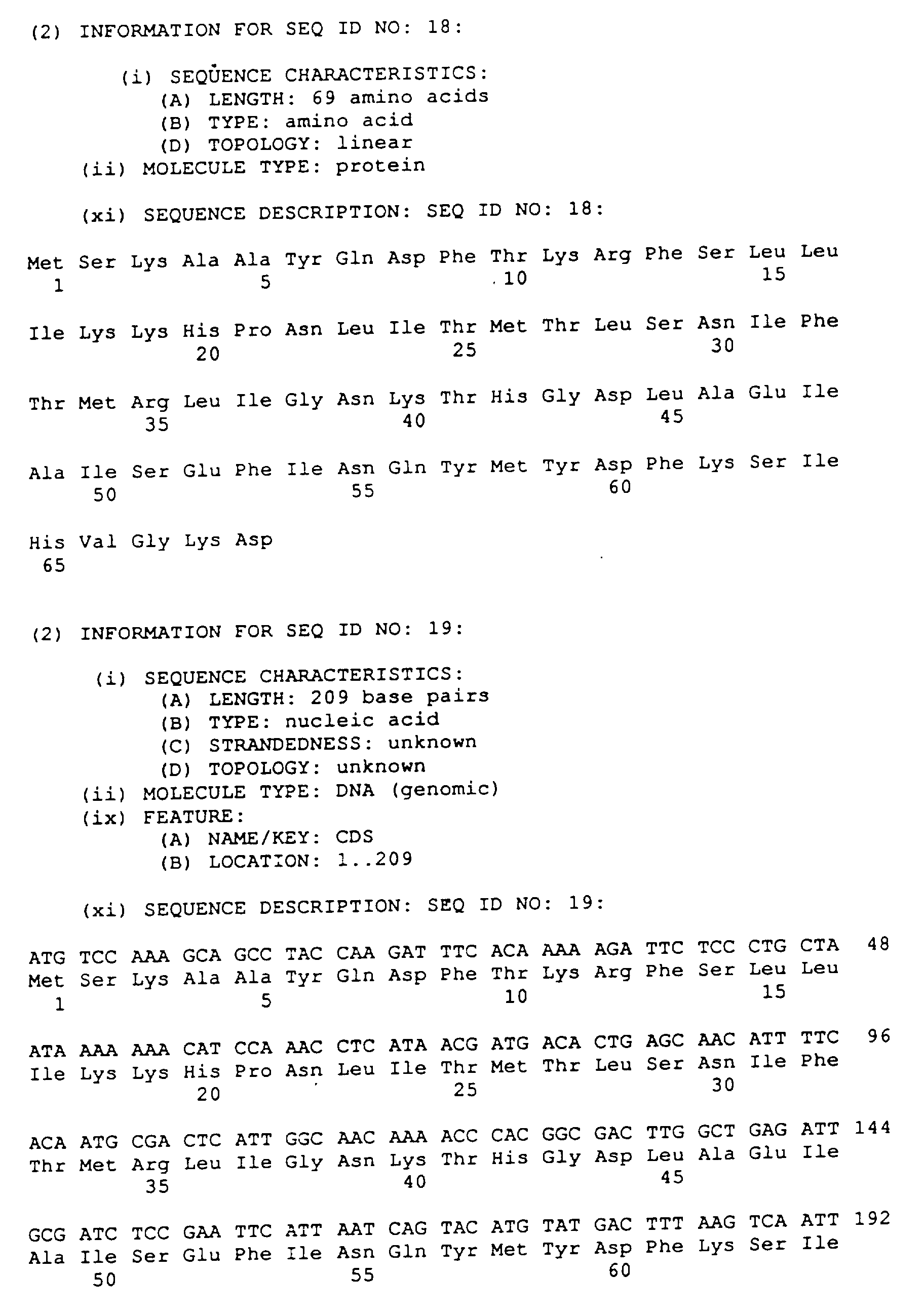


Claims (7)
- Isolated DNA coding for the SspI restriction endonuclease, wherein the isolated DNA is obtainable from the vector p(pAII17)SspR7.2-B1.
- A recombinant DNA vector comprising a vector into which a DNA segment coding for the SspI restriction endonuclease has been inserted.
- Isolated DNA coding for the SspI restriction endonuclease and methylase, wherein the isolated DNA is obtainable from the vector p(pAII17)SspR7.2-B1.
- A cloning vector which comprises the isolated DNA of claim 3.
- The cloning vector of claim 4, wherein the cloning comprises p(pAII17)SspR7.2-B1.
- A host cell transformed by the cloning vector of claim 2, 4 or 5.
- A method of producing an SspI restriction endonuclease comprising culturing a host cell transformed with the vector of claim 2, 4 or 5 under conditions suitable for expression of said endonuclease.
Applications Claiming Priority (2)
| Application Number | Priority Date | Filing Date | Title |
|---|---|---|---|
| US319621 | 1994-10-06 | ||
| US08/319,621 US5516678A (en) | 1994-10-06 | 1994-10-06 | Method for producing the SSPI restriction endonuclease and methylase |
Publications (3)
| Publication Number | Publication Date |
|---|---|
| EP0707066A2 true EP0707066A2 (en) | 1996-04-17 |
| EP0707066A3 EP0707066A3 (en) | 1997-09-10 |
| EP0707066B1 EP0707066B1 (en) | 2000-01-12 |
Family
ID=23243037
Family Applications (1)
| Application Number | Title | Priority Date | Filing Date |
|---|---|---|---|
| EP95307106A Expired - Lifetime EP0707066B1 (en) | 1994-10-06 | 1995-10-06 | Method for producing the SspI restriction endonuclease and methylase |
Country Status (4)
| Country | Link |
|---|---|
| US (1) | US5516678A (en) |
| EP (1) | EP0707066B1 (en) |
| JP (1) | JP3963971B2 (en) |
| DE (1) | DE69514477T2 (en) |
Cited By (3)
| Publication number | Priority date | Publication date | Assignee | Title |
|---|---|---|---|---|
| EP0985730A1 (en) * | 1998-08-31 | 2000-03-15 | New England Biolabs, Inc. | Method for cloning and producing the snaBI restriction endonuclease and purification thereof |
| EP0869174A3 (en) * | 1997-03-20 | 2001-03-07 | New England Biolabs, Inc. | Method for cloning and producing the SpeI restriction endonuclease |
| EP2455464A1 (en) * | 2007-07-12 | 2012-05-23 | New England Biolabs, Inc. | High fidelity restriction endonucleases |
Families Citing this family (1)
| Publication number | Priority date | Publication date | Assignee | Title |
|---|---|---|---|---|
| EP2565267A1 (en) | 2005-08-04 | 2013-03-06 | New England Biolabs, Inc. | Novel restriction endonucleases, DNA encoding these endonucleases and methods for identifying new endonucleases with the same or varied specificity |
Family Cites Families (1)
| Publication number | Priority date | Publication date | Assignee | Title |
|---|---|---|---|---|
| IN166864B (en) * | 1985-03-01 | 1990-07-28 | New England Biolabs Inc |
-
1994
- 1994-10-06 US US08/319,621 patent/US5516678A/en not_active Expired - Lifetime
-
1995
- 1995-10-06 DE DE69514477T patent/DE69514477T2/en not_active Expired - Lifetime
- 1995-10-06 JP JP29632295A patent/JP3963971B2/en not_active Expired - Fee Related
- 1995-10-06 EP EP95307106A patent/EP0707066B1/en not_active Expired - Lifetime
Non-Patent Citations (1)
| Title |
|---|
| BIOCHROMATOGRAPHY, vol. 3, no. 5, 1988, pages 225-30, XP000655221 W. WARREN AND M. MERION: "Rapid HPLC Purification of a Restriction Enzyme from Sphaerotilus sp." * |
Cited By (3)
| Publication number | Priority date | Publication date | Assignee | Title |
|---|---|---|---|---|
| EP0869174A3 (en) * | 1997-03-20 | 2001-03-07 | New England Biolabs, Inc. | Method for cloning and producing the SpeI restriction endonuclease |
| EP0985730A1 (en) * | 1998-08-31 | 2000-03-15 | New England Biolabs, Inc. | Method for cloning and producing the snaBI restriction endonuclease and purification thereof |
| EP2455464A1 (en) * | 2007-07-12 | 2012-05-23 | New England Biolabs, Inc. | High fidelity restriction endonucleases |
Also Published As
| Publication number | Publication date |
|---|---|
| JPH08266288A (en) | 1996-10-15 |
| EP0707066B1 (en) | 2000-01-12 |
| US5516678A (en) | 1996-05-14 |
| DE69514477T2 (en) | 2000-09-07 |
| EP0707066A3 (en) | 1997-09-10 |
| DE69514477D1 (en) | 2000-02-17 |
| JP3963971B2 (en) | 2007-08-22 |
Similar Documents
| Publication | Publication Date | Title |
|---|---|---|
| US5202248A (en) | Method for cloning and producing the nco i restriction endonuclease and methylase | |
| US4999294A (en) | Method for producing the FokI restriction endonuclease and methylase | |
| JP2746964B2 (en) | Method for producing XbaI restriction endonuclease and methylase | |
| EP0388212B1 (en) | Method for producing the Hinc II restriction endonuclease and methylase | |
| US5139942A (en) | Method for producing the nde i restriction endonuclease and methylase | |
| EP0343010B1 (en) | Method for producing the eag I restriction endonuclease and methylase | |
| US4999293A (en) | Method for producing the HhaI restriction endonuclease and methylase | |
| US4983522A (en) | Method for producing the HinPI restriction endonuclease and methylase | |
| EP0388136B1 (en) | Method for producing the MwoI restriction endonuclease and methylase | |
| US5278060A (en) | Method for producing the Nla III restriction endonuclease and methylase | |
| EP0707066B1 (en) | Method for producing the SspI restriction endonuclease and methylase | |
| EP0586116B1 (en) | DNA encoding the Sph I restriction endonuclease and methods for producing the same | |
| US5637476A (en) | Method for cloning and producing the SFII restriction endonuclease and methylase | |
| US5030569A (en) | Method for producing the AFL II restriction endonuclease and methylase | |
| US6238904B1 (en) | Type II restriction endonuclease, HpyCH4III, obtainable from Helicobacter pylori CH4 and a process for producing the same | |
| US5100793A (en) | Method for producing the asei restriction endonuclease and methylase | |
| EP0324248A2 (en) | Method for producing the FnuDI restriction endonuclease and methylase | |
| US5298404A (en) | Method for producing the Hpa I restriction endonuclease and methylase | |
| EP0355981A2 (en) | Method for producing the N1aIV restriction endonuclease and methylase | |
| EP0314346B1 (en) | Method for producing the Afl II restriction endonuclease |
Legal Events
| Date | Code | Title | Description |
|---|---|---|---|
| PUAI | Public reference made under article 153(3) epc to a published international application that has entered the european phase |
Free format text: ORIGINAL CODE: 0009012 |
|
| AK | Designated contracting states |
Kind code of ref document: A2 Designated state(s): DE FR GB |
|
| PUAL | Search report despatched |
Free format text: ORIGINAL CODE: 0009013 |
|
| AK | Designated contracting states |
Kind code of ref document: A3 Designated state(s): DE FR GB |
|
| 17P | Request for examination filed |
Effective date: 19980223 |
|
| 17Q | First examination report despatched |
Effective date: 19980810 |
|
| GRAG | Despatch of communication of intention to grant |
Free format text: ORIGINAL CODE: EPIDOS AGRA |
|
| GRAG | Despatch of communication of intention to grant |
Free format text: ORIGINAL CODE: EPIDOS AGRA |
|
| GRAH | Despatch of communication of intention to grant a patent |
Free format text: ORIGINAL CODE: EPIDOS IGRA |
|
| GRAH | Despatch of communication of intention to grant a patent |
Free format text: ORIGINAL CODE: EPIDOS IGRA |
|
| GRAA | (expected) grant |
Free format text: ORIGINAL CODE: 0009210 |
|
| AK | Designated contracting states |
Kind code of ref document: B1 Designated state(s): DE FR GB |
|
| XX | Miscellaneous (additional remarks) |
Free format text: DUE TO A PATENTIN ERROR THIS DOCUMENT CONTAINS AN ASTERISK AT THE END OF SEQUENCES 12 AND 14. THE ASTERISKS ARE TO BE IGNORED AND THE LENGTH OF THESE SEQUENCES TO BE ADAPTED, I.E. 1 RESIDUE LESS. |
|
| REF | Corresponds to: |
Ref document number: 69514477 Country of ref document: DE Date of ref document: 20000217 |
|
| ET | Fr: translation filed | ||
| PLBE | No opposition filed within time limit |
Free format text: ORIGINAL CODE: 0009261 |
|
| STAA | Information on the status of an ep patent application or granted ep patent |
Free format text: STATUS: NO OPPOSITION FILED WITHIN TIME LIMIT |
|
| 26N | No opposition filed | ||
| REG | Reference to a national code |
Ref country code: GB Ref legal event code: IF02 |
|
| PGFP | Annual fee paid to national office [announced via postgrant information from national office to epo] |
Ref country code: FR Payment date: 20111005 Year of fee payment: 17 |
|
| PGFP | Annual fee paid to national office [announced via postgrant information from national office to epo] |
Ref country code: DE Payment date: 20121003 Year of fee payment: 18 |
|
| PGFP | Annual fee paid to national office [announced via postgrant information from national office to epo] |
Ref country code: GB Payment date: 20121003 Year of fee payment: 18 |
|
| REG | Reference to a national code |
Ref country code: FR Ref legal event code: ST Effective date: 20130628 |
|
| PG25 | Lapsed in a contracting state [announced via postgrant information from national office to epo] |
Ref country code: FR Free format text: LAPSE BECAUSE OF NON-PAYMENT OF DUE FEES Effective date: 20121031 |
|
| GBPC | Gb: european patent ceased through non-payment of renewal fee |
Effective date: 20131006 |
|
| REG | Reference to a national code |
Ref country code: DE Ref legal event code: R119 Ref document number: 69514477 Country of ref document: DE Effective date: 20140501 |
|
| PG25 | Lapsed in a contracting state [announced via postgrant information from national office to epo] |
Ref country code: GB Free format text: LAPSE BECAUSE OF NON-PAYMENT OF DUE FEES Effective date: 20131006 |
|
| PG25 | Lapsed in a contracting state [announced via postgrant information from national office to epo] |
Ref country code: DE Free format text: LAPSE BECAUSE OF NON-PAYMENT OF DUE FEES Effective date: 20140501 |