EP0726316A2 - Expression de la protéine non-structurelle NS1 du virus de la grippe et détection des anticorps anti-NS1 dans le sérum - Google Patents
Expression de la protéine non-structurelle NS1 du virus de la grippe et détection des anticorps anti-NS1 dans le sérum Download PDFInfo
- Publication number
- EP0726316A2 EP0726316A2 EP96300681A EP96300681A EP0726316A2 EP 0726316 A2 EP0726316 A2 EP 0726316A2 EP 96300681 A EP96300681 A EP 96300681A EP 96300681 A EP96300681 A EP 96300681A EP 0726316 A2 EP0726316 A2 EP 0726316A2
- Authority
- EP
- European Patent Office
- Prior art keywords
- protein
- influenza
- virus
- infection
- equine
- Prior art date
- Legal status (The legal status is an assumption and is not a legal conclusion. Google has not performed a legal analysis and makes no representation as to the accuracy of the status listed.)
- Withdrawn
Links
Images


Classifications
-
- C—CHEMISTRY; METALLURGY
- C07—ORGANIC CHEMISTRY
- C07K—PEPTIDES
- C07K14/00—Peptides having more than 20 amino acids; Gastrins; Somatostatins; Melanotropins; Derivatives thereof
- C07K14/005—Peptides having more than 20 amino acids; Gastrins; Somatostatins; Melanotropins; Derivatives thereof from viruses
-
- C—CHEMISTRY; METALLURGY
- C07—ORGANIC CHEMISTRY
- C07K—PEPTIDES
- C07K2319/00—Fusion polypeptide
-
- C—CHEMISTRY; METALLURGY
- C12—BIOCHEMISTRY; BEER; SPIRITS; WINE; VINEGAR; MICROBIOLOGY; ENZYMOLOGY; MUTATION OR GENETIC ENGINEERING
- C12N—MICROORGANISMS OR ENZYMES; COMPOSITIONS THEREOF; PROPAGATING, PRESERVING, OR MAINTAINING MICROORGANISMS; MUTATION OR GENETIC ENGINEERING; CULTURE MEDIA
- C12N2760/00—MICROORGANISMS OR ENZYMES; COMPOSITIONS THEREOF; PROPAGATING, PRESERVING, OR MAINTAINING MICROORGANISMS; MUTATION OR GENETIC ENGINEERING; CULTURE MEDIA ssRNA viruses negative-sense
- C12N2760/00011—Details
- C12N2760/16011—Orthomyxoviridae
- C12N2760/16111—Influenzavirus A, i.e. influenza A virus
- C12N2760/16122—New viral proteins or individual genes, new structural or functional aspects of known viral proteins or genes
Definitions
- the present invention relates to a non-structural protein NS1 of the influenza virus, especially NS1 of Equine Influenza A virus, and fusion proteins incorporating part of NS1. It also relates to the detection of anti-NS1 antibodies in a diagnostic test for influenza infection.
- Virus detection can be carried out by isolation of the virus in embryonated hens eggs using material extracted from nasopharyngeal swabs (Burrows, 1968; Burrows et al , 1981).
- An alternative method is to use an antigen capture ELISA which detects influenza nucleoprotein antigen in material extracted from nasopharyngeal swabs (Cook et al , 1988). This method is rapid, giving same day diagnosis on receipt of samples and is able to detect infection in horses shedding small amounts of virus. Moreover, it does not depend upon the infectivity of the sample which may be reduced during its transit to the laboratory.
- the nucleoprotein (NP) ELISA only detects the transient shedding of virus which during an acute infection may be between five and six days post infection (Cook et al , 1988). However in a subclinical infection which may be expected in a vaccinated population it may take time to identify the problem because symptoms are less acute and it may be too late to obtain a nasal swab or an acute serum sample. In addition the virus shedding stage will be of a shorter duration and at a lower level which may mean that the NP ELISA may not detect the virus because even with samples from an acute infection it is not 100% effective (Cook et al , 1988).
- HI haemagglutin inhibition
- SSH single radial haemolysis
- RNA segment 8 of influenza A viruses encodes two overlapping proteins which are thought to be present only in virus-infected cells (Lazarowitz et al , 1971; Skehel, 1972; Inglis et al , 1980). These proteins are referred to as NS1 and NS2 and are considered to be nonstructural proteins, although Richardson & Akkina (1991) detected NS2 in purified virus confirming similar earlier observations (Lamb et al , 1978; Akkina et al , 1984).
- NS1 is synthesized in large amounts early in infection and accumulates in the nucleus of infected cells, and is present in polysomal cell fractions (Dimmock, 1969; Lazarowitz et al , 1971; Compans, 1973); two independent signals for nuclear localization have been identified in the NS1 molecule (Greenspan et al , 1988). Furthermore, NS1 has also been found in association with cellular RNA in the form of paracrystalline inclusion bodies within the cytoplasm of infected cells (Yoshida et al , 1981; Shaw et al , 1982). Considerable heterogeneity with respect to molecular size, charge and phosphorylation has been demonstrated for several NS1 proteins.
- NS1 protein stimulates the translation of the Ml protein (Enami et al , 1994) but its effect may rather be through the regulation of mRNA nuclear export (Qui and Krug., 1994; Qian et al , 1994). Conversely, Huang et al (1990) have shown that NS1 protein was not required for replication and expression of viral ribonucleoprotein.
- NS1 proteins from several subtypes Antigenic analyses of purified NS1 proteins from several subtypes has indicated cross-reactivity among all the influenza A virus strains tested (Shaw et al , 1982; Brown et al , 1983; Young et al , 1983), although antigenic drift (from the earliest to the most recent virus isolate) was detected (Shaw et al , 1982).
- the NS1 gene from various human influenza virus strains has been cloned (Baez et al , 1980; Krystal et al , 1983) and the protein successfully expressed in Escherichia coli (Young et al , 1983; Hatada et al , 1992) and vaccinia (Smith et al , 1987).
- nucleotide sequence of the nonstructural protein NS1 of the influenza virus A/equine 2/Suffolk/89 was determined and found to be 97% identical to that of A/equine 2/Miami/63. A similar level of identity was shown for the deduced NS1 amino acid sequence.
- the NS1 gene was expressed in its entirety, and in part as fusion proteins with glutathione S-transferase (GST).
- Antibodies to NS1 protein were detected in serum samples from ponies experimentally infected with influenza virus, but not in animals vaccinated with whole inactivated virus or in unprimed control animals.
- the antigenic determinant(s) of NS1 protein appear to be located in the C-terminal half of the protein.
- the NS1 protein only appears in infected cells (Dimmock, 1969; Lazarowitz et al , 1971) and is not incorporated into viral particles and therefore horses routinely vaccinated with whole inactivated virus will not be exposed to this antigen.
- the invention makes possible the use of NS1 protein as a diagnostic marker for influenza virus infection in the presence of high levels of circulating antibody to influenza haemagglutinin generated by recent vaccination.
- the invention provides for the use of NS1 protein from an equine influenza virus in its entirety or in part as a fusion protein with glutathione S-transferase (GST) in a diagnostic test for the rapid detection of equine influenza infection even following recent vaccination.
- GST glutathione S-transferase
- the invention is exemplified hereafter with reference to the cloning, sequencing and expression of the equine influenza NS1 protein and its use either complete or as part of a fusion protein in a diagnostic test for equine influenza.
- Equine influenza virus A/equine 2/Suffolk/89 was grown in embryonated chicken eggs and purified by gradient centrifugation as previously described (Cook et al , 1988). Viral RNA was extracted according to the method of Binns et al (1992). Reverse transcription (Chirnside & Spaan, 1990) was carried out using 5 ⁇ l of RNA with the oligonucleotide 5'-CGGGATCCCCATGGATTCCAACACTGTGTCA-3' (primer 21/91:SEQ.ID.1).
- PCR Polymerase chain reaction
- primer 21/91 and primer 22/91 S'CGGGATCCTCAAACTTCTGACTCAATTGT-3':SEQ.ID.2
- the DNA produced was genecleaned according to the manufacturer's instructions (Bio 101, La Jolla, CA, U.S.A.) and then digested with BamHI, sites for which had been included in primers 21/91 and 22/91 to facilitate the cloning procedure.
- the BamHI sites were preceded by two nucleotides to improve digestion efficiency and followed by 23 and 21 NS1-specific nucleotides (Nakajima et al , 1990) in 21/91 and 22/91 respectively.
- the digested DNA was ligated into BamHI-cleaved pGEX-3X or pUC 13 (Pharmacia, Uppsala, Sweden). Following transformation into E.
- coli TG1 colonies were blotted onto nitrocellulose filters (Hybond N; Amersham Life Sciences, Amersham, U.K.) according to the supplier's instructions and screened for the presence of NS1 insert using a 32P-labelled NS1 fragment (Feinberg & Vogelstein, 1984) isolated from low melting temperature agarose. DNA was prepared from positive recombinants using the Magic Miniprep System (Promega, Madison, WI, U.S.A.) and digested with HindIII and PstI to check for the correct orientation of the NS1 insert.
- the NS1 gene was also cloned into two approximate halves, referred to as NS1-N and NS1-C. This was achieved by ligating two BamHI-digested PCR products into BamHI-cleaved pGEX-3X as described above. PCR was performed using template DNA prepared from the recombinant NS1 clone in the PUC vector. The two primer pairs used were primers 22/91 and 129/92 (5'-CGGGATCCTCATGCCCAAGCAG-3':SEQ.ID.3), and primers 21/91 and 130/92 (5'-CGGGATCCTGATGTTCTTATCCA-3'sEQ.ID.4), respectively. Positive recombinants were screened as before and correct orientation determined by HindIII/PstI or BspHI/PstI digestion of the plasmid DNA.
- Recombinants of interest were sequenced using the dsDNA Cycle Sequencing System (Gibco BRL, Paisley, U.K.).
- the primers used were primer 22/91 (above), primer 36/91 (S'GCGACCATCCTCCAA-3':SEQ.ID.5; specific for pGEX vectors), as well as two primers based on primers 129/92 and 130/92 but lacking the BamHI cloning "tag".
- the annealing temperatures were 60°C, 55°C, 48°C and 40°C, respectively.
- Bacteria were pelleted, washed once with 200 ⁇ g of ice-cold STE (10mM Tris pH 8.0, 150mM NaCl, 1mMEDTA) and resuspended by repeated pipetting in 133 ⁇ l of STE containing 100 ⁇ g/ml of lysozyme (freshly made). After incubation on ice for 15min diothiothreitol (DTT) was added to a final concentration of 5mM and sarkosyl to a final concentration of 1 %. The bacteria were vortexed for 5s and then lysed by sonication in a sonic water bath (QH Kerry, Kerry Ultrasonics Ltd.) for 1 min.
- DTT diothiothreitol
- the lysate was clarified by centrifuging for 15min in a microfuge, full speed, at 4°C and the supernatant was transferred to a new eppendorf tube. Triton X 100 (BDH Chemicals Ltd., Poole, U. K.) was added to a final concentration of 1 % and the lysate was vortexed for 5s. The supernatant was then mixed with 200 ⁇ l of glutathione Sepharose 4B (Pharmacia) suspension (50% in PBS (150mM NaCl, 16mM Na2HPO4, 4mM NaH2P04, pH7.5), washed by repeated centrifugation and resuspension (3 times) for 15min.
- PBS 150mM NaCl, 16mM Na2HPO4, 4mM NaH2P04, pH7.5
- the unbound material was removed following centrifugation (full speed, l0s) and the gel was washed twice with lml PBS for 5 min on a rotator (30rpm).
- the bound fusion protein was eluted with lml elution buffer (75mM Hepes, pH7.4, 150mM NaCl, 10mM reduced glutathione (Sigma), 5mM DTT, 0.1% Triton X100) and stored at 20°C after the addition of glycerol to a final concentration of 10% (v/v).
- Blots were washed three times in PBST at 37°C (5 min/wash) and then incubated at 37°C with serum samples diluted 1:2500 in PBST containing 5% goat serum (Dilution Buffer) for 90min. Each blot was washed 3 times in PBST at 37°C (5min/wash) and then incubated with goat anti-horse IgG-biotin conjugate (Dynatech Lab Ltd, Daux Road, Billingshurst, Hampshire) diluted 1: 1000 in Dilution Buffer for 90min at 37°C.
- each blot was incubated with streptavidin-peroxidase conjugate (Dynatech Lab Ltd) diluted 1:1000 in Dilution Buffer for 30min at room temperature. After washing 6 times in PBST (room temperature, 5min/wash) the blots were developed with either 3',3' diaminobenzidine and 3 % H 2 O 2 or Amersham's ECL detection system.
- Ponies vaccinated with inactivated whole virus (Wood et al 1983; Mumford, 1992) and unvaccinated control animals were infected with A/equine 2/Newmarket/79 or A/equine 2/Suffolk/89 using nebulised aerosols of allantoic fluid as described previously (Mumford et al , 1990). Serum samples collected before and fourteen days after challenge were tested for the presence of anti-haemagglutinin antibodies using the HI and/or SRH tests (Burrows et al , 1977; Wood et al , 1983) and anti-NS1 antibodies by immunoblotting (above) and ELISA (below).
- Microtitre plates (Dynatech immulon III) were incubated at 4°C overnight with 100 ⁇ l (per well) of a solution containing 500-1500ng of purified GST or NS1-C fusion protein in 0.05M carbonate buffer (pH 9.6).
- the coated plates were washed three times in PBSJ (150mM NaCl, 16mM NA2HPO4, 4mM NaH2PO4, 0.1% v/v Tween 20, pH 7.5) then blocked for 90 minutes at 37°C with 100 ⁇ l (per well) of Dilution Buffer (PBST + 5% goat serum).
- the ELISAs were performed by incubating aliquots of horse serum diluted 1:100 in Dilution Buffer for 90 minutes at 37°C in a humid chamber. After three washes with PBST, 100 ⁇ l of goat anti-horse IgG-biotin conjugate diluted 1:1000 in Dilution Buffer was added to the wells and the plates were incubated at 37°C for 90 minutes. The plates were washed three times in PBST and then 100 ⁇ l of Streptavidin-peroxidase conjugate diluted 1:1000 in Dilution Buffer was added to the wells and the plates were incubated at room temperature for 30 minutes.
- Cloning of the Suffolk/89 NS1 gene was achieved using reverse transcription and PCR with primers based on the nucleotide sequence of the A/equine2/Miami/63 (H3N8) NS 1 gene (Nakajima et al , 1990).
- the nucleotide sequence of the Suffolk/89 NS1 gene was determined using two independently isolated clones to identify any potential artefacts generated during the PCR amplification. Both clones possessed identical sequences which are presented in Fig. 1a. Both sequences start at the initiating methionine since reverse transcription was performed with a primer based on the first seven codons of the Miami/63 NS1 gene (Nakajima et al , 1990).
- NS1-N and NS1-C Cloning smaller portions of the NS1 protein (NS1-N and NS1-C) unfortunately did not produce more soluble fusion proteins following lysozyme treatment and lysis in Triton xl00.
- Frangioni and Neel (1993) have shown that sarkosyl can be used to solubilise most GST fusion proteins and that subsequent treatment with non-ionic detergent allows the sarkosyl solubilised GST fusion protein to bind with high affinity to glutathione agarose. Therefore the smallest portion of the NS1 protein (NS1-C) was expressed in E. coli as a fusion protein with GST and purified in the presence of sarkosyl.
- Triton X-100 allowed binding of the GST fusion protein (data not shown) to glutathione agarose and purification of the NS1-C protein. Protein estimations (Sigma) showed that the yield was of the order of 2mg/ml purified protein.
- FIG 3 shows the results from one horse which are representative of those obtained for the other animals in this experimental challenge experiment.
- the lanes are as follows: lane 1, uninduced pGEX-3X; lane 2, induced pGEX-3X (GST); lane 3, NS1; lane 4, NS1-N; lane 5, NS1-C. It can be seen in Fig3 (a) that only minor non-specific reactions were observed with the high titre pre-challenged sera from vaccinated animals (tested positive for serum influenza anti-haemagglutinin antibodies).
- NS1 In order to be practically useful as a marker for influenza infection in the field, the detection of NS1 has to be performed against a background of vaccination.
- the sera in the previously described experiments were obtained from non-vaccinated horses/ponies. Therefore serum from three vaccine trials conducted at the Animal Health Trust were tested for the presence of NS1 antibodies.
- the animals had been vaccinated and then challenged with influenza and in addition a group of control animals which were not vaccinated were also challenged. Pre and post-challenge sera were taken and tested in the NS1 ELISA. For the control animals a positive NS1 ELISA result was obtained in post-challenge sera from 6 out of 10 animals (i.e. 60%) which is similar to the 79% detection rate described above for naive animals.
- the ELISA was then used to investigate an influenza outbreak in a training yard from samples submitted to the Animal Health Trust diagnostic service in an attempt to evaluate how effective it would be.
- the deduced amino acid sequence of the Suffolk/89 NS1 protein revealed a very high degree of homology with another sequenced NS1 protein from the same H3N8 subtype, A/equine 2/Miami/63 influenza virus. Furthermore, it also appears to confirm the suggestion of "the existence of constraints on the amino acid changes of the NS1 protein in evolution" (Nakajima et al , 1990) This conservation tends to imply an important functional role as has been suggested previously (Wolstenholme et al , 1980; Koennecke et al , 1981). Such a high degree of conservation also allows this protein to be utilised as a marker for viral infection.
- NS1 protein Since competition horses are routinely vaccinated with whole inactivated virus, and NS1 protein only appears in infected cells (Dimmock, 1969; Lazarowitz et al , 1971), vaccinated animals will not be exposed to this antigen whereas those suffering a viral infection will.
- This protein would therefore appear to be a suitable candidate for a differential diagnostic marker, capable of distinguishing infected from vaccinated horses if antibodies to NS1 protein are produced during influenza infection. It would also be particularly useful in cases where only a single convalescent serum sample is available.
- NS1 protein is known to be antigenic (Shaw et al , 1982; Brown et al , 1983) and the results in Figure 3(b) clearly indicate that antibodies to NS1 protein are present in the serum of influenza challenged ponies. The specificity of these reactions was further confirmed by the lack of response to the GST portion of the fusion protein (lane 2). Cross-reactivity was also observed since positive reactions were obtained with serum from ponies exposed to the Newmarket/79 strain, further supporting the high conservation of NS1 proteins. As far as we are aware this is the first report of antibodies to NS1 being identified following influenza infection. The finding that the purification of the fusion protein was difficult may be explained, at least in part, by the hydrophobic nature of the NS1 protein (see Fig. 1b).
- NS1 protein is predominantly hydrophobic with only the final one-fifth of the protein being hydrophillic. It would appear that in combination with GST, which itself is quite soluble, the hydrophobicity of these fusion proteins may lead to the formation of insoluble inclusion bodies, thus preventing the release of the vast majority of the protein upon cell lysis.
- the occurrence of insoluble GST fusion proteins has been reported previously (Smith & Johnson, 1988) with the presence of strongly hydrophobic regions the major cause of the insolubility. However the inclusion of sarkosyl in the purification method enabled the production of soluble fusion protein.
- the antigenic determinant(s) of NS1 protein are situated in the C-terminal half of the protein as judged by the responses to NS1-N and NS1-C (Fig. 3).
- a cytotoxic T lymphocyte epitope has been identified on the NS1 protein of human influenza A/PR/8/34 virus corresponding to residues 152-160 (Cossins et al , 1993).
- Highly homologous regions are present at the same position in the C-terminal portions of both the Miami/63 and Suffolk/89 NS1 proteins, as well as many other influenza NS1 proteins (Nakajima et al , 1990), clearly indicating that these proteins should be capable of eliciting a similar immune response.
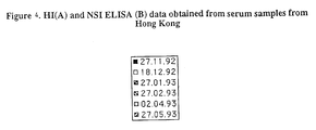
- NS1 protein as a diagnostic marker of influenza infection depends upon the duration of the antibody response to it following the initial infection. A prolonged response would obviously limit its use diagnostically. Results indicate it is short-lived, as is the case for anti-haemagglutinin antibodies (Hannant et al , 1988). Table 1. HI and ELISA data obtained from serum samples submitted to the AHT diagnostic service.
- CHIRNSIDE E. D. & SPAAN, W. J. M. (1990) Reverse transcription and cDNA amplification by the polymerase chain reaction of equine arteritis virus (EAV). Journal of Virological Methods 30, 133-140.
- HATADA E., TAKIZAWA, T. & FUKUDA, R. (1992) Specific binding of influenza A virus NS1 protein to the virus minus-sense RNA in vitro. Journal. of General Virology 73, 1725.
- KOENNECKE I., BOSCHEK, C. B. & SCHLOLTISSEK, C. (1981) Isolation and properties of a temperature-sensitive mutant (ts 412) of the influenza A virus recombinant with a ts lesion in the gene coding for nonstructural protein. Virology 110, 16-25.
- LAEMMLI U.K. (1970) Cleavage of structural proteins during the assembly of the head of bacteriophage T4. Nature (London.) 227, 680-685.
- NAKAJIMA K., NOBUSAWA, E. & NAKAJIMA, S. (1990) Evolution of the NS genes of the influenza A viruses. 11. Characteristics of the amino acid changes in the NS1 proteins of the influenza viruses. Virus Genes 4, 15-26.
- NIETZERT E., BECK, E., DE MELLO, P. A., GOMES, 1. & BERGMANN, I. E. (1991) Expression of the aphthovirus RNA polymerase gene in Escherichia coli and its use with other bioengineered nonstructural antigens in detection of late persistent infections. Virology 184, 799-804.
- the influenza virus NS1 protein is a poly(A) -binding protein that inhibits nuclear export of mRNAs containing poly(A). Journal of Virology 68 2425-2432.
Landscapes
- Chemical & Material Sciences (AREA)
- Organic Chemistry (AREA)
- Health & Medical Sciences (AREA)
- Life Sciences & Earth Sciences (AREA)
- Biophysics (AREA)
- Biochemistry (AREA)
- Gastroenterology & Hepatology (AREA)
- General Health & Medical Sciences (AREA)
- Genetics & Genomics (AREA)
- Medicinal Chemistry (AREA)
- Molecular Biology (AREA)
- Proteomics, Peptides & Aminoacids (AREA)
- Virology (AREA)
- Peptides Or Proteins (AREA)
Applications Claiming Priority (2)
| Application Number | Priority Date | Filing Date | Title |
|---|---|---|---|
| GBGB9502489.9A GB9502489D0 (en) | 1995-02-09 | 1995-02-09 | Expression of the non-structural protein NS1 of influenza virus and detection of anti-NS1 antibody in serum |
| GB9502489 | 1995-02-09 |
Publications (2)
| Publication Number | Publication Date |
|---|---|
| EP0726316A2 true EP0726316A2 (fr) | 1996-08-14 |
| EP0726316A3 EP0726316A3 (fr) | 1997-12-29 |
Family
ID=10769300
Family Applications (1)
| Application Number | Title | Priority Date | Filing Date |
|---|---|---|---|
| EP96300681A Withdrawn EP0726316A3 (fr) | 1995-02-09 | 1996-01-31 | Expression de la protéine non-structurelle NS1 du virus de la grippe et détection des anticorps anti-NS1 dans le sérum |
Country Status (2)
| Country | Link |
|---|---|
| EP (1) | EP0726316A3 (fr) |
| GB (1) | GB9502489D0 (fr) |
Cited By (19)
| Publication number | Priority date | Publication date | Assignee | Title |
|---|---|---|---|---|
| US6013632A (en) * | 1997-01-13 | 2000-01-11 | Emory University | Compounds and their combinations for the treatment of influenza infection |
| GB2412731A (en) * | 2004-04-02 | 2005-10-05 | Ilaria Capua | method for diagnosing avian influenza virus comprisisng detecting NS1 specific antibodies |
| WO2007018843A2 (fr) | 2005-07-01 | 2007-02-15 | Arbor Vita Corporation | Methodes et compositions de diagnostic et de traitement de la grippe |
| EP1843158A1 (fr) * | 2006-04-05 | 2007-10-10 | Micronas Holding GmbH | Système de vérification pour agent pathogène |
| US7595152B2 (en) | 2005-07-01 | 2009-09-29 | Arbor Vita Corporation | Detection of influenza virus |
| US7892729B1 (en) * | 2005-06-23 | 2011-02-22 | Iowa State University Research Foundation, Inc. | Universal and differential serologic assay for swine influenza virus |
| US8080645B2 (en) | 2007-10-01 | 2011-12-20 | Longhorn Vaccines & Diagnostics Llc | Biological specimen collection/transport compositions and methods |
| US8084443B2 (en) | 2007-10-01 | 2011-12-27 | Longhorn Vaccines & Diagnostics Llc | Biological specimen collection and transport system and methods of use |
| US8097419B2 (en) | 2006-09-12 | 2012-01-17 | Longhorn Vaccines & Diagnostics Llc | Compositions and method for rapid, real-time detection of influenza A virus (H1N1) swine 2009 |
| US8652782B2 (en) | 2006-09-12 | 2014-02-18 | Longhorn Vaccines & Diagnostics, Llc | Compositions and methods for detecting, identifying and quantitating mycobacterial-specific nucleic acids |
| US8697089B2 (en) | 2005-01-11 | 2014-04-15 | Wisconsin Alumni Research Foundation | H3 equine influenza A virus |
| US8821885B2 (en) | 2007-08-27 | 2014-09-02 | Longhorn Vaccines & Diagnostics, Llc | Immunogenic compositions and methods |
| US9481912B2 (en) | 2006-09-12 | 2016-11-01 | Longhorn Vaccines And Diagnostics, Llc | Compositions and methods for detecting and identifying nucleic acid sequences in biological samples |
| US9598462B2 (en) | 2012-01-26 | 2017-03-21 | Longhorn Vaccines And Diagnostics, Llc | Composite antigenic sequences and vaccines |
| US9683256B2 (en) | 2007-10-01 | 2017-06-20 | Longhorn Vaccines And Diagnostics, Llc | Biological specimen collection and transport system |
| US9976136B2 (en) | 2015-05-14 | 2018-05-22 | Longhorn Vaccines And Diagnostics, Llc | Rapid methods for the extraction of nucleic acids from biological samples |
| US10004799B2 (en) | 2007-08-27 | 2018-06-26 | Longhorn Vaccines And Diagnostics, Llc | Composite antigenic sequences and vaccines |
| US11041216B2 (en) | 2007-10-01 | 2021-06-22 | Longhorn Vaccines And Diagnostics, Llc | Compositions and methods for detecting and quantifying nucleic acid sequences in blood samples |
| US11041215B2 (en) | 2007-08-24 | 2021-06-22 | Longhorn Vaccines And Diagnostics, Llc | PCR ready compositions and methods for detecting and identifying nucleic acid sequences |
Family Cites Families (2)
| Publication number | Priority date | Publication date | Assignee | Title |
|---|---|---|---|---|
| NZ224663A (en) * | 1987-05-28 | 1990-11-27 | Amrad Corp Ltd | Fusion proteins containing glutathione-s-transferase and expression of foreign polypeptides using glutathione-s-transferase encoding vectors |
| US5136019A (en) * | 1989-05-24 | 1992-08-04 | Sri International | Synthetic peptides for diagnosis and prevention of influenza virus infection and their use |
-
1995
- 1995-02-09 GB GBGB9502489.9A patent/GB9502489D0/en active Pending
-
1996
- 1996-01-31 EP EP96300681A patent/EP0726316A3/fr not_active Withdrawn
Cited By (39)
| Publication number | Priority date | Publication date | Assignee | Title |
|---|---|---|---|---|
| US6107281A (en) * | 1997-01-13 | 2000-08-22 | Nutri-Quest, Inc. | Compounds and their combinations for the treatment of influenza infection |
| US6013632A (en) * | 1997-01-13 | 2000-01-11 | Emory University | Compounds and their combinations for the treatment of influenza infection |
| GB2412731A (en) * | 2004-04-02 | 2005-10-05 | Ilaria Capua | method for diagnosing avian influenza virus comprisisng detecting NS1 specific antibodies |
| US8697089B2 (en) | 2005-01-11 | 2014-04-15 | Wisconsin Alumni Research Foundation | H3 equine influenza A virus |
| US10369212B2 (en) | 2005-01-11 | 2019-08-06 | Wisconsin Alumni Research Foundation (Warf) | H3 influenza A virus |
| US10034932B2 (en) | 2005-01-11 | 2018-07-31 | Wisconsin Alumni Research Foundation (Warf) | H3 influenza A virus |
| US9814770B2 (en) | 2005-01-11 | 2017-11-14 | Wisconsin Alumni Research Foundation | H3 influenza A virus |
| US9492530B2 (en) | 2005-01-11 | 2016-11-15 | Wisconsin Alumni Research Foundation | H3 influenza a virus |
| US9180181B2 (en) | 2005-01-11 | 2015-11-10 | Wisconsin Alumni Research Foundation | H3 influenza A virus |
| US8784838B2 (en) | 2005-01-11 | 2014-07-22 | Wisconsin Alumni Research Foundation | H3 influenza A virus |
| US7892729B1 (en) * | 2005-06-23 | 2011-02-22 | Iowa State University Research Foundation, Inc. | Universal and differential serologic assay for swine influenza virus |
| US20100092944A1 (en) * | 2005-07-01 | 2010-04-15 | Arbor Vita Corporation | Detection of influenza virus |
| WO2007018843A2 (fr) | 2005-07-01 | 2007-02-15 | Arbor Vita Corporation | Methodes et compositions de diagnostic et de traitement de la grippe |
| WO2007018843A3 (fr) * | 2005-07-01 | 2007-06-28 | Arbor Vita Corp | Methodes et compositions de diagnostic et de traitement de la grippe |
| US7595151B2 (en) | 2005-07-01 | 2009-09-29 | Arbor Vita Corporation | Methods and compositions for diagnosis and treatment of influenza |
| US7595152B2 (en) | 2005-07-01 | 2009-09-29 | Arbor Vita Corporation | Detection of influenza virus |
| EP1843158A1 (fr) * | 2006-04-05 | 2007-10-10 | Micronas Holding GmbH | Système de vérification pour agent pathogène |
| US9481912B2 (en) | 2006-09-12 | 2016-11-01 | Longhorn Vaccines And Diagnostics, Llc | Compositions and methods for detecting and identifying nucleic acid sequences in biological samples |
| US9080204B2 (en) | 2006-09-12 | 2015-07-14 | Longhorn Vaccines And Diagnostics, Llc | Compositions and methods for rapid, real-time detection of influenza a virus (H1N1) Swine 2009 |
| US8097419B2 (en) | 2006-09-12 | 2012-01-17 | Longhorn Vaccines & Diagnostics Llc | Compositions and method for rapid, real-time detection of influenza A virus (H1N1) swine 2009 |
| US8652782B2 (en) | 2006-09-12 | 2014-02-18 | Longhorn Vaccines & Diagnostics, Llc | Compositions and methods for detecting, identifying and quantitating mycobacterial-specific nucleic acids |
| US11041215B2 (en) | 2007-08-24 | 2021-06-22 | Longhorn Vaccines And Diagnostics, Llc | PCR ready compositions and methods for detecting and identifying nucleic acid sequences |
| US9777045B2 (en) | 2007-08-27 | 2017-10-03 | Longhorn Vaccines And Diagnostics, Llc | Immunogenic compositions and methods |
| US8821885B2 (en) | 2007-08-27 | 2014-09-02 | Longhorn Vaccines & Diagnostics, Llc | Immunogenic compositions and methods |
| US10596250B2 (en) | 2007-08-27 | 2020-03-24 | Longhorn Vaccines And Diagnostics, Llc | Methods of treating and preventing influenza infections |
| US9388220B2 (en) | 2007-08-27 | 2016-07-12 | Longhorn Vaccines And Diagnostics, Llc | Immunogenic compositions and methods |
| US10004799B2 (en) | 2007-08-27 | 2018-06-26 | Longhorn Vaccines And Diagnostics, Llc | Composite antigenic sequences and vaccines |
| US9683256B2 (en) | 2007-10-01 | 2017-06-20 | Longhorn Vaccines And Diagnostics, Llc | Biological specimen collection and transport system |
| US8080645B2 (en) | 2007-10-01 | 2011-12-20 | Longhorn Vaccines & Diagnostics Llc | Biological specimen collection/transport compositions and methods |
| US8415330B2 (en) | 2007-10-01 | 2013-04-09 | Longhorn Vaccines & Diagnostics, Llc | Biological specimen collection and transport system and method of use |
| US8084443B2 (en) | 2007-10-01 | 2011-12-27 | Longhorn Vaccines & Diagnostics Llc | Biological specimen collection and transport system and methods of use |
| US8293467B2 (en) | 2007-10-01 | 2012-10-23 | Longhorn Vaccines & Diagnostics Llc | Biological specimen collection and transport system and methods of use |
| US9416416B2 (en) | 2007-10-01 | 2016-08-16 | Longhorn Vaccines And Diagnostics, Llc | Biological specimen collection/transport compositions and methods |
| US9212399B2 (en) | 2007-10-01 | 2015-12-15 | Longhorn Vaccines And Diagnostics, Llc | Biological specimen collection and transport system and method of use |
| US11041216B2 (en) | 2007-10-01 | 2021-06-22 | Longhorn Vaccines And Diagnostics, Llc | Compositions and methods for detecting and quantifying nucleic acid sequences in blood samples |
| US8669240B2 (en) | 2007-10-01 | 2014-03-11 | Longhorn Vaccines & Diagnostics, Llc | Biological specimen collection and transport system and method of use |
| US9598462B2 (en) | 2012-01-26 | 2017-03-21 | Longhorn Vaccines And Diagnostics, Llc | Composite antigenic sequences and vaccines |
| US9976136B2 (en) | 2015-05-14 | 2018-05-22 | Longhorn Vaccines And Diagnostics, Llc | Rapid methods for the extraction of nucleic acids from biological samples |
| US10087439B1 (en) | 2015-05-14 | 2018-10-02 | Longhorn Vaccines And Diagnostics, Llc | Rapid methods for the extraction of nucleic acids from biological samples |
Also Published As
| Publication number | Publication date |
|---|---|
| GB9502489D0 (en) | 1995-03-29 |
| EP0726316A3 (fr) | 1997-12-29 |
Similar Documents
| Publication | Publication Date | Title |
|---|---|---|
| Birch-Machin et al. | Expression of the nonstructural protein NS1 of equine influenza A virus: detection of anti-NS1 antibody in post infection equine sera | |
| EP0726316A2 (fr) | Expression de la protéine non-structurelle NS1 du virus de la grippe et détection des anticorps anti-NS1 dans le sérum | |
| Mebatsion et al. | Newcastle disease virus (NDV) marker vaccine: an immunodominant epitope on the nucleoprotein gene of NDV can be deleted or replaced by a foreign epitope | |
| WO2021254327A1 (fr) | Vaccin à vecteur viral de type à remplacement d'enveloppe et procédé de construction associé | |
| McGinnes et al. | Assembly and biological and immunological properties of Newcastle disease virus-like particles | |
| Dargeviciute et al. | Yeast-expressed Puumala hantavirus nucleocapsid protein induces protection in a bank vole model | |
| Palese et al. | Genetics of influenza viruses | |
| Cox et al. | Comparative studies of wild-type and cold-mutant (temperature-sensitive) influenza viruses: Nonrandom reassortment of genes during preparation of live virus vaccine candidates by recombination at 25° between recent H3N2 and H1N1 epidemic strains and cold-adapted A/Ann Arbor/6/60 | |
| US10064933B2 (en) | Recombinant rift valley fever (RVF) viruses and methods of use | |
| RU2733831C1 (ru) | Искусственный ген, кодирующий бицистронную структуру, образованную последовательностями рецептор-связующего домена гликопротеина S коронавируса SARS-CoV-2, P2A-пептида и гликопротеина G VSV, рекомбинантная плазмида pStem-rVSV-Stbl_RBD_SC2, обеспечивающая экспрессию искусственного гена и рекомбинантный штамм вируса везикулярного стоматита rVSV-Stbl_RBD_SC2, используемого для создания вакцины против коронавируса SARS-CoV-2 | |
| Nemchinov et al. | Transient expression of the ectodomain of matrix protein 2 (M2e) of avian influenza A virus in plants | |
| JP3617668B2 (ja) | 麻疹ウイルス組換え体ポックスウイルスワクチン | |
| Parida et al. | Rescue of a chimeric rinderpest virus with the nucleocapsid protein derived from peste-des-petits-ruminants virus: use as a marker vaccine | |
| CN113382748A (zh) | 用于制备和使用病毒样颗粒(vlp)的组合物和方法 | |
| Gupta et al. | Immunogenic and antigenic properties of recombinant soluble glycoprotein of rabies virus | |
| Curran et al. | Rescue of a Sendai virus DI genome by other parainfluenza viruses: implications for genome replication | |
| Dupuy et al. | Directed molecular evolution improves the immunogenicity and protective efficacy of a Venezuelan equine encephalitis virus DNA vaccine | |
| Brown et al. | Antigenic variation in the influenza A virus nonstructural protein, NS1 | |
| CN102680699B (zh) | 鉴别i群禽腺病毒感染的elisa检测方法 | |
| CN110172452B (zh) | 一种高致病性h7n9禽流感病毒、疫苗、检测试剂以及病毒、疫苗的制备方法 | |
| Rota et al. | Comparison of the immune response to variant influenza type B hemagglutinins expressed in vaccinia virus | |
| Kim et al. | Characterization of the humoral immune response of experimentally infected and vaccinated pigs to swine influenza viral proteins | |
| Hua et al. | Infectious hematopoietic necrosis virus truncated G protein effect on survival, immune response, and disease resistance in rainbow trout | |
| CN110106193B (zh) | 一种具有低受体结合活性的高致病性h7n9禽流感病毒抗原及其制备方法 | |
| Ito et al. | Rabies virus M protein expressed in Escherichia coli and its regulatory role in virion-associated transcriptase activity |
Legal Events
| Date | Code | Title | Description |
|---|---|---|---|
| PUAI | Public reference made under article 153(3) epc to a published international application that has entered the european phase |
Free format text: ORIGINAL CODE: 0009012 |
|
| AK | Designated contracting states |
Kind code of ref document: A2 Designated state(s): DE FR GB IE IT SE |
|
| PUAL | Search report despatched |
Free format text: ORIGINAL CODE: 0009013 |
|
| AK | Designated contracting states |
Kind code of ref document: A3 Designated state(s): DE FR GB IE IT SE |
|
| STAA | Information on the status of an ep patent application or granted ep patent |
Free format text: STATUS: THE APPLICATION HAS BEEN WITHDRAWN |
|
| 18W | Application withdrawn |
Withdrawal date: 19971218 |