EP0746268B1 - Greffe prothese en collagene bioremodelable - Google Patents
Greffe prothese en collagene bioremodelable Download PDFInfo
- Publication number
- EP0746268B1 EP0746268B1 EP95911032A EP95911032A EP0746268B1 EP 0746268 B1 EP0746268 B1 EP 0746268B1 EP 95911032 A EP95911032 A EP 95911032A EP 95911032 A EP95911032 A EP 95911032A EP 0746268 B1 EP0746268 B1 EP 0746268B1
- Authority
- EP
- European Patent Office
- Prior art keywords
- prosthesis
- layer
- collagen
- drug
- tunica
- Prior art date
- Legal status (The legal status is an assumption and is not a legal conclusion. Google has not performed a legal analysis and makes no representation as to the accuracy of the status listed.)
- Expired - Lifetime
Links
Images



Classifications
-
- A—HUMAN NECESSITIES
- A61—MEDICAL OR VETERINARY SCIENCE; HYGIENE
- A61L—METHODS OR APPARATUS FOR STERILISING MATERIALS OR OBJECTS IN GENERAL; DISINFECTION, STERILISATION OR DEODORISATION OF AIR; CHEMICAL ASPECTS OF BANDAGES, DRESSINGS, ABSORBENT PADS OR SURGICAL ARTICLES; MATERIALS FOR BANDAGES, DRESSINGS, ABSORBENT PADS OR SURGICAL ARTICLES
- A61L27/00—Materials for grafts or prostheses or for coating grafts or prostheses
- A61L27/50—Materials characterised by their function or physical properties, e.g. injectable or lubricating compositions, shape-memory materials, surface modified materials
- A61L27/58—Materials at least partially resorbable by the body
-
- A—HUMAN NECESSITIES
- A61—MEDICAL OR VETERINARY SCIENCE; HYGIENE
- A61F—FILTERS IMPLANTABLE INTO BLOOD VESSELS; PROSTHESES; DEVICES PROVIDING PATENCY TO, OR PREVENTING COLLAPSING OF, TUBULAR STRUCTURES OF THE BODY, e.g. STENTS; ORTHOPAEDIC, NURSING OR CONTRACEPTIVE DEVICES; FOMENTATION; TREATMENT OR PROTECTION OF EYES OR EARS; BANDAGES, DRESSINGS OR ABSORBENT PADS; FIRST-AID KITS
- A61F2/00—Filters implantable into blood vessels; Prostheses, i.e. artificial substitutes or replacements for parts of the body; Appliances for connecting them with the body; Devices providing patency to, or preventing collapsing of, tubular structures of the body, e.g. stents
- A61F2/02—Prostheses implantable into the body
- A61F2/04—Hollow or tubular parts of organs, e.g. bladders, tracheae, bronchi or bile ducts
- A61F2/06—Blood vessels
-
- A—HUMAN NECESSITIES
- A61—MEDICAL OR VETERINARY SCIENCE; HYGIENE
- A61L—METHODS OR APPARATUS FOR STERILISING MATERIALS OR OBJECTS IN GENERAL; DISINFECTION, STERILISATION OR DEODORISATION OF AIR; CHEMICAL ASPECTS OF BANDAGES, DRESSINGS, ABSORBENT PADS OR SURGICAL ARTICLES; MATERIALS FOR BANDAGES, DRESSINGS, ABSORBENT PADS OR SURGICAL ARTICLES
- A61L27/00—Materials for grafts or prostheses or for coating grafts or prostheses
- A61L27/14—Macromolecular materials
- A61L27/22—Polypeptides or derivatives thereof, e.g. degradation products
- A61L27/24—Collagen
-
- A—HUMAN NECESSITIES
- A61—MEDICAL OR VETERINARY SCIENCE; HYGIENE
- A61L—METHODS OR APPARATUS FOR STERILISING MATERIALS OR OBJECTS IN GENERAL; DISINFECTION, STERILISATION OR DEODORISATION OF AIR; CHEMICAL ASPECTS OF BANDAGES, DRESSINGS, ABSORBENT PADS OR SURGICAL ARTICLES; MATERIALS FOR BANDAGES, DRESSINGS, ABSORBENT PADS OR SURGICAL ARTICLES
- A61L27/00—Materials for grafts or prostheses or for coating grafts or prostheses
- A61L27/28—Materials for coating prostheses
- A61L27/34—Macromolecular materials
-
- A—HUMAN NECESSITIES
- A61—MEDICAL OR VETERINARY SCIENCE; HYGIENE
- A61L—METHODS OR APPARATUS FOR STERILISING MATERIALS OR OBJECTS IN GENERAL; DISINFECTION, STERILISATION OR DEODORISATION OF AIR; CHEMICAL ASPECTS OF BANDAGES, DRESSINGS, ABSORBENT PADS OR SURGICAL ARTICLES; MATERIALS FOR BANDAGES, DRESSINGS, ABSORBENT PADS OR SURGICAL ARTICLES
- A61L27/00—Materials for grafts or prostheses or for coating grafts or prostheses
- A61L27/36—Materials for grafts or prostheses or for coating grafts or prostheses containing ingredients of undetermined constitution or reaction products thereof, e.g. transplant tissue, natural bone, extracellular matrix
- A61L27/3604—Materials for grafts or prostheses or for coating grafts or prostheses containing ingredients of undetermined constitution or reaction products thereof, e.g. transplant tissue, natural bone, extracellular matrix characterised by the human or animal origin of the biological material, e.g. hair, fascia, fish scales, silk, shellac, pericardium, pleura, renal tissue, amniotic membrane, parenchymal tissue, fetal tissue, muscle tissue, fat tissue, enamel
- A61L27/3629—Intestinal tissue, e.g. small intestinal submucosa
-
- A—HUMAN NECESSITIES
- A61—MEDICAL OR VETERINARY SCIENCE; HYGIENE
- A61L—METHODS OR APPARATUS FOR STERILISING MATERIALS OR OBJECTS IN GENERAL; DISINFECTION, STERILISATION OR DEODORISATION OF AIR; CHEMICAL ASPECTS OF BANDAGES, DRESSINGS, ABSORBENT PADS OR SURGICAL ARTICLES; MATERIALS FOR BANDAGES, DRESSINGS, ABSORBENT PADS OR SURGICAL ARTICLES
- A61L27/00—Materials for grafts or prostheses or for coating grafts or prostheses
- A61L27/36—Materials for grafts or prostheses or for coating grafts or prostheses containing ingredients of undetermined constitution or reaction products thereof, e.g. transplant tissue, natural bone, extracellular matrix
- A61L27/3641—Materials for grafts or prostheses or for coating grafts or prostheses containing ingredients of undetermined constitution or reaction products thereof, e.g. transplant tissue, natural bone, extracellular matrix characterised by the site of application in the body
-
- A—HUMAN NECESSITIES
- A61—MEDICAL OR VETERINARY SCIENCE; HYGIENE
- A61L—METHODS OR APPARATUS FOR STERILISING MATERIALS OR OBJECTS IN GENERAL; DISINFECTION, STERILISATION OR DEODORISATION OF AIR; CHEMICAL ASPECTS OF BANDAGES, DRESSINGS, ABSORBENT PADS OR SURGICAL ARTICLES; MATERIALS FOR BANDAGES, DRESSINGS, ABSORBENT PADS OR SURGICAL ARTICLES
- A61L27/00—Materials for grafts or prostheses or for coating grafts or prostheses
- A61L27/36—Materials for grafts or prostheses or for coating grafts or prostheses containing ingredients of undetermined constitution or reaction products thereof, e.g. transplant tissue, natural bone, extracellular matrix
- A61L27/3683—Materials for grafts or prostheses or for coating grafts or prostheses containing ingredients of undetermined constitution or reaction products thereof, e.g. transplant tissue, natural bone, extracellular matrix subjected to a specific treatment prior to implantation, e.g. decellularising, demineralising, grinding, cellular disruption/non-collagenous protein removal, anti-calcification, crosslinking, supercritical fluid extraction, enzyme treatment
- A61L27/3687—Materials for grafts or prostheses or for coating grafts or prostheses containing ingredients of undetermined constitution or reaction products thereof, e.g. transplant tissue, natural bone, extracellular matrix subjected to a specific treatment prior to implantation, e.g. decellularising, demineralising, grinding, cellular disruption/non-collagenous protein removal, anti-calcification, crosslinking, supercritical fluid extraction, enzyme treatment characterised by the use of chemical agents in the treatment, e.g. specific enzymes, detergents, capping agents, crosslinkers, anticalcification agents
-
- A—HUMAN NECESSITIES
- A61—MEDICAL OR VETERINARY SCIENCE; HYGIENE
- A61F—FILTERS IMPLANTABLE INTO BLOOD VESSELS; PROSTHESES; DEVICES PROVIDING PATENCY TO, OR PREVENTING COLLAPSING OF, TUBULAR STRUCTURES OF THE BODY, e.g. STENTS; ORTHOPAEDIC, NURSING OR CONTRACEPTIVE DEVICES; FOMENTATION; TREATMENT OR PROTECTION OF EYES OR EARS; BANDAGES, DRESSINGS OR ABSORBENT PADS; FIRST-AID KITS
- A61F2/00—Filters implantable into blood vessels; Prostheses, i.e. artificial substitutes or replacements for parts of the body; Appliances for connecting them with the body; Devices providing patency to, or preventing collapsing of, tubular structures of the body, e.g. stents
- A61F2/02—Prostheses implantable into the body
- A61F2/30—Joints
- A61F2/3094—Designing or manufacturing processes
-
- A—HUMAN NECESSITIES
- A61—MEDICAL OR VETERINARY SCIENCE; HYGIENE
- A61F—FILTERS IMPLANTABLE INTO BLOOD VESSELS; PROSTHESES; DEVICES PROVIDING PATENCY TO, OR PREVENTING COLLAPSING OF, TUBULAR STRUCTURES OF THE BODY, e.g. STENTS; ORTHOPAEDIC, NURSING OR CONTRACEPTIVE DEVICES; FOMENTATION; TREATMENT OR PROTECTION OF EYES OR EARS; BANDAGES, DRESSINGS OR ABSORBENT PADS; FIRST-AID KITS
- A61F2/00—Filters implantable into blood vessels; Prostheses, i.e. artificial substitutes or replacements for parts of the body; Appliances for connecting them with the body; Devices providing patency to, or preventing collapsing of, tubular structures of the body, e.g. stents
- A61F2/02—Prostheses implantable into the body
- A61F2/30—Joints
- A61F2002/30001—Additional features of subject-matter classified in A61F2/28, A61F2/30 and subgroups thereof
- A61F2002/30003—Material related properties of the prosthesis or of a coating on the prosthesis
- A61F2002/3006—Properties of materials and coating materials
- A61F2002/30062—(bio)absorbable, biodegradable, bioerodable, (bio)resorbable, resorptive
-
- A—HUMAN NECESSITIES
- A61—MEDICAL OR VETERINARY SCIENCE; HYGIENE
- A61F—FILTERS IMPLANTABLE INTO BLOOD VESSELS; PROSTHESES; DEVICES PROVIDING PATENCY TO, OR PREVENTING COLLAPSING OF, TUBULAR STRUCTURES OF THE BODY, e.g. STENTS; ORTHOPAEDIC, NURSING OR CONTRACEPTIVE DEVICES; FOMENTATION; TREATMENT OR PROTECTION OF EYES OR EARS; BANDAGES, DRESSINGS OR ABSORBENT PADS; FIRST-AID KITS
- A61F2/00—Filters implantable into blood vessels; Prostheses, i.e. artificial substitutes or replacements for parts of the body; Appliances for connecting them with the body; Devices providing patency to, or preventing collapsing of, tubular structures of the body, e.g. stents
- A61F2/02—Prostheses implantable into the body
- A61F2/30—Joints
- A61F2002/30001—Additional features of subject-matter classified in A61F2/28, A61F2/30 and subgroups thereof
- A61F2002/30108—Shapes
- A61F2002/30199—Three-dimensional shapes
- A61F2002/30224—Three-dimensional shapes cylindrical
- A61F2002/30235—Three-dimensional shapes cylindrical tubular, e.g. sleeves
-
- A—HUMAN NECESSITIES
- A61—MEDICAL OR VETERINARY SCIENCE; HYGIENE
- A61F—FILTERS IMPLANTABLE INTO BLOOD VESSELS; PROSTHESES; DEVICES PROVIDING PATENCY TO, OR PREVENTING COLLAPSING OF, TUBULAR STRUCTURES OF THE BODY, e.g. STENTS; ORTHOPAEDIC, NURSING OR CONTRACEPTIVE DEVICES; FOMENTATION; TREATMENT OR PROTECTION OF EYES OR EARS; BANDAGES, DRESSINGS OR ABSORBENT PADS; FIRST-AID KITS
- A61F2/00—Filters implantable into blood vessels; Prostheses, i.e. artificial substitutes or replacements for parts of the body; Appliances for connecting them with the body; Devices providing patency to, or preventing collapsing of, tubular structures of the body, e.g. stents
- A61F2/02—Prostheses implantable into the body
- A61F2/30—Joints
- A61F2002/30001—Additional features of subject-matter classified in A61F2/28, A61F2/30 and subgroups thereof
- A61F2002/30316—The prosthesis having different structural features at different locations within the same prosthesis; Connections between prosthetic parts; Special structural features of bone or joint prostheses not otherwise provided for
- A61F2002/30535—Special structural features of bone or joint prostheses not otherwise provided for
-
- A—HUMAN NECESSITIES
- A61—MEDICAL OR VETERINARY SCIENCE; HYGIENE
- A61F—FILTERS IMPLANTABLE INTO BLOOD VESSELS; PROSTHESES; DEVICES PROVIDING PATENCY TO, OR PREVENTING COLLAPSING OF, TUBULAR STRUCTURES OF THE BODY, e.g. STENTS; ORTHOPAEDIC, NURSING OR CONTRACEPTIVE DEVICES; FOMENTATION; TREATMENT OR PROTECTION OF EYES OR EARS; BANDAGES, DRESSINGS OR ABSORBENT PADS; FIRST-AID KITS
- A61F2/00—Filters implantable into blood vessels; Prostheses, i.e. artificial substitutes or replacements for parts of the body; Appliances for connecting them with the body; Devices providing patency to, or preventing collapsing of, tubular structures of the body, e.g. stents
- A61F2/02—Prostheses implantable into the body
- A61F2/30—Joints
- A61F2/3094—Designing or manufacturing processes
- A61F2002/30971—Laminates, i.e. layered products
-
- A—HUMAN NECESSITIES
- A61—MEDICAL OR VETERINARY SCIENCE; HYGIENE
- A61F—FILTERS IMPLANTABLE INTO BLOOD VESSELS; PROSTHESES; DEVICES PROVIDING PATENCY TO, OR PREVENTING COLLAPSING OF, TUBULAR STRUCTURES OF THE BODY, e.g. STENTS; ORTHOPAEDIC, NURSING OR CONTRACEPTIVE DEVICES; FOMENTATION; TREATMENT OR PROTECTION OF EYES OR EARS; BANDAGES, DRESSINGS OR ABSORBENT PADS; FIRST-AID KITS
- A61F2210/00—Particular material properties of prostheses classified in groups A61F2/00 - A61F2/26 or A61F2/82 or A61F9/00 or A61F11/00 or subgroups thereof
- A61F2210/0004—Particular material properties of prostheses classified in groups A61F2/00 - A61F2/26 or A61F2/82 or A61F9/00 or A61F11/00 or subgroups thereof bioabsorbable
-
- A—HUMAN NECESSITIES
- A61—MEDICAL OR VETERINARY SCIENCE; HYGIENE
- A61F—FILTERS IMPLANTABLE INTO BLOOD VESSELS; PROSTHESES; DEVICES PROVIDING PATENCY TO, OR PREVENTING COLLAPSING OF, TUBULAR STRUCTURES OF THE BODY, e.g. STENTS; ORTHOPAEDIC, NURSING OR CONTRACEPTIVE DEVICES; FOMENTATION; TREATMENT OR PROTECTION OF EYES OR EARS; BANDAGES, DRESSINGS OR ABSORBENT PADS; FIRST-AID KITS
- A61F2230/00—Geometry of prostheses classified in groups A61F2/00 - A61F2/26 or A61F2/82 or A61F9/00 or A61F11/00 or subgroups thereof
- A61F2230/0063—Three-dimensional shapes
- A61F2230/0069—Three-dimensional shapes cylindrical
-
- A—HUMAN NECESSITIES
- A61—MEDICAL OR VETERINARY SCIENCE; HYGIENE
- A61F—FILTERS IMPLANTABLE INTO BLOOD VESSELS; PROSTHESES; DEVICES PROVIDING PATENCY TO, OR PREVENTING COLLAPSING OF, TUBULAR STRUCTURES OF THE BODY, e.g. STENTS; ORTHOPAEDIC, NURSING OR CONTRACEPTIVE DEVICES; FOMENTATION; TREATMENT OR PROTECTION OF EYES OR EARS; BANDAGES, DRESSINGS OR ABSORBENT PADS; FIRST-AID KITS
- A61F2240/00—Manufacturing or designing of prostheses classified in groups A61F2/00 - A61F2/26 or A61F2/82 or A61F9/00 or A61F11/00 or subgroups thereof
- A61F2240/001—Designing or manufacturing processes
-
- A—HUMAN NECESSITIES
- A61—MEDICAL OR VETERINARY SCIENCE; HYGIENE
- A61F—FILTERS IMPLANTABLE INTO BLOOD VESSELS; PROSTHESES; DEVICES PROVIDING PATENCY TO, OR PREVENTING COLLAPSING OF, TUBULAR STRUCTURES OF THE BODY, e.g. STENTS; ORTHOPAEDIC, NURSING OR CONTRACEPTIVE DEVICES; FOMENTATION; TREATMENT OR PROTECTION OF EYES OR EARS; BANDAGES, DRESSINGS OR ABSORBENT PADS; FIRST-AID KITS
- A61F2250/00—Special features of prostheses classified in groups A61F2/00 - A61F2/26 or A61F2/82 or A61F9/00 or A61F11/00 or subgroups thereof
- A61F2250/0058—Additional features; Implant or prostheses properties not otherwise provided for
-
- A—HUMAN NECESSITIES
- A61—MEDICAL OR VETERINARY SCIENCE; HYGIENE
- A61F—FILTERS IMPLANTABLE INTO BLOOD VESSELS; PROSTHESES; DEVICES PROVIDING PATENCY TO, OR PREVENTING COLLAPSING OF, TUBULAR STRUCTURES OF THE BODY, e.g. STENTS; ORTHOPAEDIC, NURSING OR CONTRACEPTIVE DEVICES; FOMENTATION; TREATMENT OR PROTECTION OF EYES OR EARS; BANDAGES, DRESSINGS OR ABSORBENT PADS; FIRST-AID KITS
- A61F2310/00—Prostheses classified in A61F2/28 or A61F2/30 - A61F2/44 being constructed from or coated with a particular material
- A61F2310/00005—The prosthesis being constructed from a particular material
- A61F2310/00365—Proteins; Polypeptides; Degradation products thereof
Definitions
- the present invention is in the field of implantable biological prostheses.
- the present invention is a resilient, biocompatible two- or three-layered tissue prosthesis which can be engineered in flat sheets or in tubes with various luminal diameters and thicknesses. At least one layer is composed of collagen or a collagenous material.
- the present invention is gradually degraded and bioremodeled by the host's cells which replace the implanted prosthesis and assume its shape.
- This invention is directed to a prosthesis, which, when implanted into a mammalian host, undergoes controlled biodegradation accompanied by adequate living cell replacement, or neo-tissue formation, such that the original implanted prosthesis is bioremodeled by the host's cells before it is digested by host enzymes.
- the prosthesis of this invention comprises at least two layers as defined in claim 1
- the two-layered prosthesis has an inner (luminal) layer which provides a smooth, thrombosis-resistant flow surface and an outer structural layer which provides structural stability, and is pliable, semi-permeable, and suturable.
- the prosthesis has three layers: an inner (luminal) layer which acts as a smooth, thrombosis-resistant flow surface; a middle structural layer which provides structural stability, and is pliable, semi-permeable, and suturable ; and, an outer (abluminal) layer.
- the outer layers of both the two-layer or the three-layer prosthesis adds strength to the graft and allows the patient's host cells to attach and grow into the graft thereby facilitating the bioremodeling.
- the inner layer may be treated with drugs for anti-thrombotic effect, such as heparin or other appropriate agent(s).
- the prosthesis can be implanted into a mammalian host where it undergoes controlled biodegradation accompanied by adequate living cell replacement, or neo-tissue formation. such that the original implanted prosthesis is bioremodeled by the host's cells.
- This invention is directed to prostheses, which, when implanted into a mammalian host, serve as a functioning replacement for a body part, or tissue structure, and will undergo controlled biodegradation occurring concomitantly with bioremodeling by the host's cells.
- the prosthesis of this invention in its various embodiments, thus has dual properties: First, it functions as a substitute body part and second, while still functioning as a substitute body party, it functions as a bioremodeling template for the ingrowth of host cells.
- the vascular graft prosthesis may be tubular or flat. Tubular grafts will be used as a conduit to bypass or replace arteries or veins. When formed into flat sheets, the prosthesis can be used as a vascular or intra-cardiac patch. In addition, the prosthesis can be implanted to replace diseased or damaged organs, including the esophagus, intestine, bowel, urethra, and fallopian tubes. These organs all have a basic tubular shape with an outer surface and a luminal surface. Further, the prosthesis can be used as a conduit for nerve regrowth and regeneration.
- the prosthesis of this invention has increased resiliency or "spring-open” or “spring-back” properties.
- Spring back properties are important for applications such as a vascular tubes or patches.
- the prosthesis is made of at least two layers: as defined in claim 1.
- the mechanical integrity means that the prosthesis is non-dilating and non-aneurysmal during bioremodeling, and additionally is pliable and suturable.
- the term “pliable” means good handling properties.
- suturable means that the mechanical properties of the layer include suture retention which permits needles and suture materials to pass through the prosthesis material at the time of suturing of the prosthesis to sections of natural vessel, a process known as anastomosis.
- vascular (blood vessel) grafts must not tear as a result of the tensile forces applied to them by the suture, nor should they tear when the suture is knotted.
- the prosthesis of this invention is particularly directed to use as a bypass or replacement of small diameter blood vessels in the host patient.
- a small diameter tube is less than 6 mm, typically around 3 to 4 mm.
- a medium diameter tube is between 6 to 12 mm.
- a large diameter tube is greater than 12 mm.
- the various vascular diameter sizes in adult humans are as follows: the diameter of aortic vessels is from about 12 to 22 mm; the diameter of the iliac vein is from 8 to 12 mm; the diameter of the superficial femoral vein is 6 mm. Above the knee, the femoral is 6 mm; across the knee, the femoral is 4 to 6 mm.
- the combination of the two layers of the prosthesis of this invention when used as a tubular vascular graft work advantageously by combining a smooth thrombosis resistant flow surface on the inner (luminal) collagenous layer with the structural layer which, in addition to its other properties, aids in preventing luminal creep, that is maintaining the nominal diameter.
- Dilatation (or aneurysmal) failure occurs when the pulsatile pressure and forces exceed the ability of the graft to resist an increase in diameter. Dilatation or aneurysm formation is an increase in diameter beyond nominal. This occurs in both prosthesis as well as in atherosclerotic arteries.
- non-dilatating means that the biomechanical properties of the prosthesis impart durability so that the diameter of the prosthesis is not stretched, distended, or expanded beyond normal limits after implantation. As is described below, total dilatation of the implanted prosthesis of this invention is within acceptable limits.
- the prosthesis of this invention acquires a resistance to dilatation as a function of post-implantation cellular bioremodeling by replacement of structural collagen by host cells at a faster rate than the loss of mechanical strength of the implanted materials due from biodegradation and remodeling.
- Figure 1A shows a two layer prosthesis with an outer collagenous layer and an inner structural layer.
- Figure 1B shows a prosthesis with three layers: inner and an outer collagenous layers and a middle structural layer.
- Figure 1C shows a two layer prosthesis with an inner collagenous layer and an outer structural layer.
- the two layer prosthesis shown by Figure 1A is useful as a replacement for vessels or hollow organs which can tolerate a less smooth inner or luminal surface, such as the esophagus, intestine, bowel, urethra, or,fallopian tubes.
- the outer collagenous layer adds strength, to the graft and allows the host's cells to attach to it, permitting ingrowth into the graft.
- the prosthesis as shown in Figure 1B and in Figure 1C with an inner, smooth collagenous layer are useful as blood vessel replacements.
- the inner collagenous layer functions as a smooth flow surface.
- the structural layer is made from bioremodelable collagen or collagenous materials.
- collagenous material from collagenous parts of tissue from the mammalian body is used to make this layer.
- tissue includes but is not limited to intestine, fascia lata, or dura mater.
- the most preferred material for use as a structural layer is the tunica submucosa layer of the small intestine, termed herein the "intestinal collagen layer.”
- the structural layer will typically have a thickness of between about 50 microns to about 150 microns, more preferably between about 75 microns to about 125 microns. These dimensions are for a intestinal collagen layer after mechanical cleaning, but before tubulation by heat welding and crosslinking, as described below; both mechanical cleaning and heat welding significantly reduce the "apparent" thickness of the intestinal collagen layer.
- the collagenous material of tissue origin used to form the structural layer may be crosslinked to provide strength to the structure.
- Crosslinking collagenous material also provides some stiffness to the material to improve handling properties. Additionally, crosslinking collagenous material on a mandrel yields a tube of a more uniform diameter than if the material had not been crosslinked. This minimizes the risk of thrombosis which can be enhanced when there is discontinuity in the geometry of the vessel.
- Crosslinking agents should be selected so as to produce a biocompatible material capable of being bioremodeled by host cells. Various types of crosslinking agents are known and can be used; this is discussed below with the preferred embodiment.
- a preferred crosslinking agent is 1-ethyl-3-(3-dimethylaminopropyl) carbodiimide hydrochloride (EDC).
- EDC 1-ethyl-3-(3-dimethylaminopropyl) carbodiimide hydrochloride
- crosslinking agents that cannot be used on the prosthesis of this invention since they will produce a. crosslinked material that will not undergo remodeling by host cells.
- Glutaraldehyde for example, is not useful for crosslinking with this invention as.the residual of the glutaraldehyde monomer and lower molecular polymers are cytotoxic. Therefore, it would prevent cell ingrowth and bioremodeling.
- the structural layer made with bioremodelable collagen or collagenous materials, such as the intestinal collagen layer, will be "semi-permeable,” that is, permitting the ingrowth of host cells for remodeling or for deposition of the collagenous layer, as described below.
- Crosslinking ICL renders the material relatively less permeable as measured by water porosity testing.
- the other layer of the prosthesis is the collagenous layer, the function of which is to act as a smooth flow surface for whatever its ultimate application.
- its function is to provide a smooth contacting surface, particularly « blood contact flow surface.
- This smooth collagenous layer is made from acid-extracted fibrillar or non-fibrillar collagen, which may predominantly be type I collagen, but may also include type 3 or 4 collagen, or both.
- the collagen used may be derived from any number of mammalian sources, typically bovine, porcine, or ovine skin and tendons.
- the collagen has been processed by acid extraction to result in a fibril dispersion or gel of high purity.
- Collagen may be acid-extracted from the collagen source using a weak acid, such as acetic, citric, or formic acid. Once extracted into solution, the collagen can be salt-precipitated using NaCl and recovered, using standard techniques such as centrifugation or filtration. Details of acid extracted collagen are described, for example, in U.S. 5.106.949
- Collagen dispersions or gels for use in the present invention are generally at a concentration of about 1 to 10 mg/ml, preferably, from about 2 to 6 mg/ml, and most preferably at about 2 to 4 mg/ml and at pH of about 2 to 4.
- a preferred solvent for the collagen is dilute acetic acid, e.g., about 0.05 to 0.1%.
- Other conventional solvents for collagen may be used as long as such solvents are compatible.
- the collagenous layer can include mechanically sheared or chopped collagen fibers.
- the chopped collagen fibers improve the spring-back performance of the collagenous layer.
- the chopped fibers can be added to the collagen solution used for formation of the acid-extracted collagen gel.
- the properties of the construct incorporating the fibers may be varied by variations in the length and diameter of the fiber; variations on the proportion of the fiber used, and partially crosslinking fibers.
- the length of the fibers can range from 5 cm to 5.0 cm, and will typically be incorporated into the collagen gel at a concentration of 5 to 60.
- the formation of the inner or outer collagenous layer can incorporate previously formed collagen threads.
- a helix, or braid of micron diameter collagen thread could be incorporated as part of the formation of the collagen inner layer.
- the diameter size of the helix or braid of collagen thread can range from 25 to 50 microns, preferably 25 to 40 microns.
- the properties of the collagen layer can be varied by the geometry of the thread used for the reinforcement. The functionality of the design is dependent on the geometry of the braid or twist. Many of these will also effect the physical properties (i.e, compliance, radial strength, kink resistance, suture retention). Physical properties of the thread may also be varied by crosslinking.
- Some portion or all of the fibers used could be polylactic acid.
- the physical and degradation properties of the lactic acid fibers themselves can be manipulated by varying the molecular weight, as well as the use of the D or L racemes or a mixture of D/L forms of lactic acid.
- Other fibers fabricated from degradable polymers could also be used, such as polyglycolic acid, caprolacatone, and polydioxinone.
- the tunica submucosa of the small intestine is harder and stiffer than the surrounding tissue, and the rollers squeeze the softer components from the submucosa.
- the submucosa may be chemically cleaned to remove substances other than collagen using, for example, by soaking in buffer solutions at 4°C, without the use of any detergents such as Triton or SDS, or by soaking with NaOH or trypsin, or other known cleaning techniques.
- the ICL may be tubulated by various alternative means or combinations thereof.
- the ICL material may be formed into a tube in either the normal or the everted position, but the everted position is preferred.
- the tube may be made mechanically by suturing, using alternating knot stitches with suitable suture material.
- the knot stitch is advantageous as it allows the tube to be trimmed and shaped by the surgeon at the time of implantation without unraveling-
- Other processes to seam the submucosa may include adhesive bonding, such as the use of fibrin-based glues or industrial-type adhesives such as polyurethane, vinyl acetate or polyepoxy. Heat bonding techniques may also be used including heat welding or laser welding of the seam, followed by quenching, to seal the sides of the thus formed tube.
- tubulation techniques the ends of the sides may be either butt ended or overlapped. If the sides are overlapped, the seam may be trimmed once the tube is formed. In addition, these tubulation techniques are typically done on a mandrel so as to determine the desired diameter.
- the thus formed structural tube can be kept on a mandrel or other suitable spindle for further processing.
- the prosthesis is preferably crosslinked, using a suitable crosslinking agent, such as (1-ethyl-3-(3-dimethylaminopropyl) carbodiimide hydrochloride (EDC).
- EDC (1-ethyl-3-(3-dimethylaminopropyl) carbodiimide hydrochloride
- Crosslinking the prosthesis also aids in prevent luminal creep, in keeping the tube diameter uniform, and in increasing the burst strength. It is believed that crosslinking the intestinal collagen layer also improves the suture retention strength by improving resistance to crack propagation.
- Bovine collagen may be deposited on the internal surface of the submucosa as described in example 5 of U.S. Patent Application 07/505,678. Briefly, the structural intestinal collagen layer is sealed at one end by luer fittings and the collagen dispersion fills the tube. This step may also be accomplished as described in the above-referenced patent application using a hydrostatic pressure head. The inner layer of collagen can also be deposited by flowing collagen into both ends of the tube simultaneously. The tube is then placed into a bath of 20% polyethylene glycol (PEG) in isotonic phosphate buffered saline (PBS), pH about 7.
- PEG polyethylene glycol
- PBS isotonic phosphate buffered saline
- the prosthesis construct is thrombogenic in small diameter blood vessel replacements. It can only be used in vascular applications in high flow (large diameter) vessels. Therefore, the prosthesis must be rendered non-thrombogenic if to be useful for small diameter blood vessel repair or replacement.
- Heparin can be applied to the prosthesis, by a variety of well-known techniques.
- heparin can be applied to the prosthesis in the following three ways.
- BA-Hep benzalkonium heparin
- EDC can be used to activate the heparin, then to covalently bond the heparin to the collagen fiber.
- EDC can be used to activate the collagen, then covalently bond protamine to the collagen and then ionically bond heparin to the protamine.
- Many other heparin coating, bonding, and attachment procedures are well known in the art which could also be used.
- the drugs may include for example, growth factors to promote vascularization and epitheliazation, such as macrophage derived growth factor (MDGF), platelet derived growth factor (PDGF), endothelial cell derived growth factor (ECDGF) ; antibiotics to fight any potential infection from the surgery implant; or nerve growth factors incorporated into the inner collagenous layer when the prosthesis is used as a conduit for nerve regeneration.
- growth factors to promote vascularization and epitheliazation such as macrophage derived growth factor (MDGF), platelet derived growth factor (PDGF), endothelial cell derived growth factor (ECDGF) ; antibiotics to fight any potential infection from the surgery implant; or nerve growth factors incorporated into the inner collagenous layer when the prosthesis is used as a conduit for nerve regeneration.
- the treatment of the abluminal (outer) layer may also be done in a manner similar to that for the luminal (inner layer).
- the completed two or three layer prosthesis can be laser drilled to create micron sized pores through the completed prosthesis for aid in cell ingrowth using an excimer laser at either KrF or XeF wavelengths.
- the pore size can vary from 20 to 100 microns, but is preferably from about 30 to 60 microns and spacing can vary, but about 500 microns on center is preferred.
- the completed graft is then sterilized.
- the preferred method is to use peracetic acid as described in U.S. patent application serial number 08/177, 618.
- Sterilization may also be accomplished by subjecting the prosthesis to a gamma radiation treatment ( 60 Co) of 10.0 to 25.0 kGy.
- the radiation dose eliminates all microorganisms without adversely affecting the biomechanical properties of the prosthesis.
- This machine cleaned intestinal collagen layer (ICL) was mounted and stretched on a frame so that it was under slight tension both radially and longitudinally.
- Coarse running basting stitches (6-0 Novafil® ) were applied to form a small diameter tube, with the submucosa in the everted position.
- the stretched ICL tissue was cut in half to overlap and form flaps.
- a fine seam through both layers of ICL was formed using 6-0 Novafil® suture with an alternating knot stitch so that a final internal diameter of 4.5 mm was obtained.
- the flaps were then removed.
- the small-diameter ICL tube, used as the structural layer was placed onto a 4.5 mm glass rod and crosslinked with 100 mmol EDC (Pierce) for 18 hours at room temperature.
- the machine cleaned intestinal collagen layer (ICL) in a fully hydrated condition was mounted and wrapped on a mandrel so that the ends overlapped.
- the ICL wrapped mandrel was heated to 62°C plus or minus 10°C for 15 minutes in a moist atmosphere, followed by quenching at 4°C in iced aqueous solution for 5 minutes.
- the tubulated ICL was then crosslinked with EDC for 6 to 18 hours, rinsed, and removed from the mandrel.
- DFC distyrene fibrillar collagen
- the collagen was allowed to fill the lumen of the ICL tube and was then placed into a stirring bath of 20% MM 8000 polyethylene glycol (Sigma Chemical Co.) for 16 hours at 4°C. The apparatus was then dismantled. To fix the luminal diameter, a 4 mm diameter glass rod was placed into the collagen-filled ICL tube. The prosthesis was then allowed to dry for 18 hours at 4°C.
- a layer of acid extracted fibrillar collagen was deposited onto a 4.0 mm diameter porous ceramic mandrel as described in Example 4, U.S. Patent Application 07/505,678 for 6 hours and dehydrated at 4°C.
- the ICL tube as described above, was placed over the dried collagen and a second layer of dense fibrillar collagen (DFC), as described above, was applied for 18 hours to the outside (abluminal) of the ICL.
- DFC dense fibrillar collagen
- Pores were drilled in the ICL/DFC or the DFC/ICL/DFC using an excimer laser at either KrF or XeF wavelengths.
- the pore size was about 50 microns and spacing was 500 microns on center.
- the construct was rehydrated in 4° C 1M PBS for 6 hours.
- the prosthesis was treated with application of benzalkonium heparin in isopropranol. Sterilization was accomplished with 0.1% peracetic acid for 18 hours at 4°C.
- the prosthesis was packaged and sterilized with 10.0 to 25.0 kGy of gamma radiation ( 60 Co). (The prosthesis can also be shipped dry and rehydrated in saline, prior to implantation.)
- Proline 7-0
- the grafts were 1.5 cm in length and 3 mm in diameter. No anti-platelet medications were administered post-operatively.
- the cellular response was more myofibroblastic than inflammatory. Significant amounts of new collagen as well as small amounts of elastin were readily identified. About 50 percent of the cast collagen had been remodeled. Endothelial cells as identified by SEM appearance, TEM (Weibel-Palade bodies) and positive Factor VIII staining, covered the surface of the remodeling construct.
- the matrix surrounding the myofibroblasts (as identified with g-actin) stained prominently for collagen.
- the stroma demonstrated well organized predominantly radially and longitudinally oriented myofibroblasts and host produced collagen. Significant amounts of elastin could be identified. Greater than 90 percent of the implanted collagen had been remodeled. No Ram-11 staining macrophages were identified.
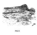
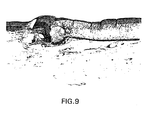
- Figures 4-9 show the results of a comparison of three-layer prothesis with a similarly configured contra-lateral reference material, e-PTFE, in a canine femoral artery study.
- the grafts were implanted in canines as femoral interposition prosthesis. Grafts were explanted from 30 to 256 days.
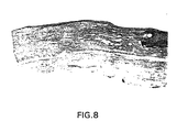
- Histological evaluation of the three-layer collagen graft demonstrated cellular ingrowth into the graft at 30 days, with more than 90 percent of the graft collagen remodeled by 90 days; and a mature 'neo-artery' at 180 days.
- Host tissue bridged the anastomosis by 60 days with the anastomosis only demarcated by the non-resorbable sutures.
- the predominant cell type in the neo-artery was a positive g-Actin staining smooth muscle like cell.
- the surface of the remodeled graft was lined by endothelial cells as demonstrated by SEM, TEM and Factor VIII staining.
- Figure 8 is a Verhoeff's elastic stain (10x) of the proximal anastomosis of a three-layer prosthesis implanted as a canine femoral interposition prosthesis at 256 days compared with Figure 9 of an e-PTFE graft.
Landscapes
- Health & Medical Sciences (AREA)
- Life Sciences & Earth Sciences (AREA)
- Chemical & Material Sciences (AREA)
- Animal Behavior & Ethology (AREA)
- General Health & Medical Sciences (AREA)
- Transplantation (AREA)
- Veterinary Medicine (AREA)
- Oral & Maxillofacial Surgery (AREA)
- Public Health (AREA)
- Medicinal Chemistry (AREA)
- Epidemiology (AREA)
- Dermatology (AREA)
- Engineering & Computer Science (AREA)
- Biomedical Technology (AREA)
- Chemical Kinetics & Catalysis (AREA)
- Botany (AREA)
- Vascular Medicine (AREA)
- Molecular Biology (AREA)
- Cardiology (AREA)
- Biophysics (AREA)
- Heart & Thoracic Surgery (AREA)
- Urology & Nephrology (AREA)
- Zoology (AREA)
- Pulmonology (AREA)
- Gastroenterology & Hepatology (AREA)
- General Chemical & Material Sciences (AREA)
- Materials For Medical Uses (AREA)
- Prostheses (AREA)
- Display Devices Of Pinball Game Machines (AREA)
Claims (20)
- Prothèse comprenant une première couche de collagène fibrillaire ou non fibrillaire extrait à l'acide et une deuxième couche provenant de la sous-muqueuse de l'intestin grêle d'un mammifère qui fournit une stabilité structurelle, est pliable, et est semi-perméable et suturable, ladite prothèse étant un modèle de bioremodelage et lorsqu'elle est implantée dans un patient mammifère, subissant une biodégradation contrôlée se produisant avec le remplacement adéquat par des cellules vivantes de telle manière que la prothèse est bioremodelée par les cellules vivantes du patient.
- Prothèse selon la revendication 1, dans laquelle la prothèse est tubulaire et sert de dérivation ou au remplacement de vaisseaux sanguins de faible diamètre dans le patient mammifère.
- Prothèse selon la revendication 2, dans laquelle la prothèse tubulaire a un diamètre de lumière de moins de 6 mm.
- Prothèse selon la revendication 1, dans laquelle la prothèse sert au remplacement d'organes malades ou endommagés tels que l'oesophage, l'intestin, l'urètre, ou les trompes de Fallope.
- Prothèse selon la revendication 1, dans laquelle ladite première couche a une surface d'écoulement lisse.
- Prothèse selon la revendication 5, dans laquelle la surface d'écoulement lisse résiste à la thrombose.
- Prothèse selon l'une quelconque des revendications précédentes, dans laquelle ladite deuxième couche est réticulée.
- Prothèse selon la revendication 7, dans laquelle ladite deuxième couche est réticulée avec du chlorhydrate de 1-éthyl-3-(3-diméthylaminopropyl)carbodiimide (EDC).
- Prothèse selon l'une quelconque des revendications précédentes, dans laquelle ladite deuxième couche a une épaisseur comprise entre environ 50 microns à environ 150 microns.
- Prothèse selon l'une quelconque des revendications précédentes, dans laquelle la sous-muqueuse de l'intestin grêle est nettoyé mécaniquement pour éliminer la tunique musculaire et la muqueuse.
- Prothèse selon la revendication 10, dans laquelle ladite sous-muqueuse nettoyée mécaniquement est nettoyée chimiquement.
- Prothèse selon l'une quelconque des revendications précédentes, dans laquelle la sous-muqueuse de l'intestin grêle est stérilisée.
- Prothèse selon l'une quelconque des revendications précédentes, ladite prothèse comprenant en outre une troisième couche.
- Prothèse selon l'une quelconque des revendications précédentes, dans laquelle la première couche se trouve du côté intérieur de la prothèse.
- Prothèse selon l'une quelconque des revendications 1 à 13, dans laquelle la deuxième couche se trouve du côté intérieur de la prothèse.
- Prothèse selon l'une quelconque des revendications précédentes, la prothèse étant traitée avec un médicament.
- Prothèse selon l'une quelconque des revendications précédentes, dans laquelle la première couche est traitée avec un médicament.
- Prothèse selon l'une quelconque des revendications précédentes, dans laquelle la deuxième couche est traitée avec un médicament.
- Prothèse selon l'une quelconque des revendications 16 à 18, dans laquelle le médicament est l'héparine.
- Prothèse selon l'une quelconque des revendications 16 à 18, dans laquelle le médicament est un facteur de croissance.
Priority Applications (1)
| Application Number | Priority Date | Filing Date | Title |
|---|---|---|---|
| EP03019053A EP1364627B1 (fr) | 1994-02-18 | 1995-02-17 | Procédé pour produire une greffe prothétique bioremodelable en collagène |
Applications Claiming Priority (3)
| Application Number | Priority Date | Filing Date | Title |
|---|---|---|---|
| US19806294A | 1994-02-18 | 1994-02-18 | |
| PCT/US1995/002025 WO1995022301A1 (fr) | 1994-02-18 | 1995-02-17 | Prothese bioremodelable pour la greffe du collagene |
| US198062 | 2000-04-18 |
Related Child Applications (1)
| Application Number | Title | Priority Date | Filing Date |
|---|---|---|---|
| EP03019053A Division EP1364627B1 (fr) | 1994-02-18 | 1995-02-17 | Procédé pour produire une greffe prothétique bioremodelable en collagène |
Publications (3)
| Publication Number | Publication Date |
|---|---|
| EP0746268A1 EP0746268A1 (fr) | 1996-12-11 |
| EP0746268A4 EP0746268A4 (fr) | 1998-04-01 |
| EP0746268B1 true EP0746268B1 (fr) | 2003-10-01 |
Family
ID=22731845
Family Applications (2)
| Application Number | Title | Priority Date | Filing Date |
|---|---|---|---|
| EP03019053A Expired - Lifetime EP1364627B1 (fr) | 1994-02-18 | 1995-02-17 | Procédé pour produire une greffe prothétique bioremodelable en collagène |
| EP95911032A Expired - Lifetime EP0746268B1 (fr) | 1994-02-18 | 1995-02-17 | Greffe prothese en collagene bioremodelable |
Family Applications Before (1)
| Application Number | Title | Priority Date | Filing Date |
|---|---|---|---|
| EP03019053A Expired - Lifetime EP1364627B1 (fr) | 1994-02-18 | 1995-02-17 | Procédé pour produire une greffe prothétique bioremodelable en collagène |
Country Status (7)
| Country | Link |
|---|---|
| EP (2) | EP1364627B1 (fr) |
| JP (1) | JP3765828B2 (fr) |
| AT (2) | ATE372747T1 (fr) |
| CA (1) | CA2183056C (fr) |
| DE (2) | DE69535595T2 (fr) |
| MX (1) | MX9603249A (fr) |
| WO (1) | WO1995022301A1 (fr) |
Families Citing this family (44)
| Publication number | Priority date | Publication date | Assignee | Title |
|---|---|---|---|---|
| US6334872B1 (en) | 1994-02-18 | 2002-01-01 | Organogenesis Inc. | Method for treating diseased or damaged organs |
| US20020095218A1 (en) | 1996-03-12 | 2002-07-18 | Carr Robert M. | Tissue repair fabric |
| US5733337A (en) * | 1995-04-07 | 1998-03-31 | Organogenesis, Inc. | Tissue repair fabric |
| SE9601243D0 (sv) * | 1996-03-29 | 1996-03-29 | Hans Arne Hansson | Promotion of regeneration of organized tissues |
| JP5053470B2 (ja) * | 1996-08-23 | 2012-10-17 | クック バイオテク インコーポレイティド | グラフト・プロテーゼとその材料及び方法 |
| ES2230627T3 (es) * | 1996-12-10 | 2005-05-01 | Cook Biotech, Inc. | Injertos tubulares a partir de submucosa purificada. |
| US5993844A (en) | 1997-05-08 | 1999-11-30 | Organogenesis, Inc. | Chemical treatment, without detergents or enzymes, of tissue to form an acellular, collagenous matrix |
| US6004333A (en) * | 1997-10-31 | 1999-12-21 | Ethicon Endo-Surgery, Inc. | Prosthetic with collagen for tissue repair |
| JP4606583B2 (ja) * | 1998-06-05 | 2011-01-05 | オルガノジェネシス インク. | 生物工学的手法による平面シート状移植補綴 |
| JP4341049B2 (ja) * | 1998-06-05 | 2009-10-07 | オルガノジェネシス インク. | 生物工学により作成したチューブ状移植片補綴 |
| JP4356053B2 (ja) | 1998-06-05 | 2009-11-04 | オルガノジェネシス インク. | 生物工学により作成した血管移植片支持体補綴 |
| MXPA00012061A (es) | 1998-06-05 | 2003-04-22 | Organogenesis Inc | Protesis de injerto vascular biodisenadas. |
| CZ301649B6 (cs) * | 2000-06-28 | 2010-05-12 | Ed. Geistlich Soehne Ag Fur Chemische Industrie Incorporated Under The Laws Of Switzerland | Trubice pro regeneraci nervu a zpusob jejich výroby |
| US6638312B2 (en) | 2000-08-04 | 2003-10-28 | Depuy Orthopaedics, Inc. | Reinforced small intestinal submucosa (SIS) |
| AU9109201A (en) * | 2000-09-18 | 2002-03-26 | Organogenesis Inc | Methods for treating a patient using a bioengineered flat sheet graft prostheses |
| AU2002354913B2 (en) | 2001-07-16 | 2008-07-17 | Depuy Products, Inc. | Unitary surgical device and method |
| ATE499908T1 (de) | 2001-07-16 | 2011-03-15 | Depuy Products Inc | Gerät zur reparatur von knorpelmaterial |
| US7201917B2 (en) | 2001-07-16 | 2007-04-10 | Depuy Products, Inc. | Porous delivery scaffold and method |
| CA2732196A1 (fr) | 2001-12-21 | 2003-07-24 | Organogenesis Inc. | Systeme et procede permettant de former des protheses tubulaires pour greffes au moyen de techniques biologiques |
| AU2003253620A1 (en) * | 2002-06-03 | 2003-12-19 | Nmt Medical, Inc. | Device with biological tissue scaffold for intracardiac defect closure |
| US7056337B2 (en) * | 2003-10-21 | 2006-06-06 | Cook Incorporated | Natural tissue stent |
| US20050283256A1 (en) * | 2004-02-09 | 2005-12-22 | Codman & Shurtleff, Inc. | Collagen device and method of preparing the same |
| US7569233B2 (en) | 2004-05-04 | 2009-08-04 | Depuy Products, Inc. | Hybrid biologic-synthetic bioabsorbable scaffolds |
| US7354627B2 (en) | 2004-12-22 | 2008-04-08 | Depuy Products, Inc. | Method for organizing the assembly of collagen fibers and compositions formed therefrom |
| WO2006102063A2 (fr) * | 2005-03-19 | 2006-09-28 | Cook Biotech Incorporated | Implants prothetiques comprenant un materiau composite a matrice extracellulaire |
| SE528738C2 (sv) * | 2005-06-16 | 2007-02-06 | Sinova Safetech Innovation Ab | Tarmimplantat |
| AU2006262178B2 (en) * | 2005-06-21 | 2012-07-05 | Cook Biotech, Inc. | Implantable graft to close a fistula |
| US7595062B2 (en) | 2005-07-28 | 2009-09-29 | Depuy Products, Inc. | Joint resurfacing orthopaedic implant and associated method |
| US20090311298A1 (en) * | 2005-10-18 | 2009-12-17 | Oranogenesis, In.C | Antimicrobial Collagenous Constructs |
| DE102005054941A1 (de) * | 2005-11-17 | 2007-05-31 | Gelita Ag | Nervenleitschiene |
| US7722665B2 (en) | 2006-07-07 | 2010-05-25 | Graft Technologies, Inc. | System and method for providing a graft in a vascular environment |
| WO2008021391A1 (fr) | 2006-08-15 | 2008-02-21 | Anthrogenesis Corporation | Biomatériau à base de cordon ombilical pour usage médical |
| US8071135B2 (en) | 2006-10-04 | 2011-12-06 | Anthrogenesis Corporation | Placental tissue compositions |
| AU2007318210B2 (en) | 2006-10-06 | 2014-02-20 | Celularity Inc. | Native (telopeptide) placental collagen compositions |
| JP5702515B2 (ja) | 2007-12-28 | 2015-04-15 | 東洋紡株式会社 | 神経再生誘導管 |
| AU2009201541B2 (en) | 2008-04-23 | 2014-12-04 | Integra Lifesciences Corporation | Flowable collagen material for dural closure |
| JP2011206203A (ja) * | 2010-03-29 | 2011-10-20 | Kawasumi Lab Inc | 神経再生チューブ及び神経再生チューブの製造方法 |
| WO2012029887A1 (fr) * | 2010-08-31 | 2012-03-08 | 独立行政法人農業生物資源研究所 | Structure cylindrique et procédé de production de cette structure |
| CA2860850C (fr) | 2011-12-20 | 2019-05-21 | Lifecell Corporation | Produits de tissu en feuille |
| CA2859657C (fr) | 2011-12-20 | 2021-03-02 | Lifecell Corporation | Particules de matrice tissulaire acellulaire et produits fluides qui lescomposent |
| EP3797801A1 (fr) | 2012-01-24 | 2021-03-31 | LifeCell Corporation | Matrices tissulaires allongées |
| US10071184B2 (en) | 2012-02-29 | 2018-09-11 | Collagen Matrix, Inc. | Collagen-coated tissue-based membranes |
| IL297786A (en) | 2020-05-01 | 2022-12-01 | Askel Healthcare Ltd | A method for preparing a three-dimensional scaffold for medical use |
| GB2616438B (en) * | 2022-03-08 | 2024-08-28 | Newtec Vascular Products Ltd | Vascular stent |
Family Cites Families (13)
| Publication number | Priority date | Publication date | Assignee | Title |
|---|---|---|---|---|
| US505678A (en) | 1893-09-26 | Brick-cutting machinery | ||
| US4539716A (en) * | 1981-03-19 | 1985-09-10 | Massachusetts Institute Of Technology | Fabrication of living blood vessels and glandular tissues |
| US4902289A (en) * | 1982-04-19 | 1990-02-20 | Massachusetts Institute Of Technology | Multilayer bioreplaceable blood vessel prosthesis |
| IL74180A (en) * | 1984-01-30 | 1992-06-21 | Meadox Medicals Inc | Drug delivery collagen-impregnated synthetic vascular graft |
| US5336256A (en) * | 1986-04-17 | 1994-08-09 | Uab Research Foundation | Elastomeric polypeptides as vascular prosthetic materials |
| DE68915540T2 (de) * | 1988-03-09 | 1994-10-13 | Terumo Corp | Für zellen eindringbares medizinisches material und kunsthaut. |
| US4902508A (en) * | 1988-07-11 | 1990-02-20 | Purdue Research Foundation | Tissue graft composition |
| US5024671A (en) * | 1988-09-19 | 1991-06-18 | Baxter International Inc. | Microporous vascular graft |
| US5026381A (en) * | 1989-04-20 | 1991-06-25 | Colla-Tec, Incorporated | Multi-layered, semi-permeable conduit for nerve regeneration comprised of type 1 collagen, its method of manufacture and a method of nerve regeneration using said conduit |
| US4990158A (en) * | 1989-05-10 | 1991-02-05 | United States Surgical Corporation | Synthetic semiabsorbable tubular prosthesis |
| US5106949A (en) * | 1989-09-15 | 1992-04-21 | Organogenesis, Inc. | Collagen compositions and methods for preparation thereof |
| US5256418A (en) * | 1990-04-06 | 1993-10-26 | Organogenesis, Inc. | Collagen constructs |
| CS277367B6 (en) * | 1990-12-29 | 1993-01-13 | Krajicek Milan | Three-layered vascular prosthesis |
-
1995
- 1995-02-17 CA CA002183056A patent/CA2183056C/fr not_active Expired - Fee Related
- 1995-02-17 AT AT03019053T patent/ATE372747T1/de not_active IP Right Cessation
- 1995-02-17 JP JP52193295A patent/JP3765828B2/ja not_active Expired - Fee Related
- 1995-02-17 MX MX9603249A patent/MX9603249A/es active IP Right Grant
- 1995-02-17 DE DE69535595T patent/DE69535595T2/de not_active Expired - Lifetime
- 1995-02-17 EP EP03019053A patent/EP1364627B1/fr not_active Expired - Lifetime
- 1995-02-17 AT AT95911032T patent/ATE250901T1/de not_active IP Right Cessation
- 1995-02-17 WO PCT/US1995/002025 patent/WO1995022301A1/fr not_active Ceased
- 1995-02-17 EP EP95911032A patent/EP0746268B1/fr not_active Expired - Lifetime
- 1995-02-17 DE DE69531859T patent/DE69531859T2/de not_active Expired - Lifetime
Also Published As
| Publication number | Publication date |
|---|---|
| MX9603249A (es) | 1997-07-31 |
| DE69535595D1 (de) | 2007-10-25 |
| DE69535595T2 (de) | 2008-06-05 |
| JP3765828B2 (ja) | 2006-04-12 |
| CA2183056A1 (fr) | 1995-08-24 |
| EP0746268A1 (fr) | 1996-12-11 |
| EP1364627A1 (fr) | 2003-11-26 |
| DE69531859T2 (de) | 2004-07-29 |
| ATE250901T1 (de) | 2003-10-15 |
| DE69531859D1 (en) | 2003-11-06 |
| EP0746268A4 (fr) | 1998-04-01 |
| EP1364627B1 (fr) | 2007-09-12 |
| ATE372747T1 (de) | 2007-09-15 |
| JPH09509085A (ja) | 1997-09-16 |
| CA2183056C (fr) | 2001-07-10 |
| WO1995022301A1 (fr) | 1995-08-24 |
Similar Documents
| Publication | Publication Date | Title |
|---|---|---|
| EP1364627B1 (fr) | Procédé pour produire une greffe prothétique bioremodelable en collagène | |
| US6334872B1 (en) | Method for treating diseased or damaged organs | |
| US5733337A (en) | Tissue repair fabric | |
| US7909886B2 (en) | Tissue repair fabric | |
| CA2267408C (fr) | Constructions tubulaires pour greffe sous-muqueuse | |
| US7887576B2 (en) | Endoluminal device with extracellular matrix material and methods | |
| US5019087A (en) | Nerve regeneration conduit | |
| RU2851556C1 (ru) | Способ формирования многослойного биологически активного сосудистого протеза | |
| MXPA97007655A (en) | Tej repair fabric | |
| AU774634B2 (en) | Tubular submucosal graft constructs | |
| Guidoin et al. | Biotextiles as medical implants: 15. Vascular prostheses for open surgery |
Legal Events
| Date | Code | Title | Description |
|---|---|---|---|
| PUAI | Public reference made under article 153(3) epc to a published international application that has entered the european phase |
Free format text: ORIGINAL CODE: 0009012 |
|
| 17P | Request for examination filed |
Effective date: 19960807 |
|
| AK | Designated contracting states |
Kind code of ref document: A1 Designated state(s): AT BE CH DE DK ES FR GB GR IE IT LI LU MC NL PT SE |
|
| A4 | Supplementary search report drawn up and despatched |
Effective date: 19971230 |
|
| AK | Designated contracting states |
Kind code of ref document: A4 Designated state(s): AT BE CH DE DK ES FR GB GR IE IT LI LU MC NL PT SE |
|
| 17Q | First examination report despatched |
Effective date: 20020314 |
|
| GRAH | Despatch of communication of intention to grant a patent |
Free format text: ORIGINAL CODE: EPIDOS IGRA |
|
| GRAS | Grant fee paid |
Free format text: ORIGINAL CODE: EPIDOSNIGR3 |
|
| GRAA | (expected) grant |
Free format text: ORIGINAL CODE: 0009210 |
|
| AK | Designated contracting states |
Kind code of ref document: B1 Designated state(s): AT BE CH DE DK ES FR GB GR IE IT LI LU MC NL PT SE |
|
| PG25 | Lapsed in a contracting state [announced via postgrant information from national office to epo] |
Ref country code: NL Free format text: LAPSE BECAUSE OF FAILURE TO SUBMIT A TRANSLATION OF THE DESCRIPTION OR TO PAY THE FEE WITHIN THE PRESCRIBED TIME-LIMIT Effective date: 20031001 Ref country code: LI Free format text: LAPSE BECAUSE OF FAILURE TO SUBMIT A TRANSLATION OF THE DESCRIPTION OR TO PAY THE FEE WITHIN THE PRESCRIBED TIME-LIMIT Effective date: 20031001 Ref country code: CH Free format text: LAPSE BECAUSE OF FAILURE TO SUBMIT A TRANSLATION OF THE DESCRIPTION OR TO PAY THE FEE WITHIN THE PRESCRIBED TIME-LIMIT Effective date: 20031001 |
|
| REG | Reference to a national code |
Ref country code: GB Ref legal event code: FG4D |
|
| REG | Reference to a national code |
Ref country code: CH Ref legal event code: EP |
|
| REG | Reference to a national code |
Ref country code: IE Ref legal event code: FG4D |
|
| REF | Corresponds to: |
Ref document number: 69531859 Country of ref document: DE Date of ref document: 20031106 Kind code of ref document: P |
|
| PG25 | Lapsed in a contracting state [announced via postgrant information from national office to epo] |
Ref country code: SE Free format text: LAPSE BECAUSE OF FAILURE TO SUBMIT A TRANSLATION OF THE DESCRIPTION OR TO PAY THE FEE WITHIN THE PRESCRIBED TIME-LIMIT Effective date: 20040101 Ref country code: GR Free format text: LAPSE BECAUSE OF FAILURE TO SUBMIT A TRANSLATION OF THE DESCRIPTION OR TO PAY THE FEE WITHIN THE PRESCRIBED TIME-LIMIT Effective date: 20040101 Ref country code: DK Free format text: LAPSE BECAUSE OF FAILURE TO SUBMIT A TRANSLATION OF THE DESCRIPTION OR TO PAY THE FEE WITHIN THE PRESCRIBED TIME-LIMIT Effective date: 20040101 |
|
| PG25 | Lapsed in a contracting state [announced via postgrant information from national office to epo] |
Ref country code: ES Free format text: LAPSE BECAUSE OF FAILURE TO SUBMIT A TRANSLATION OF THE DESCRIPTION OR TO PAY THE FEE WITHIN THE PRESCRIBED TIME-LIMIT Effective date: 20040112 |
|
| NLV1 | Nl: lapsed or annulled due to failure to fulfill the requirements of art. 29p and 29m of the patents act | ||
| REG | Reference to a national code |
Ref country code: CH Ref legal event code: PL |
|
| ET | Fr: translation filed | ||
| PLBE | No opposition filed within time limit |
Free format text: ORIGINAL CODE: 0009261 |
|
| STAA | Information on the status of an ep patent application or granted ep patent |
Free format text: STATUS: NO OPPOSITION FILED WITHIN TIME LIMIT |
|
| 26N | No opposition filed |
Effective date: 20040702 |
|
| PGFP | Annual fee paid to national office [announced via postgrant information from national office to epo] |
Ref country code: MC Payment date: 20070705 Year of fee payment: 13 |
|
| PGFP | Annual fee paid to national office [announced via postgrant information from national office to epo] |
Ref country code: LU Payment date: 20070731 Year of fee payment: 13 |
|
| PG25 | Lapsed in a contracting state [announced via postgrant information from national office to epo] |
Ref country code: PT Free format text: LAPSE BECAUSE OF NON-PAYMENT OF DUE FEES Effective date: 20040301 |
|
| PG25 | Lapsed in a contracting state [announced via postgrant information from national office to epo] |
Ref country code: MC Free format text: LAPSE BECAUSE OF NON-PAYMENT OF DUE FEES Effective date: 20080228 |
|
| PGFP | Annual fee paid to national office [announced via postgrant information from national office to epo] |
Ref country code: AT Payment date: 20080829 Year of fee payment: 14 Ref country code: IE Payment date: 20080829 Year of fee payment: 14 |
|
| PGFP | Annual fee paid to national office [announced via postgrant information from national office to epo] |
Ref country code: BE Payment date: 20080829 Year of fee payment: 14 |
|
| BERE | Be: lapsed |
Owner name: *ORGANOGENESIS INC. Effective date: 20090228 |
|
| PG25 | Lapsed in a contracting state [announced via postgrant information from national office to epo] |
Ref country code: IT Free format text: LAPSE BECAUSE OF NON-PAYMENT OF DUE FEES Effective date: 20080217 |
|
| PG25 | Lapsed in a contracting state [announced via postgrant information from national office to epo] |
Ref country code: AT Free format text: LAPSE BECAUSE OF NON-PAYMENT OF DUE FEES Effective date: 20090217 |
|
| REG | Reference to a national code |
Ref country code: IE Ref legal event code: MM4A |
|
| PGRI | Patent reinstated in contracting state [announced from national office to epo] |
Ref country code: IT Effective date: 20091001 |
|
| PG25 | Lapsed in a contracting state [announced via postgrant information from national office to epo] |
Ref country code: IE Free format text: LAPSE BECAUSE OF NON-PAYMENT OF DUE FEES Effective date: 20090217 |
|
| PG25 | Lapsed in a contracting state [announced via postgrant information from national office to epo] |
Ref country code: BE Free format text: LAPSE BECAUSE OF NON-PAYMENT OF DUE FEES Effective date: 20090228 |
|
| PG25 | Lapsed in a contracting state [announced via postgrant information from national office to epo] |
Ref country code: LU Free format text: LAPSE BECAUSE OF NON-PAYMENT OF DUE FEES Effective date: 20080217 |
|
| PGRI | Patent reinstated in contracting state [announced from national office to epo] |
Ref country code: IT Effective date: 20091001 |
|
| PGRI | Patent reinstated in contracting state [announced from national office to epo] |
Ref country code: IT Effective date: 20110616 |
|
| PGFP | Annual fee paid to national office [announced via postgrant information from national office to epo] |
Ref country code: DE Payment date: 20140228 Year of fee payment: 20 |
|
| PGFP | Annual fee paid to national office [announced via postgrant information from national office to epo] |
Ref country code: IT Payment date: 20140228 Year of fee payment: 20 Ref country code: FR Payment date: 20140228 Year of fee payment: 20 |
|
| PGFP | Annual fee paid to national office [announced via postgrant information from national office to epo] |
Ref country code: GB Payment date: 20140227 Year of fee payment: 20 |
|
| REG | Reference to a national code |
Ref country code: DE Ref legal event code: R071 Ref document number: 69531859 Country of ref document: DE |
|
| REG | Reference to a national code |
Ref country code: GB Ref legal event code: PE20 Expiry date: 20150216 |
|
| PG25 | Lapsed in a contracting state [announced via postgrant information from national office to epo] |
Ref country code: GB Free format text: LAPSE BECAUSE OF EXPIRATION OF PROTECTION Effective date: 20150216 |