EP0760476A2 - Verfahren zur quantitativen Bestimmung einer oder mehrerer Charakteristiken einer Substanz - Google Patents
Verfahren zur quantitativen Bestimmung einer oder mehrerer Charakteristiken einer Substanz Download PDFInfo
- Publication number
- EP0760476A2 EP0760476A2 EP96306179A EP96306179A EP0760476A2 EP 0760476 A2 EP0760476 A2 EP 0760476A2 EP 96306179 A EP96306179 A EP 96306179A EP 96306179 A EP96306179 A EP 96306179A EP 0760476 A2 EP0760476 A2 EP 0760476A2
- Authority
- EP
- European Patent Office
- Prior art keywords
- radiation
- locations
- substance
- detecting locations
- determining
- Prior art date
- Legal status (The legal status is an assumption and is not a legal conclusion. Google has not performed a legal analysis and makes no representation as to the accuracy of the status listed.)
- Withdrawn
Links
Images
Classifications
-
- A—HUMAN NECESSITIES
- A61—MEDICAL OR VETERINARY SCIENCE; HYGIENE
- A61B—DIAGNOSIS; SURGERY; IDENTIFICATION
- A61B5/00—Measuring for diagnostic purposes; Identification of persons
- A61B5/145—Measuring characteristics of blood in vivo, e.g. gas concentration or pH-value ; Measuring characteristics of body fluids or tissues, e.g. interstitial fluid or cerebral tissue
- A61B5/1455—Measuring characteristics of blood in vivo, e.g. gas concentration or pH-value ; Measuring characteristics of body fluids or tissues, e.g. interstitial fluid or cerebral tissue using optical sensors, e.g. spectral photometrical oximeters
- A61B5/14551—Measuring characteristics of blood in vivo, e.g. gas concentration or pH-value ; Measuring characteristics of body fluids or tissues, e.g. interstitial fluid or cerebral tissue using optical sensors, e.g. spectral photometrical oximeters for measuring blood gases
- A61B5/14553—Measuring characteristics of blood in vivo, e.g. gas concentration or pH-value ; Measuring characteristics of body fluids or tissues, e.g. interstitial fluid or cerebral tissue using optical sensors, e.g. spectral photometrical oximeters for measuring blood gases specially adapted for cerebral tissue
-
- G—PHYSICS
- G01—MEASURING; TESTING
- G01N—INVESTIGATING OR ANALYSING MATERIALS BY DETERMINING THEIR CHEMICAL OR PHYSICAL PROPERTIES
- G01N21/00—Investigating or analysing materials by the use of optical means, i.e. using sub-millimetre waves, infrared, visible or ultraviolet light
- G01N21/17—Systems in which incident light is modified in accordance with the properties of the material investigated
- G01N21/25—Colour; Spectral properties, i.e. comparison of effect of material on the light at two or more different wavelengths or wavelength bands
- G01N21/31—Investigating relative effect of material at wavelengths characteristic of specific elements or molecules, e.g. atomic absorption spectrometry
- G01N21/35—Investigating relative effect of material at wavelengths characteristic of specific elements or molecules, e.g. atomic absorption spectrometry using infrared light
- G01N21/359—Investigating relative effect of material at wavelengths characteristic of specific elements or molecules, e.g. atomic absorption spectrometry using infrared light using near infrared light
-
- G—PHYSICS
- G01—MEASURING; TESTING
- G01N—INVESTIGATING OR ANALYSING MATERIALS BY DETERMINING THEIR CHEMICAL OR PHYSICAL PROPERTIES
- G01N21/00—Investigating or analysing materials by the use of optical means, i.e. using sub-millimetre waves, infrared, visible or ultraviolet light
- G01N21/17—Systems in which incident light is modified in accordance with the properties of the material investigated
- G01N21/47—Scattering, i.e. diffuse reflection
- G01N21/49—Scattering, i.e. diffuse reflection within a body or fluid
Definitions
- This invention relates to a method of quantitatively determining one or more characteristics of a substance using near infrared spectroscopy.
- NIRS Near Infrared Spectroscopy
- the above method for measuring path length is used in a single channel tissue spectroscopy system where the measurement of quantified change is achievable.
- a dual channel tissue spectroscopy system is intended to allow dynamic cancellation of these superficially positioned effects in order to allow reliable measurement of the oxygen supply deep within the cerebral cortex.
- the known two channel device does not account for light divergence, constant background absorbers, or optical path length
- the known device relied on a calibration procedure in which a specific contribution from the venous and arterial compartments in the tissue sample volume was assumed.
- a method of quantitatively determining one or more characteristics of a substance by measurement of radiation absorption spectra of the substance comprising:
- the method includes determining the relative coupling efficiencies of detectors at the detecting locations (D1, D2) by use of an emitter positioned equidistant between the detecting locations (D1, D2) and, preferably, step vi) includes modifying the result by accounting for the effect of the relative detector coupling efficiencies.
- a factor is accounted for when determining the characteristic, the factor relating to radiation absorbers present in the substance other than that of the characteristic being measured and an absorption offset is subtracted from the detected radiation intensities.
- a ratio of the intensities of the detected radiation at the two detecting locations may be taken, removing a need for a measure of the attenuation of the radiation at the point of irradiation.
- a factor may be accounted for when determining the characteristic being measured, the factor relating to the relative coupling efficiency of detectors at the detecting locations.
- the effect of divergence of the radiation detected at the two detecting locations is 1/d 2 , wherein d is the optical path length to each detecting location from the irradiation point.
- More than one characteristic may be measured by using radiation at more than two distinct wavelengths.
- the substance may be part of a human or animal body and the radiation is near infrared radiation.
- the characteristic is the haemoglobin or cytochrome concentration of the tissue of the human or animal body.
- the substance is the tissue of the cerebral cortex.
- Figure 1 shows a simplified cross section of a sensor 10 placed on the surface of the head over the cerebral cortex 40.
- the characteristic is a single chromophore to be measured within the cerebral cortex 40.
- a second optical path (d 2 ) is defined from the same emitter 20 to detector 2 (D2). It should once again be possible to predict this different average path length, but as before unpredictable intensity reducing factors are still present.
- Figures 2a and 2b provide a representation of the two optical path lengths (d 1 , d 2 ) of Figure 1 which pass through an absorbing layer 50 made up of skin layers, bone, meninges etc. and through the cerebral cortex 40.
- This system can be mathematically represented as follows.
- I t 2 I i ⁇ K 1 ⁇ 10 - I 1 k 1 c 1 ⁇ 10 - C ⁇ d 21 B+G 2 ⁇ 10 - I 2 k 2 c 2 ⁇ K 2
- the intensity of radiation measured at detector 2 can be defined as follows:
- the path lengths used in Equation 5 are those through the brain tissue. However the path length difference is related to the calculable path difference as shown in 8.
- d 11 d 1 - (I 1 + I 2 )
- d 21 d 2 - (I 1 + I 2 )
- Equation 9 provides a relationship of the concentration of a particular chromophore C, to the measurable light intensities at detectors 1 and 2, related by its absorption coefficient and constant terms which should be fixed by the geometry of the sensor 10. This is effectively an absorption measurement which corresponds to the deep tissue of pathlength d 2 - d 1 , which is sited at the centre of path 2. (The implications of slight variations in the quality of sensor attachment will be discussed in relation to the LED coupling system described below).
- This analysis can be extended to a multiple wavelength, multiple chromophore system.
- Equations 12 and 13 can now be rewritten after taking Logarithms.
- Equation 17 defines the concentrations of the two chromophores with respect to the intensities of the two wavelengths of light measured at the two detectors. In this form it allows a solution for the concentrations of the chromophores so long as the absorption coefficients and pathlengths have been previously defined.
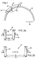
- FIG. 3 is a representation of a sensor 10 with a light source 30 in the form of a light emitting diode (LED) provided midway between the detectors D1, D2 as described below.
- LED light emitting diode
- the principle of operation of the previously described dual channel system relies on the optical coupling and the gains of the two detector channels being precisely matched, or else well defined. If matched, this allows the coupling efficiencies between the two channels to effectively cancel each other out. If the coupling efficiencies are well defined it allows them to be calculated out of the final results.
- This invention uses a light source 30 mounted midway between both detectors which is aimed directly into the tissue under investigation. (i.e. not preferentially directed towards either of the detectors.) This should create an illumination within the tissue the intensity of which should be the same at each detector. Using this even illumination on both detectors, the gains on the two detection channels can now be measured. This will be a combination of their optical gain together with their electronic amplification gain. This provides a ratio of the overall gains of the two channels, combined with a measure of the relative coupling efficiencies and relative superficial absorber concentrations under each detector, for example the effect of a freckle under one detector, which would effect the measured intensity, will be cancelled out. This ratio can then be applied to the division which is performed in the two channel processing algorithm to correct for any unequal gains which are present.
- This ratio can now be used to calibrate the relative total gains between channel 1 and 2 in the following way:
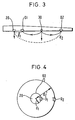
- Figure 4 is a representation of the effect of beam divergence on the detected intensity at detectors D1, D2.
- spherical shells 60 represent the detector spacings used in the two channel system.
- Equation 21 reveals that the G 2 - G 1 components within the equation are taking the form of an additional absorption.
- this additional absorption would have also included the extracerebral tissues, and emitter and detector coupling efficiencies, in addition to the fixed absorbers included within the cerebral cortex.
- the effects of extracerebral tissues and coupling efficiencies are removed by the use of the two channel system, which then only leaves the fixed absorbers present in the cerebral cortex.
- the effects of these fixed absorbers on the calculations for final chromophore concentration can be estimated. This can be done by estimating the absorption which would be caused by the quantities of substances found within the cerebral cortex, over the associated optical pathlength. This can be viewed as an absorption offset, and can be directly associated to chromophore offsets which would be measured by a dual channel tissue spectroscopy system. This absorption offset can effectively be subtracted from the signals measured by an instrument to allow a more direct measurement of the absolute chromophore concentrations within the cerebral cortex.
- the method of quantitatively determining the characteristics of a substance has been described in its application to cerebral measurements.
- the method can also be used to non-invasively monitor tissue haemoglobin concentration in other parts of the body and may be useful in fields such as plastic surgery and vascular surgery.
Landscapes
- Physics & Mathematics (AREA)
- Health & Medical Sciences (AREA)
- Life Sciences & Earth Sciences (AREA)
- Spectroscopy & Molecular Physics (AREA)
- General Health & Medical Sciences (AREA)
- Pathology (AREA)
- Chemical & Material Sciences (AREA)
- Analytical Chemistry (AREA)
- Biochemistry (AREA)
- General Physics & Mathematics (AREA)
- Immunology (AREA)
- Optics & Photonics (AREA)
- Molecular Biology (AREA)
- Biophysics (AREA)
- Engineering & Computer Science (AREA)
- Biomedical Technology (AREA)
- Heart & Thoracic Surgery (AREA)
- Medical Informatics (AREA)
- Neurology (AREA)
- Surgery (AREA)
- Animal Behavior & Ethology (AREA)
- Public Health (AREA)
- Veterinary Medicine (AREA)
- Investigating Or Analysing Materials By Optical Means (AREA)
- Measurement Of The Respiration, Hearing Ability, Form, And Blood Characteristics Of Living Organisms (AREA)
Applications Claiming Priority (2)
| Application Number | Priority Date | Filing Date | Title |
|---|---|---|---|
| GB9517366 | 1995-08-24 | ||
| GBGB9517366.2A GB9517366D0 (en) | 1995-08-24 | 1995-08-24 | Method of quantatively determining one or more characteristics of a substance |
Publications (2)
| Publication Number | Publication Date |
|---|---|
| EP0760476A2 true EP0760476A2 (de) | 1997-03-05 |
| EP0760476A3 EP0760476A3 (de) | 1998-02-04 |
Family
ID=10779712
Family Applications (1)
| Application Number | Title | Priority Date | Filing Date |
|---|---|---|---|
| EP96306179A Withdrawn EP0760476A3 (de) | 1995-08-24 | 1996-08-23 | Verfahren zur quantitativen Bestimmung einer oder mehrerer Charakteristiken einer Substanz |
Country Status (4)
| Country | Link |
|---|---|
| US (1) | US5661302A (de) |
| EP (1) | EP0760476A3 (de) |
| JP (1) | JPH09168531A (de) |
| GB (1) | GB9517366D0 (de) |
Cited By (7)
| Publication number | Priority date | Publication date | Assignee | Title |
|---|---|---|---|---|
| EP1259791A4 (de) * | 2000-05-02 | 2007-05-02 | Cas Medical Systems Inc | Verfahren zur nicht-invasiven spektrophotometrischen überwachung der sauerstoffsättigung des blutes |
| WO2010144670A1 (en) * | 2009-06-10 | 2010-12-16 | Medtronic, Inc. | Device and method for monitoring of absolute oxygen saturation and tissue hemoglobin concentration |
| US8352008B2 (en) | 2009-06-10 | 2013-01-08 | Medtronic, Inc. | Active noise cancellation in an optical sensor signal |
| US8391979B2 (en) | 2009-06-10 | 2013-03-05 | Medtronic, Inc. | Shock reduction using absolute calibrated tissue oxygen saturation and total hemoglobin volume fraction |
| US8463346B2 (en) | 2009-06-10 | 2013-06-11 | Medtronic, Inc. | Absolute calibrated tissue oxygen saturation and total hemoglobin volume fraction |
| US8515537B2 (en) | 2009-06-10 | 2013-08-20 | Medtronic, Inc. | Tissue oxygenation monitoring in heart failure |
| US8639332B2 (en) | 2009-09-11 | 2014-01-28 | Medtronic, Inc. | Method and apparatus for post-shock evaluation using tissue oxygenation measurements |
Families Citing this family (18)
| Publication number | Priority date | Publication date | Assignee | Title |
|---|---|---|---|---|
| WO1998023916A1 (fr) * | 1996-11-26 | 1998-06-04 | Omron Corporation | Procede et appareil permettant de mesurer la concentration d'une substance absorbant la lumiere dans un tissu vivant et l'epaisseur d'un tissu intercalaire |
| USRE45607E1 (en) * | 1998-10-13 | 2015-07-14 | Covidien Lp | Multi-channel non-invasive tissue oximeter |
| US7047054B2 (en) * | 1999-03-12 | 2006-05-16 | Cas Medical Systems, Inc. | Laser diode optical transducer assembly for non-invasive spectrophotometric blood oxygenation monitoring |
| US6339714B1 (en) | 1999-09-13 | 2002-01-15 | Bo Chen | Apparatus and method for measuring concentrations of a dye in a living organism |
| JP2001198112A (ja) * | 2000-01-20 | 2001-07-24 | Hitachi Medical Corp | 生体光計測装置 |
| JP4465271B2 (ja) | 2002-07-26 | 2010-05-19 | シーエーエス・メディカル・システムズ・インコーポレイテッド | 対象組織内の血液酸素飽和度を非侵襲的に決定する装置 |
| US8396524B2 (en) * | 2006-09-27 | 2013-03-12 | Covidien Lp | Medical sensor and technique for using the same |
| US8515511B2 (en) | 2009-09-29 | 2013-08-20 | Covidien Lp | Sensor with an optical coupling material to improve plethysmographic measurements and method of using the same |
| WO2012050847A2 (en) * | 2010-09-28 | 2012-04-19 | Masimo Corporation | Depth of consciousness monitor including oximeter |
| JP5849042B2 (ja) * | 2012-12-27 | 2016-01-27 | 株式会社スペクトラテック | 素子を装着するためのホルダ及び同ホルダを用いた生体情報計測装置 |
| JP6101549B2 (ja) * | 2013-04-25 | 2017-03-22 | 株式会社日立製作所 | 光学的脳表面計測用衝撃緩衝具、それを用いた光計測プローブおよびその保持具 |
| US10213550B2 (en) | 2014-01-23 | 2019-02-26 | Covidien Lp | Systems and methods for monitoring clinical procedures using regional blood oxygen saturation |
| US9867561B2 (en) | 2014-01-27 | 2018-01-16 | Covidien Lp | Systems and methods for determining whether regional oximetry sensors are properly positioned |
| US9861317B2 (en) | 2014-02-20 | 2018-01-09 | Covidien Lp | Methods and systems for determining regional blood oxygen saturation |
| JP6412956B2 (ja) * | 2014-12-22 | 2018-10-24 | 株式会社日立製作所 | 生体光計測装置、解析装置、及び方法 |
| US10328202B2 (en) | 2015-02-04 | 2019-06-25 | Covidien Lp | Methods and systems for determining fluid administration |
| EP3473172A1 (de) | 2017-10-20 | 2019-04-24 | Universität Zürich | Vorrichtung zur messung optischer parameter in streuungsmedien |
| WO2020239922A1 (en) | 2019-05-28 | 2020-12-03 | Universität Zürich | Optical apparatus comprising two self-calibrated optical measurement sets |
Family Cites Families (8)
| Publication number | Priority date | Publication date | Assignee | Title |
|---|---|---|---|---|
| DE69029152T2 (de) * | 1990-02-15 | 1997-03-06 | Hewlett Packard Gmbh | Verfahren zur nichtinvasiven Messung der Sauerstoffsättigung |
| US5379764A (en) * | 1992-12-09 | 1995-01-10 | Diasense, Inc. | Non-invasive determination of analyte concentration in body of mammals |
| EP0700267A4 (de) * | 1993-05-28 | 1998-06-24 | Somanetics Corp | Verfahren und vorrichtung zur spektrophotometrischen zerebral-oxymetrie |
| JP3433498B2 (ja) * | 1993-06-02 | 2003-08-04 | 浜松ホトニクス株式会社 | 散乱吸収体の内部情報計測方法及び装置 |
| JP3577335B2 (ja) * | 1993-06-02 | 2004-10-13 | 浜松ホトニクス株式会社 | 散乱吸収体計測方法及び装置 |
| JP2780935B2 (ja) * | 1994-09-22 | 1998-07-30 | 浜松ホトニクス株式会社 | 散乱吸収体の吸収成分の濃度計測方法及び装置 |
| JP3433534B2 (ja) * | 1994-11-07 | 2003-08-04 | 浜松ホトニクス株式会社 | 散乱吸収体内の散乱特性・吸収特性の測定方法及び装置 |
| GB9501663D0 (en) * | 1995-01-27 | 1995-03-15 | Johnson & Johnson Medical | Non-invasive medical sensor |
-
1995
- 1995-08-24 GB GBGB9517366.2A patent/GB9517366D0/en active Pending
-
1996
- 1996-08-23 US US08/697,386 patent/US5661302A/en not_active Expired - Fee Related
- 1996-08-23 JP JP8240013A patent/JPH09168531A/ja active Pending
- 1996-08-23 EP EP96306179A patent/EP0760476A3/de not_active Withdrawn
Cited By (11)
| Publication number | Priority date | Publication date | Assignee | Title |
|---|---|---|---|---|
| EP1259791A4 (de) * | 2000-05-02 | 2007-05-02 | Cas Medical Systems Inc | Verfahren zur nicht-invasiven spektrophotometrischen überwachung der sauerstoffsättigung des blutes |
| WO2010144670A1 (en) * | 2009-06-10 | 2010-12-16 | Medtronic, Inc. | Device and method for monitoring of absolute oxygen saturation and tissue hemoglobin concentration |
| US8352008B2 (en) | 2009-06-10 | 2013-01-08 | Medtronic, Inc. | Active noise cancellation in an optical sensor signal |
| US8391979B2 (en) | 2009-06-10 | 2013-03-05 | Medtronic, Inc. | Shock reduction using absolute calibrated tissue oxygen saturation and total hemoglobin volume fraction |
| US8463346B2 (en) | 2009-06-10 | 2013-06-11 | Medtronic, Inc. | Absolute calibrated tissue oxygen saturation and total hemoglobin volume fraction |
| US8515537B2 (en) | 2009-06-10 | 2013-08-20 | Medtronic, Inc. | Tissue oxygenation monitoring in heart failure |
| US8634890B2 (en) | 2009-06-10 | 2014-01-21 | Medtronic, Inc. | Device and method for monitoring of absolute oxygen saturation and tissue hemoglobin concentration |
| US9126049B2 (en) | 2009-06-10 | 2015-09-08 | Medtronic, Inc. | Shock reduction using absolute calibrated tissue oxygen saturation and total hemoglobin volume fraction |
| US10179242B2 (en) | 2009-06-10 | 2019-01-15 | Medtronic, Inc. | Tissue oxygenation monitoring in heart failure |
| US8639332B2 (en) | 2009-09-11 | 2014-01-28 | Medtronic, Inc. | Method and apparatus for post-shock evaluation using tissue oxygenation measurements |
| US8918171B2 (en) | 2009-09-11 | 2014-12-23 | Medtronic, Inc. | Method and apparatus for post-shock evaluation using tissue oxygenation measurements |
Also Published As
| Publication number | Publication date |
|---|---|
| GB9517366D0 (en) | 1995-10-25 |
| EP0760476A3 (de) | 1998-02-04 |
| JPH09168531A (ja) | 1997-06-30 |
| US5661302A (en) | 1997-08-26 |
Similar Documents
| Publication | Publication Date | Title |
|---|---|---|
| US5661302A (en) | Method of quatitatively determining one or more characteristics of a substance | |
| US6456862B2 (en) | Method for non-invasive spectrophotometric blood oxygenation monitoring | |
| JP5271700B2 (ja) | 光反射率測定値を補正するためのシステム及び方法 | |
| US8078250B2 (en) | Method for spectrophotometric blood oxygenation monitoring | |
| JP3433498B2 (ja) | 散乱吸収体の内部情報計測方法及び装置 | |
| CA2215163C (en) | Isolated layer pulse oximetry | |
| AU2006247746B2 (en) | Improved method for spectrophotometric blood oxygenation monitoring | |
| Zhang et al. | Study of near infrared technology for intracranial hematoma detection | |
| EP0352923A1 (de) | Spektralfutumetrisches Gerät und Verfahren zur Anzeige der Sauerstoffsättigung | |
| US5513642A (en) | Reflectance sensor system | |
| JP3195951B2 (ja) | 無侵襲医療用センサ | |
| US20240049996A1 (en) | Nirs / tissue oximetry based method to measure arterial blood oxygen saturation from pulsatile hemoglobin waveforms | |
| Smith et al. | Time-resolved spectroscopy and the determination of photon scattering, pathlength, and brain vascular hemoglobin saturation in a population of normal volunteers | |
| Liu et al. | Spatial sensitivity of nirs tissue oxygenation measurement using a simplified instrument |
Legal Events
| Date | Code | Title | Description |
|---|---|---|---|
| PUAI | Public reference made under article 153(3) epc to a published international application that has entered the european phase |
Free format text: ORIGINAL CODE: 0009012 |
|
| AK | Designated contracting states |
Kind code of ref document: A2 Designated state(s): DE FR GB SE |
|
| PUAL | Search report despatched |
Free format text: ORIGINAL CODE: 0009013 |
|
| AK | Designated contracting states |
Kind code of ref document: A3 Designated state(s): DE FR GB SE |
|
| 17P | Request for examination filed |
Effective date: 19980709 |
|
| RAP1 | Party data changed (applicant data changed or rights of an application transferred) |
Owner name: CRITIKON COMPANY, L.L.C. |
|
| 17Q | First examination report despatched |
Effective date: 20030417 |
|
| STAA | Information on the status of an ep patent application or granted ep patent |
Free format text: STATUS: THE APPLICATION IS DEEMED TO BE WITHDRAWN |
|
| 18D | Application deemed to be withdrawn |
Effective date: 20060301 |