EP0767378A1 - Procédé pour le diagnostic et l'analyse du tumeurs de l'estomac - Google Patents
Procédé pour le diagnostic et l'analyse du tumeurs de l'estomac Download PDFInfo
- Publication number
- EP0767378A1 EP0767378A1 EP96113179A EP96113179A EP0767378A1 EP 0767378 A1 EP0767378 A1 EP 0767378A1 EP 96113179 A EP96113179 A EP 96113179A EP 96113179 A EP96113179 A EP 96113179A EP 0767378 A1 EP0767378 A1 EP 0767378A1
- Authority
- EP
- European Patent Office
- Prior art keywords
- exon
- antibody
- exons
- variant
- antibodies
- Prior art date
- Legal status (The legal status is an assumption and is not a legal conclusion. Google has not performed a legal analysis and makes no representation as to the accuracy of the status listed.)
- Ceased
Links
Images


Classifications
-
- G—PHYSICS
- G01—MEASURING; TESTING
- G01N—INVESTIGATING OR ANALYSING MATERIALS BY DETERMINING THEIR CHEMICAL OR PHYSICAL PROPERTIES
- G01N33/00—Investigating or analysing materials by specific methods not covered by groups G01N1/00 - G01N31/00
- G01N33/48—Biological material, e.g. blood, urine; Haemocytometers
- G01N33/50—Chemical analysis of biological material, e.g. blood, urine; Testing involving biospecific ligand binding methods; Immunological testing
- G01N33/53—Immunoassay; Biospecific binding assay; Materials therefor
- G01N33/575—Immunoassay; Biospecific binding assay; Materials therefor for cancer
- G01N33/5753—Immunoassay; Biospecific binding assay; Materials therefor for cancer of the stomach or small intestine
-
- C—CHEMISTRY; METALLURGY
- C07—ORGANIC CHEMISTRY
- C07K—PEPTIDES
- C07K14/00—Peptides having more than 20 amino acids; Gastrins; Somatostatins; Melanotropins; Derivatives thereof
- C07K14/435—Peptides having more than 20 amino acids; Gastrins; Somatostatins; Melanotropins; Derivatives thereof from animals; from humans
- C07K14/705—Receptors; Cell surface antigens; Cell surface determinants
- C07K14/70585—CD44
-
- C—CHEMISTRY; METALLURGY
- C07—ORGANIC CHEMISTRY
- C07K—PEPTIDES
- C07K16/00—Immunoglobulins [IG], e.g. monoclonal or polyclonal antibodies
- C07K16/18—Immunoglobulins [IG], e.g. monoclonal or polyclonal antibodies against material from animals or humans
- C07K16/28—Immunoglobulins [IG], e.g. monoclonal or polyclonal antibodies against material from animals or humans against receptors, cell surface antigens or cell surface determinants
- C07K16/2884—Immunoglobulins [IG], e.g. monoclonal or polyclonal antibodies against material from animals or humans against receptors, cell surface antigens or cell surface determinants against CD44
-
- C—CHEMISTRY; METALLURGY
- C12—BIOCHEMISTRY; BEER; SPIRITS; WINE; VINEGAR; MICROBIOLOGY; ENZYMOLOGY; MUTATION OR GENETIC ENGINEERING
- C12Q—MEASURING OR TESTING PROCESSES INVOLVING ENZYMES, NUCLEIC ACIDS OR MICROORGANISMS; COMPOSITIONS OR TEST PAPERS THEREFOR; PROCESSES OF PREPARING SUCH COMPOSITIONS; CONDITION-RESPONSIVE CONTROL IN MICROBIOLOGICAL OR ENZYMOLOGICAL PROCESSES
- C12Q1/00—Measuring or testing processes involving enzymes, nucleic acids or microorganisms; Compositions therefor; Processes of preparing such compositions
- C12Q1/68—Measuring or testing processes involving enzymes, nucleic acids or microorganisms; Compositions therefor; Processes of preparing such compositions involving nucleic acids
- C12Q1/6876—Nucleic acid products used in the analysis of nucleic acids, e.g. primers or probes
- C12Q1/6883—Nucleic acid products used in the analysis of nucleic acids, e.g. primers or probes for diseases caused by alterations of genetic material
- C12Q1/6886—Nucleic acid products used in the analysis of nucleic acids, e.g. primers or probes for diseases caused by alterations of genetic material for cancer
-
- G—PHYSICS
- G01—MEASURING; TESTING
- G01N—INVESTIGATING OR ANALYSING MATERIALS BY DETERMINING THEIR CHEMICAL OR PHYSICAL PROPERTIES
- G01N33/00—Investigating or analysing materials by specific methods not covered by groups G01N1/00 - G01N31/00
- G01N33/48—Biological material, e.g. blood, urine; Haemocytometers
- G01N33/50—Chemical analysis of biological material, e.g. blood, urine; Testing involving biospecific ligand binding methods; Immunological testing
- G01N33/53—Immunoassay; Biospecific binding assay; Materials therefor
- G01N33/566—Immunoassay; Biospecific binding assay; Materials therefor using specific carrier or receptor proteins as ligand binding reagents where possible specific carrier or receptor proteins are classified with their target compounds
-
- G—PHYSICS
- G01—MEASURING; TESTING
- G01N—INVESTIGATING OR ANALYSING MATERIALS BY DETERMINING THEIR CHEMICAL OR PHYSICAL PROPERTIES
- G01N33/00—Investigating or analysing materials by specific methods not covered by groups G01N1/00 - G01N31/00
- G01N33/48—Biological material, e.g. blood, urine; Haemocytometers
- G01N33/50—Chemical analysis of biological material, e.g. blood, urine; Testing involving biospecific ligand binding methods; Immunological testing
- G01N33/53—Immunoassay; Biospecific binding assay; Materials therefor
- G01N33/575—Immunoassay; Biospecific binding assay; Materials therefor for cancer
- G01N33/57515—Immunoassay; Biospecific binding assay; Materials therefor for cancer of the breast
-
- G—PHYSICS
- G01—MEASURING; TESTING
- G01N—INVESTIGATING OR ANALYSING MATERIALS BY DETERMINING THEIR CHEMICAL OR PHYSICAL PROPERTIES
- G01N33/00—Investigating or analysing materials by specific methods not covered by groups G01N1/00 - G01N31/00
- G01N33/48—Biological material, e.g. blood, urine; Haemocytometers
- G01N33/50—Chemical analysis of biological material, e.g. blood, urine; Testing involving biospecific ligand binding methods; Immunological testing
- G01N33/53—Immunoassay; Biospecific binding assay; Materials therefor
- G01N33/575—Immunoassay; Biospecific binding assay; Materials therefor for cancer
- G01N33/57535—Immunoassay; Biospecific binding assay; Materials therefor for cancer of the large intestine, e.g. colon, rectum or anus
-
- G—PHYSICS
- G01—MEASURING; TESTING
- G01N—INVESTIGATING OR ANALYSING MATERIALS BY DETERMINING THEIR CHEMICAL OR PHYSICAL PROPERTIES
- G01N33/00—Investigating or analysing materials by specific methods not covered by groups G01N1/00 - G01N31/00
- G01N33/48—Biological material, e.g. blood, urine; Haemocytometers
- G01N33/50—Chemical analysis of biological material, e.g. blood, urine; Testing involving biospecific ligand binding methods; Immunological testing
- G01N33/53—Immunoassay; Biospecific binding assay; Materials therefor
- G01N33/575—Immunoassay; Biospecific binding assay; Materials therefor for cancer
- G01N33/5755—Immunoassay; Biospecific binding assay; Materials therefor for cancer of the uterine cervix, uterine corpus or endometrium
-
- G—PHYSICS
- G01—MEASURING; TESTING
- G01N—INVESTIGATING OR ANALYSING MATERIALS BY DETERMINING THEIR CHEMICAL OR PHYSICAL PROPERTIES
- G01N33/00—Investigating or analysing materials by specific methods not covered by groups G01N1/00 - G01N31/00
- G01N33/48—Biological material, e.g. blood, urine; Haemocytometers
- G01N33/50—Chemical analysis of biological material, e.g. blood, urine; Testing involving biospecific ligand binding methods; Immunological testing
- G01N33/53—Immunoassay; Biospecific binding assay; Materials therefor
- G01N33/575—Immunoassay; Biospecific binding assay; Materials therefor for cancer
- G01N33/5758—Immunoassay; Biospecific binding assay; Materials therefor for cancer involving compounds serving as markers for tumours, cancers or neoplasias, e.g. cellular determinants, receptors, heat shock/stress proteins, A-protein, oligosaccharides or metabolites
-
- G—PHYSICS
- G01—MEASURING; TESTING
- G01N—INVESTIGATING OR ANALYSING MATERIALS BY DETERMINING THEIR CHEMICAL OR PHYSICAL PROPERTIES
- G01N33/00—Investigating or analysing materials by specific methods not covered by groups G01N1/00 - G01N31/00
- G01N33/48—Biological material, e.g. blood, urine; Haemocytometers
- G01N33/50—Chemical analysis of biological material, e.g. blood, urine; Testing involving biospecific ligand binding methods; Immunological testing
- G01N33/68—Chemical analysis of biological material, e.g. blood, urine; Testing involving biospecific ligand binding methods; Immunological testing involving proteins, peptides or amino acids
-
- C—CHEMISTRY; METALLURGY
- C07—ORGANIC CHEMISTRY
- C07K—PEPTIDES
- C07K2317/00—Immunoglobulins specific features
- C07K2317/30—Immunoglobulins specific features characterized by aspects of specificity or valency
- C07K2317/34—Identification of a linear epitope shorter than 20 amino acid residues or of a conformational epitope defined by amino acid residues
-
- C—CHEMISTRY; METALLURGY
- C12—BIOCHEMISTRY; BEER; SPIRITS; WINE; VINEGAR; MICROBIOLOGY; ENZYMOLOGY; MUTATION OR GENETIC ENGINEERING
- C12Q—MEASURING OR TESTING PROCESSES INVOLVING ENZYMES, NUCLEIC ACIDS OR MICROORGANISMS; COMPOSITIONS OR TEST PAPERS THEREFOR; PROCESSES OF PREPARING SUCH COMPOSITIONS; CONDITION-RESPONSIVE CONTROL IN MICROBIOLOGICAL OR ENZYMOLOGICAL PROCESSES
- C12Q2600/00—Oligonucleotides characterized by their use
- C12Q2600/112—Disease subtyping, staging or classification
-
- C—CHEMISTRY; METALLURGY
- C12—BIOCHEMISTRY; BEER; SPIRITS; WINE; VINEGAR; MICROBIOLOGY; ENZYMOLOGY; MUTATION OR GENETIC ENGINEERING
- C12Q—MEASURING OR TESTING PROCESSES INVOLVING ENZYMES, NUCLEIC ACIDS OR MICROORGANISMS; COMPOSITIONS OR TEST PAPERS THEREFOR; PROCESSES OF PREPARING SUCH COMPOSITIONS; CONDITION-RESPONSIVE CONTROL IN MICROBIOLOGICAL OR ENZYMOLOGICAL PROCESSES
- C12Q2600/00—Oligonucleotides characterized by their use
- C12Q2600/118—Prognosis of disease development
-
- G—PHYSICS
- G01—MEASURING; TESTING
- G01N—INVESTIGATING OR ANALYSING MATERIALS BY DETERMINING THEIR CHEMICAL OR PHYSICAL PROPERTIES
- G01N2333/00—Assays involving biological materials from specific organisms or of a specific nature
- G01N2333/435—Assays involving biological materials from specific organisms or of a specific nature from animals; from humans
- G01N2333/705—Assays involving receptors, cell surface antigens or cell surface determinants
-
- G—PHYSICS
- G01—MEASURING; TESTING
- G01N—INVESTIGATING OR ANALYSING MATERIALS BY DETERMINING THEIR CHEMICAL OR PHYSICAL PROPERTIES
- G01N2333/00—Assays involving biological materials from specific organisms or of a specific nature
- G01N2333/435—Assays involving biological materials from specific organisms or of a specific nature from animals; from humans
- G01N2333/705—Assays involving receptors, cell surface antigens or cell surface determinants
- G01N2333/70585—CD44
Definitions
- the invention relates to methods for the diagnosis and / or analysis of gastric carcinomas by determining the expression of variable exons of the CD44 gene, agents for such methods and their use.
- the hematogenous spread of breast cancer occurs very early in the course of the disease and is related to the later appearance of distant metastases (Diel et al., 1992).
- the molecular mechanisms of metastatic spread are still unknown.
- the prognostic factors for the prediction of the risk of metastasis are currently mainly based on pathological criteria, the main factors being tumor stage, differentiation (grading) and lymph node metastasis (Fisher et al., 1990).
- grade differentiation
- lymph node metastasis Fisher et al., 1990.
- lymph node metastases are missing despite advanced tumor size or low differentiation (high grade).
- a subset of patients with lymph node-negative breast cancer who later develop distant metastases is poorly studied.
- stomach tumors are also examples. They can be divided into two broad histological categories, the "intestinal” and the “diffuse” type (Lauren, 1965).
- Tumors of the intestinal, but not of the diffuse type, are often accompanied by chronic gastritis B and especially by intestinal metaplasias, which are considered to be precursors of dysplastic changes and adenocarcinomas of the intestinal type (Jida et Kusama, 1982; Jass, 1983; Kato et al., 1981; Sipponen et al., 1983; Sirula et al., 1974; Strickland et Mackay, 1973).
- Pathogenetic differences between these two types of adenocarcinoma are also reflected in the observation that patients with diffuse type tumors often belong to blood group A, indicating a possible influence of genetic factors on the risk of cancer (Piper, 1978), while environmental factors, e.g.
- CD44 splice variants of CD44
- the CD44 isoforms are generated by alternative splicing in such a way that the sequences of 10 exons (v1-v10) are completely cut out in CD44s, but can occur in different combinations in the larger variants (Screaton et al., 1992; Heider et al. , 1993; Hofinann et al., 1991).
- the variants differ in that different amino acid sequences are inserted at a certain point in the extracellular part of the protein. Such variants could be detected in various human tumor cells and in human tumor tissue.
- the exposure of CD44 variants in the course of colorectal carcinogenesis was recently examined (Heider et al., 1993).
- CD44 variants are absent in normal human colonic epithelium, and only weak expression is detectable in the proliferating cells of the crypts. In later stages of tumor progression, for example in adenocarcinomas, all malignancies express variants of CD44. Furthermore, expression of CD44 splice variants in activated lymphocytes and in non-Hodgkin lymphomas has recently been shown (Koopman et al., 1993).
- the object of the present invention was to develop new methods for diagnosing and analyzing tumors and to provide means for such methods.
- This object was achieved with the present invention. It relates to a method for the diagnosis and / or analysis of tumors, in particular gastric carcinomas, which is characterized in that variant CD44 is used as a molecular marker.
- variable exons v5 and / or v6 are particularly preferred.
- the methods according to the invention can be carried out on samples outside the human or animal body or in vivo .
- the method according to the invention can advantageously be used to examine samples from patients who are suspected of having gastric carcinoma or who are already diagnosed, but who want to characterize the tumor more precisely.
- the expression of exons v5 and / or v6 can be examined.
- Variant CD44 molecules can be detected at the protein level using antibodies or at the nucleic acid level using specific nucleic acid probes or primers for the polymerase chain reaction (PCR).
- PCR polymerase chain reaction
- the invention accordingly also relates to antibodies and nucleic acids which are suitable as probes or primers for such methods, and the use of such antibodies and nucleic acids for the diagnosis and analysis of tumors, in particular gastric carcinomas.
- the invention preferably also relates to antibodies against epitopes which are encoded by exons v5 and / or v6, and nucleic acids which can hybridize with exons v5 and / or v6, furthermore primers which are used for the RT-PCR amplification of RNA which contains exons v5 and / or v6 can be used.
- the CD44v-specific antibodies can be used, for example, with a detectable marker, e.g. B. a radioisotope, linked and injected into a patient's vascular system. After the antibodies have been selectively bound to the tumor, it can be visualized using a suitable detection system. Protocols for such immunodiagnostic methods in vivo are state of the art (Thomas et al., 1989).
- the nucleic acid and amino acid sequence of the variant exons of the CD44 gene is known (Hofmann et al., 1991, Screaton et al., 1992, Tölg et al., 1993).
- the existence of degenerate or allelic variants is not important for the implementation of the invention; such variants are therefore expressly included.
- sequence of exon v5 of the human CD44 gene is (SEQ ID NO: 1):
- sequence of exon v6 of the human CD44 gene is (SEQ ID NO: 3):
- the antibodies according to the invention are therefore preferably directed against epitopes within the amino acid sequences shown or their allelic variants.
- a v6-specific antibody which is directed to an epitope within the amino acid sequence (SEQ ID NO: 10) is particularly preferred. or an allelic variant of this sequence binds.
- Monoclonal antibodies are particularly preferred.
- other antibody molecules can also be used for the method according to the invention, for example Fab or F (ab ') 2 fragments of immunoglobulins, recombinantly produced single-chain antibodies (scFv), chimeric or humanized antibodies as well as other molecules that bind specifically to epitopes that are encoded by exons v5 and / or v6.
- Antibodies against known amino acid sequences can be produced by methods known per se (Catty, 1989). For example, a peptide of this sequence can be synthesized and used as an antigen in an immunization protocol.
- Another way is to produce a fusion protein which contains the desired amino acid sequence by integrating a nucleic acid (which can be produced synthetically or, for example, from a suitable sample, for example by polymerase chain reaction (PCR)), which codes for this sequence, into an expression vector and the fusion protein is expressed in a host organism.
- the optionally purified fusion protein can then be used as an antigen in an immunization protocol and insert-specific antibodies or, in the case of monoclonal antibodies, hybridomas which express insert-specific antibodies can be selected using suitable methods.
- suitable methods are state of the art. Heider et al. (1993) and Koopman et al. (1993) describe the production of antibodies against variant epitopes of CD44.
- Nucleic acids which hybridize with variant exons, in particular v5 and / or v6, in particular those with a homology of more than 80% to the corresponding exon sequences, can also serve as a means of execution. Such nucleic acids can be produced by methods known per se. This also applies analogously to primers which can be used in the RT-PCR of RNA to detect the expression of exons v5 and / or v6.
- the method according to the invention for diagnosing and / or analyzing tumors, in particular gastric carcinomas can expediently be carried out by examining samples taken from the body, for example from biopsies.
- the agents according to the invention can be used advantageously.
- tissue sections can be examined immunohistochemically with the antibodies according to the invention using methods known per se. Extracts or body fluids obtained from tissue samples can also be examined by other immunological methods using the said antibodies, for example in Western blots, enzyme-linked immunosorbent assays (ELISA, Catty et Raykundalia, 1989), radioimmunoassays (RIA, Catty et Murphy, 1989) or related immunoassays.
- ELISA enzyme-linked immunosorbent assays
- RIA radioimmunoassays
- variant exons can also be detected at the nucleic acid level, for example by hybridization of RNA obtained from tissue samples (Northern blot) or RT-PCR-amplified RNA with suitable primers, or by hybridization of nucleic acids in tissue sections with suitable samples (in-situ hybridization).
- the examinations can be carried out qualitatively, semi-quantitatively or quantitatively.
- Data obtained through detection and / or quantification of the expression of variant CD44 epitopes can thus be incorporated into the diagnosis and prognosis.
- the combination with other prognostic parameters, such as grading, can be advantageous.
- the methods and agents according to the invention are outstandingly suitable for the diagnosis and / or analysis of tumors, in particular gastric carcinomas. This is demonstrated by the examples below.
- the comparative investigation of v5 and v6 expression allows an important contribution to the differential diagnosis of intestinal and diffuse gastric carcinoma.
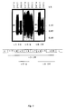
- Fig. 1 Schematic representation of a CD44 molecule that contains the variant exons v2 to v10. Hatched boxes symbolize CD44 standard sequences (CD44s).
- TM transmembrane domain. The approximate location of the epitopes recognized by different monoclonal (black bars) and polyclonal (gray bars) antibodies is indicated.
- the antibodies VFF7, VFF8 and VFF16 are each specific for one exon, whereas mAb VFF17 reacts with an epitope that is coded together by exons v7 and v8.
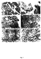
- Fig. 2 Immunohistochemistry of normal mucosa and gastric adenocarcinoma.
- a focal stressed anti-CD44v-positive reaction can be seen in tumor cells of a moderately differentiated adenocarcinoma (Lauren intestinal type) of the stomach ( a ) and in a regional lymph node metastasis ( b ) .
- the foci of intestinal metaplasias react positively with mAb VFF4 ( c , arrows) and with mAb VFF8 ( d , arrows), accompanied by an additional reaction on the mucoid surface and the foveolar epithelium ( d , arrowheads ).
- Fig. 4 Reverse transcription / PCR amplification of CD44 transcripts in gastric carcinoma cell lines.
- Upper part PCR amplification products after using CD44 standard primers. The first strand synthesis was carried out with polyadenylated RNA from the gastric carcinoma cell lines 2474, 2957 and 3051.
- Lower part GADPH-specific PCR amplification products of the same RNA samples. The samples were separated in a 1.2% agarose gel, stained with ethidium bromide and visualized under UV light.
- FIG. 11 Southern blot analysis of CD44 PCR amplification products in gastric carcinoma cell lines.
- the PCR reactions of FIG. 11 were separated in a 1.2% agarose gel, transferred to a Hybond N + membrane and successively hybridized with samples which were specific for different variant CD44 exons.
- v3-10 contains nucleotides from positions 25 to 1013;
- v5 / v6 contains nucleotides from positions 244 to 468;
- v8-10 contains nucleotides from positions 623 to 981 of the published human variant CD44 exon sequences (Hofinann et al., 1991).
- Example 1 Production of antibodies against epitopes, which are encoded by variant exon sequences of the CD44 gene
- the entire variant region of the HPKII type of CD44v was amplified from human keratinocyte cDNA by polymerase chain reaction (PCR).
- PCR polymerase chain reaction
- the two PCR primers 5 '-CAGGCTGGGAGCCAAATGAAGAAAATG- 3 ' (SEQ ID NO: 11), positions 25-52, and 5 '-TGATAAGGAACGATTGACATTAGAGTT GGA- 3' (SEQ ID NO: 12), positions 1013-984 of the LCLC97 variants Region as described by Hofinann et al. contained an Eco RI recognition site which was used to clone the PCR product directly into the vector pGEX-2T (Smith et al., 1988).
- the resulting construct (pGEX CD44v HPKII, v3-v10) codes for a fusion protein of ⁇ 70 kD consisting of glutathione-S-transferase from Schistosoma japonicum and the exons v3-v10 from human CD44 (FIG. 1; Heider et al., 1993).
- the fusion protein was expressed in E. coli and then affinity purified using glutathione agarose (Smith et al., 1988).
- fragments were cloned which contained DI (v3), DII / III (v5, v6), and DIII (v6, v7), where the appropriate restriction interfaces were used.
- Fusion protein DI contains the CD44 sequence described by Stamenkovic et al. (1989), from position 744 to position 142 of the sequence of variant CD44 as described by Hofinann et al. (1991).
- Fusion protein DII / III contains the variant sequence from position 290-460
- fusion protein DIII contains the variant sequence from position 378-638 (Hofinann et al., 1991).
- the fragments containing DI and DIII were cloned into the pGEX vector system, the DII / III fragment into the pATH vector (Angel et al., 1988).
- mice Female Balb / c mice were immunized intraperitoneally with affinity-purified fusion protein (GST-CD44v3-10). In the first immunization 90 ⁇ g fusion protein in complete Freund's adjuvant was applied, in the second and third immunization 50 ⁇ g fusion protein in incomplete Freund's adjuvant. The immunizations were carried out every 4 weeks. 14 days after the last immunization, the animals were immunized for three consecutive days with 10 ⁇ g of fusion protein in PBS. The following day, spleen cells from an animal with a high antibody titer were fused with P3.X63-Ag8.653 mouse myeloma cells using polyethylene glycol 4000. The hybridoma cells were then selected in microtiter plates in HAT medium (Köhler et Milstein, 1975; Kearney et al., 1979).
- the determination of the antibody titer in the serum or the screening of the hybridoma supernatants was carried out using an ELISA.
- microtiter plates are first coated with fusion protein (GST-CD44v3-10) or only with glutathione -S-transferase. It was then incubated with serial dilutions of serum samples or hybridoma supernatants and the specific antibodies were detected with peroxidase-conjugated antibodies against mouse immunoglobulin. Hybridomas that only reacted with glutathione-S-transferase were discarded.
- VFF7 recognizes an epitope in the amino acid sequence of exon v6.
- VFF8 recognizes an epitope in the amino acid sequence of exon v5.
- VFF-17 recognizes an epitope that is encoded jointly by exons v7 and v8.
- the antibodies NKI-P1 and SFF2 recognize epitopes in the standard portion of the extracellular domain near the N-terminus.
- Tumor samples and normal tissues were selected from the inventory of the Pathology Department at the University of Würzburg, Germany. The samples were snap frozen immediately after surgical removal and stored at -80 ° C until use. Normal tissue was taken from twelve different tumor patients from both the body and the antral region of the stomach. Pathological tissues were obtained from a total of 47 patients with an average age of 63 years. Of the primary carcinomas, 29 belonged to the intestinal and 18 to the diffuse type according to Lauren (1965). The tumor stages ranged from localized (pT1) to extensive (pT4), the histological grading from well-differentiated (G1) to poorly differentiated (G3) adenocarcinomas.
- Immunostaining was carried out according to standard protocols as previously described (Heider et al., 1993). Frozen sections (6-7 ⁇ m) were in ice-cold methanol for 4 min., Then in acetone for 1 min. fixed, washed in PBS (8 g / l NaCl, 0.2 g / l KCI, 1.44 g / l Na 2 HPO 4 , 0.24 g / l KH 2 PO 4 , pH 7.4) and preincubated with normal goat serum (10% in PBS) . Then they were washed 3 times with PBS and incubated for 1 hour with the primary antibody (in PBS, 1% BSA).
- a final concentration of 5 ⁇ g / ml was used for monoclonal antibodies.
- Affinity-purified polyclonal sera were titrated on normal skin keratinocytes for optimal staining results. Endogenous peroxidase was blocked with 0.3% H 2 O 2 in methanol and the sections with biotinylated second antibody (either anti-mouse or anti-rabbit F (ab ') 2 , DAKO Corp., Santa Barbara, CA, USA, depending on the primary antibody used ) incubated. The immune complex was visualized with horseradish peroxidase, which was coupled to biotin as a streptavidin-biotin-peroxidase complex (DAKO).
- DAKO streptavidin-biotin-peroxidase complex
- Polyclonal antisera directed against epitopes v3-v10 (CD44v in Fig. 1) stained 42/42 frozen sections of gastric tumors (Table 1, examples in Fig. 2a, b).
- the coloring was heterogeneous in terms of intensity and distribution. Between 5% and 100% of the tumor cells were stained with different intensities. This confirms the finding of Heider et al. (1993) that adenocarcinomas express CD44 splice variants. Surprisingly, however, the subsequent examinations showed that the fine analysis of the expression pattern of the various variant exons is suitable for the diagnosis and / or analysis of gastric carcinomas. Both qualitative and semi-quantitative conclusions are possible.
- the present invention thus provides for the first time a molecular marker for differentiating between different types of gastric cancer.
- Both primary tumors and lymph node metastases were available from 10 patients. Five of these pairs were of the intestinal type and five of the diffuse type. Epitopes recognized by the polyclonal antiserum were present both on the primary tumors and on the metastases from all 10 tumor pairs (Table 2; FIGS. 2a and b show the primary tumor No. 9069/90 from Table 2 and the associated lymph node metastasis). . All tumor samples (primary tumors and metastases) reacted with the mAb VFF8 specific for exon v5.
- Table 1 Expression of variant CD44 epitopes on the cell surfaces of gastric tumors Serum 1 Monoclonal antibodies Adenocarcinoma ⁇ CD44v VFF11 VFF8 VFF4 VFF9 VFF14 (v3-v10) (v3 / 4) (v5) (v6) (v7) (v8-10) Diffuse type 17/17 2 0/17 14/17 3/17 0/17 0/17 Intestinal type 25/25 0/25 25/25 23/25 0/25 0/25 total 42/42 0/42 39/42 26/42 0/42 0/42 1 polyclonal antiserum 2 Number of positive tumors / number of tumors examined
- RNA from tissues
- polyA + RNA from cell lines
- 5 ⁇ l of the first strand cDNA were amplified with Taq polymerase (Amersham) in 50 ⁇ l under the buffer conditions recommended by the manufacturer.
- Taq polymerase Amersham
- oligonucleotides were used which were homologous to positions 8-29 and 362-339 of the published cDNA sequence (Allen et al ., 1987). After 26 rounds of amplification (95 ° C. for 1 min., 62 ° C. for 1 min., 72 ° C.
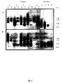
- RNA containing exon v5 or v6 was detected by hybridization with a v5-specific or a v6-specific sample. There was a marked difference in the pattern of RNA expressed in normal tissue and in tumors. The RNA patterns also differed from tumor to tumor.
- RNA from 10 of the 12 samples of normal gastric mucosa which had also been examined in immunohistochemistry, revealed two dominant bands that hybridized with exons v6 and two dominant bands of similar size that hybridized with exon v5 (four representative examples are shown in FIG. 3A and B).
- RNA from tumor samples gave a more complex and variable pattern of the splice products.
- the tumors at least partially also contained larger splice variants up to a size that suggests the presence of all variant exon sequences (FIG. 3). Obviously, not all of the epitopes encoded by these exons were accessible to immunohistochemistry.
- Both diffuse and intestinal type tumors showed strong expression of transcripts containing exon v5 (Fig. 3A, lanes 1-5).
- the present invention thus permits diagnosis and analysis of adenocarcinomas on the basis of molecular markers at the nucleic acid level and provides the means for this.
- the method according to the invention allows the distinction of gastric carcinomas of the diffuse and of the intestinal type and the analysis of the corresponding metastases at the nucleic acid level.
- glycoprotein CD44 confers metastatic potential to rat carcinoma cells. Cell 65: 13-24 (1991).
- lymphocyte molecule implicated in lymph node homing is a member ofthe cartilage link protein family. Cell 56 : 1057-1062 (1989).
Landscapes
- Health & Medical Sciences (AREA)
- Life Sciences & Earth Sciences (AREA)
- Chemical & Material Sciences (AREA)
- Immunology (AREA)
- Engineering & Computer Science (AREA)
- Molecular Biology (AREA)
- Biomedical Technology (AREA)
- Hematology (AREA)
- Urology & Nephrology (AREA)
- General Health & Medical Sciences (AREA)
- Biochemistry (AREA)
- Pathology (AREA)
- Medicinal Chemistry (AREA)
- Analytical Chemistry (AREA)
- Microbiology (AREA)
- Physics & Mathematics (AREA)
- Cell Biology (AREA)
- Biotechnology (AREA)
- General Physics & Mathematics (AREA)
- Organic Chemistry (AREA)
- Food Science & Technology (AREA)
- Proteomics, Peptides & Aminoacids (AREA)
- Genetics & Genomics (AREA)
- Zoology (AREA)
- Biophysics (AREA)
- Wood Science & Technology (AREA)
- Hospice & Palliative Care (AREA)
- Oncology (AREA)
- Bioinformatics & Cheminformatics (AREA)
- General Engineering & Computer Science (AREA)
- Toxicology (AREA)
- Gastroenterology & Hepatology (AREA)
- Measuring Or Testing Involving Enzymes Or Micro-Organisms (AREA)
- Peptides Or Proteins (AREA)
- Preparation Of Compounds By Using Micro-Organisms (AREA)
- Medicines Containing Antibodies Or Antigens For Use As Internal Diagnostic Agents (AREA)
Applications Claiming Priority (9)
| Application Number | Priority Date | Filing Date | Title |
|---|---|---|---|
| DE4320624 | 1993-06-22 | ||
| DE4320623 | 1993-06-22 | ||
| DE4320624A DE4320624A1 (de) | 1993-06-22 | 1993-06-22 | Verfahren zur Diagnose und Analyse von Mammakarzinomen |
| DE4320623A DE4320623C2 (de) | 1993-06-22 | 1993-06-22 | Verfahren zur Diagnose und Analyse von Adenokarzinomen |
| DE4321944 | 1993-07-02 | ||
| DE4321944A DE4321944A1 (de) | 1993-07-02 | 1993-07-02 | Verfahren zur Diagnose und Analyse von Kolonkarzinomen |
| DE4414787 | 1994-04-28 | ||
| DE4414787A DE4414787A1 (de) | 1994-04-28 | 1994-04-28 | Verfahren zur Diagnose und Analyse von Zervixkarzinomen |
| EP94919633A EP0705436A1 (fr) | 1993-06-22 | 1994-06-15 | Procede pour le diagnostic et l'analyse du carcinome mammair |
Related Parent Applications (2)
| Application Number | Title | Priority Date | Filing Date |
|---|---|---|---|
| EP94919633A Division EP0705436A1 (fr) | 1993-06-22 | 1994-06-15 | Procede pour le diagnostic et l'analyse du carcinome mammair |
| EP94919633.1 Division | 1995-01-05 |
Publications (1)
| Publication Number | Publication Date |
|---|---|
| EP0767378A1 true EP0767378A1 (fr) | 1997-04-09 |
Family
ID=27435773
Family Applications (4)
| Application Number | Title | Priority Date | Filing Date |
|---|---|---|---|
| EP96113180A Ceased EP0767379A1 (fr) | 1993-06-22 | 1994-06-15 | Procédé pour le diagnostic et l'analyse de carcinomes de colon |
| EP94919633A Ceased EP0705436A1 (fr) | 1993-06-22 | 1994-06-15 | Procede pour le diagnostic et l'analyse du carcinome mammair |
| EP96113181A Ceased EP0767380A1 (fr) | 1993-06-22 | 1994-06-15 | Procédé pour le diagnostic et l'analyse de carcinomes du col de l'utérus |
| EP96113179A Ceased EP0767378A1 (fr) | 1993-06-22 | 1994-06-15 | Procédé pour le diagnostic et l'analyse du tumeurs de l'estomac |
Family Applications Before (3)
| Application Number | Title | Priority Date | Filing Date |
|---|---|---|---|
| EP96113180A Ceased EP0767379A1 (fr) | 1993-06-22 | 1994-06-15 | Procédé pour le diagnostic et l'analyse de carcinomes de colon |
| EP94919633A Ceased EP0705436A1 (fr) | 1993-06-22 | 1994-06-15 | Procede pour le diagnostic et l'analyse du carcinome mammair |
| EP96113181A Ceased EP0767380A1 (fr) | 1993-06-22 | 1994-06-15 | Procédé pour le diagnostic et l'analyse de carcinomes du col de l'utérus |
Country Status (5)
| Country | Link |
|---|---|
| US (1) | US6010865A (fr) |
| EP (4) | EP0767379A1 (fr) |
| JP (1) | JPH08511624A (fr) |
| CA (1) | CA2164728A1 (fr) |
| WO (1) | WO1995000851A1 (fr) |
Cited By (3)
| Publication number | Priority date | Publication date | Assignee | Title |
|---|---|---|---|---|
| RU2164349C1 (ru) * | 1999-07-27 | 2001-03-20 | Нижегородская государственная медицинская академия | Способ определения стоматологического статуса |
| RU2428100C2 (ru) * | 2009-03-20 | 2011-09-10 | Государственное образовательное учреждение высшего профессионального образования Читинская государственная медицинская академия Федерального агентства по здравоохранению и социальному развитию | Способ определения интенсивности кариеса постоянных зубов у детей в период сменного прикуса |
| CN116496398A (zh) * | 2022-10-31 | 2023-07-28 | 南京元迈细胞生物科技有限公司 | 一种特异性结合CD44的v5外显子的抗体及其用途 |
Families Citing this family (20)
| Publication number | Priority date | Publication date | Assignee | Title |
|---|---|---|---|---|
| UA58482C2 (uk) * | 1994-06-08 | 2003-08-15 | Бьорінгер Інгельхайм Інтернаціональ Гмбх | Моноклональне антитіло vff-18 проти сd44v6 і його фрагменти |
| US5861494A (en) | 1995-06-06 | 1999-01-19 | Human Genome Sciences, Inc. | Colon specific gene and protein |
| DE19540515C1 (de) * | 1995-10-31 | 1997-02-06 | Boehringer Ingelheim Int | Tumortherapie durch adoptiven Transfer CD44v-spezifischer zytotoxischer T-Lymphozyten |
| UY24389A1 (es) * | 1995-12-06 | 2001-10-25 | Karlsruhe Forschzent | Composición farmacéutica para el tratamiento de carcinoma de epitelio plano |
| DE19545472A1 (de) * | 1995-12-06 | 1997-06-12 | Boehringer Ingelheim Int | Verfahren zur Diagnose und Therapie von Plattenepithelkarzinomen |
| DE19653607A1 (de) | 1996-12-20 | 1998-06-25 | Boehringer Ingelheim Int | Verfahren zur Diagnose und Therapie von Hodgkin-Lymphomen |
| IL133647A0 (en) * | 1999-06-08 | 2001-04-30 | Yissum Res Dev Co | Novel cd44 variant |
| US7534605B2 (en) | 1999-06-08 | 2009-05-19 | Yissum Research Development Company Of The Hebrew University Of Jerusalem | CD44 polypeptides, polynucleotides encoding same, antibodies directed thereagainst and method of using same for diagnosing and treating inflammatory diseases |
| US6972324B2 (en) | 2001-05-18 | 2005-12-06 | Boehringer Ingelheim Pharmaceuticals, Inc. | Antibodies specific for CD44v6 |
| CN1561223A (zh) * | 2001-08-24 | 2005-01-05 | 斯塔凡·斯特伦布拉德 | 新型药物 |
| US7460960B2 (en) | 2002-05-10 | 2008-12-02 | Epitome Biosystems, Inc. | Proteome epitope tags and methods of use thereof in protein modification analysis |
| US7618788B2 (en) | 2002-05-10 | 2009-11-17 | Millipore Corporation | Proteome epitope tags and methods of use thereof in protein modification analysis |
| EP1539235A2 (fr) | 2002-07-01 | 2005-06-15 | Human Genome Sciences, Inc. | Anticorps qui se lient specifiquement a reg iv |
| US20050009110A1 (en) * | 2003-07-08 | 2005-01-13 | Xiao-Jia Chang | Methods of producing antibodies for diagnostics and therapeutics |
| US7855057B2 (en) | 2006-03-23 | 2010-12-21 | Millipore Corporation | Protein splice variant/isoform discrimination and quantitative measurements thereof |
| MX2009010400A (es) | 2007-03-30 | 2010-02-18 | Chu Sainte Justine | Método para la determinacion de riesgo de escoliosis. |
| CA2914213A1 (fr) * | 2013-06-17 | 2014-12-24 | Alain Moreau | Methode d'augmentation de signalisation de gipcr dans les cellules d'un sujet scoliotique |
| ES2639855T3 (es) | 2013-12-23 | 2017-10-30 | Exchange Imaging Technologies Gmbh | Nanopartícula conjugada a péptidos de unión a CD44 |
| EP3169343B1 (fr) | 2014-07-15 | 2020-03-25 | Yissum Research and Development Company of the Hebrew University of Jerusalem Ltd. | Polypeptides isolés de cd44 et utilisations associées |
| AU2020216780B2 (en) * | 2019-01-28 | 2025-05-08 | Multitude Therapeutics Inc. | Antibodies specific to CD44 |
Citations (2)
| Publication number | Priority date | Publication date | Assignee | Title |
|---|---|---|---|---|
| WO1991017248A1 (fr) * | 1990-05-07 | 1991-11-14 | Kernforschungszentrum Karlsruhe Gmbh | Variantes de proteines superficielles cd44, sequences d'adn les codant, anticorps contre ces proteines, ainsi que leur utilisation dans le diagnostic et la therapie |
| WO1994002633A1 (fr) * | 1992-07-21 | 1994-02-03 | Isis Innovation Limited | Methode de diagnostic |
-
1994
- 1994-06-15 EP EP96113180A patent/EP0767379A1/fr not_active Ceased
- 1994-06-15 EP EP94919633A patent/EP0705436A1/fr not_active Ceased
- 1994-06-15 US US08/564,225 patent/US6010865A/en not_active Expired - Fee Related
- 1994-06-15 EP EP96113181A patent/EP0767380A1/fr not_active Ceased
- 1994-06-15 EP EP96113179A patent/EP0767378A1/fr not_active Ceased
- 1994-06-15 CA CA002164728A patent/CA2164728A1/fr not_active Abandoned
- 1994-06-15 JP JP7502412A patent/JPH08511624A/ja active Pending
- 1994-06-15 WO PCT/EP1994/001952 patent/WO1995000851A1/fr not_active Ceased
Patent Citations (2)
| Publication number | Priority date | Publication date | Assignee | Title |
|---|---|---|---|---|
| WO1991017248A1 (fr) * | 1990-05-07 | 1991-11-14 | Kernforschungszentrum Karlsruhe Gmbh | Variantes de proteines superficielles cd44, sequences d'adn les codant, anticorps contre ces proteines, ainsi que leur utilisation dans le diagnostic et la therapie |
| WO1994002633A1 (fr) * | 1992-07-21 | 1994-02-03 | Isis Innovation Limited | Methode de diagnostic |
Non-Patent Citations (7)
| Title |
|---|
| CORNELIA TÖLG ET AL.: "Splicing choice from ten variant exons establishes CD44 variability", NUCLEIC ACIDS RESEARCH, vol. 21, no. 5, 11 March 1993 (1993-03-11), OXFORD, GB, pages 1225 - 1229, XP000508272 * |
| GAVIN R. SCREATON ET AL.: "Genomic structure of DNA encoding the lymphocyte homing receptor CD44 reveals at least 12 alternatively spliced exons", PROCEEDINGS OF THE NATIONAL ACADEMY OF SCIENCES OF USA, vol. 89, no. 24, 15 December 1992 (1992-12-15), WASHINGTON US, pages 12160 - 12164, XP000470187 * |
| GERRIT KOOPMAN ET AL.: "Activated Human Lymphocytes and Aggressive Non-Hodgkin's Lymphomas Express a Homologue of the Rat Metastasis-associated Variant of CD44", JOURNAL OF EXPERIMENTAL MEDICINE, vol. 177, no. 4, 1 April 1993 (1993-04-01), NEW YORK, NY, US, pages 897 - 904, XP000471698 * |
| KARL-HEINZ HEIDER ET AL.: "A Human Homologue of the Rat Metastasis-associated Variant of CD44 Is Expressed in Colorectal Carcinomas and Adenomatous Polyps", JOURNAL OF CELL BIOLOGY, vol. 120, no. 1, January 1993 (1993-01-01), NEW YORK, NY, US, pages 227 - 233, XP000471652 * |
| MACKAY, CHARLES R. ET AL: "Expression and modulation of CD44 variant isoforms in humans", J. CELL BIOL. (1994), 124(1-2), 71-82 CODEN: JCLBA3;ISSN: 0021-9525, 1994, XP000471699 * |
| MARTIN HOFFMANN ET AL.: "CD44 Splice Variants Confer Metastatic Behavor in Rats: Homologous Sequences Are Expressed in Human Tumor Cell Lines", CANCER RESEARCH, vol. 51, no. 19, 1 October 1991 (1991-10-01), PHILADELPHIA, PA, US, pages 5292 - 5297, XP002023409 * |
| SEITER, S. ET AL: "Prevention of tumor metastasis formation by anti-variant CD44", J. EXP. MED. (1993), 177(2), 443-55 CODEN: JEMEAV;ISSN: 0022-1007, 1993, XP000471653 * |
Cited By (4)
| Publication number | Priority date | Publication date | Assignee | Title |
|---|---|---|---|---|
| RU2164349C1 (ru) * | 1999-07-27 | 2001-03-20 | Нижегородская государственная медицинская академия | Способ определения стоматологического статуса |
| RU2428100C2 (ru) * | 2009-03-20 | 2011-09-10 | Государственное образовательное учреждение высшего профессионального образования Читинская государственная медицинская академия Федерального агентства по здравоохранению и социальному развитию | Способ определения интенсивности кариеса постоянных зубов у детей в период сменного прикуса |
| CN116496398A (zh) * | 2022-10-31 | 2023-07-28 | 南京元迈细胞生物科技有限公司 | 一种特异性结合CD44的v5外显子的抗体及其用途 |
| CN116496398B (zh) * | 2022-10-31 | 2023-10-31 | 南京元迈细胞生物科技有限公司 | 一种特异性结合CD44的v5外显子的抗体及其用途 |
Also Published As
| Publication number | Publication date |
|---|---|
| EP0705436A1 (fr) | 1996-04-10 |
| US6010865A (en) | 2000-01-04 |
| EP0767379A1 (fr) | 1997-04-09 |
| WO1995000851A1 (fr) | 1995-01-05 |
| EP0767380A1 (fr) | 1997-04-09 |
| CA2164728A1 (fr) | 1995-01-05 |
| JPH08511624A (ja) | 1996-12-03 |
Similar Documents
| Publication | Publication Date | Title |
|---|---|---|
| EP0767378A1 (fr) | Procédé pour le diagnostic et l'analyse du tumeurs de l'estomac | |
| WO2005110338A2 (fr) | Identification d'antigenes de surface pour le diagnostic et la therapie de tumeurs | |
| WO2005030250A2 (fr) | Identification d'antigenes de surface cellulaire associes a une tumeur dans le cadre d'un diagnostic et d'une therapie | |
| EP2314310A2 (fr) | Identification d'antigènes de surfaces pour le diagnostic et la thérapie de tumeurs | |
| EP2171465B1 (fr) | Cadhérine-17 soluble pour le diagnostic et la stratification du risque de cancer et de tumeur du tractus gastro-intestinal | |
| EP0582582B1 (fr) | Procede de detection de la differentiation et de l'invasivite de cellules de carcinomes | |
| DE102005052384B4 (de) | Verfahren zur Erkennung, Markierung und Behandlung von epithelialen Lungentumorzellen sowie Mittel zur Durchführung des Verfahrens | |
| DE69635925T2 (de) | Verfahren und Vorrichtung zum Beurteilen der metastatischen Tendenz von Tumoren | |
| DE4321944A1 (de) | Verfahren zur Diagnose und Analyse von Kolonkarzinomen | |
| DE4320623C2 (de) | Verfahren zur Diagnose und Analyse von Adenokarzinomen | |
| DE3689084T2 (de) | Verfahren zur in vitro-Diagnose von bösartigen Zellen aus dem Verdauungstrakt. | |
| EP1960544B1 (fr) | M,arqueur moleculaire pour le diagnostic et la therapie des tumeurs | |
| DE60221123T2 (de) | Krebs-assoziiertes protein | |
| DE60223677T2 (de) | Bcmp-101, ein mit krebs assoziiertes protein | |
| DE69925477T2 (de) | Diagnose für metastasischen prostatakrebs | |
| DE4414787A1 (de) | Verfahren zur Diagnose und Analyse von Zervixkarzinomen | |
| DE4320624A1 (de) | Verfahren zur Diagnose und Analyse von Mammakarzinomen |
Legal Events
| Date | Code | Title | Description |
|---|---|---|---|
| PUAI | Public reference made under article 153(3) epc to a published international application that has entered the european phase |
Free format text: ORIGINAL CODE: 0009012 |
|
| AC | Divisional application: reference to earlier application |
Ref document number: 705436 Country of ref document: EP |
|
| AK | Designated contracting states |
Kind code of ref document: A1 Designated state(s): AT BE CH DE DK ES FR GB GR IE IT LI LU MC NL PT SE |
|
| RIN1 | Information on inventor provided before grant (corrected) |
Inventor name: HEIDER, KARL-HEINZ, DR. DIPL.-BIOL. Inventor name: HERRLICH, PETER, PROF. DR. Inventor name: PONTA, HELMUT, PROF. DR. |
|
| 17P | Request for examination filed |
Effective date: 19971009 |
|
| GRAG | Despatch of communication of intention to grant |
Free format text: ORIGINAL CODE: EPIDOS AGRA |
|
| 17Q | First examination report despatched |
Effective date: 19990315 |
|
| STAA | Information on the status of an ep patent application or granted ep patent |
Free format text: STATUS: THE APPLICATION HAS BEEN REFUSED |
|
| 18R | Application refused |
Effective date: 19990904 |