EP0768332A1 - Verfahren zu einer photooxidativen Behandlung von kollagenhaltigen Geweben - Google Patents
Verfahren zu einer photooxidativen Behandlung von kollagenhaltigen Geweben Download PDFInfo
- Publication number
- EP0768332A1 EP0768332A1 EP95810637A EP95810637A EP0768332A1 EP 0768332 A1 EP0768332 A1 EP 0768332A1 EP 95810637 A EP95810637 A EP 95810637A EP 95810637 A EP95810637 A EP 95810637A EP 0768332 A1 EP0768332 A1 EP 0768332A1
- Authority
- EP
- European Patent Office
- Prior art keywords
- tissue
- solution
- collagen
- photooxidative
- exposure
- Prior art date
- Legal status (The legal status is an assumption and is not a legal conclusion. Google has not performed a legal analysis and makes no representation as to the accuracy of the status listed.)
- Granted
Links
- 102000008186 Collagen Human genes 0.000 title claims abstract description 31
- 108010035532 Collagen Proteins 0.000 title claims abstract description 31
- 229920001436 collagen Polymers 0.000 title claims abstract description 31
- 238000000034 method Methods 0.000 title claims description 29
- 230000008569 process Effects 0.000 title description 6
- 210000001519 tissue Anatomy 0.000 claims abstract description 60
- 210000000845 cartilage Anatomy 0.000 claims abstract description 22
- 230000003750 conditioning effect Effects 0.000 claims abstract description 15
- 238000002360 preparation method Methods 0.000 claims abstract description 12
- RBTBFTRPCNLSDE-UHFFFAOYSA-N 3,7-bis(dimethylamino)phenothiazin-5-ium Chemical compound C1=CC(N(C)C)=CC2=[S+]C3=CC(N(C)C)=CC=C3N=C21 RBTBFTRPCNLSDE-UHFFFAOYSA-N 0.000 claims abstract description 11
- 238000006243 chemical reaction Methods 0.000 claims abstract description 11
- 229960000907 methylthioninium chloride Drugs 0.000 claims abstract description 11
- XLYOFNOQVPJJNP-UHFFFAOYSA-N water Substances O XLYOFNOQVPJJNP-UHFFFAOYSA-N 0.000 claims abstract description 11
- 239000000654 additive Substances 0.000 claims abstract description 9
- 230000000996 additive effect Effects 0.000 claims abstract description 8
- 210000000988 bone and bone Anatomy 0.000 claims abstract description 7
- 210000002435 tendon Anatomy 0.000 claims abstract description 6
- 210000003041 ligament Anatomy 0.000 claims abstract description 5
- 239000000243 solution Substances 0.000 claims description 39
- PEDCQBHIVMGVHV-UHFFFAOYSA-N Glycerine Chemical compound OCC(O)CO PEDCQBHIVMGVHV-UHFFFAOYSA-N 0.000 claims description 25
- 239000000835 fiber Substances 0.000 claims description 11
- 235000011187 glycerol Nutrition 0.000 claims description 10
- QVGXLLKOCUKJST-UHFFFAOYSA-N atomic oxygen Chemical compound [O] QVGXLLKOCUKJST-UHFFFAOYSA-N 0.000 claims description 9
- 229910052760 oxygen Inorganic materials 0.000 claims description 9
- 239000001301 oxygen Substances 0.000 claims description 9
- 238000004042 decolorization Methods 0.000 claims description 8
- 239000004744 fabric Substances 0.000 claims description 7
- 239000008363 phosphate buffer Substances 0.000 claims description 6
- 239000000126 substance Substances 0.000 claims description 6
- 239000007943 implant Substances 0.000 claims description 5
- LFQSCWFLJHTTHZ-UHFFFAOYSA-N Ethanol Chemical compound CCO LFQSCWFLJHTTHZ-UHFFFAOYSA-N 0.000 claims description 4
- 239000007980 Sørensen’s phosphate buffer Substances 0.000 claims description 4
- 230000003197 catalytic effect Effects 0.000 claims description 3
- 230000007123 defense Effects 0.000 claims description 3
- 230000001900 immune effect Effects 0.000 claims description 3
- 238000005406 washing Methods 0.000 claims description 3
- 238000001816 cooling Methods 0.000 claims description 2
- 239000000203 mixture Substances 0.000 claims description 2
- 239000000975 dye Substances 0.000 description 23
- 230000005540 biological transmission Effects 0.000 description 9
- 238000007539 photo-oxidation reaction Methods 0.000 description 9
- 238000010586 diagram Methods 0.000 description 6
- 238000004040 coloring Methods 0.000 description 3
- MTHSVFCYNBDYFN-UHFFFAOYSA-N diethylene glycol Chemical compound OCCOCCO MTHSVFCYNBDYFN-UHFFFAOYSA-N 0.000 description 3
- 102000029816 Collagenase Human genes 0.000 description 2
- 108060005980 Collagenase Proteins 0.000 description 2
- -1 Hydrogen ions Chemical class 0.000 description 2
- AUNGANRZJHBGPY-SCRDCRAPSA-N Riboflavin Chemical compound OC[C@@H](O)[C@@H](O)[C@@H](O)CN1C=2C=C(C)C(C)=CC=2N=C2C1=NC(=O)NC2=O AUNGANRZJHBGPY-SCRDCRAPSA-N 0.000 description 2
- 210000001188 articular cartilage Anatomy 0.000 description 2
- 239000007853 buffer solution Substances 0.000 description 2
- 230000000747 cardiac effect Effects 0.000 description 2
- 229960002424 collagenase Drugs 0.000 description 2
- 238000009792 diffusion process Methods 0.000 description 2
- 238000009826 distribution Methods 0.000 description 2
- 210000003709 heart valve Anatomy 0.000 description 2
- 238000001356 surgical procedure Methods 0.000 description 2
- IICCLYANAQEHCI-UHFFFAOYSA-N 4,5,6,7-tetrachloro-3',6'-dihydroxy-2',4',5',7'-tetraiodospiro[2-benzofuran-3,9'-xanthene]-1-one Chemical compound O1C(=O)C(C(=C(Cl)C(Cl)=C2Cl)Cl)=C2C21C1=CC(I)=C(O)C(I)=C1OC1=C(I)C(O)=C(I)C=C21 IICCLYANAQEHCI-UHFFFAOYSA-N 0.000 description 1
- 241000283690 Bos taurus Species 0.000 description 1
- AUNGANRZJHBGPY-UHFFFAOYSA-N D-Lyxoflavin Natural products OCC(O)C(O)C(O)CN1C=2C=C(C)C(C)=CC=2N=C2C1=NC(=O)NC2=O AUNGANRZJHBGPY-UHFFFAOYSA-N 0.000 description 1
- 102000004190 Enzymes Human genes 0.000 description 1
- 108090000790 Enzymes Proteins 0.000 description 1
- QIVBCDIJIAJPQS-VIFPVBQESA-N L-tryptophane Chemical compound C1=CC=C2C(C[C@H](N)C(O)=O)=CNC2=C1 QIVBCDIJIAJPQS-VIFPVBQESA-N 0.000 description 1
- OUYCCCASQSFEME-QMMMGPOBSA-N L-tyrosine Chemical compound OC(=O)[C@@H](N)CC1=CC=C(O)C=C1 OUYCCCASQSFEME-QMMMGPOBSA-N 0.000 description 1
- WDVSHHCDHLJJJR-UHFFFAOYSA-N Proflavine Chemical compound C1=CC(N)=CC2=NC3=CC(N)=CC=C3C=C21 WDVSHHCDHLJJJR-UHFFFAOYSA-N 0.000 description 1
- QIVBCDIJIAJPQS-UHFFFAOYSA-N Tryptophan Natural products C1=CC=C2C(CC(N)C(O)=O)=CNC2=C1 QIVBCDIJIAJPQS-UHFFFAOYSA-N 0.000 description 1
- 238000010521 absorption reaction Methods 0.000 description 1
- 125000000539 amino acid group Chemical group 0.000 description 1
- 238000004061 bleaching Methods 0.000 description 1
- 238000007664 blowing Methods 0.000 description 1
- 239000003054 catalyst Substances 0.000 description 1
- 230000008859 change Effects 0.000 description 1
- 210000001612 chondrocyte Anatomy 0.000 description 1
- 230000006835 compression Effects 0.000 description 1
- 238000007906 compression Methods 0.000 description 1
- 230000001419 dependent effect Effects 0.000 description 1
- 230000029087 digestion Effects 0.000 description 1
- BNIILDVGGAEEIG-UHFFFAOYSA-L disodium hydrogen phosphate Chemical compound [Na+].[Na+].OP([O-])([O-])=O BNIILDVGGAEEIG-UHFFFAOYSA-L 0.000 description 1
- 239000003814 drug Substances 0.000 description 1
- 230000000694 effects Effects 0.000 description 1
- 229940088598 enzyme Drugs 0.000 description 1
- YQGOJNYOYNNSMM-UHFFFAOYSA-N eosin Chemical compound [Na+].OC(=O)C1=CC=CC=C1C1=C2C=C(Br)C(=O)C(Br)=C2OC2=C(Br)C(O)=C(Br)C=C21 YQGOJNYOYNNSMM-UHFFFAOYSA-N 0.000 description 1
- 230000005281 excited state Effects 0.000 description 1
- GNBHRKFJIUUOQI-UHFFFAOYSA-N fluorescein Chemical compound O1C(=O)C2=CC=CC=C2C21C1=CC=C(O)C=C1OC1=CC(O)=CC=C21 GNBHRKFJIUUOQI-UHFFFAOYSA-N 0.000 description 1
- 229960002143 fluorescein Drugs 0.000 description 1
- 229910052736 halogen Inorganic materials 0.000 description 1
- 150000002367 halogens Chemical class 0.000 description 1
- 125000000487 histidyl group Chemical group [H]N([H])C(C(=O)O*)C([H])([H])C1=C([H])N([H])C([H])=N1 0.000 description 1
- 229910052739 hydrogen Inorganic materials 0.000 description 1
- 239000001257 hydrogen Substances 0.000 description 1
- 230000006872 improvement Effects 0.000 description 1
- 230000002427 irreversible effect Effects 0.000 description 1
- 210000000629 knee joint Anatomy 0.000 description 1
- 238000004519 manufacturing process Methods 0.000 description 1
- 239000000463 material Substances 0.000 description 1
- 238000005259 measurement Methods 0.000 description 1
- YYGBVRCTHASBKD-UHFFFAOYSA-M methylene green Chemical compound [Cl-].C1=CC(N(C)C)=C([N+]([O-])=O)C2=[S+]C3=CC(N(C)C)=CC=C3N=C21 YYGBVRCTHASBKD-UHFFFAOYSA-M 0.000 description 1
- 229910000402 monopotassium phosphate Inorganic materials 0.000 description 1
- 235000019796 monopotassium phosphate Nutrition 0.000 description 1
- 231100000252 nontoxic Toxicity 0.000 description 1
- 230000003000 nontoxic effect Effects 0.000 description 1
- 230000035515 penetration Effects 0.000 description 1
- 230000002572 peristaltic effect Effects 0.000 description 1
- PJNZPQUBCPKICU-UHFFFAOYSA-N phosphoric acid;potassium Chemical compound [K].OP(O)(O)=O PJNZPQUBCPKICU-UHFFFAOYSA-N 0.000 description 1
- 238000002203 pretreatment Methods 0.000 description 1
- 229960000286 proflavine Drugs 0.000 description 1
- 102000004169 proteins and genes Human genes 0.000 description 1
- 108090000623 proteins and genes Proteins 0.000 description 1
- NGVDGCNFYWLIFO-UHFFFAOYSA-N pyridoxal 5'-phosphate Chemical compound CC1=NC=C(COP(O)(O)=O)C(C=O)=C1O NGVDGCNFYWLIFO-UHFFFAOYSA-N 0.000 description 1
- 235000007682 pyridoxal 5'-phosphate Nutrition 0.000 description 1
- 239000011589 pyridoxal 5'-phosphate Substances 0.000 description 1
- 229960001327 pyridoxal phosphate Drugs 0.000 description 1
- 229960002477 riboflavin Drugs 0.000 description 1
- 235000019192 riboflavin Nutrition 0.000 description 1
- 239000002151 riboflavin Substances 0.000 description 1
- 229930187593 rose bengal Natural products 0.000 description 1
- 229940081623 rose bengal Drugs 0.000 description 1
- STRXNPAVPKGJQR-UHFFFAOYSA-N rose bengal A Natural products O1C(=O)C(C(=CC=C2Cl)Cl)=C2C21C1=CC(I)=C(O)C(I)=C1OC1=C(I)C(O)=C(I)C=C21 STRXNPAVPKGJQR-UHFFFAOYSA-N 0.000 description 1
- 210000000323 shoulder joint Anatomy 0.000 description 1
- OUYCCCASQSFEME-UHFFFAOYSA-N tyrosine Natural products OC(=O)C(N)CC1=CC=C(O)C=C1 OUYCCCASQSFEME-UHFFFAOYSA-N 0.000 description 1
Images
Classifications
-
- A—HUMAN NECESSITIES
- A61—MEDICAL OR VETERINARY SCIENCE; HYGIENE
- A61L—METHODS OR APPARATUS FOR STERILISING MATERIALS OR OBJECTS IN GENERAL; DISINFECTION, STERILISATION OR DEODORISATION OF AIR; CHEMICAL ASPECTS OF BANDAGES, DRESSINGS, ABSORBENT PADS OR SURGICAL ARTICLES; MATERIALS FOR BANDAGES, DRESSINGS, ABSORBENT PADS OR SURGICAL ARTICLES
- A61L27/00—Materials for grafts or prostheses or for coating grafts or prostheses
- A61L27/36—Materials for grafts or prostheses or for coating grafts or prostheses containing ingredients of undetermined constitution or reaction products thereof, e.g. transplant tissue, natural bone, extracellular matrix
- A61L27/3683—Materials for grafts or prostheses or for coating grafts or prostheses containing ingredients of undetermined constitution or reaction products thereof, e.g. transplant tissue, natural bone, extracellular matrix subjected to a specific treatment prior to implantation, e.g. decellularising, demineralising, grinding, cellular disruption/non-collagenous protein removal, anti-calcification, crosslinking, supercritical fluid extraction, enzyme treatment
- A61L27/3691—Materials for grafts or prostheses or for coating grafts or prostheses containing ingredients of undetermined constitution or reaction products thereof, e.g. transplant tissue, natural bone, extracellular matrix subjected to a specific treatment prior to implantation, e.g. decellularising, demineralising, grinding, cellular disruption/non-collagenous protein removal, anti-calcification, crosslinking, supercritical fluid extraction, enzyme treatment characterised by physical conditions of the treatment, e.g. applying a compressive force to the composition, pressure cycles, ultrasonic/sonication or microwave treatment, lyophilisation
-
- A—HUMAN NECESSITIES
- A61—MEDICAL OR VETERINARY SCIENCE; HYGIENE
- A61L—METHODS OR APPARATUS FOR STERILISING MATERIALS OR OBJECTS IN GENERAL; DISINFECTION, STERILISATION OR DEODORISATION OF AIR; CHEMICAL ASPECTS OF BANDAGES, DRESSINGS, ABSORBENT PADS OR SURGICAL ARTICLES; MATERIALS FOR BANDAGES, DRESSINGS, ABSORBENT PADS OR SURGICAL ARTICLES
- A61L27/00—Materials for grafts or prostheses or for coating grafts or prostheses
- A61L27/14—Macromolecular materials
- A61L27/22—Polypeptides or derivatives thereof, e.g. degradation products
- A61L27/24—Collagen
-
- A—HUMAN NECESSITIES
- A61—MEDICAL OR VETERINARY SCIENCE; HYGIENE
- A61L—METHODS OR APPARATUS FOR STERILISING MATERIALS OR OBJECTS IN GENERAL; DISINFECTION, STERILISATION OR DEODORISATION OF AIR; CHEMICAL ASPECTS OF BANDAGES, DRESSINGS, ABSORBENT PADS OR SURGICAL ARTICLES; MATERIALS FOR BANDAGES, DRESSINGS, ABSORBENT PADS OR SURGICAL ARTICLES
- A61L27/00—Materials for grafts or prostheses or for coating grafts or prostheses
- A61L27/36—Materials for grafts or prostheses or for coating grafts or prostheses containing ingredients of undetermined constitution or reaction products thereof, e.g. transplant tissue, natural bone, extracellular matrix
- A61L27/3604—Materials for grafts or prostheses or for coating grafts or prostheses containing ingredients of undetermined constitution or reaction products thereof, e.g. transplant tissue, natural bone, extracellular matrix characterised by the human or animal origin of the biological material, e.g. hair, fascia, fish scales, silk, shellac, pericardium, pleura, renal tissue, amniotic membrane, parenchymal tissue, fetal tissue, muscle tissue, fat tissue, enamel
-
- A—HUMAN NECESSITIES
- A61—MEDICAL OR VETERINARY SCIENCE; HYGIENE
- A61L—METHODS OR APPARATUS FOR STERILISING MATERIALS OR OBJECTS IN GENERAL; DISINFECTION, STERILISATION OR DEODORISATION OF AIR; CHEMICAL ASPECTS OF BANDAGES, DRESSINGS, ABSORBENT PADS OR SURGICAL ARTICLES; MATERIALS FOR BANDAGES, DRESSINGS, ABSORBENT PADS OR SURGICAL ARTICLES
- A61L27/00—Materials for grafts or prostheses or for coating grafts or prostheses
- A61L27/36—Materials for grafts or prostheses or for coating grafts or prostheses containing ingredients of undetermined constitution or reaction products thereof, e.g. transplant tissue, natural bone, extracellular matrix
- A61L27/3683—Materials for grafts or prostheses or for coating grafts or prostheses containing ingredients of undetermined constitution or reaction products thereof, e.g. transplant tissue, natural bone, extracellular matrix subjected to a specific treatment prior to implantation, e.g. decellularising, demineralising, grinding, cellular disruption/non-collagenous protein removal, anti-calcification, crosslinking, supercritical fluid extraction, enzyme treatment
- A61L27/3687—Materials for grafts or prostheses or for coating grafts or prostheses containing ingredients of undetermined constitution or reaction products thereof, e.g. transplant tissue, natural bone, extracellular matrix subjected to a specific treatment prior to implantation, e.g. decellularising, demineralising, grinding, cellular disruption/non-collagenous protein removal, anti-calcification, crosslinking, supercritical fluid extraction, enzyme treatment characterised by the use of chemical agents in the treatment, e.g. specific enzymes, detergents, capping agents, crosslinkers, anticalcification agents
-
- C—CHEMISTRY; METALLURGY
- C08—ORGANIC MACROMOLECULAR COMPOUNDS; THEIR PREPARATION OR CHEMICAL WORKING-UP; COMPOSITIONS BASED THEREON
- C08H—DERIVATIVES OF NATURAL MACROMOLECULAR COMPOUNDS
- C08H1/00—Macromolecular products derived from proteins
- C08H1/06—Macromolecular products derived from proteins derived from horn, hoofs, hair, skin or leather
-
- A—HUMAN NECESSITIES
- A61—MEDICAL OR VETERINARY SCIENCE; HYGIENE
- A61F—FILTERS IMPLANTABLE INTO BLOOD VESSELS; PROSTHESES; DEVICES PROVIDING PATENCY TO, OR PREVENTING COLLAPSING OF, TUBULAR STRUCTURES OF THE BODY, e.g. STENTS; ORTHOPAEDIC, NURSING OR CONTRACEPTIVE DEVICES; FOMENTATION; TREATMENT OR PROTECTION OF EYES OR EARS; BANDAGES, DRESSINGS OR ABSORBENT PADS; FIRST-AID KITS
- A61F2310/00—Prostheses classified in A61F2/28 or A61F2/30 - A61F2/44 being constructed from or coated with a particular material
- A61F2310/00005—The prosthesis being constructed from a particular material
- A61F2310/00365—Proteins; Polypeptides; Degradation products thereof
-
- A—HUMAN NECESSITIES
- A61—MEDICAL OR VETERINARY SCIENCE; HYGIENE
- A61L—METHODS OR APPARATUS FOR STERILISING MATERIALS OR OBJECTS IN GENERAL; DISINFECTION, STERILISATION OR DEODORISATION OF AIR; CHEMICAL ASPECTS OF BANDAGES, DRESSINGS, ABSORBENT PADS OR SURGICAL ARTICLES; MATERIALS FOR BANDAGES, DRESSINGS, ABSORBENT PADS OR SURGICAL ARTICLES
- A61L2430/00—Materials or treatment for tissue regeneration
- A61L2430/34—Materials or treatment for tissue regeneration for soft tissue reconstruction
-
- Y—GENERAL TAGGING OF NEW TECHNOLOGICAL DEVELOPMENTS; GENERAL TAGGING OF CROSS-SECTIONAL TECHNOLOGIES SPANNING OVER SEVERAL SECTIONS OF THE IPC; TECHNICAL SUBJECTS COVERED BY FORMER USPC CROSS-REFERENCE ART COLLECTIONS [XRACs] AND DIGESTS
- Y10—TECHNICAL SUBJECTS COVERED BY FORMER USPC
- Y10S—TECHNICAL SUBJECTS COVERED BY FORMER USPC CROSS-REFERENCE ART COLLECTIONS [XRACs] AND DIGESTS
- Y10S623/00—Prosthesis, i.e. artificial body members, parts thereof, or aids and accessories therefor
- Y10S623/915—Method or apparatus for preparing biological material
Definitions
- the invention relates to a method for a photooxidative treatment of collagen-containing tissues according to the preamble of claim 1.
- Collagen-containing tissues are, for example, cartilage tissue and tissue from ligaments or tendons.
- the invention also relates to tissues of this type which have been prepared by the method according to the invention.
- a photooxidative treatment of collagen-containing tissue is known from US Pat. No. 5,147,514.
- tyrosine, tryptophan and / or histidine residues of collagen fibers react with oxygen under the catalytic action of light-activated dye molecules in such a way that connections between collagen fibers form.
- Methylene blue, methylene green, rose bengal, riboflavin, proflavin, fluorescein, eosin and pyridoxal-5-phosphate are mentioned as effective dyes.
- cartilage tissue is made up of a network of collagen fibers and high molecular proteoglycan. Around 75% of the fabric consists of water. Because of this composition, the cartilage is a soft, elastic, open-pored and permeable tissue. Water in the tissue is easily interchangeable with water in the environment. In addition to diffusion, water or substances dissolved in the water can also be exchanged by convection, which is accompanied by changes in volume due to compression and expansion of the tissue. Good exchangeability is a prerequisite for conditioning the cartilage tissue and loading it with a dye.
- the known method is used in the photooxidative treatment of tissue obtained from cardiac bags, which is intended for the production of implantable heart valves.
- This fabric is flat and thin-walled - around 0.5 mm thick - and can be exposed from both sides.
- Collagenase can be used to demonstrate that the collagen fibers are changed by the photooxidative treatment. This enzyme dissolves the fibers of the untreated tissue into parts, with the result that the mechanical cohesion of the tissue is lost. After photooxidative treatment, this "digestion" by collagenase is no longer equally effective.
- the photo-oxidative treatment partially cross-links the collagen fibers with each other, which gives the fabric greater mechanical strength.
- collagen fibers are changed photooxidatively in such a way that when the treated tissue is used immunological defense reactions as an implant are absent or are only weakly expressed.
- cartilage preparations for the surgical treatment of knee joints, there is a desire to use cartilage preparations as implants - analogous to the heart valves made of cardiac bags.
- Articular cartilage is 2 to 3 mm thick and grown on bones.
- the cartilage tissue should be used together with a piece of associated bone material for use as an implant. This means that the tissue can only be exposed from the side of the exposed surface. This means that the light must be able to penetrate the tissue up to 3 mm deep. However, as has been shown experimentally, the light transmission is quite poor.
- the collagen fibers and the proteoglycan form a multitude of scattering centers, due to which the light can only penetrate effectively into the marginal areas of the cartilage tissue.
- the scattering of light is based on the fact that the refractive index at the scattering centers changes strongly locally.
- the teaching according to the invention is therefore based on the idea of increasing the refractive index of the water that fills the space between the scattering centers by adding suitable substances, so that the refractive index is leveled out in the tissue.
- the tissue is pretreated by means of an aqueous conditioning solution before irradiation with light.
- the tissue permeable to the solution is loaded with a dye contained in the solution, in particular with methylene blue. This dye, activated by light, catalyzes photooxidative reactions.
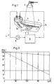
- the experimental arrangement in FIG. 1 shows a preparation 1 with a cartilage layer 2 and a bone layer 3, which is washed around and overflowed with a solution 5 in a transparent, closed vessel 4.
- the preparation 1 is irradiated with a lamp 6 (cold light halogen lamp, 75 W, 12 V, 10 ° opening angle).
- the solution 5 is circulated in the circuit 7 with a peristaltic pump 8 and kept in a cooling device 9 at a predetermined temperature (16 ° C.).
- the solution is fed into and out of the vessel 4 via the tube pieces 7a and 7b.
- Solution 5 is usually the conditioning solution with which preparation 1 has been pretreated before exposure.
- a tendon or a band can also be provided.
- the choice of methylene blue as a dye is particularly advantageous since this substance is non-toxic (can be proven by various applications in medicine). It is advantageous to provide about 30% by volume for the glycerol and 0.01% by weight for the methylene blue.
- dye molecules are put into an excited state by photons of the incident light. In this state, they can react with oxygen or water to form an intermediate. While the intermediate reacts with amino acid residues of the collagen (or other proteins), the dye is released unchanged: it acts as a catalyst during photooxidation.
- Photooxidation is slowed down at lower temperatures, and at higher temperatures the collagen-containing tissue can be damaged by the effects of heat.
- the dye absorbs light and converts it to heat.
- Photooxidation is an exothermic reaction. Therefore - in order to be able to carry out the treatment at an optimal temperature between around 14 and 18 ° C - the circulated solution 5 must be continuously cooled.
- Oxygen is consumed during photo-oxidation.
- the content of oxygen that is normally contained in the reaction solution is usually sufficient. Blowing in additional oxygen does not improve the treatment process.
- Methylene blue with a concentration of 0.01% by weight is advantageously used. At a lower concentration, the photo-oxidative treatment is too slow. At a higher concentration, too much light is absorbed in the reaction solution and in the outer regions of the tissue, so that the entire tissue and in particular its lower regions receive too little light.
- microtome sections with thicknesses between 30 and 210 ⁇ m have been made. Test results are shown in the diagram in FIG. 2.
- T is the transmission and x the thickness of a measured section.
- the values for T are plotted on a logarithmic scale.
- the empty circles indicate the measured values for cartilage tissue that has been pretreated according to the known method.
- the filled circles show the corresponding values for tissue pretreated according to the invention, glycerol with a proportion a of 80% by volume being used as the additive.
- the pretreatment with an additive according to the invention results in a considerable improvement in the transparency of the fabric.
- the light penetration depth can be increased by at least five times. This enables the photooxidative treatment of articular cartilage.
- the collagen-containing tissue is decolored, and at the same time the additive is removed. This is done by washing several times with a decolorizing solution.
- the above-mentioned S ⁇ rensen phosphate buffer is advantageously used as the decolorization solution. In order to increase the solubility of the dye, up to 50% by volume of ethanol can be added to this phosphate buffer.
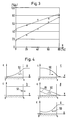
- the diagrams show the dye concentration c as a function of x.
- the cartilage tissue extends over the interval 0 ⁇ x ⁇ s.
- the reaction solution or the decolorization solution is located in the area x> s.
- FIG. 4a illustrates the initial phase of the coloring during the pretreatment.
- the arrow 50 indicates the dye transport based on diffusion. After pretreatment, there should be an equilibrium with a homogeneous dye distribution. This is shown in Fig.4b.
- FIGS. 4c and 4d The decolorization after the photooxidation is shown in FIGS. 4c and 4d. Since during the decolorization the dye concentration in the deeper areas of the tissue is still relatively high for some time, the exposure can advantageously also be continued during this process phase, up to a point in time , since the concentration c assumes, for example, the curve 55 drawn in dashed lines. This is advantageous because photooxidation has previously been far less effective in the deeper areas than in the upper areas.
- the exposure should be carried out intermittently, namely in such a way that the cartilage tissue simultaneously during the exposure phases is decolorized and that during intermediate phases the cartilage tissue is treated with conditioning solution without exposure and then stained again.
- a renewed coloring of the fabric is illustrated by the diagram in Fig.4e.
- the treatment of the tissue should be carried out to the extent that the collagen fibers change in such a way that immunological defense reactions do not occur or are only weakly pronounced in a patient.
- the cartilage tissue should gain greater mechanical strength through the photooxidative treatment.
- the exposure can only be effective from one side.
- the treatment of ligaments and tendons according to the invention exposure from both or all sides is possible.
Landscapes
- Health & Medical Sciences (AREA)
- Life Sciences & Earth Sciences (AREA)
- Chemical & Material Sciences (AREA)
- Chemical Kinetics & Catalysis (AREA)
- Engineering & Computer Science (AREA)
- Medicinal Chemistry (AREA)
- General Health & Medical Sciences (AREA)
- Dermatology (AREA)
- Biomedical Technology (AREA)
- Oral & Maxillofacial Surgery (AREA)
- Transplantation (AREA)
- Epidemiology (AREA)
- Animal Behavior & Ethology (AREA)
- Public Health (AREA)
- Veterinary Medicine (AREA)
- Botany (AREA)
- Molecular Biology (AREA)
- Biophysics (AREA)
- Urology & Nephrology (AREA)
- Zoology (AREA)
- General Chemical & Material Sciences (AREA)
- Biochemistry (AREA)
- Materials Engineering (AREA)
- Polymers & Plastics (AREA)
- Organic Chemistry (AREA)
- Materials For Medical Uses (AREA)
- Peptides Or Proteins (AREA)
- Non-Silver Salt Photosensitive Materials And Non-Silver Salt Photography (AREA)
Abstract
Description
- Die Erfindung betrifft ein Verfahren zu einer photooxidativen Behandlung von kollagenhaltigen Geweben gemäss Oberbegriff von Anspruch 1. Kollagenhaltige Gewebe sind beispielsweise Knorpelgewebe und Gewebe von Bändern oder Sehnen. Die Erfindung bezieht sich auch auf derartige Gewebe, die nach dem erfindungsgemässen Verfahren präpariert worden sind.
- Eine photooxidative Behandlung von kollagenhaltigem Gewebe (insbesondere Herzbeutelgewebe) ist aus der US-PS 5 147 514 bekannt. Bei diesem Verfahren reagieren Tyrosin-, Tryptophan- und/oder Histidinreste von Kollagenfasern unter der katalytischen Wirkung von durch Licht aktivierten Farbstoffmolekülen mit Sauerstoff derart, dass sich Verbindungen zwischen Kollagenfasern ausbilden. Als wirksame Farbstoffe werden Methylenblau, Methylengrün, Bengalrosa, Riboflavin, Proflavin, Fluoreszein, Eosin und Pyridoxal-5-phosphat genannt.
- Knorpelgewebe ist - abgesehen von vereinzelt vorliegenden Chondrocyten - aus einem Netzwerk von Kollagenfasern und hochmolekularem Proteoglykan aufgebaut. Zu rund 75% besteht das Gewebe aus Wasser. Aufgrund dieser Zusammensetzung ist der Knorpel ein weichelastisches, offenporiges und permeables Gewebe. Wasser, das sich im Gewebe befindet, ist leicht mit Wasser der Umgebung austauschbar. Ein Austausch von Wasser oder von in dem Wasser gelösten Stoffen kann ausser durch Diffusion auch durch eine Konvektion erfolgen, die mit Volumenänderungen durch Komprimieren und Expandieren des Gewebes einhergeht. Für eine Konditionierung des Knorpelgewebes und dessen Beladung mit einem Farbstoff ist die gute Austauschfähigkeit eine Voraussetzung. Diese Feststellungen gelten auch für Sehnen und Bänder.
- Das bekannte Verfahren wird bei der photooxidativen Behandlung von aus Herzbeuteln gewonnenen Gewebe angewendet, das für die Herstellung von implantierbaren Herzklappen vorgesehen ist. Dieses Gewebe ist flächenartig ausgebildet und dünnwandig - rund 0,5 mm dick - und kann von beiden Seiten her belichtet werden.
- Dass die Kollagenfasern durch die photooxidative Behandlung verändert werden, lässt sich mit Kollagenase nachweisen. Dieses Enzym löst die Fasern des unbehandelten Gewebes in Teile auf, mit der Folge, dass der mechanische Zusammenhalt des Gewebes verloren geht. Nach einer photooxidativen Behandlung ist diese "Verdauung" durch Kollagenase nicht mehr in gleichem Ausmass wirksam.
- Die Kollagenfasern werden durch die photooxidative Behandlung teilweise miteinander vernetzt, wodurch das Gewebe eine grössere mechanische Beanspruchbarkeit gewinnt. Ausserdem werden unter der katalytischen Wirkung des Farbstoffs Kollagenfasern in der Weise photooxidativ verändert, dass bei Verwendung des behandelten Gewebes als Implantat immunologische Abwehrreaktionen unterbleiben oder nur schwach ausgeprägt sind.
- Für die chirurgische Behandlung von Kniegelenken besteht der Wunsch - analog zu den Herzklappen aus Herzbeuteln - Knorpelpräparate als Implantate verwenden zu können. Gelenkknorpel sind 2 bis 3 mm dick und auf Knochen aufgewachsen. Für die Nutzung als Implantat soll das Knorpelgewebe zusammen mit einem Stück zugehörigen Knochenmaterials verwendet werden. Dies bedeutet, dass das Gewebe nur von der Seite der freiliegenden Oberfläche her belichtet werden kann. Somit muss das Licht bis 3 mm tief in das Gewebe eindringen können. Wie sich jedoch experimentell gezeigt hat, ist die Lichtdurchlässigkeit ziemlich schlecht.
- Es ist daher Aufgabe der Erfindung, ein Verfahren zu schaffen, das eine photooxidative Behandlung des Kollagennetzwerks trotz schlechter Lichtdurchlässigkeit des natürlichen Knorpelgewebes ermöglicht. Diese Aufgabe wird mit den in Anspruch 1 genannten Massnahmen gelöst.
- Die Kollagenfasern und das Proteoglykan bilden eine Vielzahl von Streuzentren, aufgrund derer das Licht nur in die Randbereiche des Knorpelgewebes wirksam eindringen kann. Die Lichtstreuung beruht darauf, dass sich der Brechungsindex bei den Streuzentren lokal stark ändert. Die erfindungsgemässe Lehre geht daher von der Idee aus, den Brechungsindex des Wassers, das den Raum zwischen den Streuzentren füllt, durch Zusetzen geeigneter Stoffe zu erhöhen, so dass der Brechungsindex eine Nivellierung seiner Werte im Gewebe erfährt. Zusatzstoffe, die sich zu diesem Zweck verwenden lassen, sind beispielsweise Diethylenglycol (Brechungsindex n = 1,447 bei 20°C) oder Glycerin (n = 1,474).
- Bei dem erfindungsgemässen Verfahren zu einer photooxidativen Behandlung von kollagenhaltigen Geweben wird das Gewebe vor einer Bestrahlung mit Licht mittels einer wässrigen Konditionierungslösung vorbehandelt. Bei dieser Vorbehandlung wird das für die Lösung permeable Gewebe mit einem in der Lösung enthaltenen Farbstoff, insbesondere mit Methylenblau, beladen. Dieser Farbstoff, durch Licht aktiviert, katalysiert photooxidative Reaktionen. Der Konditionierungslösung wird erfindungsgemäss ein Stoff zugegeben, dessen Brechungsindex grösser als jener von Wasser (n = 1,333 bei 25°C) ist. Dank dieses Zusatzstoffes nimmt das kollagenhaltige Gewebe bei der Vorbehandlung eine für das Licht vergrösserte Transparenz an.
- Die abhängigen Ansprüche 2 bis 11 beziehen sich auf vorteilhafte Ausführungsformen des erfindungsgemässen Verfahrens und/oder betreffen besondere Aspekte des Verfahrens. Die Ansprüche 11 bis 13 haben Gewebepräparate zum Gegenstand, die nach dem erfindungsgemässen Verfahren behandelt worden sind.
- Die photooxidative Behandlung wird gemäss folgendem, drei Schritte umfassenden Verfahren durchgeführt, wobei die Temperatur vorzugsweise bei rund 14 bis 18°C gehalten wird:
- 1. Vorbehandlung des kollagenhaltigen Gewebes mit der Konditionierungslösung während rund 1 bis 5 Stunden.
- 2. Belichten des kollagenhaltigen Gewebes während rund 10 bis 50 Stunden.
- 3. Entfärben des kollagenhaltigen Gewebes sowie Entfernen des Zusatzstoffs durch mehrfaches Waschen mit einer Entfärbungslösung.
- Nachfolgend wird die Erfindung anhand der Zeichnungen näher erläutert. Es zeigen:
- Fig. 1
- eine Versuchsanordnung zur Durchführung einer photooxidativen Behandlung,
- Fig. 2
- Transmission von kollagenhaltigen Geweben in Abhängigkeit von der Gewebedicke,
- Fig. 3
- Transmission von Geweben nach Vorbehandlungen mit Konditionierungslösungen, die Glycerin enthalten, und
- Fig. 4
- Diagramme zur qualitativen Darstellung der Farbstoffverteilung im Gewebe.
- Die Versuchsanordnung der Fig.1 zeigt ein Präparat 1 mit einer Knorpelschicht 2 und einer Knochenschicht 3, das in einem durchsichtigen, geschlossenen Gefäss 4 von einer Lösung 5 um- und überspült wird. Das Präparat 1 wird mit einer Lampe 6 (Kaltlichthalogenlampe, 75 W, 12 V, 10° Öffnungswinkel) bestrahlt. Die Lösung 5 wird im Kreislauf 7 mit einer peristaltischen Pumpe 8 umgewälzt und in einer Kühleinrichtung 9 auf einer vorgegebene Temperatur (16°C) gehalten. Über die Schlauchstücke 7a und 7b wird die Lösung in das Gefäss 4 zu- bzw. aus ihm abgeführt. Die Lösung 5 ist in der Regel die Konditionierungslösung, mit der das Präparat 1 vor der Belichtung vorbehandelt worden ist. Anstelle des Knorpelpräparates 1 kann auch eine Sehne oder ein Band vorgesehen sein.
- Die Lösung 5 setzt sich aus einem Phosphatpuffer nach Sørensen, Glycerin (C3H8O3, Molekulargewicht M = 92 g/mol) und Methylenblau (C16H18ClN3S.3H2O, M = 320 g/mol + aq) zusammen. Der Phosphatpuffer nach Sørensen weist eine Molarität von 1/15 und einen pH-Wert von 7,4 auf; er ist eine Lösung aus reinem Wasser ("Nanopur") und 1,787 g/l Kaliumdihydrogenphosphat (KH2PO4, M = 136 g/mol) sowie 9.532 g/l Dinatriumhydrogenphosphat (Na2HPO4, M = 178 g/mol). Die Wahl von Methylenblau als Farbstoff ist besonders vorteilhaft, da dieser Stoff nicht toxisch ist (durch verschiedene Anwendungen in der Medizin belegbar). Mit Vorteil sieht man für das Glycerin einen Anteil von rund 30 Vol.-% vor und für das Methylenblau 0,01 Gew.-%.
- Präparate 1 werden beispielsweise aus Schultergelenken geschlachteter Rinder entnommen, wobei mittels einer aus der Chirurgie bekannten oszillierenden Säge Knorpelstücke mit Knochen (ca. 10×10×5 mm) aus den Gelenken geschnitten werden. Die Präparate 1 werden in einer Pufferlösung transportiert sowie gelagert und dabei gekühlt gehalten (bei rund 4°C).
- Bei der Photooxidation werden Farbstoffmoleküle durch Photonen des eingestrahlten Lichts in einen angeregten Zustand versetzt. In diesem Zustand können sie mit Sauerstoff oder Wasser zu einem Zwischenprodukt reagieren. Während das Zwischenprodukt mit Aminosäureresten des Kollagens (oder anderer Proteine) reagiert, wird der Farbstoff unverändert freigesetzt: er wirkt bei der Photooxidation als Katalysator.
- Allerdings finden unter der Einwirkung des Lichts auch irreversible Veränderungen der Farbstoffmoleküle statt, die als Ausbleichung des Farbstoffs in Erscheinung treten. Es muss daher für einen laufenden Nachschub von Farbstoffmolekülen in das Gewebe gesorgt werden. Dies geschieht dadurch, dass das Präparat 1 während der Belichtung ständig mit Konditionierungslösung 5 umspült wird. Über einen Stoffaustausch zwischen dem Gewebe und der Lösung 5 ergibt sich eine teilweise Erneuerung des Farbstoffs im Gewebe, wobei insbesondere ausgebleichter Farbstoff durch intakten ersetzt wird. Gelegentlich - jeweils nach etwa 24 Stunden - muss die gesamte Reaktionslösung durch neue ersetzt werden.
- Bei tieferen Temperaturen ist die Photooxidation verlangsamt, und bei höheren Temperaturen kann das kollagenhaltige Gewebe durch die Wärmeeinwirkung geschädigt werden. Durch den Farbstoff wird Licht absorbiert und in Wärme umgewandelt. Die Photooxidation ist eine exotherme Reaktion. Daher muss - um die Behandlung bei einer optimalen Temperatur zwischen rund 14 und 18°C durchführen zu können - die umgewälzte Lösung 5 laufend gekühlt werden.
- Bei der Photooxidation werden Wasserstoffionen freigesetzt. Daher muss - um die Behandlung bei einem optimalen pH-Wert zwischen rund 7 und 8 durchführen zu können - eine Pufferlösung verwendet werden (Phosphatpuffer nach Sørensen).
- Bei der Photooxidation wird Sauerstoff verbraucht. Der Gehalt an Sauerstoff, der normalerweise in der Reaktionslösung enthalten ist, reicht in der Regel aus. Einblasen von zusätzlichem Sauerstoff bringt keine Verbesserung des Behandlungsverfahrens.
- Mit Vorteil wird Methylenblau mit einer Konzentration von 0,01 Gew.-% verwendet. Bei einer geringeren Konzentration ist die photooxitative Behandlung zu langsam. Bei einer höheren Konzentration wird zu viel Licht in der Reaktionslösung und in den äusseren Bereichen des Gewebes absorbiert, so dass das ganze Gewebe und insbesondere dessen tieferen Bereiche zu wenig Licht empfangen.
- Dank des relativ kleinen Molekulargewichts von Methylenblau (M = 320 g/mol) diffundiert dieser Farbstoff gut in und durch das kollagenhaltige Gewebe. Dies bestätigen Untersuchungen an Dünnschnitten.
- Es ist die Lichtdurchlässigkeit (Transmission) des kollagenhaltigen Gewebes beim Absorptionsmaximum von Methylenblau, nämlich 665 nm, bestimmt worden. Dazu sind Mikrotomschnitte mit Dicken zwischen 30 und 210 µm angefertigt worden. Versuchsergebnisse sind in dem Diagramm der Fig.2 dargestellt. T ist die Transmission und x die Dicke eines ausgemessenen Schnittes. Die Werte für T sind in einem logarithmischen Massstab aufgetragen. Die leeren Kreise geben die Messwerte für Knorpelgewebe an, das gemäss dem bekannten Verfahren vorbehandelt worden ist. Die gefüllten Kreise zeigen die entsprechenden Werte für erfindungsgemäss vorbehandeltes Gewebe, wobei als Zusatzstoff Glycerin mit einem Anteil a von 80 Vol.-% verwendet worden ist.
- In Fig.3 ist dargestellt, wie die Transmission T von dem Anteil a an Glycerin abhängt. Die zwei gezeigten Messreihen beziehen sich auf Schichtdicken von 90 µm (Dreiecke) und 180 µm (Kreise).
- Wie aus den Diagrammen der Figuren 2 und 3 hervorgeht, ergibt sich durch die erfindungsgemässe Vorbehandlung mit einem Zusatzstoff eine erhebliche Verbesserung der Transparenz des Gewebes. Die Lichteindringtiefe kann um mindestens das Fünffache vergrössert werden. Damit wird die photooxidative Behandlung von Gelenkknorpeln möglich.
- Nach der Durchführung der Photooxidation wird das kollagenhaltige Gewebe entfärbt, und gleichzeitig wird der Zusatzstoff entfernt. Dies geschieht durch mehrfaches Waschen mit einer Entfärbungslösung. Mit Vorteil wird als Entfärbungslösung der oben genannte Phosphatpuffer nach Sørensen verwendet. Um die Löslichkeit des Farbstoffs zu vergrössern, kann diesem Phosphatpuffer bis zu 50 Vol-% Ethanol zugemischt sein.
- Anhand der Diagramme der Teilfiguren a bis e in Fig.4 sollen noch einige Aspekte der Gewebeeinfärbung erläutert werden. Die Diagramme stellen die Farbstoffkonzentration c als Funktion von x dar. Durch x ist der Abstand von der Knochenoberfläche (x = 0) gegeben. Über das Intervall 0 < x < s erstreckt sich das Knorpelgewebe. Im Gebiet x > s befindet sich die Reaktionslösung bzw. die Entfärbungslösung.
- Fig.4a illustriert die Anfangsphase des Einfärbens während der Vorbehandlung. Der Pfeil 50 deutet den auf Diffusion beruhenden Farbstofftransport an. Nach erfolgter Vorbehandlung sollte ein Gleichgewicht mit einer homogenen Farbstoffverteilung vorliegen. Dies ist in Fig.4b gezeigt. Der Doppelpfeil 51 deutet an, dass weiterhin ein Stoffaustausch über die Gewebeoberfläche bei

- Das Entfärben nach der Photooxidation stellen die Figuren 4c und 4d dar. Da während des Entfärbens die Farbstoffkonzentration in den tieferen Bereichen des Gewebes einige Zeit noch relativ hoch ist, kann die Belichtung mit Vorteil auch während dieser Verfahrensphase fortgesetzt werden, und zwar bis zu einem Zeitpunkt, da die Konzentration c beispielsweise den gestrichelt gezeichneten Kurvenverlauf 55 annimmt. Dies ist vorteilhaft, da die Photooxidation zuvor in den tieferen Bereichen weit weniger wirksam als in den oberen Bereichen gewesen ist.
- Die Lehre, auch während des Entfärbens die Belichtung fortzusetzen, lässt sich zu einer vorteilhaften Verfahrensvariante weiterentwickeln: Die Belichtung soll intermittierend vorgenommen werden, nämlich derart, dass während Belichtungsphasen das Knorpelgewebe gleichzeitig entfärbt wird und dass während Zwischenphasen das Knorpelgewebe ohne Belichtung mit Konditionierungslösung behandelt und erneut eingefärbt wird. Eine erneute Einfärbung des Gewebes ist durch das Diagramm der Fig.4e illustriert.
- Ist eine Verwendung des photooxidativ behandelten Knorpelgewebes als Implantat vorgesehen, so soll die Behandlung des Gewebes in dem Ausmass durchgeführt werden, dass sich die Kollagenfasern in der Weise verändern, dass bei einem Patienten immunologische Abwehrreaktionen unterbleiben oder nur schwach ausgeprägt sind. Ausserdem soll durch die photooxidative Behandlung das Knorpelgewebe eine grössere mechanische Beanspruchbarkeit gewinnen.
- Bei der Belichtung von Knorpelpräparaten kann die Belichtung wirkungsvoll nur von der einen Seite vorgenommen werden. Bei der erfindungsgemässen Behandlung von Bändern und Sehnen ist eine Belichtung von beiden oder allen Seiten möglich.
Claims (13)
- Verfahren zu einer photooxidativen Behandlung von kollagenhaltigen Geweben, bei welchem Verfahren das Gewebe vor einer Bestrahlung mit Licht mittels einer wässrigen Konditionierungslösung vorbehandelt wird, so dass das für die Lösung permeable Gewebe mit einem in der Lösung enthaltenen Farbstoff, insbesondere Methylenblau, beladen wird, wobei dieser Farbstoff durch Licht aktiviert photooxidative Reaktionen katalysiert,
dadurch gekennzeichnet, dass in die Lösung ein Zusatzstoff zugegeben wird, dessen Brechungsindex grösser als jener von Wasser - nämlich 1,333 bei 25°C - ist und mit dem das Gewebe bei der Vorbehandlung eine für das Licht vergrösserte Transparenz annimmt. - Verfahren nach Anspruch 1, dadurch gekennzeichnet, dass der zusätzliche Stoff einen Brechungsindex grösser als 1,4 aufweist und dass dieser Stoff insbesondere Glycerin ist.
- Verfahren nach Anspruch 1 oder 2, dadurch gekennzeichnet, dass in Anwesenheit von Sauerstoff und unter der katalytischen Wirkung des Farbstoffs Kollagenfasern in der Weise photooxidativ verändert werden, dass bei Verwendung des behandelten Gewebes als Implantat in einem Patienten immunologische Abwehrreaktionen unterbleiben oder nur schwach ausgeprägt sind.
- Verfahren nach Anspruch 3, dadurch gekennzeichnet, dass die Kollagenfasern durch die photooxidative Behandlung teilweise miteinander vernetzt werden, so dass das Gewebe eine grössere mechanische Beanspruchbarkeit gewinnt.
- Verfahren nach einem der Ansprüche 1 bis 4, dadurch gekennzeichnet, dass folgende drei Schritte, vorzugsweise bei rund 14 bis 18°C, durchgeführt werden:1. Vorbehandlung des Gewebes mit der Konditionierungslösung während rund 1 bis 5 Stunden,2. Belichten des Gewebes während rund 10 bis 50 Stunden, und3. Entfärben des kollagenhaltigen Gewebes sowie Entfernen des Zusatzstoffs durch mehrfaches Waschen mit einer Entfärbungslösung.
- Verfahren nach Anspruch 5, dadurch gekennzeichnet, dass die Konditionierungslösung ein Gemisch aus einem Phosphatpuffers und Glycerin ist, wobei vorzugsweise ein Phosphatpuffer nach Sørensen verwendet wird, der eine Molarität von 1/15 und einen pH-Wert von 7,4 aufweist, der Anteil an Glycerin vorzugsweise rund 30 Vol-% beträgt und der Anteil an Methylenblau vorzugsweise rund 0,01 Gew-% beträgt.
- Verfahren nach Anspruch 5 oder 6, dadurch gekennzeichnet, dass die Entfärbungslösung ein Phosphatpuffer ist, dem 0 bis 50 Vol-% Ethanol zugemischt ist, wobei der Phosphatpuffer vorzugsweise ein Phosphatpuffer nach Sørensen ist, der eine Molarität von 1/15 und einen pH-Wert von 7,4 aufweist.
- Verfahren nach einem der Ansprüche 1 bis 7, dadurch gekennzeichnet, dass während der ganzen Belichtungsphase oder während eines grossen Teils der Belichtungsphase das Gewebe mit sauerstoffhaltiger Konditionierungslösung umspült wird.
- Verfahren nach Anspruch 8, dadurch gekennzeichnet, dass die Konditionierungslösung mit einer Pumpe in einem Kreislauf geführt wird und dass mittels einer in diesem Kreislauf vorgesehenen Kühleinrichtung eine vorgegebene Temperatur der umgewälzten Lösung eingestellt wird.
- Verfahren nach Anspruch 8 oder 9, dadurch gekennzeichnet, dass während einer die Belichtung abschliessenden Phase das Gewebe mit sauerstoffhaltiger Entfärbungslösung umspült wird.
- Verfahren nach Anspruch 5, dadurch gekennzeichnet, dass die Belichtung intermittierend vorgenommen wird, dass während Belichtungsphasen das Gewebe mit sauerstoffhaltiger Entfärbungslösung umspült wird und dass während Zwischenphasen das Gewebe ohne Belichtung mit Konditionierungslösung behandelt wird.
- Knorpelpräparat (1) mit einer Knochenschicht (3) und einer Knorpelschicht (2), die mit dem Verfahren gemäss einem der Ansprüche 1 bis 11 behandelt worden ist.
- Kollagenhaltiges Gewebe, das aus einer Sehne oder einem Band gewonnen worden ist und das mit dem Verfahren gemäss einem der Ansprüche 1 bis 11 behandelt worden ist.
Priority Applications (6)
| Application Number | Priority Date | Filing Date | Title |
|---|---|---|---|
| AT95810637T ATE200092T1 (de) | 1995-10-11 | 1995-10-11 | Verfahren zu einer photooxidativen behandlung von kollagenhaltigen geweben |
| EP95810637A EP0768332B1 (de) | 1995-10-11 | 1995-10-11 | Verfahren zu einer photooxidativen Behandlung von kollagenhaltigen Geweben |
| DE59509135T DE59509135D1 (de) | 1995-10-11 | 1995-10-11 | Verfahren zu einer photooxidativen Behandlung von kollagenhaltigen Geweben |
| JP8264397A JPH09122226A (ja) | 1995-10-11 | 1996-10-04 | 膠原含有組織の光酸化処理方法 |
| US08/729,259 US5817153A (en) | 1995-10-11 | 1996-10-09 | Method of photo-oxidative treatment of tissues containing collagen |
| JP2009023799A JP2009136699A (ja) | 1995-10-11 | 2009-02-04 | 膠原含有組織の光酸化処理方法 |
Applications Claiming Priority (1)
| Application Number | Priority Date | Filing Date | Title |
|---|---|---|---|
| EP95810637A EP0768332B1 (de) | 1995-10-11 | 1995-10-11 | Verfahren zu einer photooxidativen Behandlung von kollagenhaltigen Geweben |
Publications (2)
| Publication Number | Publication Date |
|---|---|
| EP0768332A1 true EP0768332A1 (de) | 1997-04-16 |
| EP0768332B1 EP0768332B1 (de) | 2001-03-28 |
Family
ID=8221806
Family Applications (1)
| Application Number | Title | Priority Date | Filing Date |
|---|---|---|---|
| EP95810637A Expired - Lifetime EP0768332B1 (de) | 1995-10-11 | 1995-10-11 | Verfahren zu einer photooxidativen Behandlung von kollagenhaltigen Geweben |
Country Status (5)
| Country | Link |
|---|---|
| US (1) | US5817153A (de) |
| EP (1) | EP0768332B1 (de) |
| JP (2) | JPH09122226A (de) |
| AT (1) | ATE200092T1 (de) |
| DE (1) | DE59509135D1 (de) |
Cited By (5)
| Publication number | Priority date | Publication date | Assignee | Title |
|---|---|---|---|---|
| WO2001043667A1 (de) | 1999-12-15 | 2001-06-21 | Sulzer Orthopedics Ltd. | Präparat für die reparatur von knorpel- oder knorpel/knochen-defekten in menschlichen oder tierischen gelenken |
| US8435305B2 (en) | 2010-08-31 | 2013-05-07 | Zimmer, Inc. | Osteochondral graft delivery device and uses thereof |
| US8801725B2 (en) | 2008-03-10 | 2014-08-12 | Zimmer Orthobiologics, Inc. | Instruments and methods used when repairing a defect on a tissue surface |
| US8829166B2 (en) | 2002-06-26 | 2014-09-09 | Zimmer Orthobiologics, Inc. | Rapid isolation of osteoinductive protein mixtures from mammalian bone tissue |
| US9113916B2 (en) | 2010-08-31 | 2015-08-25 | Zimmer, Inc. | Drill bit for osteochondral drilling with guiding element and uses thereof |
Families Citing this family (11)
| Publication number | Priority date | Publication date | Assignee | Title |
|---|---|---|---|---|
| EP0896825B1 (de) | 1997-08-14 | 2002-07-17 | Sulzer Innotec Ag | Zusammensetzung und Vorrichtung zur Reparatur von Knorpelgewebe in vivo bestehend aus Nanokapseln mit osteoinduktiven und/oder chondroinduktiven Faktoren |
| US20130274837A1 (en) * | 1998-10-23 | 2013-10-17 | Babak Nemati | Systems and Methods to Enhance Optical Transparency of Biological Tissues for Photobiomodulation |
| US6219575B1 (en) * | 1998-10-23 | 2001-04-17 | Babak Nemati | Method and apparatus to enhance optical transparency of biological tissues |
| US6723335B1 (en) * | 2000-04-07 | 2004-04-20 | Jeffrey William Moehlenbruck | Methods and compositions for treating intervertebral disc degeneration |
| AU2002358957A1 (en) * | 2001-12-10 | 2003-06-23 | Colbar Lifescience Ltd. | Methods, devices, and preparations for intervertebral disc treatment |
| US7232802B2 (en) | 2001-12-21 | 2007-06-19 | Zimmer Orthobiologics, Inc. | Compositions and methods for promoting myocardial and peripheral angiogenesis |
| WO2003080138A1 (de) * | 2002-03-22 | 2003-10-02 | Sulzer Orthopedics Ltd. | Präparat und verfahren zur verbesserung der regenerationsfähigkeit von durch trauma, abnützung oder krankheit geschädigtem knorpelgewebe oder zur prophylaktischen verhinderung von knorpelabbau |
| WO2006029571A1 (en) * | 2004-09-14 | 2006-03-23 | The University Of Hong Kong | Photochemically crosslinked collagen scaffolds and methods for their preparation |
| US7759120B2 (en) * | 2005-03-02 | 2010-07-20 | Kps Bay Medical, Inc. | Seeding implantable medical devices with cells |
| US20060199265A1 (en) * | 2005-03-02 | 2006-09-07 | Wolf Michael F | Seeding implantable medical devices with cells |
| US20080026032A1 (en) * | 2006-07-27 | 2008-01-31 | Zubery Yuval | Composite implants for promoting bone regeneration and augmentation and methods for their preparation and use |
Citations (2)
| Publication number | Priority date | Publication date | Assignee | Title |
|---|---|---|---|---|
| EP0411925A2 (de) * | 1989-08-02 | 1991-02-06 | University Of North Carolina At Chapel Hill | Verfahren zur Vernetzung von Kollagen und erzeugtes Produkt |
| US5147514A (en) | 1987-08-02 | 1992-09-15 | University Of North Carolina | Process for cross-linking collagenous material and resulting product |
Family Cites Families (2)
| Publication number | Priority date | Publication date | Assignee | Title |
|---|---|---|---|---|
| US3152976A (en) * | 1961-09-21 | 1964-10-13 | Ethicon Inc | Preparation of collagenous materials |
| US5409479A (en) * | 1983-10-06 | 1995-04-25 | Premier Laser Systems, Inc. | Method for closing tissue wounds using radiative energy beams |
-
1995
- 1995-10-11 EP EP95810637A patent/EP0768332B1/de not_active Expired - Lifetime
- 1995-10-11 DE DE59509135T patent/DE59509135D1/de not_active Expired - Lifetime
- 1995-10-11 AT AT95810637T patent/ATE200092T1/de not_active IP Right Cessation
-
1996
- 1996-10-04 JP JP8264397A patent/JPH09122226A/ja active Pending
- 1996-10-09 US US08/729,259 patent/US5817153A/en not_active Expired - Lifetime
-
2009
- 2009-02-04 JP JP2009023799A patent/JP2009136699A/ja not_active Withdrawn
Patent Citations (2)
| Publication number | Priority date | Publication date | Assignee | Title |
|---|---|---|---|---|
| US5147514A (en) | 1987-08-02 | 1992-09-15 | University Of North Carolina | Process for cross-linking collagenous material and resulting product |
| EP0411925A2 (de) * | 1989-08-02 | 1991-02-06 | University Of North Carolina At Chapel Hill | Verfahren zur Vernetzung von Kollagen und erzeugtes Produkt |
Non-Patent Citations (2)
| Title |
|---|
| MOORE M. A. ET AL.: "Stabilization of pericardial tissue by dye-mediated photooxidation", JOURNAL OF BIOMEDICAL MATERIALS RESEARCH, vol. 28, no. 5, US, pages 611 - 618, XP000566254, DOI: doi:10.1002/jbm.820280511 * |
| RAMSHAW ET AL.: "Methylene blue sensitized photooxidation of collagen fibrils", BIOCHIMICA ET BIOPHYSICA ACTA, vol. 1206, no. 2, NL, pages 225 - 230 * |
Cited By (9)
| Publication number | Priority date | Publication date | Assignee | Title |
|---|---|---|---|---|
| WO2001043667A1 (de) | 1999-12-15 | 2001-06-21 | Sulzer Orthopedics Ltd. | Präparat für die reparatur von knorpel- oder knorpel/knochen-defekten in menschlichen oder tierischen gelenken |
| EP1498089A1 (de) * | 1999-12-15 | 2005-01-19 | Zimmer GmbH | Präparat für die Reparatur von Knorpel- oder Knorpel/Knochen-Defekten in menschlichen oder tierischen Gelenken |
| US6858042B2 (en) | 1999-12-15 | 2005-02-22 | Zimmer Orthobiologics, Inc. | Preparation for repairing cartilage defects or cartilage/bone defects in human or animal joints |
| USRE43714E1 (en) * | 1999-12-15 | 2012-10-02 | Zimmer Orthobiologics, Inc. | Preparation for repairing cartilage defects or cartilage/bone defects in human or animal joints |
| US8829166B2 (en) | 2002-06-26 | 2014-09-09 | Zimmer Orthobiologics, Inc. | Rapid isolation of osteoinductive protein mixtures from mammalian bone tissue |
| US8801725B2 (en) | 2008-03-10 | 2014-08-12 | Zimmer Orthobiologics, Inc. | Instruments and methods used when repairing a defect on a tissue surface |
| US8435305B2 (en) | 2010-08-31 | 2013-05-07 | Zimmer, Inc. | Osteochondral graft delivery device and uses thereof |
| US8753406B2 (en) | 2010-08-31 | 2014-06-17 | Zimmer Inc. | Osteochondral graft delivery device and uses thereof |
| US9113916B2 (en) | 2010-08-31 | 2015-08-25 | Zimmer, Inc. | Drill bit for osteochondral drilling with guiding element and uses thereof |
Also Published As
| Publication number | Publication date |
|---|---|
| DE59509135D1 (de) | 2001-05-03 |
| JPH09122226A (ja) | 1997-05-13 |
| US5817153A (en) | 1998-10-06 |
| ATE200092T1 (de) | 2001-04-15 |
| JP2009136699A (ja) | 2009-06-25 |
| EP0768332B1 (de) | 2001-03-28 |
Similar Documents
| Publication | Publication Date | Title |
|---|---|---|
| EP0768332B1 (de) | Verfahren zu einer photooxidativen Behandlung von kollagenhaltigen Geweben | |
| Heilbrunn | The action of calcium on muscle protoplasm | |
| Haug | Electron microscopical localization of the zinc in hippocampal mossy fibre synapses by a modified sulfide silver procedure | |
| DE69019512T2 (de) | Verhinderung von verkalkung in bioprosthetischen implantaten. | |
| EP0237037B1 (de) | Mit vernetzter Gelatine imprägnierte Gefässprothese und Verfahren zu ihrer Herstellung | |
| EP0401199B1 (de) | Verfahren zur Präparierung von biologischem Implantationsmaterial | |
| DE69031483T2 (de) | Verfahren zur Vernetzung von Kollagen und dadurch erzeugtes Produkt | |
| DE2515697A1 (de) | Fotochemische stimulierung von nerven | |
| DE69124129T2 (de) | Hydroxyapatitbeschichtung von Substraten | |
| DE69835011T2 (de) | Unterschiedliche behandlung von prothesenteilen | |
| DE69429667T2 (de) | Verfahren zur herstellung von verkalkungsbeständigem bioprosthetischem gewebe | |
| DE69925632T2 (de) | Verfahren zur gewebefixierung mittels epoxiden | |
| EP2654815B1 (de) | Herstellung eines transplantats aus tierischer dermis mit natriumsulfidlösung | |
| DE2657370A1 (de) | Hilfsmittel zum bedecken und/oder ausfuellen von knochendefekten und verfahren zur herstellung desselben | |
| DE1496645A1 (de) | Verfahren zur Verstaerkung eines Glasgegenstandes | |
| DE2854490C2 (de) | Knochenersatzmaterial mit verbesserter biologischer Stabilität auf Basis von Kollagen | |
| DE3435771A1 (de) | Knochenmatrix und verfahren zu ihrer herstellung | |
| DE1620795A1 (de) | Verfahren zur Herstellung von Kollagenschwaemmen | |
| EP0364871A1 (de) | Verfahren zur Herstellung von Rinder-Pericard-Materialien und ihre Verwendung | |
| EP1124593B1 (de) | Verfahren zur präparation von knochenmaterial | |
| DE3104815A1 (de) | Verfahren zur herstellung eines verbundstoffs mit langzeit-freigabe | |
| DE19832221C2 (de) | Verfahren und Vorrichtung zur kosmetischen Haarentfernung | |
| EP3541441B1 (de) | Verfahren zum aufbereiten eines transplantates | |
| EP3110246B1 (de) | Verfahren zur konservierung von porcinem knorpelgewebe | |
| EP0012443B1 (de) | Zahnpulpahöhlenfüllmaterial, bestehend aus Kollagen und Verfahren zu dessen Herstellung |
Legal Events
| Date | Code | Title | Description |
|---|---|---|---|
| PUAI | Public reference made under article 153(3) epc to a published international application that has entered the european phase |
Free format text: ORIGINAL CODE: 0009012 |
|
| AK | Designated contracting states |
Kind code of ref document: A1 Designated state(s): AT CH DE FR GB IT LI |
|
| AX | Request for extension of the european patent |
Free format text: LT;SI |
|
| RBV | Designated contracting states (corrected) |
Designated state(s): AT CH DE FR GB IT LI |
|
| 17P | Request for examination filed |
Effective date: 19970918 |
|
| GRAG | Despatch of communication of intention to grant |
Free format text: ORIGINAL CODE: EPIDOS AGRA |
|
| 17Q | First examination report despatched |
Effective date: 20000616 |
|
| GRAG | Despatch of communication of intention to grant |
Free format text: ORIGINAL CODE: EPIDOS AGRA |
|
| GRAH | Despatch of communication of intention to grant a patent |
Free format text: ORIGINAL CODE: EPIDOS IGRA |
|
| GRAH | Despatch of communication of intention to grant a patent |
Free format text: ORIGINAL CODE: EPIDOS IGRA |
|
| RAP1 | Party data changed (applicant data changed or rights of an application transferred) |
Owner name: SULZER MARKETS AND TECHNOLOGY AG |
|
| GRAA | (expected) grant |
Free format text: ORIGINAL CODE: 0009210 |
|
| AK | Designated contracting states |
Kind code of ref document: B1 Designated state(s): AT CH DE FR GB IT LI |
|
| REF | Corresponds to: |
Ref document number: 200092 Country of ref document: AT Date of ref document: 20010415 Kind code of ref document: T |
|
| REG | Reference to a national code |
Ref country code: CH Ref legal event code: NV Representative=s name: SULZER MANAGEMENT AG Ref country code: CH Ref legal event code: EP |
|
| REF | Corresponds to: |
Ref document number: 59509135 Country of ref document: DE Date of ref document: 20010503 |
|
| GBT | Gb: translation of ep patent filed (gb section 77(6)(a)/1977) |
Effective date: 20010501 |
|
| ITF | It: translation for a ep patent filed | ||
| ET | Fr: translation filed | ||
| REG | Reference to a national code |
Ref country code: GB Ref legal event code: IF02 |
|
| PLBE | No opposition filed within time limit |
Free format text: ORIGINAL CODE: 0009261 |
|
| STAA | Information on the status of an ep patent application or granted ep patent |
Free format text: STATUS: NO OPPOSITION FILED WITHIN TIME LIMIT |
|
| 26N | No opposition filed | ||
| REG | Reference to a national code |
Ref country code: CH Ref legal event code: PUE Owner name: ZIMMER GMBH Free format text: SULZER MARKETS AND TECHNOLOGY AG#ZUERCHERSTRASSE 12#8401 WINTERTHUR (CH) -TRANSFER TO- ZIMMER GMBH#SULZER ALLEE 8#8404 WINTERTHUR (CH) |
|
| REG | Reference to a national code |
Ref country code: GB Ref legal event code: 732E |
|
| REG | Reference to a national code |
Ref country code: GB Ref legal event code: 732E |
|
| REG | Reference to a national code |
Ref country code: FR Ref legal event code: TP |
|
| PGFP | Annual fee paid to national office [announced via postgrant information from national office to epo] |
Ref country code: AT Payment date: 20080919 Year of fee payment: 14 |
|
| PG25 | Lapsed in a contracting state [announced via postgrant information from national office to epo] |
Ref country code: AT Free format text: LAPSE BECAUSE OF NON-PAYMENT OF DUE FEES Effective date: 20091011 |
|
| PGFP | Annual fee paid to national office [announced via postgrant information from national office to epo] |
Ref country code: FR Payment date: 20101004 Year of fee payment: 16 |
|
| PGFP | Annual fee paid to national office [announced via postgrant information from national office to epo] |
Ref country code: GB Payment date: 20100923 Year of fee payment: 16 |
|
| PGFP | Annual fee paid to national office [announced via postgrant information from national office to epo] |
Ref country code: DE Payment date: 20101029 Year of fee payment: 16 Ref country code: CH Payment date: 20101026 Year of fee payment: 16 |
|
| PGFP | Annual fee paid to national office [announced via postgrant information from national office to epo] |
Ref country code: IT Payment date: 20101016 Year of fee payment: 16 |
|
| REG | Reference to a national code |
Ref country code: CH Ref legal event code: PL |
|
| GBPC | Gb: european patent ceased through non-payment of renewal fee |
Effective date: 20111011 |
|
| REG | Reference to a national code |
Ref country code: FR Ref legal event code: ST Effective date: 20120629 |
|
| PG25 | Lapsed in a contracting state [announced via postgrant information from national office to epo] |
Ref country code: LI Free format text: LAPSE BECAUSE OF NON-PAYMENT OF DUE FEES Effective date: 20111031 Ref country code: CH Free format text: LAPSE BECAUSE OF NON-PAYMENT OF DUE FEES Effective date: 20111031 Ref country code: DE Free format text: LAPSE BECAUSE OF NON-PAYMENT OF DUE FEES Effective date: 20120501 |
|
| REG | Reference to a national code |
Ref country code: DE Ref legal event code: R119 Ref document number: 59509135 Country of ref document: DE Effective date: 20120501 |
|
| PG25 | Lapsed in a contracting state [announced via postgrant information from national office to epo] |
Ref country code: GB Free format text: LAPSE BECAUSE OF NON-PAYMENT OF DUE FEES Effective date: 20111011 Ref country code: FR Free format text: LAPSE BECAUSE OF NON-PAYMENT OF DUE FEES Effective date: 20111102 Ref country code: IT Free format text: LAPSE BECAUSE OF NON-PAYMENT OF DUE FEES Effective date: 20111011 |