EP0769691A2 - Dispositif pour l'analyse d'urine - Google Patents
Dispositif pour l'analyse d'urine Download PDFInfo
- Publication number
- EP0769691A2 EP0769691A2 EP96116693A EP96116693A EP0769691A2 EP 0769691 A2 EP0769691 A2 EP 0769691A2 EP 96116693 A EP96116693 A EP 96116693A EP 96116693 A EP96116693 A EP 96116693A EP 0769691 A2 EP0769691 A2 EP 0769691A2
- Authority
- EP
- European Patent Office
- Prior art keywords
- urogenous
- component
- excitation light
- urine
- raman
- Prior art date
- Legal status (The legal status is an assumption and is not a legal conclusion. Google has not performed a legal analysis and makes no representation as to the accuracy of the status listed.)
- Granted
Links
- 238000005353 urine analysis Methods 0.000 title 1
- 210000002700 urine Anatomy 0.000 claims abstract description 93
- 238000001069 Raman spectroscopy Methods 0.000 claims abstract description 80
- 230000005284 excitation Effects 0.000 claims abstract description 63
- 230000003595 spectral effect Effects 0.000 claims abstract description 63
- 238000005259 measurement Methods 0.000 claims abstract description 35
- 238000012545 processing Methods 0.000 claims abstract description 30
- 230000003287 optical effect Effects 0.000 claims description 31
- 238000012314 multivariate regression analysis Methods 0.000 claims description 22
- DDRJAANPRJIHGJ-UHFFFAOYSA-N creatinine Chemical compound CN1CC(=O)NC1=N DDRJAANPRJIHGJ-UHFFFAOYSA-N 0.000 claims description 16
- 238000011088 calibration curve Methods 0.000 claims description 13
- 239000013307 optical fiber Substances 0.000 claims description 13
- 229940109239 creatinine Drugs 0.000 claims description 8
- 239000007864 aqueous solution Substances 0.000 claims description 5
- 238000007405 data analysis Methods 0.000 claims description 3
- 230000001678 irradiating effect Effects 0.000 claims description 3
- 230000005540 biological transmission Effects 0.000 claims 1
- 239000004065 semiconductor Substances 0.000 claims 1
- 239000000835 fiber Substances 0.000 abstract description 38
- 238000000034 method Methods 0.000 description 27
- CSCPPACGZOOCGX-UHFFFAOYSA-N Acetone Chemical compound CC(C)=O CSCPPACGZOOCGX-UHFFFAOYSA-N 0.000 description 15
- 238000012937 correction Methods 0.000 description 15
- 238000006243 chemical reaction Methods 0.000 description 14
- 238000011835 investigation Methods 0.000 description 13
- 238000001228 spectrum Methods 0.000 description 13
- XSQUKJJJFZCRTK-UHFFFAOYSA-N Urea Chemical compound NC(N)=O XSQUKJJJFZCRTK-UHFFFAOYSA-N 0.000 description 12
- 239000000126 substance Substances 0.000 description 12
- XLYOFNOQVPJJNP-UHFFFAOYSA-N water Substances O XLYOFNOQVPJJNP-UHFFFAOYSA-N 0.000 description 11
- 238000012360 testing method Methods 0.000 description 10
- 239000003153 chemical reaction reagent Substances 0.000 description 9
- 238000002562 urinalysis Methods 0.000 description 9
- WQZGKKKJIJFFOK-GASJEMHNSA-N Glucose Natural products OC[C@H]1OC(O)[C@H](O)[C@@H](O)[C@@H]1O WQZGKKKJIJFFOK-GASJEMHNSA-N 0.000 description 8
- 239000008103 glucose Substances 0.000 description 8
- LPXPTNMVRIOKMN-UHFFFAOYSA-M sodium nitrite Chemical compound [Na+].[O-]N=O LPXPTNMVRIOKMN-UHFFFAOYSA-M 0.000 description 8
- 238000011156 evaluation Methods 0.000 description 7
- WHBMMWSBFZVSSR-UHFFFAOYSA-N 3-hydroxybutyric acid Chemical compound CC(O)CC(O)=O WHBMMWSBFZVSSR-UHFFFAOYSA-N 0.000 description 6
- BPYKTIZUTYGOLE-IFADSCNNSA-N Bilirubin Chemical compound N1C(=O)C(C)=C(C=C)\C1=C\C1=C(C)C(CCC(O)=O)=C(CC2=C(C(C)=C(\C=C/3C(=C(C=C)C(=O)N\3)C)N2)CCC(O)=O)N1 BPYKTIZUTYGOLE-IFADSCNNSA-N 0.000 description 6
- 239000004202 carbamide Substances 0.000 description 6
- 238000012628 principal component regression Methods 0.000 description 6
- UNKYOXKQMHLGPW-UHFFFAOYSA-N Urobilin IXalpha Natural products CCC1=C(C)C(=O)NC1CC2=NC(=Cc3[nH]c(CC4NC(=O)C(=C4C)CC)c(C)c3CCC(=O)O)C(=C2C)CCC(=O)O UNKYOXKQMHLGPW-UHFFFAOYSA-N 0.000 description 5
- 238000010586 diagram Methods 0.000 description 5
- 238000006911 enzymatic reaction Methods 0.000 description 5
- 238000000611 regression analysis Methods 0.000 description 5
- KDCCOOGTVSRCHX-UHFFFAOYSA-N urobilin Chemical compound CCC1=C(C)C(=O)NC1CC1=C(C)C(CCC(O)=O)=C(C=C2C(=C(C)C(CC3C(=C(CC)C(=O)N3)C)=N2)CCC(O)=O)N1 KDCCOOGTVSRCHX-UHFFFAOYSA-N 0.000 description 5
- 238000004737 colorimetric analysis Methods 0.000 description 4
- 201000010099 disease Diseases 0.000 description 4
- 208000037265 diseases, disorders, signs and symptoms Diseases 0.000 description 4
- 238000003672 processing method Methods 0.000 description 4
- 102000004169 proteins and genes Human genes 0.000 description 4
- 108090000623 proteins and genes Proteins 0.000 description 4
- 238000004445 quantitative analysis Methods 0.000 description 4
- 235000010288 sodium nitrite Nutrition 0.000 description 4
- OBHRVMZSZIDDEK-UHFFFAOYSA-N urobilinogen Chemical compound CCC1=C(C)C(=O)NC1CC1=C(C)C(CCC(O)=O)=C(CC2=C(C(C)=C(CC3C(=C(CC)C(=O)N3)C)N2)CCC(O)=O)N1 OBHRVMZSZIDDEK-UHFFFAOYSA-N 0.000 description 4
- 102000009027 Albumins Human genes 0.000 description 3
- 108010088751 Albumins Proteins 0.000 description 3
- 102000004190 Enzymes Human genes 0.000 description 3
- 108090000790 Enzymes Proteins 0.000 description 3
- 229930091371 Fructose Natural products 0.000 description 3
- RFSUNEUAIZKAJO-ARQDHWQXSA-N Fructose Chemical compound OC[C@H]1O[C@](O)(CO)[C@@H](O)[C@@H]1O RFSUNEUAIZKAJO-ARQDHWQXSA-N 0.000 description 3
- 239000005715 Fructose Substances 0.000 description 3
- 102000006395 Globulins Human genes 0.000 description 3
- 108010044091 Globulins Proteins 0.000 description 3
- 102000001554 Hemoglobins Human genes 0.000 description 3
- 108010054147 Hemoglobins Proteins 0.000 description 3
- GUBGYTABKSRVRQ-QKKXKWKRSA-N Lactose Natural products OC[C@H]1O[C@@H](O[C@H]2[C@H](O)[C@@H](O)C(O)O[C@@H]2CO)[C@H](O)[C@@H](O)[C@H]1O GUBGYTABKSRVRQ-QKKXKWKRSA-N 0.000 description 3
- 238000001237 Raman spectrum Methods 0.000 description 3
- 238000004458 analytical method Methods 0.000 description 3
- WQZGKKKJIJFFOK-VFUOTHLCSA-N beta-D-glucose Chemical compound OC[C@H]1O[C@@H](O)[C@H](O)[C@@H](O)[C@@H]1O WQZGKKKJIJFFOK-VFUOTHLCSA-N 0.000 description 3
- 230000015572 biosynthetic process Effects 0.000 description 3
- 238000001514 detection method Methods 0.000 description 3
- 239000005357 flat glass Substances 0.000 description 3
- 229930182830 galactose Natural products 0.000 description 3
- 230000005484 gravity Effects 0.000 description 3
- 239000008101 lactose Substances 0.000 description 3
- UTLRZTUJSMCBHB-UHFFFAOYSA-M lithium;3-oxobutanoate Chemical compound [Li+].CC(=O)CC([O-])=O UTLRZTUJSMCBHB-UHFFFAOYSA-M 0.000 description 3
- 239000013076 target substance Substances 0.000 description 3
- LOUPRKONTZGTKE-WZBLMQSHSA-N Quinine Chemical compound C([C@H]([C@H](C1)C=C)C2)C[N@@]1[C@@H]2[C@H](O)C1=CC=NC2=CC=C(OC)C=C21 LOUPRKONTZGTKE-WZBLMQSHSA-N 0.000 description 2
- LEHOTFFKMJEONL-UHFFFAOYSA-N Uric Acid Chemical compound N1C(=O)NC(=O)C2=C1NC(=O)N2 LEHOTFFKMJEONL-UHFFFAOYSA-N 0.000 description 2
- TVWHNULVHGKJHS-UHFFFAOYSA-N Uric acid Natural products N1C(=O)NC(=O)C2NC(=O)NC21 TVWHNULVHGKJHS-UHFFFAOYSA-N 0.000 description 2
- 230000000903 blocking effect Effects 0.000 description 2
- 210000001124 body fluid Anatomy 0.000 description 2
- 239000010839 body fluid Substances 0.000 description 2
- 230000006870 function Effects 0.000 description 2
- 230000014509 gene expression Effects 0.000 description 2
- 239000011521 glass Substances 0.000 description 2
- 238000009499 grossing Methods 0.000 description 2
- 230000002440 hepatic effect Effects 0.000 description 2
- 230000002452 interceptive effect Effects 0.000 description 2
- 238000010606 normalization Methods 0.000 description 2
- 238000004321 preservation Methods 0.000 description 2
- 229940116269 uric acid Drugs 0.000 description 2
- GUBGYTABKSRVRQ-XLOQQCSPSA-N Alpha-Lactose Chemical compound O[C@@H]1[C@@H](O)[C@@H](O)[C@@H](CO)O[C@H]1O[C@@H]1[C@@H](CO)O[C@H](O)[C@H](O)[C@H]1O GUBGYTABKSRVRQ-XLOQQCSPSA-N 0.000 description 1
- 235000001258 Cinchona calisaya Nutrition 0.000 description 1
- 229910000530 Gallium indium arsenide Inorganic materials 0.000 description 1
- 206010018364 Glomerulonephritis Diseases 0.000 description 1
- 206010061218 Inflammation Diseases 0.000 description 1
- 206010023379 Ketoacidosis Diseases 0.000 description 1
- 208000007976 Ketosis Diseases 0.000 description 1
- 206010029164 Nephrotic syndrome Diseases 0.000 description 1
- IOVCWXUNBOPUCH-UHFFFAOYSA-M Nitrite anion Chemical compound [O-]N=O IOVCWXUNBOPUCH-UHFFFAOYSA-M 0.000 description 1
- 208000001647 Renal Insufficiency Diseases 0.000 description 1
- 241001601725 Sthenias Species 0.000 description 1
- 230000002159 abnormal effect Effects 0.000 description 1
- 238000010521 absorption reaction Methods 0.000 description 1
- WQZGKKKJIJFFOK-PHYPRBDBSA-N alpha-D-galactose Chemical compound OC[C@H]1O[C@H](O)[C@H](O)[C@@H](O)[C@H]1O WQZGKKKJIJFFOK-PHYPRBDBSA-N 0.000 description 1
- 238000005452 bending Methods 0.000 description 1
- 239000003809 bile pigment Substances 0.000 description 1
- 238000004364 calculation method Methods 0.000 description 1
- 235000009120 camo Nutrition 0.000 description 1
- 244000213578 camo Species 0.000 description 1
- 230000015556 catabolic process Effects 0.000 description 1
- LOUPRKONTZGTKE-UHFFFAOYSA-N cinchonine Natural products C1C(C(C2)C=C)CCN2C1C(O)C1=CC=NC2=CC=C(OC)C=C21 LOUPRKONTZGTKE-UHFFFAOYSA-N 0.000 description 1
- 238000002790 cross-validation Methods 0.000 description 1
- 238000013016 damping Methods 0.000 description 1
- 206010012601 diabetes mellitus Diseases 0.000 description 1
- 230000004069 differentiation Effects 0.000 description 1
- 238000007598 dipping method Methods 0.000 description 1
- 201000006549 dyspepsia Diseases 0.000 description 1
- 238000002474 experimental method Methods 0.000 description 1
- 238000000605 extraction Methods 0.000 description 1
- 230000008570 general process Effects 0.000 description 1
- 229910052736 halogen Inorganic materials 0.000 description 1
- 150000002367 halogens Chemical class 0.000 description 1
- 230000036541 health Effects 0.000 description 1
- 230000004054 inflammatory process Effects 0.000 description 1
- 150000002500 ions Chemical class 0.000 description 1
- 235000021174 kaiseki Nutrition 0.000 description 1
- 150000002576 ketones Chemical class 0.000 description 1
- 201000006370 kidney failure Diseases 0.000 description 1
- 230000003907 kidney function Effects 0.000 description 1
- 210000004185 liver Anatomy 0.000 description 1
- 208000019423 liver disease Diseases 0.000 description 1
- 230000003908 liver function Effects 0.000 description 1
- 238000004519 manufacturing process Methods 0.000 description 1
- 230000007246 mechanism Effects 0.000 description 1
- 230000003340 mental effect Effects 0.000 description 1
- 230000027939 micturition Effects 0.000 description 1
- 238000004848 nephelometry Methods 0.000 description 1
- 208000009928 nephrosis Diseases 0.000 description 1
- 231100001027 nephrosis Toxicity 0.000 description 1
- 230000007935 neutral effect Effects 0.000 description 1
- 230000010355 oscillation Effects 0.000 description 1
- 230000003204 osmotic effect Effects 0.000 description 1
- 230000003647 oxidation Effects 0.000 description 1
- 238000007254 oxidation reaction Methods 0.000 description 1
- 239000002244 precipitate Substances 0.000 description 1
- 238000002360 preparation method Methods 0.000 description 1
- 230000008569 process Effects 0.000 description 1
- 230000022558 protein metabolic process Effects 0.000 description 1
- 238000012797 qualification Methods 0.000 description 1
- 229960000948 quinine Drugs 0.000 description 1
- 238000006479 redox reaction Methods 0.000 description 1
- 238000012764 semi-quantitative analysis Methods 0.000 description 1
- 230000035945 sensitivity Effects 0.000 description 1
- 208000011580 syndromic disease Diseases 0.000 description 1
- 238000002560 therapeutic procedure Methods 0.000 description 1
- 230000002485 urinary effect Effects 0.000 description 1
- 201000000365 urinary system benign neoplasm Diseases 0.000 description 1
- 208000029584 urinary system neoplasm Diseases 0.000 description 1
Images







Classifications
-
- A—HUMAN NECESSITIES
- A61—MEDICAL OR VETERINARY SCIENCE; HYGIENE
- A61B—DIAGNOSIS; SURGERY; IDENTIFICATION
- A61B10/00—Instruments for taking body samples for diagnostic purposes; Other methods or instruments for diagnosis, e.g. for vaccination diagnosis, sex determination or ovulation-period determination; Throat striking implements
- A61B10/0045—Devices for taking samples of body liquids
- A61B10/007—Devices for taking samples of body liquids for taking urine samples
-
- G—PHYSICS
- G01—MEASURING; TESTING
- G01N—INVESTIGATING OR ANALYSING MATERIALS BY DETERMINING THEIR CHEMICAL OR PHYSICAL PROPERTIES
- G01N21/00—Investigating or analysing materials by the use of optical means, i.e. using sub-millimetre waves, infrared, visible or ultraviolet light
- G01N21/62—Systems in which the material investigated is excited whereby it emits light or causes a change in wavelength of the incident light
- G01N21/63—Systems in which the material investigated is excited whereby it emits light or causes a change in wavelength of the incident light optically excited
- G01N21/65—Raman scattering
Definitions
- the present invention relates to a urogenous component measuring apparatus which is employed for urinalysis, and more particularly, it relates to a urogenous component measuring apparatus which is integrated with a stool, having a function of automatically collecting urine from the stool and qualitatively/quantitatively measuring a plurality of urogenous components at the same time.
- urinalysis is employed not only for decision of various types of diseases but as an index for judgement of aftercare or therapy, to serve as means for grasping daily health.
- a stool comprising a urinalysis test paper or a measurer has been developed as means for simplifying personal urinalysis.
- An apparatus for collecting part of urine in a prescribed portion of a stool and dipping a test paper therein for measuring urogenous glucose (refer to Japanese Patent Publication No. 5-39552 (1993))
- an apparatus providing a urine collection chamber in a stool for measuring urogenous glucose or bilirubin with a reagent (refer to Japanese Patent Laying-Open Gazette No.
- a decision method in the test paper method is mainly colorimetry, and a main reaction mechanism thereof is chemical reaction such as enzyme reaction or oxidation-reduction reaction.
- colorimetry through an indicator or enzyme/chemical reaction or nephelometry employed for protein measurement is principal.
- every method has potential errors due to an indirect method through mediational reaction.
- errors are readily caused in decision of results, and the method through chemical reaction has low specificity.
- a number of substances interfering with such examination are present in urine, and hence these interfering substances may inhibit reaction, cause false-positive reaction, or present a tone different from positive reaction to conceal the positive reaction.
- a system employing an enzyme is originally instable, and a system through chemical reaction is readily influenced by a coexistent substance. While the test paper method can measure a number of items at the same time, only semiquantitative analysis is possible.
- the biosensor is also through enzyme reaction, resulting in a similar phenomenon. Further, although its technique is substantially established in measurement of glucose or uric acid, there are also substances which are still unmeasurable, and sensitivity and accuracy come into question.
- a patient himself In nosocomial examination, a patient himself must collect urine in a vessel such as a urine collection cup and hand it to a nurse or an examiner. In this case, the patient may feel ashamed.
- An object of the present invention is to provide a urogenous component measuring apparatus for urinalysis which requires no expendables such as a reagent or a test paper, solves problems of preservation stability of an unused reagent and disposal of a biohazard substance, eliminates interference by a coexistent substance, can simultaneously determine various types of components contained in urine in a short time, requires no urine collection, can directly measure urine immediately after urination, and inflicts no mental pain on a patient.
- an optical technique is employed in place of the reagent method or the test paper method generally employed as measuring means.
- no expendables such as a reagent or a test paper are necessary, whereby the problems of preservation stability of an unused reagent and disposal of a biohazard substance are solved, and a urogenous component measuring apparatus hardly subjected to interference by a coexistent substance due to no intermediate reaction such as enzyme reaction or chemical reaction with no concealment of a positive color by the tone of urine has been invented.
- Raman light which is specific to a target substance.
- the apparatus can be manufactured without extremely changing the structure of a conventional stool.
- a specific Raman spectrum is obtained from each urogenous component in a target substance, for quantitatively measuring each component concentration from its spectral intensity and spectral pattern.
- the term "Raman” employed in the present invention includes not only Raman scattering in a narrow meaning but fluorescence, since fluorescence is also detected when fluorescence is generated.
- a shift wavenumber having a correlation coefficient R of at least 0.8, preferably at least 0.9, between the concentration and the specific Raman spectral intensity is selected as a measuring shift wavenumber which is specific to the component, a urine specimen is irradiated with Raman excitation light, a Raman spectral intensity at the measuring shift wavenumber selected for each of a plurality of urogenous components to be measured under the aforementioned condition is measured, and the plurality of urogenous components are simultaneously quantitatively analyzed by a previously formed calibration curve or multivariate regression analysis.
- Fig. 25 is a graph taking correlation coefficients R between the spectrum and the concentration on the axis of ordinates while taking shift wavenumber positions of 0 to 4000 cm -1 of the spectrum on the axis of abscissas as to glucose.
- a shift wavenumber of at least 0.8, preferably at least 0.9, on the axis of ordinates may be selected as a measuring shift wavenumber.
- a urine specimen contains at least two components in the aforementioned components, however, respective component concentrations are calculated on the basis of spectral intensities with measuring shift wavenumbers which are set for the respective components in the aforementioned manner and at shift wavenumbers not overlapping with each other among Raman spectra obtained by irradiating the urine specimen with excitation light of a single wavelength and receiving scattered light from the urine specimen, in order to make determination without employing data processing such as multivariate regression analysis.
- the multivariate regression analysis operation is adapted to make data analysis through multivariate regression analysis such as principal component regression analysis (PCR) or a partial least square method (PLS method).
- PCR principal component regression analysis
- PLS method partial least square method
- regression analysis can be made by employing a number of spectral intensities at once, whereby quantitative analysis of higher accuracy as compared with single regression analysis is possible. While multiple regression analysis is most generally employed, a number of samples are required and its quantitative analysis accuracy is reduced if correlation between spectral intensities at respective shift wavenumbers is high.
- PCR which is multivariate regression analysis can intensify spectral intensities at a plurality of shift wavenumber regions to principal components which are irrelevant to each other and delete unnecessary noise data, whereby high quantitative analysis accuracy can be attained.
- the PLS method can also utilize data of sample concentration in extraction of principal components, whereby high quantitative analysis accuracy can be attained similarly to the PCR.
- "Tahenryo Kaiseki” by Kazuo Nakatani, Shinyo-Sha
- a typical processing method is stored in processing software provided in a commercially available near infrared apparatus or the like.
- processing software there is "Unscramber" by CAMO Company or the like.
- Typical processing methods are the aforementioned multiple regression analysis, PLS, the principal component regression analysis etc.
- the calibration is processing of constructing mathematical relational expressions between spectral data and analytical values of target characteristics, i.e. models. Formation of models is performed by a statistical technique by employing analytical values of samples for forming a calibration curve and spectral data. In order to correctly evaluate accuracy of prediction of the prepared calibration curve with respect to an unknown sample, measurement errors with respect to the unknown sample are obtained through an evaluation sample. When the accuracy of the calibration curve is decided as being insufficient, the type of the processing method or parameters are changed at need, to correct the calibration curve.
- a calibration curve which is recognized as having sufficient accuracy is employed as a relational expression for predicting values of target characteristics from spectral data in analysis of the unknown sample, to be used for determination of the unknown sample concentration.
- Fig. 23 is a flow chart of a general procedure of multivariate regression analysis.
- Raman spectral measurement of a sample for preparing a calibration curve (having known analytical value) is performed, and preprocesses such as smoothing and normalization are performed at need, for thereafter forming a calibration model from obtained Raman spectral data (Raman spectral intensity at each wavenumber) by performing calibration through multivariate regression analysis.
- Raman spectral measurement of an evaluation sample (having known analytical value) for evaluating this calibration model is performed and preprocesses are performed at need, for thereafter substituting obtained Raman spectral data in the calibration model and comparing a measured value and a calculated value from the calibration model with each other, thereby evaluating accuracy of the calibration model.
- the process returns to the stages of the spectral measurement of the sample for preparing a calibration curve, preprocesses and formation of a calibration model, for correcting the calibration model and repeating evaluation by Raman spectral data of the evaluation sample if the accuracy is insufficient. If it is decided that sufficient accuracy is attained, on the other hand, Raman spectral data obtained by Raman spectral measurement of an unknown sample (having unknown analytical value) is substituted in this calibration model, for calculating the concentration.
- a shift wavenumber having a correlation coefficient of at least 0.8, preferably at least 0.9, between the concentration and the Raman spectral intensity can be selected as described above, while the shift wavenumber employed for measurement can alternatively be selected in an arbitrary wavelength range when multivariate regression analysis is employed.
- the concentration of each urogenous component fluctuates with the urine volume.
- creatinine has a constant discharge volume per time. Therefore, it is preferable to contain creatinine as a urogenous component to be measured, for correcting other urogenous component concentrations with reference to a measured creatinine concentration. Thus, measured values correcting fluctuation of the urine volume can be obtained.
- Urine passed by a user in a stool is collected in a concave urine collector in the stool.
- This urine is irradiated with Raman excitation light from an excitation laser, for example, to obtain Raman light.
- Obtained Raman spectral data are subjected to arithmetic processing, whereby a number of urogenous components can be qualitatively/quantitatively measured.
- urine collected in the urine collector is irradiated with Raman excitation light so that intensities at proper shift wavenumber positions or total spectral intensities are obtained as to a plurality of body fluid components to be measured from the obtained Raman light and a technique such as multivariate regression analysis is utilized, whereby a plurality of urogenous components can be simultaneously quantitatively analyzed in a short time, expendables such as a test paper or a reagent are unnecessary, and no problem of disposal after use arises. It is not necessary to collect urine, and an individual can readily make urinalysis. Further, influence by a coexistent substance is hardly exerted due to no enzyme reaction or chemical reaction, and measurement can be made with high accuracy.
- a small hole for setting a scupper, an excitation light source, a Raman light receiving fiber member and a condensing fiber member may be provided in an existent stool while it is possible to cope with a spectral detection part, a data processing part and a data output part which are set in the exterior of the stool body by extremely simple ones, whereby no wide place is required. Therefore, it can be set in a space which is existent in a toilet of a house or a hospital. Further, the cost related to manufacturing/working may be reasonable since a relatively low-priced detector and the like are employed.
- Figs. 1A and 1B are a side sectional view and a plan sectional view of a stool body according to an embodiment of the present invention respectively.
- a stool hole 2 is formed in an upper surface of a stool body 1 to present a western-style stool shape similarly to the prior art
- a water tank 7 storing wash water is set above a rear upper surface of the stool body 1 similarly to the prior art
- a water hole 9 is provided in an upper portion of the stool body 1.
- the wash water passes through a passage 8 from the water tank 7 and flows out from the water hole 9, to wash a bowl 3 and a urine collector 4.
- the concave urine collector 4 is set under a front end portion of the stool body 1, so that urine can be collected in the bowl 3.
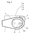
- Single fiber holes 13L and 13R are set on the front end and the left side of the urine collector 4 respectively, and condenser lenses 15L and 15R for condensing light are set on bowl 3 sides of these fiber holes 13L and 13R respectively, while an excitation light fiber member 5 for carrying excitation light from an excitation light source 14 (described later with reference to Figs. 5 and 6) and a Raman light receiving fiber member 6 for receiving Raman light are set in the fiber holes 13L and 13R respectively.
- Fig. 1B illustrates the inner parts of the fiber holes 13L and 13R in detail.
- the condenser lens 15L is set on the bowl 3 side of the fiber hole member 13L, in order to condense the excitation light outgoing from the excitation light fiber member 5 which is inserted in the stool body 1 outer side of the fiber hole 13L.
- the condenser lens 15R is set on the bowl 3 side of the fiber hole 13R for condensing the Raman light from a urine specimen in the urine collector 4, so that Raman light condensed here is received by the Raman light receiving fiber member 6 and guided to an optical system including a spectrodetector.
- a scupper 35 is provided on the bottom portion of the urine collector 4, and the diameter of the scupper 35 is set at a size having such a passage resistance that the urine specimen remains in the urine collector 4 for at least a time necessary for measurement. While the urine flows from the urine collector 4 also during measurement, the urine does not disappear during the measurement. After the measurement, the urine remaining in the urine collector 4 continuously flows through the scupper 35 so that the urine collector 4 is vacated after a constant time. When the urine collector 4 is washed in a state storing the urine, the remaining urine is washed away with wash water, which in turn is collected in the urine collector 4. The wash water collected in the urine collector 4 also flows through the scupper 35, so that the urine collector 4 is vacated after a constant time.
- the excitation light fiber member 5 is connected with the excitation light source 14 which is set in the exterior of the stool body 1, while the Raman light receiving fiber member 6 communicates with a spectrodetector 10 which is set in the exterior of the stool body 1.
- a Raman signal which is separated into its spectral components detected by the spectrodetector 10 is data-processed by a data processing part 11, and outputted from a data output part 12 as a measured value.
- Respective parts of the excitation light source 14, the spectrodetector 10, the data processing part 11 and the data output part 12 are described later with reference to Figs. 5 and 6.
- Figs. 3 and 4 are a side elevational view and a front elevational view showing a stool body according to another embodiment of the present invention respectively.
- a stool hole 2 is formed on a front surface of the stool body 1 to present the shape of a urinal similarly to the prior art, and a water hole 9 is provided in an upper portion of the stool body 1, similarly to the prior art. Wash water flowing into a urine collector 4 from a bowl 3 flows through a scupper 35, to wash the urine collector 4.
- the concave urine collector 4 is set in an inner lower portion of the stool body 1, so that urine can be collected in the bowl 3.
- Single fiber holes 13L and 13R are set on the lower end and the left side of the urine collector 4 respectively, and condenser lenses 15L and 15R for condensing light are set on bowl 3 sides of the fiber holes 13L and 13R respectively, while an excitation light fiber member 5 for carrying excitation light from an excitation light source 14 (described later with reference to Figs. 5 and 6) and a Raman light receiving fiber member 6 for receiving Raman light are set in the fiber holes 13L and 13R respectively.
- the excitation light fiber member 5 is connected with the excitation light source 14 which is set in the exterior of the stool body 1, while the Raman light receiving fiber member 6 communicates with a spectrodetector 10 which is set in the exterior of the stool body 1, similarly to Fig. 2.
- a Raman signal which is separated into its spectral components detected by the spectrodetector 10 is data-processed by a data processing part 11, and outputted from a data output part 12 as a measured value.
- Fig. 5 is a block diagram showing mainly an optical system in an embodiment of the present invention, which consists of a light source part 16, a measuring/photoreceiving part 17, an optical target adjusting part 18, a spectro-detector part 19, a data processing and output part 20, and an optical correction adjusting part 21.
- the measuring/photoreceiving part 17 is set in a stool body 1, while the light source part 16, the optical target adjusting part 18, the spectro-detector part 19, the data processing and output part 20 and the optical correction adjusting part 21 are set in the exterior of the stool body 1.
- the light source part 16 comprises an excitation light source 14 generating single wavelength light, a bandpass filter 23 for transmitting only an excitation wavelength from the excitation light source 14 while reflecting the remaining light, a beam splitter 24 for dividing a beam from the excitation light source 14 into a sample beam 31s and a correction beam 31r, and a light source condenser lens 22 and a convergent lens 32 which are set on both sides of the beam splitter 24 for converging the sample beam 31s on a urine specimen in a urine collector 4 of the measuring/photoreceiving part 17.
- the excitation light source 14 is a laser unit, it is preferable to provide a bandpass filter 23 for blocking off a sideband from its laser beam.
- a laser unit for example, is employed as the excitation light source 14.
- a continuously oscillating Kr ion laser, an He-Ne laser, a laser diode of InGaAs or the like, an Nd:YAG laser, or a pulse laser can be employed as the laser unit, and can be selected from lasers of a wide wavelength range over near ultraviolet to near infrared regions and utilized.
- a light source other than the laser unit a light source such as a halogen lamp generating multiwavelength light can be employed in combination with a spectroscope or an optical filter.
- the wavelength of the excitation light is preferably at least 800 nm, i.e., in a longer wavelength region beyond near infrared.
- the reason for this is as follows: A vital component has large fluorescence, fluorescent luminous efficiency is high when excited with visible light of an Ar laser (514.5 nm) or an He-Ne laser (632.8 nm), and a spectrum is readily influenced by fluorescence, while the fluorescent luminous efficiency is reduced and influence of fluorescence can be reduced when excited with light of a longer wavelength region beyond near infrared and influence by external light of a fluorescent lamp forming stray light can be reduced.
- the measuring/photoreceiving part 17 comprises an excitation light fiber member 5 for guiding the sample beam 31s, a condenser lens 15L for condensing the sample beam 31s from the excitation light fiber member 5 and irradiating the urine sample in the urine collector 4 with the sample beam 31s, the urine collector 4 for collecting the urine specimen and serving as a sample cell, a condenser lens 15R for converging Raman light generated from the urine specimen irradiated with the sample beam 31s, and a Raman light receiving fiber member 6 for receiving the Raman light obtained through the condenser lens 15R and guiding the Raman light to the spectro-detector part 19 through the optical target adjusting part 18.
- the excitation light fiber member 5 and the Raman light receiving fiber member 6 may be single-core optical fiber members, or optical fiber bundles formed by bundling plural optical fiber members respectively.
- the optical target adjusting part 18 comprises a filter 26 for removing the same wavelength component (Rayleigh light) as the excitation light from the obtained Raman light and taking out target light including fluorescence and Raman scattered light, and an optical system (condenser lenses 25 and 27) for adjusting beams.
- a filter 26 for removing the same wavelength component (Rayleigh light) as the excitation light from the obtained Raman light and taking out target light including fluorescence and Raman scattered light
- an optical system condenser lenses 25 and 27 for adjusting beams.
- the filter 26 is preferably either a holographic notch filter including the Rayleigh light light wavelength in its notch region, or a cut filter for blocking off the Rayleigh light wavelength and a shorter wavelength side.
- the holographic notch filter blocks only a desired wavelength region and transmits wavelength light of another region. It is possible to remove an excitation light component by employing that including the excitation light wavelength in the blocked region (notch region).
- the holographic notch filter is available on Kaiser Optical Systems, Inc. (U.S.A.), for example.
- a beam splitter 28 is set between the filter 26 of the optical target adjusting part 18 and the condenser lens 27 as wave combining means, so that the target light is transmitted through the beam splitter 28 and guided to an inlet slit of a spectroscope of the spectro-detector 10 through the condenser lens 27.
- the optical correction adjusting part 21 is adapted to guide the correction beam 31r divided by the beam splitter 24 to the beam splitter 28 of the wave combining means, and comprises a neutral density filter 29 for damping the light quantity and a reflecting mirror 30 for bending an optical path, in order to adjust the correction beam 31r.
- the correction beam 31r from the optical correction adjusting part 21 is reflected by the beam splitter 28, and guided to the inlet slit of the spectroscope of the spectro-detector 10 through the condenser lens 27.
- the correction beam 31r includes only the excitation light from the excitation light source 14 and not through a sample, and hence the correction beam 31r does not depend on the sample but expresses intensity fluctuation from the light source in fidelity.
- the correction beam 31r is adapted to correct fluctuation of a spectral light intensity by fluctuation of the excitation light intensity from the excitation light source 14, and if such correction is unnecessary, the beam splitter 24 of the light source part 16, the optical correction adjusting part 21 and the beam splitter 28 which is wave combining means are unnecessary.
- the spectro-detector part 19 comprises the single spectro-detector 10 comprising the spectroscope for incorporating a beam outgoing from the optical target adjusting part 18 and the correction beam 31r outgoing from the optical correction adjusting part 21 through the beam splitter 28 serving as wave combining means and separating the beam into spectral components thereof, and a detector for detecting the spectral components separated by the spectroscope.
- the spectro-detector 10 is preferably a polychrometer comprising a multichannel photodetector for simultaneously detecting wavelength regions to be measured.
- the wavelength regions to be measured can be simultaneously detected, and a target light spectrum of a prescribed region and the excitation light can be simultaneously detected. Consequently, no difference is caused between detection times for the respective wavelengths of the target light and the excitation light. If difference may be permitted between the detection times for the respective wavelengths of the target light and the excitation light, however, the spectro-detector part 19 may employ a wavelength scan type spectroscope or optical filters and a single channel photodetector for successively detecting the wavelength regions to be measured.
- the data processing and output part 20 comprises a data processing part 11 for data-processing spectra detected by the detector of the spectro-detector 10, and an output part 12 for outputting data processed in the data processing part 11.
- the data processing part 11 is adapted to control operations of the respective parts as well as perform data processing such as multivariate regression analysis on a signal detected by the spectro-detector 10 while including a function of correcting a detected intensity of the target light with reference to a detected intensity of the excitation light component in the spectra detected by the spectro-detector 10, forms a Raman scattering spectrum in which fluctuation of the light source is corrected, and performs qualification and determination of the sample from the target light intensity.
- the output part 12 is a printer or a display outputting the data processed in the data processing part 11.
- ⁇ 90 ° in this embodiment assuming that ⁇ represents an angle formed by scattered light from a target substance with respect to incident light, the present invention is not restricted to this so far as 0 ° ⁇ ⁇ ⁇ 360 °.
- a sample beam 31s from an excitation light source part 16 is transmitted through a central transmitting reflecting mirror 33 which has a central hole and is arranged in an optical target adjusting part 18, to be applied to a urine specimen in a measuring/photoreceiving part 17 through an optical fiber member 5a.
- the optical target adjusting part 18 is similar to that shown in Fig.
- condenser lenses 25 and 27 for converging Raman light from the urine specimen passing through the optical fiber member 5a and reflected by the mirror 33 in an inlet slit of a spectroscope of a spectro-detector 10, while a holographic notch filter which is set to include a wavelength of excitation light in its notch region is arranged between the condenser lenses 25 and 27 as a filter 26 for removing the same wavelength component as the excitation light and taking out target light.
- the remaining structure other than the measuring/photoreceiving part 17 is identical to that of Fig. 5.
- the optical fiber member 5a serves both as an excitation light fiber member and a Raman light receiving fiber member.
- the optical fiber member 5a may be a single-core optical fiber member, or an optical fiber bundle formed by bundling a plurality of optical fiber members. In case of an optical fiber bundle, it is possible to distinguishedly use the same for using a part as an excitation light fiber member and the remaining part as a Raman light receiving fiber member.
- transparent plate glass members such as slide glass can be employed as the beam splitters 24, 28 and 33.
- transparent plate glass members When transparent plate glass members are obliquely arranged on optical paths, it is possible to separate single incident light into reflected light and transmitted light, and combine transmitted incident light and reflected incident light on the same optical axis.
- Half mirrors may be employed in place of the transparent plate members.
- transparent plate glass members When transparent plate glass members are employed, the transmitted light quantity can be increased since there is no absorption as to transmitted light, dissimilarly to the half mirrors.
- Figs. 7A and 7B are flow charts showing the overall flow of an operation for measuring the urine specimen in the urine collector 4 by the urinalysis stool according to the present invention and showing operations of the respective parts respectively.
- the apparatus operation is started from a measurement preparation state. Oscillation of the excitation light and measurement are started. Raman light of the urine specimen in the urine collector 4 excited by the excitation light source 14 is received by the Raman light receiving fiber member 6, and the measurement is ended after Raman spectrum measurement of a prescribed time. Results obtained by processing the obtained Raman spectral data by performing proper data analysis operation are outputted as concentrations of respective components etc.
- the power source is automatically turned off after completion of data output.
- the data processing part 11 analyzes the measured data by mathematical arithmetic processing such as multivariate regression analysis on the basis of intensities of Raman light separated into its spectral components by the spectro-detector 10 or spectral patterns and obtains respective component concentrations.
- the analytical results are outputted to the data output part 12.
- the data processing part 11 can also perform operation of correcting concentrations of other components with reference to a creatinine concentration.
- the scupper 35 in Fig. 1A may be formed by a transparent glass tube so that the measurement is automatically started when the urine specimen passes through the scupper 35, in place of a method of manually pushing a start button.
- Results of measurement on some components contained in urine are shown.
- Data shown in Figs. 8 to 22 are results of measurement as to aqueous solution samples of respective single components.
- Fig. 8 shows a result of investigation of correlation between spectral intensities and concentrations of creatinine, and a multiple correlation coefficient R 2 (square of correlation coefficient R) is 0.961.
- Figs. 9 to 11 show results of determination as to protein in urine.
- Albumin and globulin serve as indices for nephrosis syndrome or glomerulonephritis, and hemoglobin serves as an index for inflammation or tumor of the urinary system or the like.
- Fig. 9 shows the result of investigation of correlation between spectral intensities and concentrations of albumin, and the multiple correlation coefficient R 2 is 0.999.
- Fig. 10 shows the result of investigation of correlation between spectral intensities and concentrations of globulin, and the multiple correlation coefficient R 2 is 0.994.
- Fig. 11 shows the result of investigation of correlation between spectral intensities and concentrations of hemoglobin, and the multiple correlation coefficient R 2 is 0.998.
- Figs. 12 to 15 show results of determination as to sugar in urine.
- Glucose, lactose, fructose and galactose serve as indices for diabetes mellitus, infantile dyspepsia, alimentary levulosemia and critical hepatopathy or infantile trophopathy respectively.
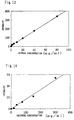
- Fig. 12 shows the result of investigation of correlation between spectral intensities and concentrations of glucose, and the multiple correlation coefficient R 2 is 0.964.
- Fig. 13 shows the result of investigation of correlation between spectral intensities and concentrations of lactose, and the multiple correlation coefficient R 2 is 0.999.
- Fig. 14 shows the result of investigation of correlation between spectral intensities and concentrations of fructose, and the multiple correlation coefficient R 2 is 0.990.
- Fig. 15 shows the result of investigation of correlation between spectral intensities and concentrations of galactose, and the multiple correlation coefficient R 2 is 0.974.
- Figs. 16 to 18 show results of determination as to ketone bodies in urine.
- Lithium acetoacetate, ⁇ -hydroxybutyric acid and acetone serve as indices for ketoacidosis or the like.
- Fig. 16 shows the result of investigation of correlation between spectral intensities and concentrations of lithium acetoacetate, and the multiple correlation coefficient R 2 is 0.998.
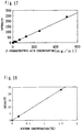
- Fig. 17 shows the result of investigation of correlation between spectral intensities and concentrations of ⁇ -hydroxybutyric acid, and the multiple correlation coefficient R 2 is 0.997.
- Fig. 18 shows the result of investigation of correlation between spectral intensities and concentrations of acetone, and the multiple correlation coefficient R 2 is 0.999.
- Figs. 19 and 20 show results of determination as to bile pigments in urine.
- Bilirubin and urobilinogen serve as indices for hepatic/biliary disease and hepatic/biliary disease or cythemolytic disease respectively. Since bilirubin is insoluble in water, ditaurobilirubin was employed in this example. As to urobilin, measurement was made with urobilin since urobilinogen is instable and readily becomes urobilin by air oxidation.
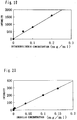
- Fig. 19 shows the result of investigation of correlation between spectral intensities and concentrations of ditaurobilirubin, and the multiple correlation coefficient R 2 is 0.994.
- Fig. 20 shows the result of investigation of correlation between spectral intensities and concentrations of urobilin, and the multiple correlation coefficient R 2 is 0.995.
- Fig. 21 shows the result of determination as to nitrite in urine, and the multiple correlation coefficient R 2 of its spectral intensities and concentrations is 0.998.
- Sodium nitrite serves as an index for microbism of a urinary system.
- Fig. 22 shows the result of determination as to urea in urine, and the multiple correlation coefficient R 2 of its spectral intensities and concentrations is 0.998.
- Urogenous urea is useful for recognizing internal protein metabolism, liver/renal function or the like. Sthenia of body protein catabolism or dosage of quinine is conceivable if it is increased, while hepatocytic affection or renal insufficiency is conceivable if it is reduced.
- the urogenous urea volume serves as an index of urine specific gravity ("Shintei Rinsho Kensa Kenshu Handbook 3" by Nozomu Kosakai, Yakujinippo-Sha), while osmotic pressure* refractive index and urine specific gravity are generally excellent in relation, and determination of the three is substituted by specific gravity ("Rinsho Kensaho Teiyo" by Masamitsu Kanai, Kinbara Shuppan Kabushiki Kaisha).
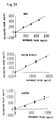
- Figs. 23 and 24 illustrate examples employing multivariate regression analysis.
- 150 mg/dl, 300 mg/dl and 450 mg/dl of urea, 600 mg/dl, 1.2 g/dl and 1.8 g/dl of sodium nitrite and 300 mg/dl, 600 mg/dl and 900 mg/dl of acetone were added to normal urine in arbitrary combinations respectively, spectra were measured, and concentrations were determined by multivariate regression analysis.
- the value k( ⁇ i) is decided in the procedure of multivariate regression analysis, so that the correlation between the concentration of a sample having a known analytical value and an estimated concentration is maximized. This calculation is previously integrated into commercially available processing software and automatically performed, so that this equation is formed every urogenous substance whose concentration is to be obtained.
- Fig. 23 is a flow chart showing the procedure from spectral measurement to concentration determination.
- no pretreatment was performed and QUANT+ by Perkin Elmer Co., Ltd. was employed as processing software.
- the PCR method was employed as the processing method. When full cross validation is performed, evaluation of accuracy of a calibration model can be omitted.
- Fig. 24 shows correlation diagrams of calculated values and actual values (reference values) obtained in this example.
- Multiple correlation coefficients R 2 and SEP of urea, sodium nitrite and acetone were 0.986 and 18. 13, 0.992 and 57.17, and 0.956 and 69.74 respectively.
Landscapes
- Health & Medical Sciences (AREA)
- Life Sciences & Earth Sciences (AREA)
- Molecular Biology (AREA)
- Surgery (AREA)
- Engineering & Computer Science (AREA)
- Biomedical Technology (AREA)
- Heart & Thoracic Surgery (AREA)
- Medical Informatics (AREA)
- Hematology (AREA)
- Pathology (AREA)
- Animal Behavior & Ethology (AREA)
- General Health & Medical Sciences (AREA)
- Public Health (AREA)
- Veterinary Medicine (AREA)
- Investigating Or Analysing Biological Materials (AREA)
- Investigating, Analyzing Materials By Fluorescence Or Luminescence (AREA)
Applications Claiming Priority (2)
| Application Number | Priority Date | Filing Date | Title |
|---|---|---|---|
| JP29622795 | 1995-10-18 | ||
| JP296227/95 | 1995-10-18 |
Publications (4)
| Publication Number | Publication Date |
|---|---|
| EP0769691A2 true EP0769691A2 (fr) | 1997-04-23 |
| EP0769691A3 EP0769691A3 (fr) | 1997-07-02 |
| EP0769691B1 EP0769691B1 (fr) | 2006-02-15 |
| EP0769691B8 EP0769691B8 (fr) | 2006-06-07 |
Family
ID=17830832
Family Applications (1)
| Application Number | Title | Priority Date | Filing Date |
|---|---|---|---|
| EP96116693A Expired - Lifetime EP0769691B8 (fr) | 1995-10-18 | 1996-10-17 | Dispositif pour l'analyse d'urine |
Country Status (3)
| Country | Link |
|---|---|
| US (1) | US5815260A (fr) |
| EP (1) | EP0769691B8 (fr) |
| DE (1) | DE69635819T2 (fr) |
Cited By (7)
| Publication number | Priority date | Publication date | Assignee | Title |
|---|---|---|---|---|
| US6087182A (en) * | 1998-08-27 | 2000-07-11 | Abbott Laboratories | Reagentless analysis of biological samples |
| WO2000049948A3 (fr) * | 1999-02-22 | 2000-12-07 | Univ Ulster | Collecteur d'urine |
| ES2176062A1 (es) * | 1999-07-28 | 2002-11-16 | Samsung Electronics Co Ltd | Unidad interior para un aparato acondicionador de aire de tipo separado. |
| EP2518498A1 (fr) * | 2011-04-25 | 2012-10-31 | Arkray, Inc. | Système d'analyse, procédé d'analyse et programme d'analyse |
| CN110101414A (zh) * | 2019-05-15 | 2019-08-09 | 许昌学院 | 一种糖尿病孕妇尿糖测试装置 |
| CN110353736A (zh) * | 2019-08-20 | 2019-10-22 | 南通大学附属医院 | 尿液标本自动采集系统装置 |
| EP4052654A1 (fr) * | 2016-12-30 | 2022-09-07 | Withings | Appareil de mesure de contenu d'urine |
Families Citing this family (24)
| Publication number | Priority date | Publication date | Assignee | Title |
|---|---|---|---|---|
| WO2003083458A2 (fr) * | 2002-04-03 | 2003-10-09 | Johann Wolfgang Goethe-Universität Frankfurt am Main | Dispositif de mesure a infrarouge, conçu en particulier pour la spectrometrie de systemes aqueux, de preference de systemes a composants multiples |
| US7002670B2 (en) * | 2002-06-12 | 2006-02-21 | Baxter International Inc. | Optical sensor and method for measuring concentration of a chemical constituent using its intrinsic optical absorbance |
| US7326576B2 (en) * | 2003-04-09 | 2008-02-05 | Prescient Medical, Inc. | Raman spectroscopic monitoring of hemodialysis |
| US20060063992A1 (en) * | 2004-08-09 | 2006-03-23 | Dejin Yu | Method and apparatus for non-invasive measurement of blood analytes |
| US20060063991A1 (en) * | 2004-08-09 | 2006-03-23 | Dejin Yu | Method and apparatus for non-invasive measurement of blood analytes with dynamic spectral calibration |
| US20060063993A1 (en) * | 2004-08-09 | 2006-03-23 | Dejin Yu | Method and apparatus for non-invasive measurement of blood analytes |
| US7688440B2 (en) * | 2005-01-27 | 2010-03-30 | Prescient Medical, Inc. | Raman spectroscopic test strip systems |
| US7651851B2 (en) * | 2005-01-27 | 2010-01-26 | Prescient Medical, Inc. | Handheld Raman body fluid analyzer |
| US7524671B2 (en) * | 2005-01-27 | 2009-04-28 | Prescient Medical, Inc. | Handheld raman blood analyzer |
| US20060211926A1 (en) * | 2005-03-21 | 2006-09-21 | Dejin Yu | Non-invasive Raman measurement apparatus with broadband spectral correction |
| WO2007092173A2 (fr) * | 2006-02-06 | 2007-08-16 | Prescient Medical, Inc. | Analyses des bandes de flux lateral par spectroscopie de raman |
| EP2133478A3 (fr) * | 2008-02-27 | 2011-10-05 | Jsm Healthcare Inc | Appareil d'analyse de composants d'urine en utilisant ATR et procédé associé |
| DE102011012674B3 (de) * | 2011-02-28 | 2012-04-26 | Ldiamon As | Verfahren und Nachweisvorrichtung zur Bestimmung eines Nierenfunktionsparameters |
| EP2856933A1 (fr) * | 2013-10-02 | 2015-04-08 | Nederlandse Organisatie voor toegepast -natuurwetenschappelijk onderzoek TNO | Sonde, système et procédé de mesure non invasive des analytes sanguins |
| US20170322197A1 (en) * | 2015-05-02 | 2017-11-09 | David R. Hall | Health Monitoring Toilet System |
| US20180080923A1 (en) * | 2016-09-19 | 2018-03-22 | David R. Hall | Toilet Bowl Optical Engine |
| WO2020071542A1 (fr) * | 2018-10-04 | 2020-04-09 | アトナープ株式会社 | Système d'acquisition d'informations biologiques et serveur et système de gestion de santé |
| CN111948191B (zh) * | 2020-07-30 | 2022-10-18 | 季华实验室 | 一种多光源拉曼光谱分析方法及其应用 |
| US20220047249A1 (en) * | 2020-08-13 | 2022-02-17 | Taiwan Redeye Biomedical Inc. | Biological detection device and method for toilet water solution |
| CN111965163B (zh) * | 2020-08-18 | 2025-01-21 | 付朝品 | 一种智能全自动无人操控拉曼光谱分析设备 |
| CN111965162B (zh) * | 2020-08-18 | 2025-01-21 | 付朝品 | 一种智能全自动无人操控多探头拉曼光谱分析设备 |
| CN117627127A (zh) * | 2022-08-12 | 2024-03-01 | 瑞爱生医股份有限公司 | 内嵌式尿液检测的便器 |
| CN117969484A (zh) * | 2023-02-21 | 2024-05-03 | 瀚诚星科技(杭州)有限公司 | 一种尿液检测系统 |
| US20240426757A1 (en) * | 2023-06-26 | 2024-12-26 | Mariposa Technology, Inc. | Spectroscopic Methods and Systems for the Qualitative and Quantitative Analysis of Samples |
Family Cites Families (4)
| Publication number | Priority date | Publication date | Assignee | Title |
|---|---|---|---|---|
| JPS63290961A (ja) * | 1987-05-22 | 1988-11-28 | Inax Corp | 尿中成分検出便器 |
| US5376556A (en) * | 1989-10-27 | 1994-12-27 | Abbott Laboratories | Surface-enhanced Raman spectroscopy immunoassay |
| JPH0749309A (ja) * | 1993-08-05 | 1995-02-21 | Kyoto Daiichi Kagaku:Kk | 光散乱式成分濃度測定装置および方法 |
| US5553616A (en) * | 1993-11-30 | 1996-09-10 | Florida Institute Of Technology | Determination of concentrations of biological substances using raman spectroscopy and artificial neural network discriminator |
-
1996
- 1996-10-17 EP EP96116693A patent/EP0769691B8/fr not_active Expired - Lifetime
- 1996-10-17 US US08/733,637 patent/US5815260A/en not_active Expired - Fee Related
- 1996-10-17 DE DE69635819T patent/DE69635819T2/de not_active Expired - Lifetime
Cited By (13)
| Publication number | Priority date | Publication date | Assignee | Title |
|---|---|---|---|---|
| US6087182A (en) * | 1998-08-27 | 2000-07-11 | Abbott Laboratories | Reagentless analysis of biological samples |
| US6365109B1 (en) | 1998-08-27 | 2002-04-02 | Abbott Laboratories | Reagentless analysis of biological samples |
| US6426045B1 (en) | 1998-08-27 | 2002-07-30 | Abbott Laboratories | Reagentless analysis of biological samples |
| US6773922B2 (en) | 1998-08-27 | 2004-08-10 | Abbott Laboratories | Reagentless analysis of biological samples |
| WO2000049948A3 (fr) * | 1999-02-22 | 2000-12-07 | Univ Ulster | Collecteur d'urine |
| ES2176062A1 (es) * | 1999-07-28 | 2002-11-16 | Samsung Electronics Co Ltd | Unidad interior para un aparato acondicionador de aire de tipo separado. |
| EP2518498A1 (fr) * | 2011-04-25 | 2012-10-31 | Arkray, Inc. | Système d'analyse, procédé d'analyse et programme d'analyse |
| US8884766B2 (en) | 2011-04-25 | 2014-11-11 | Arkray, Inc. | Analysis system, analysis method and analysis program |
| EP4052654A1 (fr) * | 2016-12-30 | 2022-09-07 | Withings | Appareil de mesure de contenu d'urine |
| CN110101414A (zh) * | 2019-05-15 | 2019-08-09 | 许昌学院 | 一种糖尿病孕妇尿糖测试装置 |
| CN110101414B (zh) * | 2019-05-15 | 2021-10-15 | 许昌学院 | 一种糖尿病孕妇尿糖测试装置 |
| CN110353736A (zh) * | 2019-08-20 | 2019-10-22 | 南通大学附属医院 | 尿液标本自动采集系统装置 |
| CN110353736B (zh) * | 2019-08-20 | 2022-02-08 | 南通大学附属医院 | 尿液标本自动采集系统 |
Also Published As
| Publication number | Publication date |
|---|---|
| EP0769691B1 (fr) | 2006-02-15 |
| EP0769691A3 (fr) | 1997-07-02 |
| DE69635819T2 (de) | 2006-12-21 |
| DE69635819D1 (de) | 2006-04-20 |
| US5815260A (en) | 1998-09-29 |
| EP0769691B8 (fr) | 2006-06-07 |
Similar Documents
| Publication | Publication Date | Title |
|---|---|---|
| US5815260A (en) | Urogenous component measuring apparatus for qualitatively/quantitatively measuring a plurality of urogenous components | |
| US5796476A (en) | Method of optically measuring component in solution | |
| US6365109B1 (en) | Reagentless analysis of biological samples | |
| US6582964B1 (en) | Method and apparatus for rapid measurement of HbA1c | |
| EP1214579B1 (fr) | Procede d'etalonnage d'un dispositif spectroscopique | |
| EP0670492B1 (fr) | Méthode et appareil pour la mesure de composants d'urine | |
| US7098037B2 (en) | Accommodating subject and instrument variations in spectroscopic determinations | |
| US6441388B1 (en) | Methods and apparatus for spectroscopic calibration model transfer | |
| Hall et al. | Near-infrared spectrophotometry: a new dimension in clinical chemistry | |
| RU2400733C2 (ru) | Система для спектроскопии пропускания для использования при определении анализируемых веществ в жидкости организма | |
| JP4672147B2 (ja) | 分光学的な較正モデルを作るための方法 | |
| EP3111216A2 (fr) | Méthode et appareil de détermination de marqueurs de santé par analyse d'un échantillon de sang | |
| US6882425B1 (en) | Method and apparatus for examining fluids of biological origin | |
| EP1023583B1 (fr) | Methode pour doser des succedanes du sang | |
| Koo et al. | Reagentless blood analysis by near-infrared Raman spectroscopy | |
| JP7373315B2 (ja) | 体液試料中の脂質、ヘモグロビンおよびビリルビンを決定するための方法 | |
| JP3774001B2 (ja) | 尿中成分測定装置 | |
| JPH07301630A (ja) | 尿中成分測定装置を備えた便器 | |
| JPH07318564A (ja) | 血液分析計 | |
| JP3642602B2 (ja) | 尿中成分の測定装置 | |
| JPH11342123A (ja) | 非侵襲生体成分測定装置 | |
| US7198955B1 (en) | Method and apparatus for measurement of blood substitutes | |
| EP0762108A2 (fr) | Méthode et dispositif de mesure de la concentration du cétone dans un organisme | |
| JPH09145619A (ja) | 散乱光等の分光測定方法及び装置 | |
| GB2632096A (en) | A method and a device for testing a model |
Legal Events
| Date | Code | Title | Description |
|---|---|---|---|
| PUAI | Public reference made under article 153(3) epc to a published international application that has entered the european phase |
Free format text: ORIGINAL CODE: 0009012 |
|
| AK | Designated contracting states |
Kind code of ref document: A2 Designated state(s): DE FR GB IT |
|
| PUAL | Search report despatched |
Free format text: ORIGINAL CODE: 0009013 |
|
| AK | Designated contracting states |
Kind code of ref document: A3 Designated state(s): DE FR GB IT |
|
| 17P | Request for examination filed |
Effective date: 19971021 |
|
| 17Q | First examination report despatched |
Effective date: 20030612 |
|
| GRAJ | Information related to disapproval of communication of intention to grant by the applicant or resumption of examination proceedings by the epo deleted |
Free format text: ORIGINAL CODE: EPIDOSDIGR1 |
|
| GRAP | Despatch of communication of intention to grant a patent |
Free format text: ORIGINAL CODE: EPIDOSNIGR1 |
|
| GRAP | Despatch of communication of intention to grant a patent |
Free format text: ORIGINAL CODE: EPIDOSNIGR1 |
|
| GRAC | Information related to communication of intention to grant a patent modified |
Free format text: ORIGINAL CODE: EPIDOSCIGR1 |
|
| GRAS | Grant fee paid |
Free format text: ORIGINAL CODE: EPIDOSNIGR3 |
|
| GRAA | (expected) grant |
Free format text: ORIGINAL CODE: 0009210 |
|
| AK | Designated contracting states |
Kind code of ref document: B1 Designated state(s): DE FR GB IT |
|
| PG25 | Lapsed in a contracting state [announced via postgrant information from national office to epo] |
Ref country code: IT Free format text: LAPSE BECAUSE OF FAILURE TO SUBMIT A TRANSLATION OF THE DESCRIPTION OR TO PAY THE FEE WITHIN THE PRE;WARNING: LAPSES OF ITALIAN PATENTS WITH EFFECTIVE DATE BEFORE 2007 MAY HAVE OCCURRED AT ANY TIME BEFORE 2007. THE CORRECT EFFECTIVE DATE MAY BE DIFFERENT FROM THE ONE RECORDED.SCRIBED TIME-LIMIT Effective date: 20060215 |
|
| REG | Reference to a national code |
Ref country code: GB Ref legal event code: FG4D |
|
| RAP2 | Party data changed (patent owner data changed or rights of a patent transferred) |
Owner name: ARKRAY, INC |
|
| REF | Corresponds to: |
Ref document number: 69635819 Country of ref document: DE Date of ref document: 20060420 Kind code of ref document: P |
|
| ET | Fr: translation filed | ||
| PLBE | No opposition filed within time limit |
Free format text: ORIGINAL CODE: 0009261 |
|
| STAA | Information on the status of an ep patent application or granted ep patent |
Free format text: STATUS: NO OPPOSITION FILED WITHIN TIME LIMIT |
|
| 26N | No opposition filed |
Effective date: 20061116 |
|
| PGFP | Annual fee paid to national office [announced via postgrant information from national office to epo] |
Ref country code: DE Payment date: 20091026 Year of fee payment: 14 |
|
| PGFP | Annual fee paid to national office [announced via postgrant information from national office to epo] |
Ref country code: IT Payment date: 20091028 Year of fee payment: 14 Ref country code: GB Payment date: 20091022 Year of fee payment: 14 Ref country code: FR Payment date: 20091110 Year of fee payment: 14 |
|
| GBPC | Gb: european patent ceased through non-payment of renewal fee |
Effective date: 20101017 |
|
| PG25 | Lapsed in a contracting state [announced via postgrant information from national office to epo] |
Ref country code: FR Free format text: LAPSE BECAUSE OF NON-PAYMENT OF DUE FEES Effective date: 20101102 |
|
| REG | Reference to a national code |
Ref country code: FR Ref legal event code: ST Effective date: 20110630 |
|
| PG25 | Lapsed in a contracting state [announced via postgrant information from national office to epo] |
Ref country code: GB Free format text: LAPSE BECAUSE OF NON-PAYMENT OF DUE FEES Effective date: 20101017 |
|
| REG | Reference to a national code |
Ref country code: DE Ref legal event code: R119 Ref document number: 69635819 Country of ref document: DE Effective date: 20110502 |
|
| PG25 | Lapsed in a contracting state [announced via postgrant information from national office to epo] |
Ref country code: IT Free format text: LAPSE BECAUSE OF NON-PAYMENT OF DUE FEES Effective date: 20101017 |
|
| PG25 | Lapsed in a contracting state [announced via postgrant information from national office to epo] |
Ref country code: DE Free format text: LAPSE BECAUSE OF NON-PAYMENT OF DUE FEES Effective date: 20110502 |