EP0790498A1 - Elektrochemische Sensoren mit verbesserter Selektivität und erhöhter Empfindlichkeit - Google Patents
Elektrochemische Sensoren mit verbesserter Selektivität und erhöhter Empfindlichkeit Download PDFInfo
- Publication number
- EP0790498A1 EP0790498A1 EP97101600A EP97101600A EP0790498A1 EP 0790498 A1 EP0790498 A1 EP 0790498A1 EP 97101600 A EP97101600 A EP 97101600A EP 97101600 A EP97101600 A EP 97101600A EP 0790498 A1 EP0790498 A1 EP 0790498A1
- Authority
- EP
- European Patent Office
- Prior art keywords
- agent according
- test agent
- fleece
- graphite
- fabric
- Prior art date
- Legal status (The legal status is an assumption and is not a legal conclusion. Google has not performed a legal analysis and makes no representation as to the accuracy of the status listed.)
- Granted
Links
- 230000035945 sensitivity Effects 0.000 title description 6
- 239000003153 chemical reaction reagent Substances 0.000 claims abstract description 29
- 238000012360 testing method Methods 0.000 claims abstract description 24
- OKTJSMMVPCPJKN-UHFFFAOYSA-N Carbon Chemical compound [C] OKTJSMMVPCPJKN-UHFFFAOYSA-N 0.000 claims description 34
- 239000012528 membrane Substances 0.000 claims description 31
- 229910002804 graphite Inorganic materials 0.000 claims description 30
- 239000010439 graphite Substances 0.000 claims description 30
- 239000003795 chemical substances by application Substances 0.000 claims description 14
- 239000011888 foil Substances 0.000 claims description 14
- 239000004744 fabric Substances 0.000 claims description 11
- 239000007788 liquid Substances 0.000 claims description 9
- 239000011148 porous material Substances 0.000 claims description 3
- -1 for example fleece Substances 0.000 claims description 2
- 239000002657 fibrous material Substances 0.000 claims 2
- 239000002184 metal Substances 0.000 claims 1
- 239000000123 paper Substances 0.000 claims 1
- 239000000463 material Substances 0.000 abstract description 7
- 210000004369 blood Anatomy 0.000 description 27
- 239000008280 blood Substances 0.000 description 27
- 108010015776 Glucose oxidase Proteins 0.000 description 22
- 239000004366 Glucose oxidase Substances 0.000 description 22
- 229940116332 glucose oxidase Drugs 0.000 description 22
- 235000019420 glucose oxidase Nutrition 0.000 description 22
- 239000010410 layer Substances 0.000 description 20
- 239000011159 matrix material Substances 0.000 description 20
- 238000000034 method Methods 0.000 description 15
- WQZGKKKJIJFFOK-GASJEMHNSA-N Glucose Natural products OC[C@H]1OC(O)[C@H](O)[C@@H](O)[C@@H]1O WQZGKKKJIJFFOK-GASJEMHNSA-N 0.000 description 13
- 239000008103 glucose Substances 0.000 description 13
- AWDBHOZBRXWRKS-UHFFFAOYSA-N tetrapotassium;iron(6+);hexacyanide Chemical compound [K+].[K+].[K+].[K+].[Fe+6].N#[C-].N#[C-].N#[C-].N#[C-].N#[C-].N#[C-] AWDBHOZBRXWRKS-UHFFFAOYSA-N 0.000 description 12
- 238000001514 detection method Methods 0.000 description 10
- 238000006243 chemical reaction Methods 0.000 description 9
- 239000000306 component Substances 0.000 description 9
- 238000004519 manufacturing process Methods 0.000 description 9
- 239000000243 solution Substances 0.000 description 9
- 239000010408 film Substances 0.000 description 7
- 230000008569 process Effects 0.000 description 7
- CIWBSHSKHKDKBQ-JLAZNSOCSA-N Ascorbic acid Chemical compound OC[C@H](O)[C@H]1OC(=O)C(O)=C1O CIWBSHSKHKDKBQ-JLAZNSOCSA-N 0.000 description 6
- 238000005266 casting Methods 0.000 description 6
- 238000011156 evaluation Methods 0.000 description 6
- 238000007650 screen-printing Methods 0.000 description 6
- 238000000926 separation method Methods 0.000 description 6
- BQCADISMDOOEFD-UHFFFAOYSA-N Silver Chemical compound [Ag] BQCADISMDOOEFD-UHFFFAOYSA-N 0.000 description 5
- 239000004745 nonwoven fabric Substances 0.000 description 5
- 229910052709 silver Inorganic materials 0.000 description 5
- 239000004332 silver Substances 0.000 description 5
- VTLYFUHAOXGGBS-UHFFFAOYSA-N Fe3+ Chemical compound [Fe+3] VTLYFUHAOXGGBS-UHFFFAOYSA-N 0.000 description 4
- VYPSYNLAJGMNEJ-UHFFFAOYSA-N Silicium dioxide Chemical compound O=[Si]=O VYPSYNLAJGMNEJ-UHFFFAOYSA-N 0.000 description 4
- 210000004027 cell Anatomy 0.000 description 4
- 238000005516 engineering process Methods 0.000 description 4
- 210000003743 erythrocyte Anatomy 0.000 description 4
- 230000003647 oxidation Effects 0.000 description 4
- 238000007254 oxidation reaction Methods 0.000 description 4
- BASFCYQUMIYNBI-UHFFFAOYSA-N platinum Chemical compound [Pt] BASFCYQUMIYNBI-UHFFFAOYSA-N 0.000 description 4
- 229920000728 polyester Polymers 0.000 description 4
- 229920000642 polymer Polymers 0.000 description 4
- YPZRHBJKEMOYQH-UYBVJOGSSA-N FADH2 Chemical compound C1=NC2=C(N)N=CN=C2N1[C@@H]([C@H](O)[C@@H]1O)O[C@@H]1COP(O)(=O)OP(O)(=O)OC[C@@H](O)[C@@H](O)[C@@H](O)CN1C(NC(=O)NC2=O)=C2NC2=C1C=C(C)C(C)=C2 YPZRHBJKEMOYQH-UYBVJOGSSA-N 0.000 description 3
- 239000002390 adhesive tape Substances 0.000 description 3
- 235000010323 ascorbic acid Nutrition 0.000 description 3
- 229960005070 ascorbic acid Drugs 0.000 description 3
- 239000011668 ascorbic acid Substances 0.000 description 3
- 239000004020 conductor Substances 0.000 description 3
- 239000013039 cover film Substances 0.000 description 3
- 238000007872 degassing Methods 0.000 description 3
- 238000003745 diagnosis Methods 0.000 description 3
- 238000009792 diffusion process Methods 0.000 description 3
- 238000005470 impregnation Methods 0.000 description 3
- 238000010348 incorporation Methods 0.000 description 3
- 238000005259 measurement Methods 0.000 description 3
- 238000009832 plasma treatment Methods 0.000 description 3
- 239000002985 plastic film Substances 0.000 description 3
- 229920006255 plastic film Polymers 0.000 description 3
- 239000000047 product Substances 0.000 description 3
- AZUYLZMQTIKGSC-UHFFFAOYSA-N 1-[6-[4-(5-chloro-6-methyl-1H-indazol-4-yl)-5-methyl-3-(1-methylindazol-5-yl)pyrazol-1-yl]-2-azaspiro[3.3]heptan-2-yl]prop-2-en-1-one Chemical compound ClC=1C(=C2C=NNC2=CC=1C)C=1C(=NN(C=1C)C1CC2(CN(C2)C(C=C)=O)C1)C=1C=C2C=NN(C2=CC=1)C AZUYLZMQTIKGSC-UHFFFAOYSA-N 0.000 description 2
- RZVAJINKPMORJF-UHFFFAOYSA-N Acetaminophen Chemical compound CC(=O)NC1=CC=C(O)C=C1 RZVAJINKPMORJF-UHFFFAOYSA-N 0.000 description 2
- 229910002016 Aerosil® 200 Inorganic materials 0.000 description 2
- 108090000790 Enzymes Proteins 0.000 description 2
- 102000004190 Enzymes Human genes 0.000 description 2
- MHAJPDPJQMAIIY-UHFFFAOYSA-N Hydrogen peroxide Chemical compound OO MHAJPDPJQMAIIY-UHFFFAOYSA-N 0.000 description 2
- SECXISVLQFMRJM-UHFFFAOYSA-N N-Methylpyrrolidone Chemical compound CN1CCCC1=O SECXISVLQFMRJM-UHFFFAOYSA-N 0.000 description 2
- 239000004677 Nylon Substances 0.000 description 2
- KDLHZDBZIXYQEI-UHFFFAOYSA-N Palladium Chemical compound [Pd] KDLHZDBZIXYQEI-UHFFFAOYSA-N 0.000 description 2
- 229920003171 Poly (ethylene oxide) Polymers 0.000 description 2
- 239000004952 Polyamide Substances 0.000 description 2
- 239000004372 Polyvinyl alcohol Substances 0.000 description 2
- 230000004523 agglutinating effect Effects 0.000 description 2
- 239000012491 analyte Substances 0.000 description 2
- 230000008901 benefit Effects 0.000 description 2
- 239000011230 binding agent Substances 0.000 description 2
- HVYWMOMLDIMFJA-DPAQBDIFSA-N cholesterol Chemical compound C1C=C2C[C@@H](O)CC[C@]2(C)[C@@H]2[C@@H]1[C@@H]1CC[C@H]([C@H](C)CCCC(C)C)[C@@]1(C)CC2 HVYWMOMLDIMFJA-DPAQBDIFSA-N 0.000 description 2
- 239000007979 citrate buffer Substances 0.000 description 2
- 239000011248 coating agent Substances 0.000 description 2
- 238000000576 coating method Methods 0.000 description 2
- 238000001035 drying Methods 0.000 description 2
- 239000007772 electrode material Substances 0.000 description 2
- 230000002255 enzymatic effect Effects 0.000 description 2
- 229940088598 enzyme Drugs 0.000 description 2
- 230000007717 exclusion Effects 0.000 description 2
- PCHJSUWPFVWCPO-UHFFFAOYSA-N gold Chemical compound [Au] PCHJSUWPFVWCPO-UHFFFAOYSA-N 0.000 description 2
- 229910052737 gold Inorganic materials 0.000 description 2
- 239000010931 gold Substances 0.000 description 2
- 230000002209 hydrophobic effect Effects 0.000 description 2
- 230000010354 integration Effects 0.000 description 2
- 230000002452 interceptive effect Effects 0.000 description 2
- 230000007774 longterm Effects 0.000 description 2
- 229920001778 nylon Polymers 0.000 description 2
- 235000011837 pasties Nutrition 0.000 description 2
- 229910052697 platinum Inorganic materials 0.000 description 2
- 229920002647 polyamide Polymers 0.000 description 2
- 229920006289 polycarbonate film Polymers 0.000 description 2
- 229920005597 polymer membrane Polymers 0.000 description 2
- 229920002451 polyvinyl alcohol Polymers 0.000 description 2
- 238000002360 preparation method Methods 0.000 description 2
- 238000007639 printing Methods 0.000 description 2
- 230000008961 swelling Effects 0.000 description 2
- 239000012085 test solution Substances 0.000 description 2
- 241001552669 Adonis annua Species 0.000 description 1
- 229910002012 Aerosil® Inorganic materials 0.000 description 1
- 102000009027 Albumins Human genes 0.000 description 1
- 108010088751 Albumins Proteins 0.000 description 1
- RYGMFSIKBFXOCR-UHFFFAOYSA-N Copper Chemical compound [Cu] RYGMFSIKBFXOCR-UHFFFAOYSA-N 0.000 description 1
- PHOQVHQSTUBQQK-SQOUGZDYSA-N D-glucono-1,5-lactone Chemical compound OC[C@H]1OC(=O)[C@H](O)[C@@H](O)[C@@H]1O PHOQVHQSTUBQQK-SQOUGZDYSA-N 0.000 description 1
- SXRSQZLOMIGNAQ-UHFFFAOYSA-N Glutaraldehyde Chemical compound O=CCCCC=O SXRSQZLOMIGNAQ-UHFFFAOYSA-N 0.000 description 1
- 239000004831 Hot glue Substances 0.000 description 1
- 102000004856 Lectins Human genes 0.000 description 1
- 108090001090 Lectins Proteins 0.000 description 1
- 229920002266 Pluriol® Polymers 0.000 description 1
- 239000004698 Polyethylene Substances 0.000 description 1
- 229920003291 Ultrason® E Polymers 0.000 description 1
- 239000013543 active substance Substances 0.000 description 1
- 239000000654 additive Substances 0.000 description 1
- 238000004026 adhesive bonding Methods 0.000 description 1
- 230000032683 aging Effects 0.000 description 1
- 238000004458 analytical method Methods 0.000 description 1
- 239000007864 aqueous solution Substances 0.000 description 1
- 238000004159 blood analysis Methods 0.000 description 1
- 239000012503 blood component Substances 0.000 description 1
- 210000001124 body fluid Anatomy 0.000 description 1
- 239000010839 body fluid Substances 0.000 description 1
- 239000012876 carrier material Substances 0.000 description 1
- 239000000919 ceramic Substances 0.000 description 1
- 230000008859 change Effects 0.000 description 1
- 235000012000 cholesterol Nutrition 0.000 description 1
- 238000010276 construction Methods 0.000 description 1
- 238000011109 contamination Methods 0.000 description 1
- 238000007796 conventional method Methods 0.000 description 1
- 229910052802 copper Inorganic materials 0.000 description 1
- 239000010949 copper Substances 0.000 description 1
- 230000003247 decreasing effect Effects 0.000 description 1
- 230000007547 defect Effects 0.000 description 1
- 239000003599 detergent Substances 0.000 description 1
- 231100000673 dose–response relationship Toxicity 0.000 description 1
- 230000009977 dual effect Effects 0.000 description 1
- 230000005611 electricity Effects 0.000 description 1
- 238000003411 electrode reaction Methods 0.000 description 1
- 239000003792 electrolyte Substances 0.000 description 1
- KTWOOEGAPBSYNW-UHFFFAOYSA-N ferrocene Chemical compound [Fe+2].C=1C=C[CH-]C=1.C=1C=C[CH-]C=1 KTWOOEGAPBSYNW-UHFFFAOYSA-N 0.000 description 1
- 239000000835 fiber Substances 0.000 description 1
- 238000009472 formulation Methods 0.000 description 1
- 235000012209 glucono delta-lactone Nutrition 0.000 description 1
- 229960003681 gluconolactone Drugs 0.000 description 1
- 238000005534 hematocrit Methods 0.000 description 1
- 230000000984 immunochemical effect Effects 0.000 description 1
- 230000006872 improvement Effects 0.000 description 1
- 239000002346 layers by function Substances 0.000 description 1
- 239000002523 lectin Substances 0.000 description 1
- 238000001471 micro-filtration Methods 0.000 description 1
- 239000000203 mixture Substances 0.000 description 1
- 229910000510 noble metal Inorganic materials 0.000 description 1
- 229910052763 palladium Inorganic materials 0.000 description 1
- 229960005489 paracetamol Drugs 0.000 description 1
- 230000035699 permeability Effects 0.000 description 1
- 239000012466 permeate Substances 0.000 description 1
- 229920001983 poloxamer Polymers 0.000 description 1
- 229920002492 poly(sulfone) Polymers 0.000 description 1
- 229920000515 polycarbonate Polymers 0.000 description 1
- 239000004417 polycarbonate Substances 0.000 description 1
- 229920002959 polymer blend Polymers 0.000 description 1
- 229920003009 polyurethane dispersion Polymers 0.000 description 1
- 229920000915 polyvinyl chloride Polymers 0.000 description 1
- 229920002620 polyvinyl fluoride Polymers 0.000 description 1
- 102000004169 proteins and genes Human genes 0.000 description 1
- 108090000623 proteins and genes Proteins 0.000 description 1
- 230000035484 reaction time Effects 0.000 description 1
- 230000001105 regulatory effect Effects 0.000 description 1
- 230000004044 response Effects 0.000 description 1
- 238000007086 side reaction Methods 0.000 description 1
- 239000000377 silicon dioxide Substances 0.000 description 1
- 239000002356 single layer Substances 0.000 description 1
- 238000002791 soaking Methods 0.000 description 1
- 239000000758 substrate Substances 0.000 description 1
- 239000004094 surface-active agent Substances 0.000 description 1
- 239000013008 thixotropic agent Substances 0.000 description 1
- XLYOFNOQVPJJNP-UHFFFAOYSA-N water Substances O XLYOFNOQVPJJNP-UHFFFAOYSA-N 0.000 description 1
- 238000003466 welding Methods 0.000 description 1
Images
Classifications
-
- C—CHEMISTRY; METALLURGY
- C12—BIOCHEMISTRY; BEER; SPIRITS; WINE; VINEGAR; MICROBIOLOGY; ENZYMOLOGY; MUTATION OR GENETIC ENGINEERING
- C12Q—MEASURING OR TESTING PROCESSES INVOLVING ENZYMES, NUCLEIC ACIDS OR MICROORGANISMS; COMPOSITIONS OR TEST PAPERS THEREFOR; PROCESSES OF PREPARING SUCH COMPOSITIONS; CONDITION-RESPONSIVE CONTROL IN MICROBIOLOGICAL OR ENZYMOLOGICAL PROCESSES
- C12Q1/00—Measuring or testing processes involving enzymes, nucleic acids or microorganisms; Compositions therefor; Processes of preparing such compositions
- C12Q1/001—Enzyme electrodes
- C12Q1/002—Electrode membranes
-
- Y—GENERAL TAGGING OF NEW TECHNOLOGICAL DEVELOPMENTS; GENERAL TAGGING OF CROSS-SECTIONAL TECHNOLOGIES SPANNING OVER SEVERAL SECTIONS OF THE IPC; TECHNICAL SUBJECTS COVERED BY FORMER USPC CROSS-REFERENCE ART COLLECTIONS [XRACs] AND DIGESTS
- Y10—TECHNICAL SUBJECTS COVERED BY FORMER USPC
- Y10T—TECHNICAL SUBJECTS COVERED BY FORMER US CLASSIFICATION
- Y10T436/00—Chemistry: analytical and immunological testing
- Y10T436/14—Heterocyclic carbon compound [i.e., O, S, N, Se, Te, as only ring hetero atom]
- Y10T436/142222—Hetero-O [e.g., ascorbic acid, etc.]
- Y10T436/143333—Saccharide [e.g., DNA, etc.]
- Y10T436/144444—Glucose
Definitions
- the present invention describes electrochemical sensors, preferably electrochemical biosensors.
- a method for producing electrochemical, preferably amperometric, biosensors for the field of diagnosis of body fluids is also described.
- amperometric biosensors in particular in the field of blood sugar diagnosis, has been state of the art for several years.
- test systems are commercially available under the product names MediSense®, ExacTex® and Glucocard®. They allow simple blood sugar diagnosis under home user conditions.
- the amperometric biosensors with glucose oxidase as a receptor component have become particularly important. As in anal. Chem. 1990, 62 1111 to 1117 described in detail, the reaction of glucose with glucose oxidase produces an amount of hydrogen peroxide proportional to the sugar concentration.
- mediators were therefore developed with a view to improving the selectivity of amperometric sensors.
- Commonly used mediators in the so-called 2nd generation biosensors are, for example, ferrocene or potassium hexacyanoferrate K 3 Fe (CN) 6 .
- the amperometric blood sugar determination follows the following reaction scheme: Glucose + GO (FAD) ⁇ gluconolactone + GO (FADH 2nd ) GO (FADH 2nd ) + Fe (III) (CN) 6 3- ⁇ Fe (II) (CN) 6 4- + GO (FAD) + H + Fe (II) (CN) 6 4- ⁇ Fe (III) (CN) 6 3- + e -
- amperometric blood sugar determination is therefore limited to the anodic oxidation described in (3), which takes place at a potential of +360 mV.
- mediator-modified biosensors thus have an increased selectivity.
- test agent In addition to the necessary detection reagents, for example glucose oxidase and potassium hexacyanoferrate, the construction of a suitable test agent also contains at least two electrodes (working electrode and reference electrode), which must be in contact via an electrolyte bridge.
- noble metals such as palladium, platinum, gold, silver and copper or graphite are suitable as electrode materials, the anode (working electrode) and cathode (reference electrode) being able to be produced from different materials or from the same material and of the same size or different can have large surfaces.
- test sequence in the commercially available systems is limited to the application of the sample liquid (whole blood), the analysis value being displayed digitally within at least one minute.
- Test agents that enable the mediator system, for example potassium hexacyanoferrate (K 3 Fe (CN) 6 ), to be applied separately to the counter electrode and the enzymatic receptor (GO) to the working electrode - if appropriate fixed by immobilization - should accordingly be advantageous.
- K 3 Fe (CN) 6 potassium hexacyanoferrate
- GO enzymatic receptor
- Test systems with separate reagent zones can also be advantageous with regard to the long-term stability of the enzymatic reagent system.
- Europ. Pat. Spec. 0 276 782 describes enzyme electrodes with albumin layers cross-linked by glutardialdehyde, which, due to their permeability, protect the working electrode from electroactive interference components, especially protect against higher molecular weight proteins.
- WO 94/27140 describes electrochemical sensors with erythrocyte exclusion membranes which contain mobile erythrocyte agglutinating agents.
- Nankai et al. describe in WO 86/07 642 a three-electrode system which, in addition to the working and reference electrodes, also contains a reference electrode which compensates for the dependence of the cell voltage on the cell current.
- Nankai et al. describe in Eur. Appl. 0 471 986 the production of an amperometric blood sugar test system with disposable sensors, this system being particularly easy to handle.
- the disposable sensor in the amperometric evaluation device is tipped with the sensor tip on the blood drop to be analyzed.
- Whole blood is transported into the sensor workspace (working or reference electrode plus detection reagents) via a microcapillary (capillary flow system).
- the detection reagents GO / K 3 Fe (CN) 6
- the device can therefore be operated without any control button.
- the amount of blood required is kept as low as possible and the volume of the microcapillary system is therefore limited to approximately 5 ⁇ l.
- Conductor tracks lead from the reaction space delimited by the microcapillary via the extended sensor part to the plug contacts, so contamination of important functional parts in the evaluation device is excluded.
- the method of screen printing technology is usually used to produce the blood glucose biosensors cited.
- the electrode part for example based on graphite or silver (from Acheson), are used, which are printed on carrier materials, such as ceramic or plastic films. This requires several successive printing and drying steps (conductor tracks, working electrodes, reference electrodes, dielectric layers).
- the screen printing pastes which contain a number of different additives such as defoamers, thixotropic agents and detergents with regard to processability, often have considerable defects with regard to reproducibility.
- the screen-printed electrode surfaces still have to be activated by plasma treatment. Because of the high, relatively hydrophobic proportion of binder, their surfaces are rather hydrophobic, poorly wetted and their conductivity is greatly reduced compared to the pure conductor material, for example graphite or silver.
- the detection reagent recipe is applied, for example glucose oxidase (GO) and potassium hexacyanoferrate in the case of blood sugar detection.
- GO glucose oxidase
- potassium hexacyanoferrate in the case of blood sugar detection.
- Each individual sensor work surface must be doped individually, either using the process of screen printing technology or the complex process of micropipetting is used.
- the microcapillary system is applied by gluing correspondingly preformed foils, which may have to be provided with hydrophilic layers with a view to good wettability.
- the method according to the invention in particular allows the desired properties described in the text, which should lead to a product improvement, to be combined in one system. At the current status, these combined and integral property profiles have not yet been implemented.
- sensitivity an increase in sensitivity (sensitivity) is possible in a simple manner by increasing the area of the reagent matrix without the required sample volumes (e.g. blood drops) having to be increased significantly, as in the conventional system.
- selectivity-increasing separation processes can namely be integrated in various sensor layers, for example porous reference electrodes, membranes as a reagent matrix and, if appropriate, via membrane coating of the working electrode.
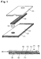
- a reagent matrix (membrane) (3) with an area in the range of a few mm 2 is applied to a graphite foil (2) which is fixed on a base foil (1).
- a strip of graphite fleece (5) is fastened over it with a perforated double-sided adhesive tape (4).
- a perforated film (6) is finally glued on as the top cover.
- the required sample volume can be obtained through Integration of a liquid stop zone in the graphite fleece, for example behind the reagent membrane (10), can be defined.
- the potentiostat is contacted at (7) to the graphite fleece layer (reference electrode, cathode) and at (8) to the graphite foil (working electrode, anode).
- the sample can be applied via the front edge (9) of the graphite fleece, which - as described in the examples - leads to a liquid transport in the direction of the reagent matrix.
- the graphite surface facing the reagent matrix (3) can be provided with an integral membrane layer, which can either be a microporous pore membrane or a pore-free, swelling membrane layer.
- a plasma treatment to improve the wettability or to increase the conductivity is not necessary.
- Porous fabrics that come into question here can be selected from the group of polymer nonwovens, for example from polyester or polyvinyl alcohol, polymer fabric, for example from polyamide or polyester, or preferably from the group of polymer membranes.
- Preferred polymer membranes are those which are assigned to the group of microfiltration and are in the pore range from approximately 0.1 to 10 ⁇ m, particularly preferably in the range from 0.3 to 5 ⁇ m and are distinguished by good wettability. Examples of this are polyamide membranes (Biodyne®) from PALL, hydrophilized polyvinyl fluoride membranes (Durapore®) from Millipore, hydrophilized polysulfone membranes (Supor®) from Gelmann or polymer blend membranes as described in US Pat. No. 5,124,128.
- the membrane types used can be self-supporting or support-supported, the support material being made of polymer fleece or polymer fabric and being able to be integrated into the membrane layer in the middle or on one side.
- the membranes used can have an asymmetrical or symmetrical structure.
- a particular advantage of the sensors according to the invention is based on the dual character of these special reagent matrices, which can perform reagent carrier functions as well as separation functions.
- porous carrier matrix The selection of the most suitable porous carrier matrix depends on the specific application. For example, such membranes are particularly suitable for the blood sugar test, which allow the plasma to permeate well, but retain the red blood cells.
- the carrier matrix can be impregnated using conventional impregnation procedures, for example with glucose oxidase and potassium hexacyanoferrate.
- the carrier matrix is impregnated with potassium hexacyanoferrate, while a pasty glucose oxidase formulation is applied to the side facing the working electrode.
- the reagent matrix areas used for the individual sensor can also be varied within a relatively wide range. For example, for less sensitive analytes or those in the lower concentration range, larger response matrix areas according to equation (A) can still be used to generate dose response signals that are easy to interpret without requiring disproportionately large sample volumes (for example blood).
- the sensors according to the invention are of great interest in particular for immunochemical detection systems.
- graphite nonwovens were used as the preferred material, which can be obtained, for example, from the SGL Carbon Group under the brand name Sigrafil® SPC 7011.
- this material is characterized by two special properties that are of particular interest for the production of electronic biosensors. These are the capabilities for very fast and gentle liquid transport in both the vertical and horizontal directions as well as its electrical conductivity, the specific electrical resistance being in the range of approx. 10 ⁇ m.
- This graphite fleece layer can thus simultaneously perform the function of capillary liquid transport and of the reference electrode.
- Such graphite fleece layers in combination with agglutinating agents, such as lectins, can also excellently bring about blood / plasma separation.
- An example of a suitable electrically conductive fabric is the Metalen 120 to 34 T from SEFAR.
- Conductive membranes can be obtained, for example, from Millipore based on pure silver.
- the base film (1) or the top cover film (6) can in principle be selected from the wide range of plastic films without a large selection process.
- polyester, polycarbonate and PVC films in the thickness range of approx. 100 to 300 ⁇ m were used, some of which were transparent or pigmented.
- the inside of the cover film carries a hydrophilic substrate layer.
- Films modified in this way are included, for example, in the standard film range from ICI or Du-Pont.
- the individual layers can be glued or laminated with the aid of adhesive tapes, hot-melt adhesives or one of the known welding processes.
- An amperometric test device according to Fig. 1 was set up: (1) Base film (Polycarbonate film, 250 ⁇ m thick) (2) Graphite foil (Sigraflex®, attached to (1) with double-sided adhesive tape) (3) Reagent membrane (Biodyne® from PALL, impregnated with glucose oxidase and potassium hexacyanoferrate) (4) Double-sided tape (5) Graphite fleece (Sigratex® SPC 7011) (6) Cover film (Polycarbonate film, 250 ⁇ m thick)
- the contact to the amperometer was made on the graphite fleece at (7) (cathode, reference electrode) or on the graphite foil at (8) (anode, working electrode)
- the sample application (3 ⁇ l) was carried out on the front side (9) of the graphite fleece with the aid of a pipette. This resulted in capillary liquid transport in the direction of the reagent matrix.
- this casting solution was coated on the nylon membrane impregnated with potassium hexacyanoferrate using a doctor blade (wet application 50 ⁇ m) and dried with warm air.
- a casting solution was prepared from the following components: Dralon L (Bayer AG) 50.0 g Ultrason E (BASF) 50.0 g Aerosil 200 (Degussa) 30.0 g Pluriol P 600 (BASF) 90.0 g N-methylpyrrolidone (NMP) 484.0 g After degassing, this casting solution was coated on a graphite foil (Sigraflex) using a doctor blade (wet application 150 ⁇ m) and immersed in a water bath.
- Example 1 After drying and impregnation with glucose oxidase and potassium hexacyanoferrate, a membrane disk with a diameter of 3 mm was punched out, as described in Example 1, and the format also described in Example 1 was built up. In the chronamperometric evaluation, analogous to Example 1a, increasing current densities were obtained with increasing glucose concentrations.
- this casting solution was coated on a graphite foil (Sigraflex®) using a doctor knife (wet application 100 ⁇ m), dried and punched out (3 mm circular disk).
- the working electrode coated in this way was used in accordance with (3) Fig. 1.
- increasing current strengths were measured with increasing glucose concentrations.
Landscapes
- Chemical & Material Sciences (AREA)
- Organic Chemistry (AREA)
- Life Sciences & Earth Sciences (AREA)
- Zoology (AREA)
- Wood Science & Technology (AREA)
- Proteomics, Peptides & Aminoacids (AREA)
- Health & Medical Sciences (AREA)
- Engineering & Computer Science (AREA)
- Bioinformatics & Cheminformatics (AREA)
- Genetics & Genomics (AREA)
- Microbiology (AREA)
- Molecular Biology (AREA)
- Analytical Chemistry (AREA)
- Biotechnology (AREA)
- Physics & Mathematics (AREA)
- Biochemistry (AREA)
- Biophysics (AREA)
- General Engineering & Computer Science (AREA)
- General Health & Medical Sciences (AREA)
- Immunology (AREA)
- Investigating Or Analysing Biological Materials (AREA)
- Measuring Or Testing Involving Enzymes Or Micro-Organisms (AREA)
- Secondary Cells (AREA)
- Investigating Or Analyzing Materials By The Use Of Fluid Adsorption Or Reactions (AREA)
- Bipolar Transistors (AREA)
- Investigating Or Analysing Materials By The Use Of Chemical Reactions (AREA)
- Inert Electrodes (AREA)
- Measuring Oxygen Concentration In Cells (AREA)
- Measurement Of The Respiration, Hearing Ability, Form, And Blood Characteristics Of Living Organisms (AREA)
- Battery Electrode And Active Subsutance (AREA)
- Hybrid Cells (AREA)
Abstract
Description
- Die vorliegende Erfindung beschreibt elektrochemische Sensoren, vorzugsweise elektrochemische Biosensoren. Es wird auch ein Verfahren zur Herstellung von elektrochemischen vorzugsweise amperometrischen Biosensoren für den Bereich der Diagnose von Körperflüssigkeiten beschrieben.
- Die Verwendung von amperometrischen Biosensoren, insbesondere im Bereich der Blutzuckerdiagnose ist seit einigen Jahren Stand der Technik.
- Beschrieben sind solche Produkte beispielsweise in US-Patent 45 45 382, in EP 127 958, EP 351 891 und EP Appl. 0 47 1 986. Die entsprechenden Testsysteme sind unter den Produktnamen MediSense®, ExacTex® und Glucocard® kommerziell erhältlich. Sie erlauben die einfache Blutzuckerdiagnose unter home user-Bedingungen.
- Eine besondere Bedeutung haben die amperometrischen Biosensoren mit Glucoseoxidase als Rezeptorkomponente erlangt. Wie in Anal. Chem. 1990, 62 1111 bis 1117 detailliert beschrieben, wird durch die Umsetzung von Glucose mit Glucoseoxidase eine der Zuckerkonzentration proportionale Wasserstoffperoxidmenge erzeugt.
- Da die anodische Oxidation

- Im Hinblick auf Selektivitätsverbesserung bei amperometrischen Sensoren wurde daher das Konzept der Mediatoren entwickelt. Häufig eingesetzte Mediatoren bei den Biosensoren der sogenannten 2. Generation sind beispielsweise Ferrocene oder Kaliumhexacyanoferrat K3Fe (CN)6. Die amperometrische Blutzuckerbestimmung verläuft in diesem Falle nach dem folgenden Reaktionsschema:


- Die amperometrische Blutzuckerbestimmung beschränkt sich meßtechnisch somit auf die in (3) beschriebene anodische Oxidation, die bei einem Potential von +360 mV abläuft. Derartige Mediator modifizierte Biosensoren weisen damit eine erhöhte Selektivität auf.
- Im Hinblick auf reproduzierbare Ergebnisse muß die O2-gesteuerte Nebenreaktion

- Der Aufbau eines geeigneten Testmittels enthält neben den erforderlichen Nachweisreagenzien, beispielsweise Glucoseoxidase und Kaliumhexacyanoferrat, noch mindestens zwei Elektroden (Arbeitselektrode und Referenzelektrode), die über eine Elektrolytbrücke in Kontakt stehen müssen.
- Als Elektrodenmaterialien kommen dem Stand der Technik entsprechend Edelmetalle, wie Palladium, Platin, Gold, Silber und Kupfer oder Graphit in Frage, wobei Anode (Arbeitselektrode) und Kathode (Referenzelektrode) aus verschiedenen Materialien oder aus demselben Material hergestellt werden können und gleich große oder verschieden große Oberflächen besitzen können.
- Der Testablauf bei den kommerziell verfügbaren Systemen beschränkt sich für den Patienten auf die Aufgabe der Probenflüssigkeit (Vollblut), wobei der Analysenwert digital innerhalb von wenigstens einer Minute angezeigt wird.
- Der tatsächliche Reaktionsablauf, bei dem der Analyt (Glucose) oxidiert und der Mediator reduziert wird, ist jedoch meßtechnisch so geregelt, daß die folgenden Schritte eingehalten werden:
- a) Blutaufgabe und Reaktionsablauf entsprechend (1) bis (2).
- b) Nach Einhalten einer gewissen Reaktionszeit von ca. 5 bis 30 sec wird eine konstante Spannung von ca. 400 mV angelegt, wobei die in (3) beschriebene anodische Oxidation stattfindet.
- c) Nach einer kurzen Verzögerungszeit wird der Strom gemessen.
- Die analytische Auswertung erfolgt im Bereich der Diffusionsgrenzströme, wobei nach der sogenannten Cottrell-Gleichung

- i =
- Strom
- n =
- Zahl der Elektronen bei der Elektrodenreaktion
- F =
- Faraday-Konstante
- D =
- Diffusionskoeffizient
- C =
- Konzentration des Analyten
- A =
- Fläche der Arbeitselektrode
- D =
- Dicke der Diffusionsgrenzschicht a.d. Arbeitselektrode
- t =
- Zeit
- Um diese Bedingungen zu erfüllen, ist es erforderlich, daß die oxidierte Form des Redox-Mediators (K3Fe(CN)6) an der Gegenelektrode die Konzentration der reduzierten Mediatorform (K4Fe(CN)6) an der Arbeitselektrode deutlich überbietet.
- Testmittel, die eine separate Auftragung des Mediatorsystems beispielsweise Kaliumhexacyanoferrat (K3Fe(CN)6) auf die Gegenelektrode sowie des enzymatischen Rezeptors (GO) auf die Arbeitselektrode ermöglichen - gegebenenfalls durch Immobilisierung fixiert - sollten dementsprechend von Vorteil sein.
- Testsysteme mit separierten Reagenzzonen können auch im Hinblick auf Langzeitstabilität des enzymatischen Reagenzsystems von Vorteil sein.
- In einer Reihe von verschiedenen Publikationen sind weitere erstrebenswerte Eigenschaftsmerkmale für elektrochemische Biosensoren aufgeführt, die zu einer Optimierung des Gesamtsystems beitragen können.
- Einige wichtige seien im folgenden aufgeführt:
- Europ. Pat. Spez. 0 276 782 beschreibt Enzymelektroden mit durch Glutardialdehyd vernetzten Albuminschichten, die aufgrund ihrer Permeabilität die Arbeitselektrode vor elektroaktiven Störkomponenten, v.a. vor höhermolekularen Proteinen schützen.
- Die Verwendung von synthetischen Membranen für den Ausschluß der roten Blutzellen bei elektrochemischen Zellen ist in Europ. Pat. Appl. 0 289 265 beschrieben.
- In WO 94/27140 sind elektrochemische Sensoren mit Erythrocyten-Exklusionsmembranen beschrieben, die mobile Erythrocyten Agglutinationsmittel enthalten.
- In Europ. Pat. Appl. 0 546 536 ist ein System mit einer zweigeteilten Arbeitselektrode bestehend aus einem enzymfreien und einem enzymhaltigen Feld beschrieben, wobei ersteres die nicht enzymatisch umsetzbaren oxidierbaren Störkomponenten, wie Ascorbinsäure, erfaßt. Der korrigierte tatsächliche Blutzuckerwert wird dann kalkulatorisch aus den Einzelpotentialmessungen ermittelt.
- Nankai et al. beschreiben in WO 86/07 642 ein Dreielektrodensystem, das neben Arbeits- und Referenzelektrode noch eine Bezugselektrode enthält, die die Abhängigkeit der Zellspannung vom Zellstrom ausgleicht.
- Die Erhöhung der Empfindlichkeit durch Vergrößern der Elektrodenoberflächen entsprechend Gleichung (A) ist in EP 0 385 964 beschrieben.
- Nankai et al. beschreiben in Eur. Appl. 0 471 986 die Herstellung eines amperometrischen Blutzuckertestsystems mit Einmalsensoren, wobei sich dieses System durch eine besonders gute Handhabbarkeit auszeichnet. Der im amperometrischen Auswertegerät steckende Einmalsensor wird mit der Sensorspitze an den zu analysierenden Bluttropfen getippt. Über eine Mikrokapillare (kapillares Fließsystem) wird Vollblut in den Sensorarbeitsraum (Arbeits- bzw. Referenzelektrode plus Nachweisreagenzien) transportiert. Hierbei lösen sich die Nachweisreagenzien (GO/K3Fe(CN)6) in der zu analysierenden Flüssigkeit (Blut) auf und es läuft die bereits zitierte Nachweisreaktion ab. Sind beide Elektroden mit Blut benetzt - Voraussetzung für störungsfreie Funktionsfähigkeit - so erfolgt aufgrund des verringerten Widerstandswertes automatisch der Start des Auswertegerätes. Das Gerät kann deshalb ohne jeglichen Bedienungsknopf gehandhabt werden. Im Hinblick auf schmerzarme Blutgewinnung wird die erforderliche Blutmenge möglichst gering gehalten und daher das Volumen des Mikrokapillarsystems auf ca. 5 µl beschränkt. Aus dem durch die Mikrokapillare begrenzten Reaktionsraum führen Leiterbahnen über den verlängerten Sensorteil zu den Steckkontakten, eine Verunreinigung von wichtigen Funktionsteilen im Auswertegerät ist somit ausgeschlossen.
- Zur Herstellung der zitierten Blutglucose-Biosensoren wird üblicherweise das Verfahren der Siebdrucktechnologie eingesetzt.
- Für den Druckprozeß des Elektrodenteils (Transducer) werden kommerziell erhältliche Siebdruckpasten, beispielsweise auf Graphit- oder Silberbasis (Fa. Acheson) eingesetzt, die auf Trägermaterialien, wie Keramik oder Kunststoff-Folien aufgedruckt werden. Hierbei sind mehrere aufeinanderfolgende Druck- und Trocknungsschritte (Leiterbahnen, Arbeitselektroden, Referenzelektroden, Dielektrikumschichten) erforderlich.
- Die Siebdruckpasten, die hinsichtlich Verarbeitbarkeit eine Reihe verschiedener Additive, wie Entschäumer, Thixotropiemittel und Detergentien, enthalten, weisen hinsichtlich Reproduzierbarkeit oft erhebliche Mängel auf.
- Häufig müssen die siebgedruckten Elektrodenoberflächen noch durch Plasmabehandlung aktiviert werden. Wegen des hohen, relativ hydrophoben Bindemittelanteils sind nämlich deren Oberflächen eher hydrophob, schlecht benetzt und in der Leitfähigkeit gegenüber dem reinen Leitermaterial, beispielsweise Graphit oder Silber stark reduziert.
- Weitere Nachteile der Plasmabehandlung, wie Alterung oder Erzeugung unerwünschter redoxaktiver Oberflächengruppen müssen einkalkuliert werden. Im Anschluß an die Herstellung des Elektrodenteils erfolgt die Auftragung der Nachweisreagenz-Rezeptur, beispielsweise Glucoseoxidase (GO) und Kaliumhexacyanoferrat im Falle des Blutzuckernachweises. Hierbei muß jede einzelne Sensor-Arbeitsfläche einzeln dotiert werden, wobei entweder ebenfalls das Verfahren der Siebdrucktechnologie oder das aufwendige Verfahren der Mikropipettierung angewandt wird.
- In einem dritten Verfahrensprozeß wird schließlich das Mikrokapillarsystem durch Verkleben entsprechend vorgeformter Folien, die gegebenenfalls im Hinblick auf gute Benetzbarkeit mit hydrophilen Schichten versehen werden müssen, aufgebracht.
- Insgesamt gesehen handelt es sich also um einen relativ komplizierten Herstellungsprozeß.
- Es wurde nun überraschenderweise ein Verfahren zur Herstellung elektrochemischer Sensoren gefunden, das hinsichtlich Herstellung deutlich vereinfacht und hinsichtlich Reproduzierbarkeit sicherer ist.
- Das erfindungsgemäße Verfahren erlaubt es insbesondere, die im Text beschriebenen angestrebten Eigenschaften, die zu einer Produktverbesserung führen sollten, in einem System zu vereinigen. Beim bisherigen Stand sind diese kombinierten und integralen Eigenschaftsprofile noch nicht realisiert worden.
- So ist eine Erhöhung der Sensitivität (Empfindlichkeit) in einfacher Weise durch eine Vergrößerung der Reagenzmatrix-Fläche möglich, ohne daß die benötigten Probenvolumina (z.B. Bluttropfen) - wie beim konventionellen System - deutlich erhöht werden müssen.
- Eine Selektivitätssteigerung ist durch die Integration von porösen Trenn-Schichten möglich. Wie im folgenden näher beschrieben, können nämlich in verschiedenen Sensorschichten, beispielsweise poröse Referenzelektrode, Membranen als Reagenzmatrix und gegebenenfalls über Membranbeschichtung der Arbeitselektrode, selektivitätssteigernde Trennprozesse integriert werden.
- Zur Verdeutlichung der erfindungsgemäßen Sensorsysteme soll die Fig. 1 dienen:
- Auf eine Graphit-Folie (2), die auf eine Basisfolie (1) fixiert ist, wird eine Reagenzmatrix (Membran) (3) mit einer Fläche im Bereich von einigen mm2 aufgebracht. Darüber wird mittels eines durchlochten Doppelklebebandes (4) ein Streifen eines Graphitvlieses (5) befestigt. Als obere Abdeckung wird schließlich eine durchlochte Folie (6) aufgeklebt. Das erforderliche Probevolumen kann durch Integration einer Flüssigkeitsstopzone im Graphitvlies, beispielsweise hinter der Reagenzmembran (10), definiert werden.
- Die Kontaktierung des Potentiostaten erfolgt an (7) zur Graphitvliesschicht (Referenzelektrode, Kathode) sowie an (8) zur Graphitfolie (Arbeitselektrode, Anode). Die Probenaufgabe kann über die Stirnkante (9) des Graphitvlieses erfolgen, wobei es - wie in den Beispielen beschrieben - zu einem Flüssigkeitstransport in Richtung der Reagenzmatrix kommt.
- Im folgenden sind die für die einzelnen Funktionsschichten verwendeten bzw. möglichen Komponenten näher beschrieben:
- Vorzugsweise eingesetzt wurden Graphit-Folien, die unter dem Markennamen Sigraflex® von der Fa. SGL CarbonGroup erhältlich sind.
Die für diesen Einsatzzweck wichtigen Kenngrößen sind: - Spezifischer elektrischer Widerstand:
- 8 bis 10 Ωµm parallel zur Schicht
600 bis 650 Ωµm senkrecht zur Schicht - Schichtdicke:
- 0,25 bis 1,00 mm
- Reinheit:
- >99,85 %
- Im Hinblick auf Steigerung der Reaktionsselektivität kann wie später in den Beispielen beschrieben wird, die der Reagenzmatrix (3) zugewandte Graphit-Oberfläche mit einer integralen Membranschicht versehen sein, wobei es sich entweder um eine mikroporöse Porenmembran oder um eine porenfreie, quellende Membranschicht handeln kann.
- Als Alternative zu Graphit-Folien können andere bekannte Elektroden-Materialien, wie Gold, Silber oder Platin eingesetzt werden.
- Eine Plasmabehandlung zur Verbesserung der Benetzbarkeit bzw. zur Erhöhung der Leitfähigkeit ist nicht erforderlich.
- Poröse Flächengebilde, die hierzu in Frage kommen, können ausgewählt sein aus der Gruppe der Polymervliese, beispielsweise aus Polyester oder Polyvinylalkohol, Polymergewebe, beispielsweise aus Polyamid oder Polyester oder vorzugsweise aus der Gruppe der Polymermembranen.
- Bevorzugte Polymermembranen sind solche, die der Gruppe der Mikrofiltration zugeordnet werden und im Porenbereich von ca. 0,1 bis 10 µm, besonders bevorzugt im Bereich von 0,3 bis 5 µm liegen und sich durch gute Benetzbarkeit auszeichnen. Beispiele hierfür sind Polyamidmembranen (Biodyne®) der Fa. PALL, hydrophilisierte Polyvinylfluoridmembranen (Durapore®) der Fa. Millipore, hydrophilisierte Polysulfonmembranen (Supor®) der Fa. Gelmann oder Polymerblendmembranen wie sie in US 5 124 128 beschrieben sind.
- Die eingesetzten Membrantypen können freitragend oder trägergestutzt sein, wobei das Trägermaterial aus Polymervlies oder Polymergewebe bestehen kann und mittig oder einseitig in die Membranschicht integriert sein kann. Vom strukturellen Aufbau her können die eingesetzten Membranen asymmetrisch oder symmetrisch sein.
- Ein besonderer Vorteil der erfindungsgemäßen Sensoren beruht auf dem dualen Charakter dieser speziellen Reagenzmatrizen, die sowohl Reagenz-Trägerfunktionen als auch Separationsfunktionen ausüben können.
- Die Auswahl der geeignetsten porösen Trägermatrix richtet sich nach dem speziellen Anwendungsfall. So sind beispielsweise für den Blutzuckertest solche Membranen besonders gut geeignet, die Plasma gut permeieren lassen, die roten Blutzellen jedoch zurückhalten.
- Es ist auch möglich, solche Trägermatrixsysteme einzusetzen, die eine Immobilisierung der Nachweis-Reagenzien, beispielsweise Glucoseoxidase ermöglichen. Denkbare Ausführungsformen wären dann beispielsweise kontinuierlich arbeitende oder wiederverwendbare Biosensoren.
- Im Hinblick auf mehrstufige Nachweisreaktionen, beispielsweise Cholesterintest, können auch mehrere Trägermatrixsysteme übereinanderliegend kombiniert werden. Es ergeben sich dann mannigfache Möglichkeiten hinsichtlich Einarbeitung der Nachweisreagenzien sowie zur Abtrennung von unerwünschten Störkomponenten.
- Wie im folgenden am Beispiel des Blutzuckertests etwas detaillierter beschrieben, bestehen bereits bei einschichtigen Matrix-Varianten im Vergleich zu Siebdruck oder Micropipetting verschiedenartige Reagenzien-Einarbeitungsmöglichkeiten, die beispielsweise hinsichtlich Reaktionsperformance oder Langzeitstabilität von Interesse sind:
Die Trägermatrix kann im einfachsten Falle über konventionelle Tränkprozeduren, beispielsweise mit Glucoseoxidase und Kaliumhexacyanoferrat imprägniert werden. - Es ist auch möglich, eine pastöse Reagenzzubereitung auf eine oder beide Seiten der Matrix zu beschichten, wobei Tränk- und Beschichtungsprozeduren auch kombiniert werden können.
- So wird beispielsweise wie in den Beispielen detailliert beschrieben, in einer bevorzugten Prozedur für den amperometrischen Blutzuckertest die Trägermatrix mit Kaliumhexacyanoferrat imprägniert, während eine pastöse Glucoseoxidase-Rezeptur auf die der Arbeitselektrode zugewandten Seite aufgetragen wird.
- Bei allen Formen der Reagenzieneinarbeitung ist jedoch evident, daß im Vergleich zu Verfahren wie Siebdruck oder Micropipetting keine individuelle Sensordotierung sondern konventionelle in der Teststreifendiagnostik etablierte Verfahren eingesetzt werden können, woraus erhebliche produktionstechnische Vorteile resultieren.
- Die für den individuellen Sensor eingesetzten Reagenzmatrix-Flächen können ebenfalls in einem relativ breiten Rahmen variiert werden. So können beispielsweise für weniger sensitive oder im niedrigeren Konzentrationsbereich liegende Analyten mit Hilfe größerer Reagenz-Matrixflächen entsprechend Gleichung (A) noch gut zu interpretierende dose response-Signale erzeugt werden, ohne daß unverhältnismäßig große Probevolumina (beispielsweise Blut) benötigt werden.
- Wegen der Möglichkeit, über die Reagenzmatrixfläche die Empfindlichkeit steigern zu können, sind die erfindungsgemäßen Sensoren insbesondere auch für immunchemische Nachweissysteme von großem Interesse.
- Praktikable Matrixflächen liegen im Bereich von einigen mm2. Wie in den Beispielen beschrieben, wurden zur Evaluierung des Blutzuckertests kreisförmige Matrix-Scheiben mit einem Durchmesser von 3 mm, was einer Fläche von etwa 7 mm2 entspricht, eingesetzt.
- Für die Funktion der damit hergestellten Biosensoren waren überraschenderweise Probevolumina im Bereich von ca. 2 µl ausreichend, wobei im Vergleich zu den bisher bekannten Blutzuckerbiosensoren deutlich höhere Stromausbeuten (ca. 8 fach erhöhte Werte) erzielt werden konnten.
- Konventionelle Biosensoren beinhalten Arbeitselektrodenflächen von ca. 1 bis 2 mm2, benötigen Probenvoluminamengen (Blut) von wenigstens 5 µl und erzielen eine Stromausbeute im Bereich von ca. 0,1 bis 20 µA.
- Sensorsysteme, die mit minimalen Probevolumina arbeiten können, sind insbesondere im Rahmen der sogenannten "minimal invasive" Konzeote (PCT WO 95/10223) von Interesse, wobei Werte von 2 µl oder weniger angestrebt werden.
- Als bevorzugtes Material wurden, wie bereits erwähnt Graphitvliese eingesetzt, die beispielsweise unter dem Markennamen Sigrafil® SPC 7011 von der Fa. SGL Carbon Group zu beziehen sind.
- Es handelt sich hierbei um schwarze, sehr reißfeste Vliese mit einem Flächengewicht von 30 g/m2, einer Dicke von 0,5 mm, einem mittleren Faserdurchmesser von 7 µm und einem Bindemittelsystem aus vernetztem Polyvinylalkohol, dessen Anteil bei ca. 20 bis 24 Gew.-% liegt.
- Wie bereits angedeutet zeichnet sich dieses Material durch zwei besondere Eigenschaften aus, die für die Herstellung von elektronischen Biosensoren von besonderem Interesse sind. Es sind dies die Fähigkeiten zum sehr schnellen und schonenden Flüssigkeitstransport sowohl in vertikaler als auch in horizontaler Richtung sowie seine elektrische Leitfähigkeit, wobei der spezifische elektrische Widerstand im Bereich von ca. 10 Ωµm liegt.
- Diese Graphitvlies-Schicht kann somit gleichzeitig die Funktion des kapillaren Flüssigkeitstransports als auch der Referenzelektrode ausüben.
- Solche Graphitvliesschichten in Kombination mit Agglutinationsmitteln, wie Lectinen können auch in hervorragender Weise eine Blut/Plasmaseparation bewirken.
- Derartige Separationsprozesse sind bei der Analyse von Blutproben im Hinblick auf Reduzierung des Hämatokriteinflusses von großem Interesse.
- Als Alternative zu den erwähnten Graphitvliesen können auch andere elektrisch leitende poröse Flächengebilde, wie metallisierte Gewebe, Vliese oder Membranen eingesetzt werden, die zur Verbesserung der Benetzbarkeit mit oberflächenaktiven Substanzen behandelt werden können.
- Als Beispiel für ein geeignetes elektrisch leitendes Gewebe sei die Type Metalen 120 bis 34 T der Fa. SEFAR angeführt.
- Es handelt sich hierbei um ein vernickeltes multifiles Polyestergewebe.
- Leitfähige Membranen sind beispielsweise von der Fa. Millipore auf Basis von reinem Silber zu erhalten.
- Es ist auch möglich, für diesen Zweck der leitfähigen, porösen Referenzelektrode konventionelle Membranen, die nach einem der gängigen Verfahren metallisiert worden sind, einzusetzen.
- Die Basisfolie (1) bzw. die obere Abdeckfolie (6) kann prinzipiell ohne größeres Auswahlverfahren aus dem breiten Sortiment der Kunststoff-Folien ausgewählt werden.
- Im Hinblick auf die mechanische Stabilität des Biosensorstreifens sind jedoch Folien mit bestimmten Steifigkeiten und Schichtdicken bevorzugt.
- Eingesetzt wurden beispielsweise Polyester-, Polycarbonat- und PVC-Folien im Dickenbereich von ca. 100 bis 300 µm, die teilweise transparent oder pigmentiert waren.
- Im Hinblick auf einen verbesserten Flüssigkeitstransport kann es von Vorteil sein, wenn die Innenseite der Deckfolie eine hydrophile Substratschicht trägt. Derartig modifizierte Folien sind beispielsweise im Standardfoliensortiment der Firmen ICI oder Du-Pont enthalten.
- Das Verkleben bzw. Laminieren der einzelnen Schichten kann, wie erwähnt, mit Hilfe von Klebebändern, Heißschmelzklebern oder einem der bekannten Schweißverfahren erfolgen.
- Es wurde ein amperometrisches Testmittel gemäß Abb. 1 aufgebaut:
(1) Basisfolie (Polycarbonatfolie, 250 µm dick) (2) Graphitfolie (Sigraflex®, mit Doppelklebeband auf (1) befestigt) (3) Reagenzmembran (Biodyne® Fa. PALL, imprägniert mit Glucoseoxidase und Kaliumhexacyanoferrat) (4) Doppelklebeband (5) Graphitvlies (Sigratex® SPC 7011) (6) Deckfolie (Polycarbonatfolie, 250 µm dick) - Die Kontaktierung zum Amperometer erfolgte am Graphitvlies bei (7) (Kathode, Referenzelektrode) bzw. an der Graphitfolie bei (8) (Anode, Arbeitselektrode)
- Die Probenaufgabe (3 µl) erfolgte an der Stirnseite (9) des Graphitvlieses mit Hilfe einer Pipette. Dabei kam es zu einem kapillaren Flüssigkeitstransport in Richtung der Reagenzmatrix.
- Präparation der Reagenzmatrix:
a) Imprägnierung mit Kaliumhexacyanoferrat
Eine Nylon-Membran der Fa. PALL (Biodyne, 0,45 µm) wurde mit einer 20 % Kaliumhexacyanoferratlösung imprägniert und getrocknet.
b) Einarbeitung von Glucoseoxidase
Mit Hilfe eines schnelldrehenden Rührers (Dissolver) wurde aus den folgenden Komponenten eine Glucoseoxidase enthaltende Gießlösung hergestellt:4,42 g Polyethylenoxid 300 000 (Union Carbide) 84,08 g Citratpuffer (0,01 molar pH = 6,5) 0,58 g Octanol-1 3,84 g Aerosil® (hochdisperse Kieselsäure, Fa. Degussa) 0,12 g Tensid FC-170 C (Fa. 3M) 7,00 g Glucoseoxidase (150 µ/mg) - Nach Entgasen wurde diese Gießlösung mit Hilfe eines Rakels (Naßauftrag 50 µm) auf die mit Kaliumhexacyanoferrat imprägnierte Nylon-Membran beschichtet und mit Warmluft getrocknet.
- Mit Hilfe einer Revolverzange wurden aus der so hergestellten Reagenzmembran kreisrunde Scheiben mit einem Durchmesser von 3 mm ausgestanzt.
- Nach Aufbau des in Abb. 1 dargestellten Formats wurden amperometrische Testserien bei 400 mV mit folgenden Testlösungen durchgeführt:
- a) Wäßrige Generallösungen
0, 25, 50, 100, 200, 300, 400, 500 mg/dl Glucose
Es wurden Meßzeiten von 30 sec eingehalten, wobei entsprechend der Cottrell-Gleichung mit 1/t abfallende chronamperometrische Kurven erhalten wurden. Entsprechend der zunehmenden Glucosekonzentration wurden Werte mit zunehmend erhöhten Stromdichten erhalten. - b) Vollblut
Es wurde frisches Vollblut mit einem Glucosewert von 104 mg/dl in Analogie zu a) appliziert. Es resultierte eine Meßkurve, die mit der 100 mg/dl Kurve aus a) weitgehend deckungsgleich war. -
a) Poröse Membranschicht
Mit Hilfe eines schnelldrehenden Rührers (Dissolver) wurden aus den folgenden Komponenten eine Gießlösung hergestellt:Dralon L (Bayer AG) 50,0 g Ultrason E (BASF) 50,0 g Aerosil 200 (Degussa) 30,0 g Pluriol P 600 (BASF) 90,0 g N-Methylpyrrolidon (NMP) 484,0 g
Nach Entgasen wurde diese Gießlösung mit Hilfe eines Rakels (Naßauftrag 150 µm) auf eine Graphit-Folie (Sigraflex) beschichtet und in ein Wasserbad getaucht. Nach Trocknen und Imprägnieren mit Glucoseoxidase und Kaliumhexacyanoferrat wurde, wie in Beispiel 1 beschrieben, eine Membranscheibe von 3 mm Durchmesser ausgestanzt und das ebenfalls in Beispiel 1 beschriebene Format aufgebaut.
Bei der chronamperometrischen Auswertung wurden in Analogie zu Beispiel 1a mit steigenden Glucosekonzentrationen steigende Stromdichten erhalten.
b) Nichtporöse, quellende Membranschicht auf Graphitfolie
Mit Hilfe eines schnelldrehenden Rührers wurde aus dem folgenden Komponenten eine Gießlösung hergestellt:8,77 g wäßrige Polyurethandispersion DLS (Bayer AG) 9,66 g Polyethylenoxid 300 000 (Union Carbide) 0,06 g Pluronic PE 6400 (BASF) 1,20 g Citratpuffer (0,1 m, pH = 6,5) 0,34 g Aerosil 200 (Degussa) 1,00 g Glucoseoxidase (154 U/mg) - Nach Entgasen wurde diese Gießlösung mit Hilfe eines Rakels (Naßauftrag 100 µm) auf eine Graphitfolie (Sigraflex®) beschichtet, getrocknet und ausgestanzt (3 mm Kreisscheibe).
- Die so beschichtete Arbeitselektrode wurde entsprechend (3) Abb. 1 eingesetzt. Bei der Auswertung mit wäßrigen Glucoselösungen wurden bei einer angelegten Spannung von 600 mV in Analogie zu Beispiel 1a mit zunehmenden Glucosekonzentrationen zunehmende Stromstärken gemessen.
- Ergebnisse der Glucosesensoren mit membranbeschichteten Graphitfolien: Während mit reinen, wäßrigen Glucoselösungen weitgehend analoge Ergebnisse wie in Beispiel 1 resultierten, wurden mit den membranmodifizierten Sensorsystemen im Hinblick auf Testlösungen, die interferierende Komponenten mitenthielten (Ascorbinsäure, Acetaminophen) verbesserte Ergebnisse erhalten.
- Eine falsch positive Veränderung durch die Interferenzverbindung war praktisch vollständig eliminiert.
Claims (9)
- Amperometrisches Testmittel, dadurch gekennzeichnet, daß die Arbeitselektrode und die Referenzelektrode durch ein elektrisch nicht leitendes permeables Flächengebilde voneinander getrennt sind.
- Testmittel gemäß Anspruch 1, dadurch gekennzeichnet, daß das permeable Flächengebilde aus einem porösen Material, beispielsweise Vlies, Papier, Gewebe, Membran oder aus einer nichtporösen, quellfähigen Membranschicht besteht.
- Testmittel gemäß den Ansprüchen 1 und 2, dadurch gekennzeichnet, daß das Flächengebilde Reagenzien enthält.
- Testmittel gemäß den Ansprüchen 1 bis 3, dadurch gekennzeichnet, daß das Flächengebilde aus mehreren Schichten aufgebaut ist.
- Testmittel gemäß Anspruch 4, dadurch gekennzeichnet, daß die einzelnen Schichten unterschiedliche Reagenzien enthalten.
- Testmittel gemäß den Anspruch 4, dadurch gekennzeichnet, daß die Probenaufgabenzone aus einem elektrisch leitenden Vlies besteht, das sowohl die Funktion des kapillaren Flüssigkeitstransports als auch der Referenzelektrode ausüben kann.
- Testmittel gemäß Anspruch 6, dadurch gekennzeichnet, daß das elektrisch leitende Vlies aus einem hydrophilen Graphitfasermaterial, vorzugsweise Graphitvlies, besteht.
- Testmittel gemäß Anspruch 7, dadurch gekennzeichnet, daß anstelle des hydrophilen Graphitfasermaterials ein nichtleitendes hydrophiles Vlies mit einer darüberliegenden leitfähigen Metall- oder Graphitfolie eingesetzt wird.
- Testmittel gemäß den Ansprüchen 6 bis 8, dadurch gekennzeichnet, daß das kapillare Transportvlies weitere Reagenzien enthält.
Applications Claiming Priority (2)
| Application Number | Priority Date | Filing Date | Title |
|---|---|---|---|
| DE19605583A DE19605583A1 (de) | 1996-02-15 | 1996-02-15 | Elektrochemische Sensoren mit verbesserter Selektivität und erhöhter Empfindlichkeit |
| DE19605583 | 1996-02-15 |
Publications (2)
| Publication Number | Publication Date |
|---|---|
| EP0790498A1 true EP0790498A1 (de) | 1997-08-20 |
| EP0790498B1 EP0790498B1 (de) | 2006-05-03 |
Family
ID=7785472
Family Applications (1)
| Application Number | Title | Priority Date | Filing Date |
|---|---|---|---|
| EP97101600A Expired - Lifetime EP0790498B1 (de) | 1996-02-15 | 1997-02-03 | Elektrochemische Sensoren mit verbesserter Selektivität und erhöhter Empfindlichkeit |
Country Status (8)
| Country | Link |
|---|---|
| US (1) | US5916156A (de) |
| EP (1) | EP0790498B1 (de) |
| JP (2) | JPH09229894A (de) |
| AT (1) | ATE325339T1 (de) |
| CA (1) | CA2197385C (de) |
| DE (2) | DE19605583A1 (de) |
| DK (1) | DK0790498T3 (de) |
| ES (1) | ES2264149T3 (de) |
Cited By (2)
| Publication number | Priority date | Publication date | Assignee | Title |
|---|---|---|---|---|
| EP2264442A1 (de) * | 2002-01-04 | 2010-12-22 | Lifescan, Inc. | Stecker einer elektrochemischen Zelle |
| WO2021151132A1 (de) | 2020-01-27 | 2021-08-05 | Universität Linz | Durchdringbares element |
Families Citing this family (127)
| Publication number | Priority date | Publication date | Assignee | Title |
|---|---|---|---|---|
| DE19747875A1 (de) * | 1997-10-20 | 1999-05-06 | Meinhard Prof Dr Knoll | Verfahren zum Messen veränderlicher Größen und Vorrichtung zum Durchführen des Verfahrens |
| DE19822123C2 (de) * | 1997-11-21 | 2003-02-06 | Meinhard Knoll | Verfahren und Vorrichtung zum Nachweis von Analyten |
| US6036924A (en) | 1997-12-04 | 2000-03-14 | Hewlett-Packard Company | Cassette of lancet cartridges for sampling blood |
| US7407811B2 (en) | 1997-12-22 | 2008-08-05 | Roche Diagnostics Operations, Inc. | System and method for analyte measurement using AC excitation |
| US7390667B2 (en) | 1997-12-22 | 2008-06-24 | Roche Diagnostics Operations, Inc. | System and method for analyte measurement using AC phase angle measurements |
| US7494816B2 (en) | 1997-12-22 | 2009-02-24 | Roche Diagnostic Operations, Inc. | System and method for determining a temperature during analyte measurement |
| US8071384B2 (en) | 1997-12-22 | 2011-12-06 | Roche Diagnostics Operations, Inc. | Control and calibration solutions and methods for their use |
| US6391005B1 (en) | 1998-03-30 | 2002-05-21 | Agilent Technologies, Inc. | Apparatus and method for penetration with shaft having a sensor for sensing penetration depth |
| FR2781885B1 (fr) * | 1998-07-28 | 2000-09-08 | Jacques Toledano | Dispositif et procede electrostatiques de detection immunologique fine |
| DE19917830A1 (de) | 1999-04-13 | 2000-10-19 | Senslab Gmbh | Planare offene Referenzelektrode zur Anwendung in voltammetrischen Meßketten |
| US20050103624A1 (en) | 1999-10-04 | 2005-05-19 | Bhullar Raghbir S. | Biosensor and method of making |
| US6627057B1 (en) | 1999-12-23 | 2003-09-30 | Roche Diagnostic Corporation | Microsphere containing sensor |
| US6562210B1 (en) | 1999-12-30 | 2003-05-13 | Roche Diagnostics Corporation | Cell for electrochemical anaylsis of a sample |
| WO2001088526A1 (en) * | 2000-05-16 | 2001-11-22 | Arkray, Inc. | Biosensor and method for manufacturing the same |
| KR100446468B1 (ko) * | 2001-05-18 | 2004-09-01 | 주식회사 아이센스 | 시료도입의 능력을 향상시킨 크로마토그래피 기능의다공성 박막이 구비된 바이오센서 |
| JP4797300B2 (ja) * | 2000-09-04 | 2011-10-19 | 東レ株式会社 | 液体展開用シート |
| US8641644B2 (en) | 2000-11-21 | 2014-02-04 | Sanofi-Aventis Deutschland Gmbh | Blood testing apparatus having a rotatable cartridge with multiple lancing elements and testing means |
| AU2002320094A1 (en) | 2001-06-12 | 2002-12-23 | Pelikan Technologies, Inc. | Integrated blood sampling analysis system with multi-use sampling module |
| US9226699B2 (en) | 2002-04-19 | 2016-01-05 | Sanofi-Aventis Deutschland Gmbh | Body fluid sampling module with a continuous compression tissue interface surface |
| WO2002100252A2 (en) | 2001-06-12 | 2002-12-19 | Pelikan Technologies, Inc. | Blood sampling apparatus and method |
| US9427532B2 (en) | 2001-06-12 | 2016-08-30 | Sanofi-Aventis Deutschland Gmbh | Tissue penetration device |
| US8337419B2 (en) | 2002-04-19 | 2012-12-25 | Sanofi-Aventis Deutschland Gmbh | Tissue penetration device |
| AU2002315180A1 (en) | 2001-06-12 | 2002-12-23 | Pelikan Technologies, Inc. | Electric lancet actuator |
| US9795747B2 (en) | 2010-06-02 | 2017-10-24 | Sanofi-Aventis Deutschland Gmbh | Methods and apparatus for lancet actuation |
| EP1404235A4 (de) | 2001-06-12 | 2008-08-20 | Pelikan Technologies Inc | Verfahren und gerät für eine auf einer blutentnahmekartusche integrierte lanzettenvorrichtung |
| US7981056B2 (en) | 2002-04-19 | 2011-07-19 | Pelikan Technologies, Inc. | Methods and apparatus for lancet actuation |
| DE60239132D1 (de) | 2001-06-12 | 2011-03-24 | Pelikan Technologies Inc | Gerät zur erhöhung der erfolgsrate im hinblick auf die durch einen fingerstich erhaltene blutausbeute |
| DE60234598D1 (de) | 2001-06-12 | 2010-01-14 | Pelikan Technologies Inc | Selbstoptimierende lanzettenvorrichtung mit adaptationsmittel für zeitliche schwankungen von hauteigenschaften |
| US7025774B2 (en) | 2001-06-12 | 2006-04-11 | Pelikan Technologies, Inc. | Tissue penetration device |
| US7344894B2 (en) | 2001-10-16 | 2008-03-18 | Agilent Technologies, Inc. | Thermal regulation of fluidic samples within a diagnostic cartridge |
| US6952604B2 (en) | 2001-12-21 | 2005-10-04 | Becton, Dickinson And Company | Minimally-invasive system and method for monitoring analyte levels |
| US20030215358A1 (en) * | 2002-01-15 | 2003-11-20 | Schulman Lloyd S. | Liquid permeable composition in dry reagent devices |
| US7713474B2 (en) * | 2002-01-15 | 2010-05-11 | Siemens Healthcare Diagnostics Inc. | Liquid permeable composition in dry reagent devices |
| US7547287B2 (en) | 2002-04-19 | 2009-06-16 | Pelikan Technologies, Inc. | Method and apparatus for penetrating tissue |
| US9248267B2 (en) | 2002-04-19 | 2016-02-02 | Sanofi-Aventis Deustchland Gmbh | Tissue penetration device |
| US8579831B2 (en) | 2002-04-19 | 2013-11-12 | Sanofi-Aventis Deutschland Gmbh | Method and apparatus for penetrating tissue |
| US7371247B2 (en) | 2002-04-19 | 2008-05-13 | Pelikan Technologies, Inc | Method and apparatus for penetrating tissue |
| US7674232B2 (en) | 2002-04-19 | 2010-03-09 | Pelikan Technologies, Inc. | Method and apparatus for penetrating tissue |
| US7258693B2 (en) | 2002-04-19 | 2007-08-21 | Pelikan Technologies, Inc. | Device and method for variable speed lancet |
| US7410468B2 (en) | 2002-04-19 | 2008-08-12 | Pelikan Technologies, Inc. | Method and apparatus for penetrating tissue |
| US7374544B2 (en) | 2002-04-19 | 2008-05-20 | Pelikan Technologies, Inc. | Method and apparatus for penetrating tissue |
| US7491178B2 (en) | 2002-04-19 | 2009-02-17 | Pelikan Technologies, Inc. | Method and apparatus for penetrating tissue |
| US8784335B2 (en) | 2002-04-19 | 2014-07-22 | Sanofi-Aventis Deutschland Gmbh | Body fluid sampling device with a capacitive sensor |
| US7901362B2 (en) | 2002-04-19 | 2011-03-08 | Pelikan Technologies, Inc. | Method and apparatus for penetrating tissue |
| US7909778B2 (en) | 2002-04-19 | 2011-03-22 | Pelikan Technologies, Inc. | Method and apparatus for penetrating tissue |
| US7524293B2 (en) | 2002-04-19 | 2009-04-28 | Pelikan Technologies, Inc. | Method and apparatus for penetrating tissue |
| US7485128B2 (en) | 2002-04-19 | 2009-02-03 | Pelikan Technologies, Inc. | Method and apparatus for penetrating tissue |
| US7198606B2 (en) | 2002-04-19 | 2007-04-03 | Pelikan Technologies, Inc. | Method and apparatus for a multi-use body fluid sampling device with analyte sensing |
| US7232451B2 (en) | 2002-04-19 | 2007-06-19 | Pelikan Technologies, Inc. | Method and apparatus for penetrating tissue |
| US7297122B2 (en) | 2002-04-19 | 2007-11-20 | Pelikan Technologies, Inc. | Method and apparatus for penetrating tissue |
| US7229458B2 (en) | 2002-04-19 | 2007-06-12 | Pelikan Technologies, Inc. | Method and apparatus for penetrating tissue |
| US7717863B2 (en) | 2002-04-19 | 2010-05-18 | Pelikan Technologies, Inc. | Method and apparatus for penetrating tissue |
| US7648468B2 (en) | 2002-04-19 | 2010-01-19 | Pelikon Technologies, Inc. | Method and apparatus for penetrating tissue |
| US8360992B2 (en) | 2002-04-19 | 2013-01-29 | Sanofi-Aventis Deutschland Gmbh | Method and apparatus for penetrating tissue |
| US7175642B2 (en) | 2002-04-19 | 2007-02-13 | Pelikan Technologies, Inc. | Methods and apparatus for lancet actuation |
| US7892183B2 (en) | 2002-04-19 | 2011-02-22 | Pelikan Technologies, Inc. | Method and apparatus for body fluid sampling and analyte sensing |
| US7291117B2 (en) | 2002-04-19 | 2007-11-06 | Pelikan Technologies, Inc. | Method and apparatus for penetrating tissue |
| US7563232B2 (en) | 2002-04-19 | 2009-07-21 | Pelikan Technologies, Inc. | Method and apparatus for penetrating tissue |
| US7976476B2 (en) | 2002-04-19 | 2011-07-12 | Pelikan Technologies, Inc. | Device and method for variable speed lancet |
| US7141058B2 (en) | 2002-04-19 | 2006-11-28 | Pelikan Technologies, Inc. | Method and apparatus for a body fluid sampling device using illumination |
| US8702624B2 (en) | 2006-09-29 | 2014-04-22 | Sanofi-Aventis Deutschland Gmbh | Analyte measurement device with a single shot actuator |
| US9795334B2 (en) | 2002-04-19 | 2017-10-24 | Sanofi-Aventis Deutschland Gmbh | Method and apparatus for penetrating tissue |
| US8221334B2 (en) | 2002-04-19 | 2012-07-17 | Sanofi-Aventis Deutschland Gmbh | Method and apparatus for penetrating tissue |
| US7331931B2 (en) | 2002-04-19 | 2008-02-19 | Pelikan Technologies, Inc. | Method and apparatus for penetrating tissue |
| US9314194B2 (en) | 2002-04-19 | 2016-04-19 | Sanofi-Aventis Deutschland Gmbh | Tissue penetration device |
| US8267870B2 (en) | 2002-04-19 | 2012-09-18 | Sanofi-Aventis Deutschland Gmbh | Method and apparatus for body fluid sampling with hybrid actuation |
| US7244265B2 (en) | 2002-04-19 | 2007-07-17 | Pelikan Technologies, Inc. | Method and apparatus for penetrating tissue |
| US7892185B2 (en) | 2002-04-19 | 2011-02-22 | Pelikan Technologies, Inc. | Method and apparatus for body fluid sampling and analyte sensing |
| AU2003245808A1 (en) * | 2002-04-22 | 2003-11-03 | Bcs Bio- Und Chemosensoren Gmbh | Device for retaining samples for biodetectors |
| US6830668B2 (en) * | 2002-04-30 | 2004-12-14 | Conductive Technologies, Inc. | Small volume electrochemical sensor |
| AU2003284512A1 (en) * | 2002-12-02 | 2004-06-23 | Arkray, Inc. | Analysis instrument |
| US7175897B2 (en) * | 2002-12-17 | 2007-02-13 | Avery Dennison Corporation | Adhesive articles which contain at least one hydrophilic or hydrophobic layer, method for making and uses for same |
| US8574895B2 (en) | 2002-12-30 | 2013-11-05 | Sanofi-Aventis Deutschland Gmbh | Method and apparatus using optical techniques to measure analyte levels |
| KR20040069476A (ko) * | 2003-01-29 | 2004-08-06 | 엘지전자 주식회사 | 직냉식 냉장고용 열교환기 |
| DE602004028463D1 (de) | 2003-05-30 | 2010-09-16 | Pelikan Technologies Inc | Verfahren und vorrichtung zur injektion von flüssigkeit |
| WO2004107964A2 (en) | 2003-06-06 | 2004-12-16 | Pelikan Technologies, Inc. | Blood harvesting device with electronic control |
| WO2006001797A1 (en) | 2004-06-14 | 2006-01-05 | Pelikan Technologies, Inc. | Low pain penetrating |
| US7604592B2 (en) | 2003-06-13 | 2009-10-20 | Pelikan Technologies, Inc. | Method and apparatus for a point of care device |
| US7645373B2 (en) | 2003-06-20 | 2010-01-12 | Roche Diagnostic Operations, Inc. | System and method for coding information on a biosensor test strip |
| US8206565B2 (en) | 2003-06-20 | 2012-06-26 | Roche Diagnostics Operation, Inc. | System and method for coding information on a biosensor test strip |
| US8071030B2 (en) | 2003-06-20 | 2011-12-06 | Roche Diagnostics Operations, Inc. | Test strip with flared sample receiving chamber |
| US8058077B2 (en) | 2003-06-20 | 2011-11-15 | Roche Diagnostics Operations, Inc. | Method for coding information on a biosensor test strip |
| US7597793B2 (en) | 2003-06-20 | 2009-10-06 | Roche Operations Ltd. | System and method for analyte measurement employing maximum dosing time delay |
| US8679853B2 (en) | 2003-06-20 | 2014-03-25 | Roche Diagnostics Operations, Inc. | Biosensor with laser-sealed capillary space and method of making |
| JP2007524816A (ja) | 2003-06-20 | 2007-08-30 | エフ ホフマン−ラ ロッシュ アクチェン ゲゼルシャフト | 細い均一な試薬ストリップの製造方法およびその試薬 |
| US7718439B2 (en) | 2003-06-20 | 2010-05-18 | Roche Diagnostics Operations, Inc. | System and method for coding information on a biosensor test strip |
| US7604721B2 (en) | 2003-06-20 | 2009-10-20 | Roche Diagnostics Operations, Inc. | System and method for coding information on a biosensor test strip |
| US7645421B2 (en) | 2003-06-20 | 2010-01-12 | Roche Diagnostics Operations, Inc. | System and method for coding information on a biosensor test strip |
| US8148164B2 (en) | 2003-06-20 | 2012-04-03 | Roche Diagnostics Operations, Inc. | System and method for determining the concentration of an analyte in a sample fluid |
| US7488601B2 (en) | 2003-06-20 | 2009-02-10 | Roche Diagnostic Operations, Inc. | System and method for determining an abused sensor during analyte measurement |
| US7452457B2 (en) | 2003-06-20 | 2008-11-18 | Roche Diagnostics Operations, Inc. | System and method for analyte measurement using dose sufficiency electrodes |
| US8282576B2 (en) | 2003-09-29 | 2012-10-09 | Sanofi-Aventis Deutschland Gmbh | Method and apparatus for an improved sample capture device |
| JP2007514460A (ja) * | 2003-09-30 | 2007-06-07 | エフ ホフマン−ラ ロッシュ アクチェン ゲゼルシャフト | 増大した生物適合性を示すセンサー |
| EP1680014A4 (de) | 2003-10-14 | 2009-01-21 | Pelikan Technologies Inc | Verfahren und gerät für eine variable anwenderschnittstelle |
| US7943089B2 (en) * | 2003-12-19 | 2011-05-17 | Kimberly-Clark Worldwide, Inc. | Laminated assay devices |
| US7822454B1 (en) | 2005-01-03 | 2010-10-26 | Pelikan Technologies, Inc. | Fluid sampling device with improved analyte detecting member configuration |
| EP1706026B1 (de) | 2003-12-31 | 2017-03-01 | Sanofi-Aventis Deutschland GmbH | Verfahren und vorrichtung zur verbesserung der fluidströmung und der probennahme |
| BRPI0507376A (pt) | 2004-02-06 | 2007-07-10 | Bayer Healthcare Llc | espécie oxidável como uma referência interna para biossensores e método de uso |
| MXPA06013234A (es) | 2004-05-14 | 2007-02-28 | Bayer Healthcare Llc | Tiras de prueba de diagnostico para la coleccion y deteccion de un analito en una muestra de fluido y metodo de utilizacion de la misma. |
| WO2006011062A2 (en) | 2004-05-20 | 2006-02-02 | Albatros Technologies Gmbh & Co. Kg | Printable hydrogel for biosensors |
| US9775553B2 (en) | 2004-06-03 | 2017-10-03 | Sanofi-Aventis Deutschland Gmbh | Method and apparatus for a fluid sampling device |
| WO2005120365A1 (en) | 2004-06-03 | 2005-12-22 | Pelikan Technologies, Inc. | Method and apparatus for a fluid sampling device |
| US7569126B2 (en) | 2004-06-18 | 2009-08-04 | Roche Diagnostics Operations, Inc. | System and method for quality assurance of a biosensor test strip |
| US7556723B2 (en) | 2004-06-18 | 2009-07-07 | Roche Diagnostics Operations, Inc. | Electrode design for biosensor |
| EP1761383B1 (de) * | 2004-06-23 | 2009-04-01 | tesa AG | Medizinischer biosensor, mittels dem biologische flussigkeiten untersucht werden |
| US7964372B2 (en) * | 2004-12-13 | 2011-06-21 | Bayer Healthcare Llc | Size self-limiting compositions and test devices for measuring analytes in biological fluids |
| US8652831B2 (en) | 2004-12-30 | 2014-02-18 | Sanofi-Aventis Deutschland Gmbh | Method and apparatus for analyte measurement test time |
| JP5385607B2 (ja) | 2005-07-20 | 2014-01-08 | バイエル・ヘルスケア・エルエルシー | ゲート化電流測定器 |
| KR20130100022A (ko) | 2005-09-30 | 2013-09-06 | 바이엘 헬스케어 엘엘씨 | 게이트형 전압 전류 측정 분석물 결정 방법 |
| WO2008119039A2 (en) * | 2007-03-27 | 2008-10-02 | Paul Wessel | Test strip and monitoring device |
| US8083677B2 (en) * | 2007-09-24 | 2011-12-27 | Baxter International Inc. | Access disconnect detection using glucose |
| JP4947373B2 (ja) * | 2007-10-04 | 2012-06-06 | 独立行政法人産業技術総合研究所 | 電極ならびにそれを用いた転写装置および転写方法 |
| WO2009076302A1 (en) | 2007-12-10 | 2009-06-18 | Bayer Healthcare Llc | Control markers for auto-detection of control solution and methods of use |
| WO2009126900A1 (en) | 2008-04-11 | 2009-10-15 | Pelikan Technologies, Inc. | Method and apparatus for analyte detecting device |
| EP2145683A1 (de) | 2008-07-14 | 2010-01-20 | F.Hoffmann-La Roche Ag | Analytisches Testelement mit hydrophil modifizierter Oberfläche |
| US9375169B2 (en) | 2009-01-30 | 2016-06-28 | Sanofi-Aventis Deutschland Gmbh | Cam drive for managing disposable penetrating member actions with a single motor and motor and control system |
| EP2459730B1 (de) * | 2009-07-27 | 2016-12-07 | Suresensors LTD | Verbesserungen im zusammenhang mit sensorvorrichtungen |
| KR101097238B1 (ko) | 2009-10-06 | 2011-12-21 | 주식회사 케이씨텍 | 혈당 측정 패치 및 이를 이용한 혈당 측정 장치 |
| US8965476B2 (en) | 2010-04-16 | 2015-02-24 | Sanofi-Aventis Deutschland Gmbh | Tissue penetration device |
| JP6300057B2 (ja) * | 2013-03-18 | 2018-03-28 | 大日本印刷株式会社 | 濃度測定センサおよびその製造方法、濃度測定センサ用シートおよびフラッシュアニール処理方法 |
| US10161897B2 (en) * | 2015-01-09 | 2018-12-25 | Xerox Corporation | Sensors incorporating palladium electrodes |
| DE102015005781A1 (de) * | 2015-05-08 | 2016-11-10 | Forschungszentrum Jülich GmbH | Verfahren zur Herstellung einer Vorrichtung zum elektrochemischen Nachweis von Molekülen mittels Redox-Cycling, sowie Vorrichtung hierzu und deren Verwendung |
| US10203302B2 (en) | 2015-08-13 | 2019-02-12 | Carrier Corporation | State of health monitoring and restoration of electrochemical sensor |
| CN105973946A (zh) * | 2016-05-05 | 2016-09-28 | 四川拉雅微科技有限公司 | 一种带红细胞压积测量和补偿的高精度血糖试纸 |
| CN110398620B (zh) * | 2019-06-11 | 2024-03-15 | 南京邮电大学 | 一种基于蛋白质液芯回音壁模共振的毛细管微腔电流传感器 |
| JP7730784B2 (ja) * | 2022-04-23 | 2025-08-28 | キヤノン株式会社 | マイクロ分析チップ |
| JP7730785B2 (ja) * | 2022-04-28 | 2025-08-28 | キヤノン株式会社 | マイクロ流路デバイス |
Citations (12)
| Publication number | Priority date | Publication date | Assignee | Title |
|---|---|---|---|---|
| DE2021285A1 (de) * | 1970-04-30 | 1971-11-11 | Varta Gmbh | Zylinderfoermiges galvanisches Primaerelement |
| EP0127958A2 (de) | 1983-05-05 | 1984-12-12 | MediSense, Inc. | Sensor-Elektroden |
| US4545382A (en) | 1981-10-23 | 1985-10-08 | Genetics International, Inc. | Sensor for components of a liquid mixture |
| WO1986007642A1 (en) | 1985-06-19 | 1986-12-31 | British Telecommunications Public Limited Company | Optical coupling assembly |
| EP0276782A2 (de) | 1987-01-24 | 1988-08-03 | Kanzaki Paper Manufacturing Co., Ltd. | Verfahren zur Herstellung von Enzymelektroden |
| EP0289265A2 (de) | 1987-04-28 | 1988-11-02 | Rank Pullin Controls Limited | Wärmebildgerät |
| EP0385964A2 (de) | 1989-01-27 | 1990-09-05 | AVL Medical Instruments AG | Biosensoranordnung |
| EP0471986A2 (de) | 1990-07-20 | 1992-02-26 | Matsushita Electric Industrial Co., Ltd. | Quantitatives Analysenverfahren und dazugehörendes, mit einem Wegwerffühler versehenes System |
| US5124128A (en) | 1988-03-22 | 1992-06-23 | Miles Inc. | Process for the production of porous membranes, the membranes produced thereby and their use as supporting matrices in test strips |
| EP0546536A1 (de) | 1991-12-12 | 1993-06-16 | Kyoto Daiichi Kagaku Co., Ltd. | Biosensor und Verfahren zur quantitativen Analyse |
| WO1994027140A1 (en) | 1993-05-12 | 1994-11-24 | Medisense, Inc. | Electrochemical sensors |
| WO1995010223A2 (en) | 1993-10-13 | 1995-04-20 | Inomet, Inc. | Interstitial fluid collection and constituent measurement |
Family Cites Families (6)
| Publication number | Priority date | Publication date | Assignee | Title |
|---|---|---|---|---|
| US4774039A (en) * | 1980-03-14 | 1988-09-27 | Brunswick Corporation | Dispersing casting of integral skinned highly asymmetric polymer membranes |
| US4629563B1 (en) * | 1980-03-14 | 1997-06-03 | Memtec North America | Asymmetric membranes |
| US5066372A (en) * | 1986-05-02 | 1991-11-19 | Ciba Corning Diagnostics Corp. | Unitary multiple electrode sensor |
| GB9019126D0 (en) * | 1990-09-01 | 1990-10-17 | Cranfield Biotech Ltd | Electrochemical biosensor stability |
| US5385846A (en) * | 1993-06-03 | 1995-01-31 | Boehringer Mannheim Corporation | Biosensor and method for hematocrit determination |
| AUPM506894A0 (en) * | 1994-04-14 | 1994-05-05 | Memtec Limited | Novel electrochemical cells |
-
1996
- 1996-02-15 DE DE19605583A patent/DE19605583A1/de not_active Withdrawn
-
1997
- 1997-02-03 DK DK97101600T patent/DK0790498T3/da active
- 1997-02-03 ES ES97101600T patent/ES2264149T3/es not_active Expired - Lifetime
- 1997-02-03 DE DE59712636T patent/DE59712636D1/de not_active Expired - Lifetime
- 1997-02-03 AT AT97101600T patent/ATE325339T1/de not_active IP Right Cessation
- 1997-02-03 EP EP97101600A patent/EP0790498B1/de not_active Expired - Lifetime
- 1997-02-07 US US08/798,387 patent/US5916156A/en not_active Expired - Lifetime
- 1997-02-12 CA CA002197385A patent/CA2197385C/en not_active Expired - Lifetime
- 1997-02-12 JP JP9041400A patent/JPH09229894A/ja active Pending
-
2009
- 2009-02-04 JP JP2009023599A patent/JP4590477B2/ja not_active Expired - Lifetime
Patent Citations (13)
| Publication number | Priority date | Publication date | Assignee | Title |
|---|---|---|---|---|
| DE2021285A1 (de) * | 1970-04-30 | 1971-11-11 | Varta Gmbh | Zylinderfoermiges galvanisches Primaerelement |
| US4545382A (en) | 1981-10-23 | 1985-10-08 | Genetics International, Inc. | Sensor for components of a liquid mixture |
| EP0351891A2 (de) | 1983-05-05 | 1990-01-24 | MediSense, Inc. | Aufgedruckte Elektroden |
| EP0127958A2 (de) | 1983-05-05 | 1984-12-12 | MediSense, Inc. | Sensor-Elektroden |
| WO1986007642A1 (en) | 1985-06-19 | 1986-12-31 | British Telecommunications Public Limited Company | Optical coupling assembly |
| EP0276782A2 (de) | 1987-01-24 | 1988-08-03 | Kanzaki Paper Manufacturing Co., Ltd. | Verfahren zur Herstellung von Enzymelektroden |
| EP0289265A2 (de) | 1987-04-28 | 1988-11-02 | Rank Pullin Controls Limited | Wärmebildgerät |
| US5124128A (en) | 1988-03-22 | 1992-06-23 | Miles Inc. | Process for the production of porous membranes, the membranes produced thereby and their use as supporting matrices in test strips |
| EP0385964A2 (de) | 1989-01-27 | 1990-09-05 | AVL Medical Instruments AG | Biosensoranordnung |
| EP0471986A2 (de) | 1990-07-20 | 1992-02-26 | Matsushita Electric Industrial Co., Ltd. | Quantitatives Analysenverfahren und dazugehörendes, mit einem Wegwerffühler versehenes System |
| EP0546536A1 (de) | 1991-12-12 | 1993-06-16 | Kyoto Daiichi Kagaku Co., Ltd. | Biosensor und Verfahren zur quantitativen Analyse |
| WO1994027140A1 (en) | 1993-05-12 | 1994-11-24 | Medisense, Inc. | Electrochemical sensors |
| WO1995010223A2 (en) | 1993-10-13 | 1995-04-20 | Inomet, Inc. | Interstitial fluid collection and constituent measurement |
Cited By (4)
| Publication number | Priority date | Publication date | Assignee | Title |
|---|---|---|---|---|
| EP2264442A1 (de) * | 2002-01-04 | 2010-12-22 | Lifescan, Inc. | Stecker einer elektrochemischen Zelle |
| WO2021151132A1 (de) | 2020-01-27 | 2021-08-05 | Universität Linz | Durchdringbares element |
| AT523450A1 (de) * | 2020-01-27 | 2021-08-15 | Univ Linz | Durchdringbares Element |
| AT523450B1 (de) * | 2020-01-27 | 2025-04-15 | Univ Linz | Durchdringbares Element |
Also Published As
| Publication number | Publication date |
|---|---|
| US5916156A (en) | 1999-06-29 |
| DE59712636D1 (de) | 2006-06-08 |
| DE19605583A1 (de) | 1997-08-21 |
| EP0790498B1 (de) | 2006-05-03 |
| ES2264149T3 (es) | 2006-12-16 |
| JPH09229894A (ja) | 1997-09-05 |
| CA2197385A1 (en) | 1997-08-16 |
| ATE325339T1 (de) | 2006-06-15 |
| CA2197385C (en) | 2002-12-10 |
| JP4590477B2 (ja) | 2010-12-01 |
| JP2009139388A (ja) | 2009-06-25 |
| DK0790498T3 (da) | 2006-08-21 |
Similar Documents
| Publication | Publication Date | Title |
|---|---|---|
| EP0790498B1 (de) | Elektrochemische Sensoren mit verbesserter Selektivität und erhöhter Empfindlichkeit | |
| DE69427353T2 (de) | Potentiometrischer biosensor und verfahren zu dessen gebrauch | |
| DE68928266T2 (de) | Verfahren und vorrichtung zur amperometrischen diagnostischen analyse | |
| EP1307583B1 (de) | Elektrochemischer einwegbiosensor für die quantitative bestimmung von analytkonzentrationen in flüssigkeiten | |
| DE69020908T2 (de) | Redox-vermittlungs-reagenz und biosensor. | |
| DE69617464T2 (de) | Teststreifen für einen elektochemischen biosensor | |
| DE60025334T2 (de) | Wegwerfteststreifen mit einer intgrierten reagenz/blut trennschicht | |
| DE69531938T2 (de) | Elektrochemische zellen | |
| EP0470290B1 (de) | Elektrochemisch-enzymatischer Sensor | |
| DE69422687T2 (de) | Biosensor zur hämatokritbestimmung | |
| DE2903216C2 (de) | Enzymelektrode und immobilisiertes Enzym enthaltende Membran | |
| DE60317422T2 (de) | Mediator-stabilisierende Verbindung und Methoden zur Verwendung zur Detektion elektrochemischer Analyte | |
| DE3805773A1 (de) | Enzymelektrodensensoren | |
| DE69820471T2 (de) | Membranbasierte elektrochemische testvorrichtung und zugehörige verfahren | |
| DE68918602T2 (de) | Elektroanalytisches Verfahren. | |
| DE69204450T2 (de) | Sensorgeräte. | |
| DE68922414T2 (de) | Enzymelektrode. | |
| DD283683A5 (de) | Verfahren und apparat fuer amperometrische diagnostische analysen | |
| EP0790494A1 (de) | Graphit-Vliese als funktionelle Schichten in diagnostischen Testkits | |
| DE69304921T2 (de) | Sensor-vorrichtungen | |
| DE4013593C2 (de) | ||
| WO2005073705A1 (de) | Elektrochemisches transducer-array und dessen verwendung | |
| DE29610450U1 (de) | Sensorelektrode zur Detektion von elektrochemisch umsetzbaren Substanzen | |
| DE20022642U1 (de) | Elektrochemischer Einwegbiosensor für die quantitative Bestimmung von Analytkonzentrationen in Flüssigkeiten | |
| DE19632778A1 (de) | Dickschichtsensor zur Bestimmung der Peroxidaseaktivität |
Legal Events
| Date | Code | Title | Description |
|---|---|---|---|
| PUAI | Public reference made under article 153(3) epc to a published international application that has entered the european phase |
Free format text: ORIGINAL CODE: 0009012 |
|
| AK | Designated contracting states |
Kind code of ref document: A1 Designated state(s): AT BE CH DE DK ES FI FR GB IT LI |
|
| 17P | Request for examination filed |
Effective date: 19980220 |
|
| RAP1 | Party data changed (applicant data changed or rights of an application transferred) |
Owner name: BAYER HEALTHCARE AG |
|
| 17Q | First examination report despatched |
Effective date: 20030827 |
|
| RIC1 | Information provided on ipc code assigned before grant |
Ipc: 7C 12Q 1/00 B Ipc: 7G 01N 27/327 A |
|
| GRAP | Despatch of communication of intention to grant a patent |
Free format text: ORIGINAL CODE: EPIDOSNIGR1 |
|
| GRAS | Grant fee paid |
Free format text: ORIGINAL CODE: EPIDOSNIGR3 |
|
| GRAA | (expected) grant |
Free format text: ORIGINAL CODE: 0009210 |
|
| AK | Designated contracting states |
Kind code of ref document: B1 Designated state(s): AT BE CH DE DK ES FI FR GB IT LI |
|
| PG25 | Lapsed in a contracting state [announced via postgrant information from national office to epo] |
Ref country code: IT Free format text: LAPSE BECAUSE OF FAILURE TO SUBMIT A TRANSLATION OF THE DESCRIPTION OR TO PAY THE FEE WITHIN THE PRESCRIBED TIME-LIMIT;WARNING: LAPSES OF ITALIAN PATENTS WITH EFFECTIVE DATE BEFORE 2007 MAY HAVE OCCURRED AT ANY TIME BEFORE 2007. THE CORRECT EFFECTIVE DATE MAY BE DIFFERENT FROM THE ONE RECORDED. Effective date: 20060503 |
|
| REG | Reference to a national code |
Ref country code: GB Ref legal event code: FG4D Free format text: NOT ENGLISH |
|
| REG | Reference to a national code |
Ref country code: CH Ref legal event code: NV Representative=s name: E. BLUM & CO. PATENTANWAELTE Ref country code: CH Ref legal event code: EP |
|
| REF | Corresponds to: |
Ref document number: 59712636 Country of ref document: DE Date of ref document: 20060608 Kind code of ref document: P |
|
| REG | Reference to a national code |
Ref country code: DK Ref legal event code: T3 |
|
| GBT | Gb: translation of ep patent filed (gb section 77(6)(a)/1977) |
Effective date: 20060822 |
|
| ET | Fr: translation filed | ||
| REG | Reference to a national code |
Ref country code: ES Ref legal event code: FG2A Ref document number: 2264149 Country of ref document: ES Kind code of ref document: T3 |
|
| PLBE | No opposition filed within time limit |
Free format text: ORIGINAL CODE: 0009261 |
|
| STAA | Information on the status of an ep patent application or granted ep patent |
Free format text: STATUS: NO OPPOSITION FILED WITHIN TIME LIMIT |
|
| 26N | No opposition filed |
Effective date: 20070206 |
|
| REG | Reference to a national code |
Ref country code: CH Ref legal event code: PFA Owner name: BAYER HEALTHCARE AG Free format text: BAYER HEALTHCARE AG# #51368 LEVERKUSEN (DE) -TRANSFER TO- BAYER HEALTHCARE AG# #51368 LEVERKUSEN (DE) |
|
| PGFP | Annual fee paid to national office [announced via postgrant information from national office to epo] |
Ref country code: ES Payment date: 20100225 Year of fee payment: 14 Ref country code: DK Payment date: 20100211 Year of fee payment: 14 Ref country code: CH Payment date: 20100215 Year of fee payment: 14 |
|
| PGFP | Annual fee paid to national office [announced via postgrant information from national office to epo] |
Ref country code: IT Payment date: 20100213 Year of fee payment: 14 |
|
| PGFP | Annual fee paid to national office [announced via postgrant information from national office to epo] |
Ref country code: BE Payment date: 20100223 Year of fee payment: 14 Ref country code: AT Payment date: 20100120 Year of fee payment: 14 |
|
| BERE | Be: lapsed |
Owner name: *BAYER HEALTHCARE A.G. Effective date: 20110228 |
|
| REG | Reference to a national code |
Ref country code: CH Ref legal event code: PL |
|
| REG | Reference to a national code |
Ref country code: DK Ref legal event code: EBP |
|
| PG25 | Lapsed in a contracting state [announced via postgrant information from national office to epo] |
Ref country code: LI Free format text: LAPSE BECAUSE OF NON-PAYMENT OF DUE FEES Effective date: 20110228 Ref country code: CH Free format text: LAPSE BECAUSE OF NON-PAYMENT OF DUE FEES Effective date: 20110228 |
|
| PG25 | Lapsed in a contracting state [announced via postgrant information from national office to epo] |
Ref country code: BE Free format text: LAPSE BECAUSE OF NON-PAYMENT OF DUE FEES Effective date: 20110228 Ref country code: AT Free format text: LAPSE BECAUSE OF NON-PAYMENT OF DUE FEES Effective date: 20110203 |
|
| PG25 | Lapsed in a contracting state [announced via postgrant information from national office to epo] |
Ref country code: IT Free format text: LAPSE BECAUSE OF NON-PAYMENT OF DUE FEES Effective date: 20110203 |
|
| REG | Reference to a national code |
Ref country code: ES Ref legal event code: FD2A Effective date: 20120411 |
|
| PG25 | Lapsed in a contracting state [announced via postgrant information from national office to epo] |
Ref country code: ES Free format text: LAPSE BECAUSE OF NON-PAYMENT OF DUE FEES Effective date: 20110204 |
|
| REG | Reference to a national code |
Ref country code: FR Ref legal event code: PLFP Year of fee payment: 20 |
|
| PGFP | Annual fee paid to national office [announced via postgrant information from national office to epo] |
Ref country code: DE Payment date: 20160226 Year of fee payment: 20 |
|
| PGFP | Annual fee paid to national office [announced via postgrant information from national office to epo] |
Ref country code: GB Payment date: 20160226 Year of fee payment: 20 Ref country code: FR Payment date: 20160217 Year of fee payment: 20 Ref country code: FI Payment date: 20160226 Year of fee payment: 20 |
|
| REG | Reference to a national code |
Ref country code: DE Ref legal event code: R071 Ref document number: 59712636 Country of ref document: DE |
|
| REG | Reference to a national code |
Ref country code: GB Ref legal event code: PE20 Expiry date: 20170202 |
|
| PG25 | Lapsed in a contracting state [announced via postgrant information from national office to epo] |
Ref country code: GB Free format text: LAPSE BECAUSE OF EXPIRATION OF PROTECTION Effective date: 20170202 |