EP0826220B1 - Appareil d'examen radiologique avec filtre a rayons x - Google Patents
Appareil d'examen radiologique avec filtre a rayons x Download PDFInfo
- Publication number
- EP0826220B1 EP0826220B1 EP97901213A EP97901213A EP0826220B1 EP 0826220 B1 EP0826220 B1 EP 0826220B1 EP 97901213 A EP97901213 A EP 97901213A EP 97901213 A EP97901213 A EP 97901213A EP 0826220 B1 EP0826220 B1 EP 0826220B1
- Authority
- EP
- European Patent Office
- Prior art keywords
- ray
- filter
- examination apparatus
- absorbing liquid
- suspension
- Prior art date
- Legal status (The legal status is an assumption and is not a legal conclusion. Google has not performed a legal analysis and makes no representation as to the accuracy of the status listed.)
- Expired - Lifetime
Links
Images


Classifications
-
- G—PHYSICS
- G21—NUCLEAR PHYSICS; NUCLEAR ENGINEERING
- G21K—HANDLING OF PARTICLES OR IONISING RADIATION NOT OTHERWISE PROVIDED FOR; IRRADIATION DEVICES; GAMMA RAY OR X-RAY MICROSCOPES
- G21K1/00—Arrangements for handling particles or ionising radiation, e.g. focusing or moderating
- G21K1/10—Scattering devices; Absorbing devices; Ionising radiation filters
Definitions
- the invention relates to an X-ray examination apparatus with
- the known X-ray apparatus comprises a X-ray filter for limiting the dynamic range of an X-ray image, being the interval between the extremes of the brightness values.
- An X-ray image is formed on the X-ray detector by arranging an object, for example a patient to be examined, between the X-ray source and the X-ray detector and by irradiating said object by means of X-rays emitted by the X-ray source. If no steps are taken, the dynamic range of the X-ray image may be large. For some parts of the object, for example lung tissue, the X-ray transmittance will be high whereas other parts of the object, for example bone tissue, can hardly be penetrated by X-rays.
- Lead shutters which are used to intercept parts of the X-ray beam emitted by the X-ray source in order to shield parts of the object to be examined from the X-rays are imaged with a uniform, very low brightness. Lead shutters are also used to prevent X-rays which do not pass through the object from reaching the X-ray detector, thus causing overexposure in the X-ray image.
- An image intensifier pick-up chain comprises an image intensifier tube for converting an incident X-ray image into a light image and a video camera for deriving an electronic image signal from the light image.
- regions of very high and very low brightness, respectively, are formed in the light image. If no further steps are taken, the dynamic range of the light image could be larger than the range of brightness values that can be handled by the video camera without causing disturbances in the electronic image signal.
- the known X-ray examination apparatus comprises a X-ray filter with X-ray filter elements provided with a bundle of parallel capillary tubes, each of which is connected, via a valve, to a reservoir containing an X-ray absorbing liquid which suitably wets the inner walls of the capillary tubes.
- the valve of the relevant capillary tube is opened, after which the capillary tube is filled with the X-ray absorbing liquid by the capillary effect.
- Such a filled capillary tube has a high X-ray absorptivity for X-rays passing through such a filled capillary tube in a direction approximately parallel to its longitudinal direction
- the valves are controlled so as to ensure that the amount of X-ray absorbing liquid in the capillary tubes is adjusted so that filter elements in parts of the X-ray beam which pass through object parts of low absorptivity are adjusted to a high X-ray absorptivity and filter elements in parts of the X-ray beam which pass through object parts of high absorptivity, or are intercepted by a lead shutter, are adjusted to a low X-ray absorptivity.
- the known X-ray apparatus is not suitable for forming successive X-ray images at a high image rate where the setting of the X-ray filter is changed between the formation of successive X-ray images.
- Switching over the known X-ray filter is rather time-consuming because it is necessary to empty all capillary tubes before the filter elements can be adjusted to new X-ray absorptivities and because the X-ray absorbing liquid suitably wets the inner wall of the capillary tube so that emptying requires a substantial period of time, i.e. several seconds or even tens of seconds.
- the capillary tube cannot be readily made completely empty by application of the magnetic field, because a layer of X-ray absorbing liquid will adhere to the inner walls of the capillary tubes.
- the X-ray absorbing liquid contains a suspension of very small X-ray absorbing particles in a solvent, which particles have a diameter less than 1 ⁇ m.
- the X-ray examination apparatus is provided with an adjusting circuit for supplying electric voltages to separate filter elements.
- the relative amount of X-ray absorbing liquid in the separate filter elements is controlled by the electric voltage applied to the relevant filter elements.
- the relative amount of X-ray absorbing liquid is to be under-stood to mean the amount of X-ray absorbing liquid in the filter element relative to the amount of X-ray absorbing liquid in such a filter element when that filter element is completely filled with X-ray absorbing liquid. For example, in the case of a first value of the voltage the adhesion of the X-ray absorbing liquid to the inner side is increased and the relevant filter element is filled with the X-ray absorbing liquid from a reservoir.
- the adhesion is decreased and the X-ray absorbing liquid is drained from the filter element to the reservoir.
- Filter elements are adjusted to a high X-ray absorptivity by filling with an X-ray absorbing liquid; they are adjusted to a low X-ray absorptivity by emptying them.
- the suspension of very small particles comprises a plurality of very small X-ray absorbing bodies which are suspended in a solvent.
- Such a suspension forms an X-ray absorbing liquid as the very small particles (VSPs) are X-ray absorbing and, like any ordinary liquid, the suspension has a consistency of flowing substantially freely, but has a constant volume.
- VSP-suspension Only a small relative amount of the suspension of very small particles (VSP-suspension) is required to achieve a high X-ray absorption for individual filter elements, because such a VSP-suspension has a very high specific X-ray absorptivity.
- the X-ray absorption occurs in the material with a high atomic number which is included in the very small particles (VSPs).
- VSP-suspension Since not much X-ray absorbing liquid is required to be moved into or out of individual filter elements for adjustment of the X-ray absorption of such filter elements, a brief time of only about 1 s or even less is required to adjust the setting of the X-ray absorption of the X-ray filter. It has been found notably that a VSP-suspension has a high specific X-ray absorptivity without causing an substantial increase in the viscosity of the X-ray absorbing liquid. It appears that a suspension can be formed which has a volume fraction of VSPs of about 40%. A volume-fraction of about 10% of the VSP in the suspen-sion does not give rise to a significant increase of the viscosity of the VSP-suspension.
- VSP-suspension having a volume fraction in the range of between 0.5% and 5% is employed.
- a VSP-suspension combines a high specific X-ray absorptiv-ity with a low viscosity, therefore, such a preferred VSP-suspension flows easily into and out of the filter elements that are adjusted.
- a preferred embodiment of an X-ray examination apparatus is characterized in that the very small particles have a diameter substantially less than 1 ⁇ m, in particular in the range of between 5 nm and 100 nm.
- the diameter of individual VSPs is substantially less than 1 ⁇ m. Particularly good results in respect of stability against sedimentation of the VSP-suspension are achieved when the diameter of the VSPs is in the range of between 5nm and 100nm.
- a further preferred embodiment of an X-ray examination apparatus according to the invention is characterized in that
- VSPs contain a heavy element, in particular the specific X-ray absorption of an individual VSP is adequate when a material with an atomic number at least 72 (Hf) is employed for forming the VSPs.
- a further preferred embodiment of an X-ray examination apparatus according to the invention is characterized in that the solvent is water.
- Water is substantially insensitive to X-radiation and is also non-toxic. Moreover, it appears to be practical to employ such materials for the inner walls of the filter elements and such a voltage range that the contact angle of a suspension in water with the wall may be adjusted around the value 90°. When the contact angle is larger than 90° the X-ray absorbing liquid doesn't enter the relevant filter element, when the contact angle is reduced to less than 90° due to the supply of an electric voltage, the X-ray absorbing liquid enters that filter element.
- a further preferred embodiment of an X-ray examination apparatus according to the invention is characterized in that the solvent is water with a surface active addition.
- a surface active addition enhances the stability of the suspension against sedimentation and/or formation of agglomerations of VSPs.
- examples of such surface active additions are polyvinyl alcohol, aminomethylacrylates etc.
- uniformity of the density of the suspension is improved by employing a surface active addition. Any residual sedimentation may be counteracted by stirring the VSP-suspension in the reservoir or by applying ultra-sound pulses to the VSP-suspension.
- a further preferred embodiment of an X-ray examination apparatus is characterized in that the very small particles comprise a nucleus containing an element having a high atomic number and the nucleus being coated with a layer which is chemically inert with respect to the solvent.
- the coating layer is chosen such that the suspension is substantially stable.
- the material properties of the X-ray absorbing material of the nucleus are of no concern with respect to ensuring stability of the VSP-suspension.
- the X-ray absorbing material of the nucleus can be chosen independently of the solvent.
- X-ray absorbing materials having a very high specific X-ray absorptivity may be employed despite of such materials being more or less toxic. Toxicity of the X-ray absorbing material of the nucleus is of no concern because the nucleus is isolated from the surrounding by the coating layer.
- X-ray absorbing materials that are only very poorly or not at all soluble may be employed.
- FIG. 1 is a schematic representation of an X-ray examination apparatus 1 according to the invention.
- the X-ray source 2 emits an X-ray beam 11 so as to irradiate an object 12, notably a patient who is to be radiologically examined. Owing to local differences in the X-ray absorption within the patient an X-ray image is formed on an X-ray sensitive face 13 of the X-ray detector 3 which faces the X-ray source.
- the patient 12 is positioned between the X-ray source 2 and the X-ray detector 3.
- the X-ray detector is an image intensifier television chain which comprises an X-ray image intensifier 14 for converting the X-ray image into a light-optical image on the exit window 15 and a television camera 16 for picking-up the light-optical image.
- the entrance screen 13 of the X-ray image intensifier 14 functions as the X-ray sensitive face that converts incident X-rays into an electron beam that is imaged by way of an electron-optical system 17 onto a phosphor layer 18 on the exit window.
- the incident electrons generate the light-optical image on the phosphor layer 18.
- the television camera 16 is optically coupled to the X-ray image intensifier by way of an optical coupling 19 which, for example comprises a system of lenses or an optical fibre-coupling.
- the television camera derives an electronic image signal from the light-optical image and the electronic image is applied to a monitor 20 to display the image information in the X-ray image.
- the electronic image signal may also be applied to an image processing unit 21 to be processed
- the X-ray filter 4 is positioned between the X-ray source 2 and the object 12 for local attenuation of the X-ray beam.
- the X-ray filter 4 comprises a plurality of filter elements 5 in the form of capillary tubes.
- the X-ray absorptivity of separate filter elements is controllable by means of an electrical voltage which is applied to the relevant filter element by means of an adjusting unit 25.
- the electrical voltage is applied to the inner wall of the capillary tubes.
- the capillary tubes may be glass tubes that are coated on the inside with a conductive, preferably metal coating, or metal tubes may be employed.
- the adhesion of the X-ray absorbing liquid to the inner wall of the capillary tubes is controllable by means of the voltage.
- the capillary tubes communicate at one end with a reservoir for X-ray absorbing liquid. Under the control of the electrical voltages applied to separate capillary tubes, these tubes are filled with a given amount of X-ray absorbing liquid.
- the capillary tubes extend about parallel to the X-ray beam and, therefore, the X-ray absorptivity depends on the amount of X-ray absorbing liquid in the relevant capillary tube.
- the voltages are adjusted by the adjusting unit 25 under the control of brightness values of the X-ray image or of the setting of the X-ray source. To that end the adjusting unit is coupled to an output 26 of the television camera and to the high-voltage generator 27 of the X-ray source.
- the X-ray absorbing liquid comprises a VSP-suspension which contains very small X-ray absorbing particles suspended in a solvent.
- the VSPs preferably have a diameter in the range of from 5 nm to 50 nm so as to achieve good stability of the suspension against sedimentation and/or formation of aggregates.
- Such a VSP-suspension may include a volume fraction of VSPs up to about 40%. Consequently, such VSP-suspensions show a very high specific X-ray absorptivity.
- the capillary tubes need to be filled with the VSP-suspension only for a rather small portion in order to achieve a high X-ray absorptiv-ity.
- a capillary tube needs to be filled with a column of a height of only 1 cm or less.
- Such a small amount of VSP solution required to fill the capillary tubes contributes signifi-cant-ly to reducing the time required for adjusting the X-ray filter.
- the X-ray filter can be read-justed within about one second.
- the elements Hf,Ta,W,Re,Os,Ir,Pa,Hg,Tl,Pb and Bi have a relatively high specific X-ray absorptivity.
- the toxicity of Hg,Ti,Pb and Bi is of no concern when VSPs of such elements are provided with a protective coating layer.
- the protective coating is preferably an anorganic coating that is not deteriorated by X-rays.
- silicon dioxide SiO2 and aluminium oxide Al2O3 are suitable materials for such a protective coating.
- Some elements, viz. Hf,Ta,W and Os, are not soluble in water, or there are even no chemical compounds of such elements that are soluble in water. The solubility is of no concern when a suspension of VSPs containing such elements is employed. Good results are found for the VSP-suspension of a high specific X-ray absorptivity when VSPs of W with an Au protective coating are used.
- a surface active agent is added to the solvent.
- the solvent is water and as surface active agents, for example polyvinyl alcohol or aminomethylacrylates may be used.
- surface active agents for example polyvinyl alcohol or aminomethylacrylates may be used.
- the skilled person will know that the field of colloid chemistry provides a broad class of suitable surface active agents.
- the relative amount of X-ray absorbing liquid is adjusted by changing the electrical voltages applied to separate filter elements. These voltages may be DC or AC in a range of up to a few hundreds of volts.
- FIG. 2 is a schematic side elevation of the X-ray filter incorporated in the X-ray examination apparatus of Figure 1.
- Seven capillary tubes 5 are shown, by way of example, but in practice the X-ray filter of the invention may be provided with a vast number of capillary tubes, for example 200 ⁇ 200 tubes arranged in a matrix.
- One end 31 of the capillary tubes communicates with the X-ray absorbing liquid 6.
- the capillary tubes may be metal tubes or glass tubes provided with a metal coating e.g. a gold or platinum coating.
- the capillary tubes comprise a conductive layer 32, either as the inner metal surface of the metal tube or the metal coating.
- the conductive layer of separate capillary tubes is coupled to a voltage line 34 by way of a switching element 33.
- the relevant switching element In order to apply the electrical voltage to the conductive layer of a relevant capillary tube, the relevant switching element is closed and simultaneously the electrical voltage is applied to the voltage line that is in electrical contact with the capillary tube concerned.
- the switching elements are controlled by means of an addressing line 35.
- voltage pulses When voltage pulses are applied voltages in the range of from 0V to 400 V may be applied.
- ⁇ -Si thin-film transistors may be employed in such a voltage range.
- a dielectric layer having a thickness sufficient to ensure that the electrical capacitance remains sufficiently low to enable a fast response of the capillary tubes to a change of the applied electrical voltage.
- a cover layer may be disposed on the dielectric layer. A cover layer having suitable hydrophobic/hydrophilic properties is employed for this purpose.
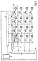
- FIG 3 is a schematic plan view of the X-ray filter incorporated in the X-ray examination apparatus of Figure 1.
- figure 3 shows an X-ray filter with a 4 ⁇ 4 matrix arrangement of capillary tubes, but in practice an X-ray filter having a much larger number such as 200 ⁇ 200 capillary tubes may be employed.
- Each of the capillary tubes has its conductive layer 32 coupled to the drain contact 40 of a field-effect transistor 33 which acts as a switching element and whose source contact 41 is coupled to a voltage line 34.
- an addressing line 35 which is coupled to the gate contacts of the field-effect transistors in that row so as to control these field-effect transistors.
- the X-ray filter comprises an adjusting unit 25 which incorporates a voltage generator 36 for applying the electrical voltage to a row driver 8 that applies the addressing signals to respective addressing lines.
- the electrical voltages to be applied to the capillary tubes are supplied by way of a column driver circuit 37.
- the addressing signals select capillary tubes that are to be supplied with the electrical voltage.
- the voltage generator produces the addressing signals as well as the electrical voltages applied to the capillary tubes so as to control the amount of X-ray absorbing liquid in the capillary tubes.
Landscapes
- Physics & Mathematics (AREA)
- Spectroscopy & Molecular Physics (AREA)
- Engineering & Computer Science (AREA)
- General Engineering & Computer Science (AREA)
- High Energy & Nuclear Physics (AREA)
- Apparatus For Radiation Diagnosis (AREA)
- Analysing Materials By The Use Of Radiation (AREA)
- X-Ray Techniques (AREA)
Abstract
Claims (6)
- Appareil d'examen à rayons X (1) étant pourvucaractérisé en ce qued'une source à rayons X (2),d'un détecteur à rayons X (3),d'un filtre à rayons X (4) qui se situe entre la source à rayons X et le détecteur à rayons X, le filtre à rayons X comportantune pluralité d'éléments filtrants (5) ayant une absorptivité de rayons X que l'on peut ajuster en commandant une quantité de liquide absorbant des rayons X dans des éléments filtrants séparés,le filtre à rayons X comporte un circuit d'ajustage pour délivrer des tensions électriques à des éléments filtrants respectifs pour commander la quantité de liquide absorbant des rayons X dans des éléments filtrants individuels et en ce quele liquide absorbant des rayons X contient une suspension de très petites particules absorbant des rayons X dans un solvant, lesdites très petites particules ayant un diamètre inférieur à 1 µm.
- Appareil d'examen à rayons X selon la revendication 1, caractérisé en ce que le diamètre des très petites particules se situe dans la gamme comprise entre 5 nm et 100 nm.
- Appareil d'examen à rayons X selon la revendication 1 ou 2, caractérisé en ce que les très petites particles se composent d'un ou de plusieurs éléments dont le nombre atomique est au moins égal à 72.
- Appareil d'examen à rayons X selon l'une quelconque des revendications précédentes 1 à 3, caractérisé en ce que le solvant est de l'eau.
- Appareil d'examen à rayons X selon la revendication 4, caractérisé en ce que le solvant est de l'eau avec une addition active à la surface.
- Appareil d'examen à rayons X selon l'une quelconque des revendications précédentes 1 à 5, caractérisé en ce que les très petites particules comprennent un noyau contenant un élément qui présente un nombre atomique élevé, le noyau étant revêtu d'une couche qui est chimiquement inerte par rapport au solvant.
Priority Applications (1)
| Application Number | Priority Date | Filing Date | Title |
|---|---|---|---|
| EP97901213A EP0826220B1 (fr) | 1996-02-14 | 1997-02-07 | Appareil d'examen radiologique avec filtre a rayons x |
Applications Claiming Priority (4)
| Application Number | Priority Date | Filing Date | Title |
|---|---|---|---|
| EP96200360 | 1996-02-14 | ||
| EP96200360 | 1996-02-14 | ||
| EP97901213A EP0826220B1 (fr) | 1996-02-14 | 1997-02-07 | Appareil d'examen radiologique avec filtre a rayons x |
| PCT/IB1997/000089 WO1997030459A1 (fr) | 1996-02-14 | 1997-02-07 | Appareil d'examen radiologique et filtre a rayons x |
Publications (2)
| Publication Number | Publication Date |
|---|---|
| EP0826220A1 EP0826220A1 (fr) | 1998-03-04 |
| EP0826220B1 true EP0826220B1 (fr) | 2002-05-29 |
Family
ID=8223665
Family Applications (1)
| Application Number | Title | Priority Date | Filing Date |
|---|---|---|---|
| EP97901213A Expired - Lifetime EP0826220B1 (fr) | 1996-02-14 | 1997-02-07 | Appareil d'examen radiologique avec filtre a rayons x |
Country Status (5)
| Country | Link |
|---|---|
| US (1) | US5768340A (fr) |
| EP (1) | EP0826220B1 (fr) |
| JP (1) | JP3839059B2 (fr) |
| DE (1) | DE69712840T2 (fr) |
| WO (1) | WO1997030459A1 (fr) |
Families Citing this family (22)
| Publication number | Priority date | Publication date | Assignee | Title |
|---|---|---|---|---|
| US5878111A (en) * | 1996-09-20 | 1999-03-02 | Siemens Aktiengesellschaft | X-ray absorption filter having a field generating matrix and field sensitive liquids |
| WO1998052468A2 (fr) * | 1997-05-23 | 1998-11-26 | Koninklijke Philips Electronics N.V. | Appareil d'examen aux rayons x comprenant un filtre |
| DE69908494T2 (de) | 1998-01-23 | 2004-05-06 | Koninklijke Philips Electronics N.V. | Röntgenstrahlung-prüfungsvorrichtung enthaltend ein filter |
| EP1040653A2 (fr) * | 1998-07-01 | 2000-10-04 | Koninklijke Philips Electronics N.V. | Appareil d'examen aux rayons x comportant un filtre de rayons x |
| DE69908751T2 (de) * | 1998-08-04 | 2004-05-13 | Koninklijke Philips Electronics N.V. | Röntgenstrahlung-prüfungsvorrichtung enthaltend ein einstellbares röntgenstrahlungsfilter |
| US6215852B1 (en) | 1998-12-10 | 2001-04-10 | General Electric Company | Thermal energy storage and transfer assembly |
| JP2002535626A (ja) * | 1999-01-13 | 2002-10-22 | コーニンクレッカ フィリップス エレクトロニクス エヌ ヴィ | X線検査装置及びこれを調整する方法 |
| WO2001043145A1 (fr) * | 1999-12-08 | 2001-06-14 | Koninklijke Philips Electronics N.V. | Dispositif de radiographie avec filtre comprenant des elements filtrants a absorption reglable des rayons x et detecteur d'absorption des rayons x |
| EP1169715A1 (fr) * | 2000-02-04 | 2002-01-09 | Koninklijke Philips Electronics N.V. | Appareil a rayons x comprenant un filtre pourvu d'elements de filtration a absorption reglable |
| US6519313B2 (en) * | 2001-05-30 | 2003-02-11 | General Electric Company | High-Z cast reflector compositions and method of manufacture |
| US6920203B2 (en) * | 2002-12-02 | 2005-07-19 | General Electric Company | Method and apparatus for selectively attenuating a radiation source |
| US7308073B2 (en) * | 2005-10-20 | 2007-12-11 | General Electric Company | X-ray filter having dynamically displaceable x-ray attenuating fluid |
| DE102008055921B4 (de) * | 2008-11-05 | 2010-11-11 | Siemens Aktiengesellschaft | Modulierbarer Strahlenkollimator |
| JP2011145162A (ja) * | 2010-01-14 | 2011-07-28 | Japan Atomic Energy Agency | 流体中微粒子のx線検出法 |
| DE102012201856B4 (de) | 2012-02-08 | 2015-04-02 | Siemens Aktiengesellschaft | Konturkollimator und adaptives Filter mit elektroaktiven Polymerelementen und zugehöriges Verfahren |
| DE102012220750B4 (de) | 2012-02-08 | 2015-06-03 | Siemens Aktiengesellschaft | Konturkollimator mit einer magnetischen, Röntgenstrahlung absorbierenden Flüssigkeit und zugehöriges Verfahren |
| DE102012206953B3 (de) * | 2012-04-26 | 2013-05-23 | Siemens Aktiengesellschaft | Adaptives Röntgenfilter und Verfahren zur adaptiven Schwächung einer Röntgenstrahlung |
| DE102012207627B3 (de) * | 2012-05-08 | 2013-05-02 | Siemens Aktiengesellschaft | Adaptives Röntgenfilter zur Veränderung der lokalen Intensität einer Röntgenstrahlung |
| DE102012209150B3 (de) | 2012-05-31 | 2013-04-11 | Siemens Aktiengesellschaft | Adaptives Röntgenfilter und Verfahren zur Veränderung der lokalen Intensität einer Röntgenstrahlung |
| DE102012217616B4 (de) * | 2012-09-27 | 2017-04-06 | Siemens Healthcare Gmbh | Anordnung und Verfahren zur Veränderung der lokalen Intensität einer Röntgenstrahlung |
| CN107003420B (zh) | 2014-10-04 | 2020-03-24 | Ibex创新有限责任公司 | 关于x射线装置中散射的改进及其使用方法 |
| GB2576772B (en) | 2018-08-31 | 2023-01-25 | Ibex Innovations Ltd | X-ray Imaging system |
Citations (4)
| Publication number | Priority date | Publication date | Assignee | Title |
|---|---|---|---|---|
| US4310507A (en) * | 1978-08-02 | 1982-01-12 | Eastman Kodak Company | Contrast agent for radiography |
| FR2601493A1 (fr) * | 1986-07-08 | 1988-01-15 | Thomson Csf | Dispositif pour former des images par deplacement de fluides et son utilisation a la realisation de filtres spatiaux a rayons x |
| EP0498482A2 (fr) * | 1991-01-25 | 1992-08-12 | NanoSystems L.L.C. | Agents de contraste pour application en imagerie médicale |
| EP0645146A1 (fr) * | 1990-03-28 | 1995-03-29 | Nycomed Imaging As | Milieux de contraste |
Family Cites Families (6)
| Publication number | Priority date | Publication date | Assignee | Title |
|---|---|---|---|---|
| SE347859B (fr) * | 1970-11-30 | 1972-08-14 | Medinova Ab | |
| FR2599886B1 (fr) * | 1986-06-06 | 1988-08-19 | Thomson Csf | Dispositif d'affichage d'image a fluide paramagnetique et son utilisation pour la realisation de filtres spatiaux de rayons x en imagerie medicale |
| NL8903110A (nl) * | 1989-12-20 | 1991-07-16 | Philips Nv | Roentgenonderzoekapparaat met dynamisch filter. |
| JP4043511B2 (ja) * | 1994-06-30 | 2008-02-06 | コーニンクレッカ フィリップス エレクトロニクス エヌ ヴィ | フィルタよりなるx線検査装置 |
| EP0740839B1 (fr) * | 1994-10-25 | 1998-09-23 | Koninklijke Philips Electronics N.V. | Appareil a rayons x comportant un filtre |
| JPH10506039A (ja) * | 1995-07-13 | 1998-06-16 | フィリップス エレクトロニクス エヌ ベー | フィルタを含むx線検査装置 |
-
1997
- 1997-02-07 JP JP52914797A patent/JP3839059B2/ja not_active Expired - Fee Related
- 1997-02-07 EP EP97901213A patent/EP0826220B1/fr not_active Expired - Lifetime
- 1997-02-07 DE DE69712840T patent/DE69712840T2/de not_active Expired - Fee Related
- 1997-02-07 WO PCT/IB1997/000089 patent/WO1997030459A1/fr not_active Ceased
- 1997-02-13 US US08/800,250 patent/US5768340A/en not_active Expired - Fee Related
Patent Citations (4)
| Publication number | Priority date | Publication date | Assignee | Title |
|---|---|---|---|---|
| US4310507A (en) * | 1978-08-02 | 1982-01-12 | Eastman Kodak Company | Contrast agent for radiography |
| FR2601493A1 (fr) * | 1986-07-08 | 1988-01-15 | Thomson Csf | Dispositif pour former des images par deplacement de fluides et son utilisation a la realisation de filtres spatiaux a rayons x |
| EP0645146A1 (fr) * | 1990-03-28 | 1995-03-29 | Nycomed Imaging As | Milieux de contraste |
| EP0498482A2 (fr) * | 1991-01-25 | 1992-08-12 | NanoSystems L.L.C. | Agents de contraste pour application en imagerie médicale |
Also Published As
| Publication number | Publication date |
|---|---|
| JPH11506691A (ja) | 1999-06-15 |
| DE69712840T2 (de) | 2002-12-12 |
| EP0826220A1 (fr) | 1998-03-04 |
| US5768340A (en) | 1998-06-16 |
| WO1997030459A1 (fr) | 1997-08-21 |
| JP3839059B2 (ja) | 2006-11-01 |
| DE69712840D1 (de) | 2002-07-04 |
Similar Documents
| Publication | Publication Date | Title |
|---|---|---|
| US5768340A (en) | X-ray examination apparatus with x-ray filter | |
| US5625665A (en) | X-ray apparatus comprising a filter | |
| EP0970479B1 (fr) | Appareil d'analyse aux rayons x comportant un filtre | |
| US6061426A (en) | X-ray examination apparatus including an x-ray filter | |
| US5751786A (en) | X-ray examination apparatus comprising a filter | |
| US5666396A (en) | X-Ray examination apparatus comprising a filter | |
| US5559853A (en) | X-ray examination apparatus comprising a filter | |
| US5966426A (en) | X-ray examination apparatus including an x-ray filter | |
| US6269147B1 (en) | X-ray examination apparatus and method for adjusting the same | |
| DE19719718B4 (de) | Szintillator, Bildaufnahmevorrichtung unter Verwendung desselben sowie Untersuchungs-Vorrichtung | |
| US6438211B1 (en) | X-ray apparatus including a filter with filter elements having an adjustable absorptivity | |
| US6453013B2 (en) | X-ray apparatus provided with a filter with a dynamically adjustable absorption | |
| EP0740883A1 (fr) | Appareil de prise d'images |
Legal Events
| Date | Code | Title | Description |
|---|---|---|---|
| PUAI | Public reference made under article 153(3) epc to a published international application that has entered the european phase |
Free format text: ORIGINAL CODE: 0009012 |
|
| AK | Designated contracting states |
Kind code of ref document: A1 Designated state(s): DE FR GB NL |
|
| 17P | Request for examination filed |
Effective date: 19980223 |
|
| 17Q | First examination report despatched |
Effective date: 19990805 |
|
| RAP3 | Party data changed (applicant data changed or rights of an application transferred) |
Owner name: PHILIPS CORPORATE INTELLECTUAL PROPERTY GMBH Owner name: KONINKLIJKE PHILIPS ELECTRONICS N.V. |
|
| GRAG | Despatch of communication of intention to grant |
Free format text: ORIGINAL CODE: EPIDOS AGRA |
|
| GRAG | Despatch of communication of intention to grant |
Free format text: ORIGINAL CODE: EPIDOS AGRA |
|
| GRAH | Despatch of communication of intention to grant a patent |
Free format text: ORIGINAL CODE: EPIDOS IGRA |
|
| GRAH | Despatch of communication of intention to grant a patent |
Free format text: ORIGINAL CODE: EPIDOS IGRA |
|
| GRAA | (expected) grant |
Free format text: ORIGINAL CODE: 0009210 |
|
| AK | Designated contracting states |
Kind code of ref document: B1 Designated state(s): DE FR GB NL |
|
| PG25 | Lapsed in a contracting state [announced via postgrant information from national office to epo] |
Ref country code: NL Free format text: LAPSE BECAUSE OF FAILURE TO SUBMIT A TRANSLATION OF THE DESCRIPTION OR TO PAY THE FEE WITHIN THE PRESCRIBED TIME-LIMIT Effective date: 20020529 |
|
| REG | Reference to a national code |
Ref country code: GB Ref legal event code: FG4D |
|
| REF | Corresponds to: |
Ref document number: 69712840 Country of ref document: DE Date of ref document: 20020704 |
|
| ET | Fr: translation filed | ||
| RAP2 | Party data changed (patent owner data changed or rights of a patent transferred) |
Owner name: PHILIPS CORPORATE INTELLECTUAL PROPERTY GMBH Owner name: KONINKLIJKE PHILIPS ELECTRONICS N.V. |
|
| NLV1 | Nl: lapsed or annulled due to failure to fulfill the requirements of art. 29p and 29m of the patents act |
Owner name: PHILIPS CORPORATE INTELLECTUAL PROPERTY GMBH |
|
| REG | Reference to a national code |
Ref country code: FR Ref legal event code: D6 |
|
| PG25 | Lapsed in a contracting state [announced via postgrant information from national office to epo] |
Ref country code: GB Free format text: LAPSE BECAUSE OF NON-PAYMENT OF DUE FEES Effective date: 20030207 |
|
| PLBE | No opposition filed within time limit |
Free format text: ORIGINAL CODE: 0009261 |
|
| STAA | Information on the status of an ep patent application or granted ep patent |
Free format text: STATUS: NO OPPOSITION FILED WITHIN TIME LIMIT |
|
| RAP2 | Party data changed (patent owner data changed or rights of a patent transferred) |
Owner name: PHILIPS INTELLECTUAL PROPERTY & STANDARDS GMBH Owner name: KONINKLIJKE PHILIPS ELECTRONICS N.V. |
|
| 26N | No opposition filed |
Effective date: 20030303 |
|
| GBPC | Gb: european patent ceased through non-payment of renewal fee | ||
| PGFP | Annual fee paid to national office [announced via postgrant information from national office to epo] |
Ref country code: DE Payment date: 20070410 Year of fee payment: 11 |
|
| PGFP | Annual fee paid to national office [announced via postgrant information from national office to epo] |
Ref country code: FR Payment date: 20070226 Year of fee payment: 11 |
|
| REG | Reference to a national code |
Ref country code: FR Ref legal event code: ST Effective date: 20081031 |
|
| PG25 | Lapsed in a contracting state [announced via postgrant information from national office to epo] |
Ref country code: DE Free format text: LAPSE BECAUSE OF NON-PAYMENT OF DUE FEES Effective date: 20080902 |
|
| PG25 | Lapsed in a contracting state [announced via postgrant information from national office to epo] |
Ref country code: FR Free format text: LAPSE BECAUSE OF NON-PAYMENT OF DUE FEES Effective date: 20080229 |