EP0832210B1 - Procede dans lequel une enzyme de reparation d'acide nucleique est utilisee pour la detection de mutation ponctuelle et une mutagenese in vitro - Google Patents
Procede dans lequel une enzyme de reparation d'acide nucleique est utilisee pour la detection de mutation ponctuelle et une mutagenese in vitro Download PDFInfo
- Publication number
- EP0832210B1 EP0832210B1 EP96921269A EP96921269A EP0832210B1 EP 0832210 B1 EP0832210 B1 EP 0832210B1 EP 96921269 A EP96921269 A EP 96921269A EP 96921269 A EP96921269 A EP 96921269A EP 0832210 B1 EP0832210 B1 EP 0832210B1
- Authority
- EP
- European Patent Office
- Prior art keywords
- probe
- enzyme
- oligonucleotide
- cleaving
- mismatch
- Prior art date
- Legal status (The legal status is an assumption and is not a legal conclusion. Google has not performed a legal analysis and makes no representation as to the accuracy of the status listed.)
- Expired - Lifetime
Links
- 102000004190 Enzymes Human genes 0.000 title claims abstract description 197
- 108090000790 Enzymes Proteins 0.000 title claims abstract description 197
- 238000000034 method Methods 0.000 title claims abstract description 87
- 230000008439 repair process Effects 0.000 title claims abstract description 69
- 230000035772 mutation Effects 0.000 title claims abstract description 61
- 150000007523 nucleic acids Chemical class 0.000 title claims abstract description 61
- 102000039446 nucleic acids Human genes 0.000 title claims abstract description 55
- 108020004707 nucleic acids Proteins 0.000 title claims abstract description 55
- 238000001514 detection method Methods 0.000 title abstract description 17
- 238000000338 in vitro Methods 0.000 title abstract description 13
- 238000002703 mutagenesis Methods 0.000 title abstract description 13
- 231100000350 mutagenesis Toxicity 0.000 title abstract description 13
- 239000000523 sample Substances 0.000 claims abstract description 249
- 102000040430 polynucleotide Human genes 0.000 claims abstract description 150
- 108091033319 polynucleotide Proteins 0.000 claims abstract description 150
- 239000002157 polynucleotide Substances 0.000 claims abstract description 150
- 108020005187 Oligonucleotide Probes Proteins 0.000 claims abstract description 47
- 239000002751 oligonucleotide probe Substances 0.000 claims abstract description 47
- 108091034117 Oligonucleotide Proteins 0.000 claims description 139
- 108020004414 DNA Proteins 0.000 claims description 62
- 238000003776 cleavage reaction Methods 0.000 claims description 62
- 230000007017 scission Effects 0.000 claims description 62
- 239000012634 fragment Substances 0.000 claims description 39
- 241000588724 Escherichia coli Species 0.000 claims description 31
- 230000000295 complement effect Effects 0.000 claims description 31
- 101150029137 mutY gene Proteins 0.000 claims description 30
- 230000000694 effects Effects 0.000 claims description 27
- 108010063362 DNA-(Apurinic or Apyrimidinic Site) Lyase Proteins 0.000 claims description 21
- 102000010719 DNA-(Apurinic or Apyrimidinic Site) Lyase Human genes 0.000 claims description 21
- 108700034637 EC 3.2.-.- Proteins 0.000 claims description 15
- 102000004317 Lyases Human genes 0.000 claims description 5
- 108090000856 Lyases Proteins 0.000 claims description 5
- 108091028043 Nucleic acid sequence Proteins 0.000 claims description 4
- 240000004808 Saccharomyces cerevisiae Species 0.000 claims description 3
- 101710190882 Endonuclease V Proteins 0.000 claims description 2
- 238000010396 two-hybrid screening Methods 0.000 claims description 2
- 238000006243 chemical reaction Methods 0.000 abstract description 55
- 238000009396 hybridization Methods 0.000 abstract description 27
- 230000000368 destabilizing effect Effects 0.000 abstract description 14
- 230000010355 oscillation Effects 0.000 abstract description 10
- 238000002372 labelling Methods 0.000 abstract description 3
- JLCPHMBAVCMARE-UHFFFAOYSA-N [3-[[3-[[3-[[3-[[3-[[3-[[3-[[3-[[3-[[3-[[3-[[5-(2-amino-6-oxo-1H-purin-9-yl)-3-[[3-[[3-[[3-[[3-[[3-[[5-(2-amino-6-oxo-1H-purin-9-yl)-3-[[5-(2-amino-6-oxo-1H-purin-9-yl)-3-hydroxyoxolan-2-yl]methoxy-hydroxyphosphoryl]oxyoxolan-2-yl]methoxy-hydroxyphosphoryl]oxy-5-(5-methyl-2,4-dioxopyrimidin-1-yl)oxolan-2-yl]methoxy-hydroxyphosphoryl]oxy-5-(6-aminopurin-9-yl)oxolan-2-yl]methoxy-hydroxyphosphoryl]oxy-5-(6-aminopurin-9-yl)oxolan-2-yl]methoxy-hydroxyphosphoryl]oxy-5-(6-aminopurin-9-yl)oxolan-2-yl]methoxy-hydroxyphosphoryl]oxy-5-(6-aminopurin-9-yl)oxolan-2-yl]methoxy-hydroxyphosphoryl]oxyoxolan-2-yl]methoxy-hydroxyphosphoryl]oxy-5-(5-methyl-2,4-dioxopyrimidin-1-yl)oxolan-2-yl]methoxy-hydroxyphosphoryl]oxy-5-(4-amino-2-oxopyrimidin-1-yl)oxolan-2-yl]methoxy-hydroxyphosphoryl]oxy-5-(5-methyl-2,4-dioxopyrimidin-1-yl)oxolan-2-yl]methoxy-hydroxyphosphoryl]oxy-5-(5-methyl-2,4-dioxopyrimidin-1-yl)oxolan-2-yl]methoxy-hydroxyphosphoryl]oxy-5-(6-aminopurin-9-yl)oxolan-2-yl]methoxy-hydroxyphosphoryl]oxy-5-(6-aminopurin-9-yl)oxolan-2-yl]methoxy-hydroxyphosphoryl]oxy-5-(4-amino-2-oxopyrimidin-1-yl)oxolan-2-yl]methoxy-hydroxyphosphoryl]oxy-5-(4-amino-2-oxopyrimidin-1-yl)oxolan-2-yl]methoxy-hydroxyphosphoryl]oxy-5-(4-amino-2-oxopyrimidin-1-yl)oxolan-2-yl]methoxy-hydroxyphosphoryl]oxy-5-(6-aminopurin-9-yl)oxolan-2-yl]methoxy-hydroxyphosphoryl]oxy-5-(4-amino-2-oxopyrimidin-1-yl)oxolan-2-yl]methyl [5-(6-aminopurin-9-yl)-2-(hydroxymethyl)oxolan-3-yl] hydrogen phosphate Polymers Cc1cn(C2CC(OP(O)(=O)OCC3OC(CC3OP(O)(=O)OCC3OC(CC3O)n3cnc4c3nc(N)[nH]c4=O)n3cnc4c3nc(N)[nH]c4=O)C(COP(O)(=O)OC3CC(OC3COP(O)(=O)OC3CC(OC3COP(O)(=O)OC3CC(OC3COP(O)(=O)OC3CC(OC3COP(O)(=O)OC3CC(OC3COP(O)(=O)OC3CC(OC3COP(O)(=O)OC3CC(OC3COP(O)(=O)OC3CC(OC3COP(O)(=O)OC3CC(OC3COP(O)(=O)OC3CC(OC3COP(O)(=O)OC3CC(OC3COP(O)(=O)OC3CC(OC3COP(O)(=O)OC3CC(OC3COP(O)(=O)OC3CC(OC3COP(O)(=O)OC3CC(OC3COP(O)(=O)OC3CC(OC3COP(O)(=O)OC3CC(OC3CO)n3cnc4c(N)ncnc34)n3ccc(N)nc3=O)n3cnc4c(N)ncnc34)n3ccc(N)nc3=O)n3ccc(N)nc3=O)n3ccc(N)nc3=O)n3cnc4c(N)ncnc34)n3cnc4c(N)ncnc34)n3cc(C)c(=O)[nH]c3=O)n3cc(C)c(=O)[nH]c3=O)n3ccc(N)nc3=O)n3cc(C)c(=O)[nH]c3=O)n3cnc4c3nc(N)[nH]c4=O)n3cnc4c(N)ncnc34)n3cnc4c(N)ncnc34)n3cnc4c(N)ncnc34)n3cnc4c(N)ncnc34)O2)c(=O)[nH]c1=O JLCPHMBAVCMARE-UHFFFAOYSA-N 0.000 description 43
- XSQUKJJJFZCRTK-UHFFFAOYSA-N Urea Chemical compound NC(N)=O XSQUKJJJFZCRTK-UHFFFAOYSA-N 0.000 description 28
- 239000000499 gel Substances 0.000 description 25
- 239000002773 nucleotide Substances 0.000 description 22
- 125000003729 nucleotide group Chemical group 0.000 description 22
- 108090000623 proteins and genes Proteins 0.000 description 22
- FAPWRFPIFSIZLT-UHFFFAOYSA-M Sodium chloride Chemical compound [Na+].[Cl-] FAPWRFPIFSIZLT-UHFFFAOYSA-M 0.000 description 18
- RWQNBRDOKXIBIV-UHFFFAOYSA-N thymine Chemical compound CC1=CNC(=O)NC1=O RWQNBRDOKXIBIV-UHFFFAOYSA-N 0.000 description 18
- 238000003752 polymerase chain reaction Methods 0.000 description 16
- 102000004169 proteins and genes Human genes 0.000 description 15
- 239000004202 carbamide Substances 0.000 description 14
- 210000004027 cell Anatomy 0.000 description 14
- 239000000126 substance Substances 0.000 description 12
- 102000053602 DNA Human genes 0.000 description 11
- 108010014303 DNA-directed DNA polymerase Proteins 0.000 description 10
- 102000016928 DNA-directed DNA polymerase Human genes 0.000 description 10
- OPTASPLRGRRNAP-UHFFFAOYSA-N cytosine Chemical compound NC=1C=CNC(=O)N=1 OPTASPLRGRRNAP-UHFFFAOYSA-N 0.000 description 10
- 229920002401 polyacrylamide Polymers 0.000 description 10
- 239000000872 buffer Substances 0.000 description 9
- 239000007795 chemical reaction product Substances 0.000 description 9
- 150000008300 phosphoramidites Chemical class 0.000 description 9
- 239000011780 sodium chloride Substances 0.000 description 9
- KCXVZYZYPLLWCC-UHFFFAOYSA-N EDTA Chemical compound OC(=O)CN(CC(O)=O)CCN(CC(O)=O)CC(O)=O KCXVZYZYPLLWCC-UHFFFAOYSA-N 0.000 description 8
- 239000013611 chromosomal DNA Substances 0.000 description 8
- 238000010790 dilution Methods 0.000 description 8
- 239000012895 dilution Substances 0.000 description 8
- 239000003446 ligand Substances 0.000 description 8
- 108700025694 p53 Genes Proteins 0.000 description 8
- 239000013612 plasmid Substances 0.000 description 8
- 229940113082 thymine Drugs 0.000 description 8
- 230000006870 function Effects 0.000 description 7
- 208000025113 myeloid leukemia Diseases 0.000 description 7
- 241000894006 Bacteria Species 0.000 description 6
- PEDCQBHIVMGVHV-UHFFFAOYSA-N Glycerine Chemical compound OCC(O)CO PEDCQBHIVMGVHV-UHFFFAOYSA-N 0.000 description 6
- NYHBQMYGNKIUIF-UUOKFMHZSA-N Guanosine Chemical compound C1=NC=2C(=O)NC(N)=NC=2N1[C@@H]1O[C@H](CO)[C@@H](O)[C@H]1O NYHBQMYGNKIUIF-UUOKFMHZSA-N 0.000 description 6
- 108010021757 Polynucleotide 5'-Hydroxyl-Kinase Proteins 0.000 description 6
- 102000008422 Polynucleotide 5'-hydroxyl-kinase Human genes 0.000 description 6
- HEMHJVSKTPXQMS-UHFFFAOYSA-M Sodium hydroxide Chemical compound [OH-].[Na+] HEMHJVSKTPXQMS-UHFFFAOYSA-M 0.000 description 6
- 238000003556 assay Methods 0.000 description 6
- HVYWMOMLDIMFJA-DPAQBDIFSA-N cholesterol Chemical group C1C=C2C[C@@H](O)CC[C@]2(C)[C@@H]2[C@@H]1[C@@H]1CC[C@H]([C@H](C)CCCC(C)C)[C@@]1(C)CC2 HVYWMOMLDIMFJA-DPAQBDIFSA-N 0.000 description 6
- 229930024421 Adenine Natural products 0.000 description 5
- GFFGJBXGBJISGV-UHFFFAOYSA-N Adenine Chemical compound NC1=NC=NC2=C1N=CN2 GFFGJBXGBJISGV-UHFFFAOYSA-N 0.000 description 5
- 101100512049 Arabidopsis thaliana LWD2 gene Proteins 0.000 description 5
- 108010061982 DNA Ligases Proteins 0.000 description 5
- 102000012410 DNA Ligases Human genes 0.000 description 5
- 102100031780 Endonuclease Human genes 0.000 description 5
- 101710081048 Endonuclease III Proteins 0.000 description 5
- 108010042407 Endonucleases Proteins 0.000 description 5
- 102000003960 Ligases Human genes 0.000 description 5
- 108090000364 Ligases Proteins 0.000 description 5
- 108091093037 Peptide nucleic acid Proteins 0.000 description 5
- 101100226885 Phomopsis amygdali PaP450-4 gene Proteins 0.000 description 5
- 108020004682 Single-Stranded DNA Proteins 0.000 description 5
- 101100350254 Streptomyces antibioticus oleV gene Proteins 0.000 description 5
- 229960000643 adenine Drugs 0.000 description 5
- 230000003321 amplification Effects 0.000 description 5
- 229940104302 cytosine Drugs 0.000 description 5
- UYTPUPDQBNUYGX-UHFFFAOYSA-N guanine Chemical class O=C1NC(N)=NC2=C1N=CN2 UYTPUPDQBNUYGX-UHFFFAOYSA-N 0.000 description 5
- 239000000203 mixture Substances 0.000 description 5
- 238000003199 nucleic acid amplification method Methods 0.000 description 5
- 230000008569 process Effects 0.000 description 5
- 239000000758 substrate Substances 0.000 description 5
- YBJHBAHKTGYVGT-ZKWXMUAHSA-N (+)-Biotin Chemical group N1C(=O)N[C@@H]2[C@H](CCCCC(=O)O)SC[C@@H]21 YBJHBAHKTGYVGT-ZKWXMUAHSA-N 0.000 description 4
- UFBJCMHMOXMLKC-UHFFFAOYSA-N 2,4-dinitrophenol Chemical compound OC1=CC=C([N+]([O-])=O)C=C1[N+]([O-])=O UFBJCMHMOXMLKC-UHFFFAOYSA-N 0.000 description 4
- 108091026890 Coding region Proteins 0.000 description 4
- ZHNUHDYFZUAESO-UHFFFAOYSA-N Formamide Chemical compound NC=O ZHNUHDYFZUAESO-UHFFFAOYSA-N 0.000 description 4
- 206010028980 Neoplasm Diseases 0.000 description 4
- 101710124239 Poly(A) polymerase Proteins 0.000 description 4
- 238000009825 accumulation Methods 0.000 description 4
- 201000010099 disease Diseases 0.000 description 4
- 208000037265 diseases, disorders, signs and symptoms Diseases 0.000 description 4
- 238000001502 gel electrophoresis Methods 0.000 description 4
- FDGQSTZJBFJUBT-UHFFFAOYSA-N hypoxanthine Chemical compound O=C1NC=NC2=C1NC=N2 FDGQSTZJBFJUBT-UHFFFAOYSA-N 0.000 description 4
- 108091032973 (ribonucleotides)n+m Proteins 0.000 description 3
- QKNYBSVHEMOAJP-UHFFFAOYSA-N 2-amino-2-(hydroxymethyl)propane-1,3-diol;hydron;chloride Chemical compound Cl.OCC(N)(CO)CO QKNYBSVHEMOAJP-UHFFFAOYSA-N 0.000 description 3
- 208000035657 Abasia Diseases 0.000 description 3
- 101000748061 Acholeplasma phage L2 Uncharacterized 16.1 kDa protein Proteins 0.000 description 3
- HRPVXLWXLXDGHG-UHFFFAOYSA-N Acrylamide Chemical compound NC(=O)C=C HRPVXLWXLXDGHG-UHFFFAOYSA-N 0.000 description 3
- 101000947615 Clostridium perfringens Uncharacterized 38.4 kDa protein Proteins 0.000 description 3
- 229920000742 Cotton Polymers 0.000 description 3
- MIKUYHXYGGJMLM-GIMIYPNGSA-N Crotonoside Natural products C1=NC2=C(N)NC(=O)N=C2N1[C@H]1O[C@@H](CO)[C@H](O)[C@@H]1O MIKUYHXYGGJMLM-GIMIYPNGSA-N 0.000 description 3
- NYHBQMYGNKIUIF-UHFFFAOYSA-N D-guanosine Natural products C1=2NC(N)=NC(=O)C=2N=CN1C1OC(CO)C(O)C1O NYHBQMYGNKIUIF-UHFFFAOYSA-N 0.000 description 3
- 108020001738 DNA Glycosylase Proteins 0.000 description 3
- 102000028381 DNA glycosylase Human genes 0.000 description 3
- 102100033215 DNA nucleotidylexotransferase Human genes 0.000 description 3
- 108010008286 DNA nucleotidylexotransferase Proteins 0.000 description 3
- 101000964391 Enterococcus faecalis UPF0145 protein Proteins 0.000 description 3
- 102100026406 G/T mismatch-specific thymine DNA glycosylase Human genes 0.000 description 3
- 101000748063 Haemophilus phage HP1 (strain HP1c1) Uncharacterized 11.1 kDa protein in rep-hol intergenic region Proteins 0.000 description 3
- 101000790840 Klebsiella pneumoniae Uncharacterized 49.5 kDa protein in cps region Proteins 0.000 description 3
- 238000012408 PCR amplification Methods 0.000 description 3
- 108010035344 Thymine DNA Glycosylase Proteins 0.000 description 3
- 239000002253 acid Substances 0.000 description 3
- 150000007513 acids Chemical class 0.000 description 3
- 235000012000 cholesterol Nutrition 0.000 description 3
- 238000013461 design Methods 0.000 description 3
- VHJLVAABSRFDPM-ZXZARUISSA-N dithioerythritol Chemical compound SC[C@H](O)[C@H](O)CS VHJLVAABSRFDPM-ZXZARUISSA-N 0.000 description 3
- 238000002474 experimental method Methods 0.000 description 3
- 239000007850 fluorescent dye Substances 0.000 description 3
- 229940029575 guanosine Drugs 0.000 description 3
- 239000000463 material Substances 0.000 description 3
- 230000004048 modification Effects 0.000 description 3
- 238000012986 modification Methods 0.000 description 3
- 239000011541 reaction mixture Substances 0.000 description 3
- 208000007056 sickle cell anemia Diseases 0.000 description 3
- 235000000346 sugar Nutrition 0.000 description 3
- 239000006228 supernatant Substances 0.000 description 3
- 238000003786 synthesis reaction Methods 0.000 description 3
- XLYOFNOQVPJJNP-UHFFFAOYSA-N water Chemical compound O XLYOFNOQVPJJNP-UHFFFAOYSA-N 0.000 description 3
- 102100025064 Cellular tumor antigen p53 Human genes 0.000 description 2
- HMFHBZSHGGEWLO-SOOFDHNKSA-N D-ribofuranose Chemical compound OC[C@H]1OC(O)[C@H](O)[C@@H]1O HMFHBZSHGGEWLO-SOOFDHNKSA-N 0.000 description 2
- 108010017826 DNA Polymerase I Proteins 0.000 description 2
- 102000004594 DNA Polymerase I Human genes 0.000 description 2
- UGQMRVRMYYASKQ-UHFFFAOYSA-N Hypoxanthine nucleoside Natural products OC1C(O)C(CO)OC1N1C(NC=NC2=O)=C2N=C1 UGQMRVRMYYASKQ-UHFFFAOYSA-N 0.000 description 2
- 101710163270 Nuclease Proteins 0.000 description 2
- 102000043276 Oncogene Human genes 0.000 description 2
- 108700020796 Oncogene Proteins 0.000 description 2
- 108091000080 Phosphotransferase Proteins 0.000 description 2
- PYMYPHUHKUWMLA-LMVFSUKVSA-N Ribose Natural products OC[C@@H](O)[C@@H](O)[C@@H](O)C=O PYMYPHUHKUWMLA-LMVFSUKVSA-N 0.000 description 2
- 108700025716 Tumor Suppressor Genes Proteins 0.000 description 2
- 102000044209 Tumor Suppressor Genes Human genes 0.000 description 2
- ISAKRJDGNUQOIC-UHFFFAOYSA-N Uracil Chemical compound O=C1C=CNC(=O)N1 ISAKRJDGNUQOIC-UHFFFAOYSA-N 0.000 description 2
- 108010072685 Uracil-DNA Glycosidase Proteins 0.000 description 2
- 102000006943 Uracil-DNA Glycosidase Human genes 0.000 description 2
- OIRDTQYFTABQOQ-KQYNXXCUSA-N adenosine Chemical compound C1=NC=2C(N)=NC=NC=2N1[C@@H]1O[C@H](CO)[C@@H](O)[C@H]1O OIRDTQYFTABQOQ-KQYNXXCUSA-N 0.000 description 2
- 125000003275 alpha amino acid group Chemical group 0.000 description 2
- HMFHBZSHGGEWLO-UHFFFAOYSA-N alpha-D-Furanose-Ribose Natural products OCC1OC(O)C(O)C1O HMFHBZSHGGEWLO-UHFFFAOYSA-N 0.000 description 2
- 238000013459 approach Methods 0.000 description 2
- 238000000376 autoradiography Methods 0.000 description 2
- 230000008901 benefit Effects 0.000 description 2
- 239000012472 biological sample Substances 0.000 description 2
- 238000001574 biopsy Methods 0.000 description 2
- 230000015572 biosynthetic process Effects 0.000 description 2
- 229960002685 biotin Drugs 0.000 description 2
- 235000020958 biotin Nutrition 0.000 description 2
- 239000011616 biotin Substances 0.000 description 2
- UDSAIICHUKSCKT-UHFFFAOYSA-N bromophenol blue Chemical compound C1=C(Br)C(O)=C(Br)C=C1C1(C=2C=C(Br)C(O)=C(Br)C=2)C2=CC=CC=C2S(=O)(=O)O1 UDSAIICHUKSCKT-UHFFFAOYSA-N 0.000 description 2
- 201000011510 cancer Diseases 0.000 description 2
- 239000012830 cancer therapeutic Substances 0.000 description 2
- 238000005251 capillar electrophoresis Methods 0.000 description 2
- 150000001718 carbodiimides Chemical class 0.000 description 2
- ZYGHJZDHTFUPRJ-UHFFFAOYSA-N coumarin Chemical compound C1=CC=C2OC(=O)C=CC2=C1 ZYGHJZDHTFUPRJ-UHFFFAOYSA-N 0.000 description 2
- 230000003247 decreasing effect Effects 0.000 description 2
- 230000001419 dependent effect Effects 0.000 description 2
- 230000001687 destabilization Effects 0.000 description 2
- 238000006073 displacement reaction Methods 0.000 description 2
- 239000000975 dye Substances 0.000 description 2
- 239000000284 extract Substances 0.000 description 2
- 238000011049 filling Methods 0.000 description 2
- 238000002866 fluorescence resonance energy transfer Methods 0.000 description 2
- -1 fpy Proteins 0.000 description 2
- 230000002068 genetic effect Effects 0.000 description 2
- 238000010353 genetic engineering Methods 0.000 description 2
- 238000010438 heat treatment Methods 0.000 description 2
- 238000007834 ligase chain reaction Methods 0.000 description 2
- 239000012160 loading buffer Substances 0.000 description 2
- 238000011068 loading method Methods 0.000 description 2
- 239000003550 marker Substances 0.000 description 2
- 125000002496 methyl group Chemical group [H]C([H])([H])* 0.000 description 2
- YBYRMVIVWMBXKQ-UHFFFAOYSA-N phenylmethanesulfonyl fluoride Chemical compound FS(=O)(=O)CC1=CC=CC=C1 YBYRMVIVWMBXKQ-UHFFFAOYSA-N 0.000 description 2
- 102000020233 phosphotransferase Human genes 0.000 description 2
- 229920000642 polymer Polymers 0.000 description 2
- 238000011160 research Methods 0.000 description 2
- 108091008146 restriction endonucleases Proteins 0.000 description 2
- VVLFAAMTGMGYBS-UHFFFAOYSA-M sodium;4-[[4-(ethylamino)-3-methylphenyl]-(4-ethylimino-3-methylcyclohexa-2,5-dien-1-ylidene)methyl]-3-sulfobenzenesulfonate Chemical compound [Na+].C1=C(C)C(NCC)=CC=C1C(C=1C(=CC(=CC=1)S([O-])(=O)=O)S(O)(=O)=O)=C1C=C(C)C(=NCC)C=C1 VVLFAAMTGMGYBS-UHFFFAOYSA-M 0.000 description 2
- 229940124597 therapeutic agent Drugs 0.000 description 2
- 210000001519 tissue Anatomy 0.000 description 2
- 241001515965 unidentified phage Species 0.000 description 2
- 0 *C1CCCCC1 Chemical compound *C1CCCCC1 0.000 description 1
- VUDQSRFCCHQIIU-UHFFFAOYSA-N 1-(3,5-dichloro-2,6-dihydroxy-4-methoxyphenyl)hexan-1-one Chemical compound CCCCCC(=O)C1=C(O)C(Cl)=C(OC)C(Cl)=C1O VUDQSRFCCHQIIU-UHFFFAOYSA-N 0.000 description 1
- JKMHFZQWWAIEOD-UHFFFAOYSA-N 2-[4-(2-hydroxyethyl)piperazin-1-yl]ethanesulfonic acid Chemical compound OCC[NH+]1CCN(CCS([O-])(=O)=O)CC1 JKMHFZQWWAIEOD-UHFFFAOYSA-N 0.000 description 1
- VKIGAWAEXPTIOL-UHFFFAOYSA-N 2-hydroxyhexanenitrile Chemical compound CCCCC(O)C#N VKIGAWAEXPTIOL-UHFFFAOYSA-N 0.000 description 1
- JDBGXEHEIRGOBU-UHFFFAOYSA-N 5-hydroxymethyluracil Chemical compound OCC1=CNC(=O)NC1=O JDBGXEHEIRGOBU-UHFFFAOYSA-N 0.000 description 1
- 206010000830 Acute leukaemia Diseases 0.000 description 1
- 108700015125 Adenovirus DBP Proteins 0.000 description 1
- 229920000936 Agarose Polymers 0.000 description 1
- 108090001008 Avidin Proteins 0.000 description 1
- 235000014469 Bacillus subtilis Nutrition 0.000 description 1
- 239000002126 C01EB10 - Adenosine Substances 0.000 description 1
- 208000005623 Carcinogenesis Diseases 0.000 description 1
- 101100185881 Clostridium tetani (strain Massachusetts / E88) mutS2 gene Proteins 0.000 description 1
- 108020004635 Complementary DNA Proteins 0.000 description 1
- KQLDDLUWUFBQHP-UHFFFAOYSA-N Cordycepin Natural products C1=NC=2C(N)=NC=NC=2N1C1OCC(CO)C1O KQLDDLUWUFBQHP-UHFFFAOYSA-N 0.000 description 1
- 101150026402 DBP gene Proteins 0.000 description 1
- 239000003298 DNA probe Substances 0.000 description 1
- 238000001712 DNA sequencing Methods 0.000 description 1
- 102100037373 DNA-(apurinic or apyrimidinic site) endonuclease Human genes 0.000 description 1
- 108010046855 DNA-deoxyinosine glycosidase Proteins 0.000 description 1
- 108010046331 Deoxyribodipyrimidine photo-lyase Proteins 0.000 description 1
- 108010036364 Deoxyribonuclease IV (Phage T4-Induced) Proteins 0.000 description 1
- 241000224495 Dictyostelium Species 0.000 description 1
- 101710094010 Endonuclease II Proteins 0.000 description 1
- 241000230501 Equine herpesvirus sp. Species 0.000 description 1
- 101710088570 Flagellar hook-associated protein 1 Proteins 0.000 description 1
- 208000011616 HELIX syndrome Diseases 0.000 description 1
- 239000007995 HEPES buffer Substances 0.000 description 1
- 101100501326 Haloarcula marismortui (strain ATCC 43049 / DSM 3752 / JCM 8966 / VKM B-1809) nfo gene Proteins 0.000 description 1
- 208000009889 Herpes Simplex Diseases 0.000 description 1
- 101000806846 Homo sapiens DNA-(apurinic or apyrimidinic site) endonuclease Proteins 0.000 description 1
- 101001137256 Homo sapiens DNA-(apurinic or apyrimidinic site) lyase Proteins 0.000 description 1
- 240000005979 Hordeum vulgare Species 0.000 description 1
- 235000007340 Hordeum vulgare Nutrition 0.000 description 1
- 241000701085 Human alphaherpesvirus 3 Species 0.000 description 1
- 241000701024 Human betaherpesvirus 5 Species 0.000 description 1
- 241000701044 Human gammaherpesvirus 4 Species 0.000 description 1
- 101000829171 Hypocrea virens (strain Gv29-8 / FGSC 10586) Effector TSP1 Proteins 0.000 description 1
- OUYCCCASQSFEME-QMMMGPOBSA-N L-tyrosine Chemical compound OC(=O)[C@@H](N)CC1=CC=C(O)C=C1 OUYCCCASQSFEME-QMMMGPOBSA-N 0.000 description 1
- KDXKERNSBIXSRK-UHFFFAOYSA-N Lysine Natural products NCCCCC(N)C(O)=O KDXKERNSBIXSRK-UHFFFAOYSA-N 0.000 description 1
- 239000004472 Lysine Substances 0.000 description 1
- 241001599018 Melanogaster Species 0.000 description 1
- 241000988893 Mesoplasma lactucae Species 0.000 description 1
- 108060004795 Methyltransferase Proteins 0.000 description 1
- 102000016397 Methyltransferase Human genes 0.000 description 1
- 101000777240 Micrococcus luteus (strain ATCC 4698 / DSM 20030 / JCM 1464 / NBRC 3333 / NCIMB 9278 / NCTC 2665 / VKM Ac-2230) Ultraviolet N-glycosylase/AP lyase Proteins 0.000 description 1
- 244000128833 Mimulus luteus Species 0.000 description 1
- 108700019961 Neoplasm Genes Proteins 0.000 description 1
- 102000048850 Neoplasm Genes Human genes 0.000 description 1
- 102100021010 Nucleolin Human genes 0.000 description 1
- 229920000776 Poly(Adenosine diphosphate-ribose) polymerase Polymers 0.000 description 1
- KDCGOANMDULRCW-UHFFFAOYSA-N Purine Natural products N1=CNC2=NC=NC2=C1 KDCGOANMDULRCW-UHFFFAOYSA-N 0.000 description 1
- 241000700159 Rattus Species 0.000 description 1
- 241001466077 Salina Species 0.000 description 1
- 229920002684 Sepharose Polymers 0.000 description 1
- 241000700584 Simplexvirus Species 0.000 description 1
- 108091081024 Start codon Proteins 0.000 description 1
- 108010090804 Streptavidin Proteins 0.000 description 1
- 101710137500 T7 RNA polymerase Proteins 0.000 description 1
- 101710145783 TATA-box-binding protein Proteins 0.000 description 1
- 108010006785 Taq Polymerase Proteins 0.000 description 1
- 101710193546 Tegument protein VP16 homolog Proteins 0.000 description 1
- 241000229714 Thermothrix Species 0.000 description 1
- 102000004357 Transferases Human genes 0.000 description 1
- 108090000992 Transferases Proteins 0.000 description 1
- 244000098338 Triticum aestivum Species 0.000 description 1
- 240000008042 Zea mays Species 0.000 description 1
- 235000007244 Zea mays Nutrition 0.000 description 1
- 229960000276 acetophenazine Drugs 0.000 description 1
- 229960005305 adenosine Drugs 0.000 description 1
- 150000003838 adenosines Chemical class 0.000 description 1
- 238000001042 affinity chromatography Methods 0.000 description 1
- 238000001261 affinity purification Methods 0.000 description 1
- 101150019302 alkA gene Proteins 0.000 description 1
- 125000000539 amino acid group Chemical group 0.000 description 1
- AVKUERGKIZMTKX-NJBDSQKTSA-N ampicillin Chemical compound C1([C@@H](N)C(=O)N[C@H]2[C@H]3SC([C@@H](N3C2=O)C(O)=O)(C)C)=CC=CC=C1 AVKUERGKIZMTKX-NJBDSQKTSA-N 0.000 description 1
- 229960000723 ampicillin Drugs 0.000 description 1
- 239000002246 antineoplastic agent Substances 0.000 description 1
- 210000003969 blast cell Anatomy 0.000 description 1
- 238000009835 boiling Methods 0.000 description 1
- 238000010804 cDNA synthesis Methods 0.000 description 1
- 244000309466 calf Species 0.000 description 1
- 230000036952 cancer formation Effects 0.000 description 1
- 231100000504 carcinogenesis Toxicity 0.000 description 1
- 239000006285 cell suspension Substances 0.000 description 1
- 238000005119 centrifugation Methods 0.000 description 1
- 230000008859 change Effects 0.000 description 1
- 231100000481 chemical toxicant Toxicity 0.000 description 1
- 229940044683 chemotherapy drug Drugs 0.000 description 1
- 238000004587 chromatography analysis Methods 0.000 description 1
- 239000002299 complementary DNA Substances 0.000 description 1
- 239000012141 concentrate Substances 0.000 description 1
- 238000007796 conventional method Methods 0.000 description 1
- OFEZSBMBBKLLBJ-BAJZRUMYSA-N cordycepin Chemical compound C1=NC=2C(N)=NC=NC=2N1[C@@H]1O[C@H](CO)C[C@H]1O OFEZSBMBBKLLBJ-BAJZRUMYSA-N 0.000 description 1
- OFEZSBMBBKLLBJ-UHFFFAOYSA-N cordycepine Natural products C1=NC=2C(N)=NC=NC=2N1C1OC(CO)CC1O OFEZSBMBBKLLBJ-UHFFFAOYSA-N 0.000 description 1
- 229960000956 coumarin Drugs 0.000 description 1
- 235000001671 coumarin Nutrition 0.000 description 1
- 239000000287 crude extract Substances 0.000 description 1
- 230000001351 cycling effect Effects 0.000 description 1
- 230000009089 cytolysis Effects 0.000 description 1
- 230000007812 deficiency Effects 0.000 description 1
- 230000002950 deficient Effects 0.000 description 1
- 239000008367 deionised water Substances 0.000 description 1
- 229910021641 deionized water Inorganic materials 0.000 description 1
- 238000010586 diagram Methods 0.000 description 1
- 238000010494 dissociation reaction Methods 0.000 description 1
- 230000005593 dissociations Effects 0.000 description 1
- 229940079593 drug Drugs 0.000 description 1
- 239000003814 drug Substances 0.000 description 1
- 238000001962 electrophoresis Methods 0.000 description 1
- 238000010828 elution Methods 0.000 description 1
- 101150100366 end gene Proteins 0.000 description 1
- 230000002255 enzymatic effect Effects 0.000 description 1
- 108010052305 exodeoxyribonuclease III Proteins 0.000 description 1
- 239000013604 expression vector Substances 0.000 description 1
- GNBHRKFJIUUOQI-UHFFFAOYSA-N fluorescein Chemical compound O1C(=O)C2=CC=CC=C2C21C1=CC=C(O)C=C1OC1=CC(O)=CC=C21 GNBHRKFJIUUOQI-UHFFFAOYSA-N 0.000 description 1
- 238000001215 fluorescent labelling Methods 0.000 description 1
- 101150117187 glmS gene Proteins 0.000 description 1
- 239000011544 gradient gel Substances 0.000 description 1
- 102000050321 human APEX1 Human genes 0.000 description 1
- 230000006872 improvement Effects 0.000 description 1
- 238000011534 incubation Methods 0.000 description 1
- 230000006698 induction Effects 0.000 description 1
- 230000010354 integration Effects 0.000 description 1
- BPHPUYQFMNQIOC-NXRLNHOXSA-N isopropyl beta-D-thiogalactopyranoside Chemical compound CC(C)S[C@@H]1O[C@H](CO)[C@H](O)[C@H](O)[C@H]1O BPHPUYQFMNQIOC-NXRLNHOXSA-N 0.000 description 1
- 210000002311 liver mitochondria Anatomy 0.000 description 1
- 239000011159 matrix material Substances 0.000 description 1
- 238000002844 melting Methods 0.000 description 1
- 230000008018 melting Effects 0.000 description 1
- 101150094317 mfd gene Proteins 0.000 description 1
- 101150031310 mutH gene Proteins 0.000 description 1
- 101150049514 mutL gene Proteins 0.000 description 1
- 101150013854 mutS gene Proteins 0.000 description 1
- 230000007935 neutral effect Effects 0.000 description 1
- 101150007306 nfo gene Proteins 0.000 description 1
- 101150030229 nth gene Proteins 0.000 description 1
- 239000002853 nucleic acid probe Substances 0.000 description 1
- 108010044762 nucleolin Proteins 0.000 description 1
- 229940124276 oligodeoxyribonucleotide Drugs 0.000 description 1
- 239000008188 pellet Substances 0.000 description 1
- 210000002826 placenta Anatomy 0.000 description 1
- 229920001184 polypeptide Polymers 0.000 description 1
- GUUBJKMBDULZTE-UHFFFAOYSA-M potassium;2-[4-(2-hydroxyethyl)piperazin-1-yl]ethanesulfonic acid;hydroxide Chemical compound [OH-].[K+].OCCN1CCN(CCS(O)(=O)=O)CC1 GUUBJKMBDULZTE-UHFFFAOYSA-M 0.000 description 1
- 238000002360 preparation method Methods 0.000 description 1
- 108090000765 processed proteins & peptides Proteins 0.000 description 1
- 102000004196 processed proteins & peptides Human genes 0.000 description 1
- 239000000047 product Substances 0.000 description 1
- 238000000746 purification Methods 0.000 description 1
- 238000000163 radioactive labelling Methods 0.000 description 1
- 239000011535 reaction buffer Substances 0.000 description 1
- 101150056906 recJ gene Proteins 0.000 description 1
- 230000009467 reduction Effects 0.000 description 1
- 230000004044 response Effects 0.000 description 1
- 230000000717 retained effect Effects 0.000 description 1
- 238000012552 review Methods 0.000 description 1
- PYWVYCXTNDRMGF-UHFFFAOYSA-N rhodamine B Chemical compound [Cl-].C=12C=CC(=[N+](CC)CC)C=C2OC2=CC(N(CC)CC)=CC=C2C=1C1=CC=CC=C1C(O)=O PYWVYCXTNDRMGF-UHFFFAOYSA-N 0.000 description 1
- 239000012266 salt solution Substances 0.000 description 1
- 150000003839 salts Chemical class 0.000 description 1
- 230000035945 sensitivity Effects 0.000 description 1
- 238000002741 site-directed mutagenesis Methods 0.000 description 1
- 238000001542 size-exclusion chromatography Methods 0.000 description 1
- 238000002415 sodium dodecyl sulfate polyacrylamide gel electrophoresis Methods 0.000 description 1
- 239000002904 solvent Substances 0.000 description 1
- 108010068698 spleen exonuclease Proteins 0.000 description 1
- 238000010561 standard procedure Methods 0.000 description 1
- 238000006467 substitution reaction Methods 0.000 description 1
- 150000008163 sugars Chemical class 0.000 description 1
- 230000004083 survival effect Effects 0.000 description 1
- 239000000725 suspension Substances 0.000 description 1
- 230000002194 synthesizing effect Effects 0.000 description 1
- 210000001541 thymus gland Anatomy 0.000 description 1
- 231100000331 toxic Toxicity 0.000 description 1
- 230000002588 toxic effect Effects 0.000 description 1
- 239000003440 toxic substance Substances 0.000 description 1
- 231100000419 toxicity Toxicity 0.000 description 1
- 230000001988 toxicity Effects 0.000 description 1
- 230000009466 transformation Effects 0.000 description 1
- 230000001131 transforming effect Effects 0.000 description 1
- 230000014616 translation Effects 0.000 description 1
- AVBGNFCMKJOFIN-UHFFFAOYSA-N triethylammonium acetate Chemical compound CC(O)=O.CCN(CC)CC AVBGNFCMKJOFIN-UHFFFAOYSA-N 0.000 description 1
- 239000012137 tryptone Substances 0.000 description 1
- OUYCCCASQSFEME-UHFFFAOYSA-N tyrosine Natural products OC(=O)C(N)CC1=CC=C(O)C=C1 OUYCCCASQSFEME-UHFFFAOYSA-N 0.000 description 1
- 238000000825 ultraviolet detection Methods 0.000 description 1
- 229940035893 uracil Drugs 0.000 description 1
- 101150073340 uvrD gene Proteins 0.000 description 1
- 101150097442 xthA gene Proteins 0.000 description 1
Images





Classifications
-
- C—CHEMISTRY; METALLURGY
- C12—BIOCHEMISTRY; BEER; SPIRITS; WINE; VINEGAR; MICROBIOLOGY; ENZYMOLOGY; MUTATION OR GENETIC ENGINEERING
- C12Q—MEASURING OR TESTING PROCESSES INVOLVING ENZYMES, NUCLEIC ACIDS OR MICROORGANISMS; COMPOSITIONS OR TEST PAPERS THEREFOR; PROCESSES OF PREPARING SUCH COMPOSITIONS; CONDITION-RESPONSIVE CONTROL IN MICROBIOLOGICAL OR ENZYMOLOGICAL PROCESSES
- C12Q1/00—Measuring or testing processes involving enzymes, nucleic acids or microorganisms; Compositions therefor; Processes of preparing such compositions
- C12Q1/68—Measuring or testing processes involving enzymes, nucleic acids or microorganisms; Compositions therefor; Processes of preparing such compositions involving nucleic acids
- C12Q1/6813—Hybridisation assays
- C12Q1/6816—Hybridisation assays characterised by the detection means
- C12Q1/6823—Release of bound markers
-
- C—CHEMISTRY; METALLURGY
- C12—BIOCHEMISTRY; BEER; SPIRITS; WINE; VINEGAR; MICROBIOLOGY; ENZYMOLOGY; MUTATION OR GENETIC ENGINEERING
- C12N—MICROORGANISMS OR ENZYMES; COMPOSITIONS THEREOF; PROPAGATING, PRESERVING, OR MAINTAINING MICROORGANISMS; MUTATION OR GENETIC ENGINEERING; CULTURE MEDIA
- C12N15/00—Mutation or genetic engineering; DNA or RNA concerning genetic engineering, vectors, e.g. plasmids, or their isolation, preparation or purification; Use of hosts therefor
- C12N15/09—Recombinant DNA-technology
- C12N15/10—Processes for the isolation, preparation or purification of DNA or RNA
- C12N15/102—Mutagenizing nucleic acids
-
- C—CHEMISTRY; METALLURGY
- C12—BIOCHEMISTRY; BEER; SPIRITS; WINE; VINEGAR; MICROBIOLOGY; ENZYMOLOGY; MUTATION OR GENETIC ENGINEERING
- C12Q—MEASURING OR TESTING PROCESSES INVOLVING ENZYMES, NUCLEIC ACIDS OR MICROORGANISMS; COMPOSITIONS OR TEST PAPERS THEREFOR; PROCESSES OF PREPARING SUCH COMPOSITIONS; CONDITION-RESPONSIVE CONTROL IN MICROBIOLOGICAL OR ENZYMOLOGICAL PROCESSES
- C12Q1/00—Measuring or testing processes involving enzymes, nucleic acids or microorganisms; Compositions therefor; Processes of preparing such compositions
- C12Q1/68—Measuring or testing processes involving enzymes, nucleic acids or microorganisms; Compositions therefor; Processes of preparing such compositions involving nucleic acids
- C12Q1/6813—Hybridisation assays
- C12Q1/6816—Hybridisation assays characterised by the detection means
- C12Q1/6818—Hybridisation assays characterised by the detection means involving interaction of two or more labels, e.g. resonant energy transfer
-
- C—CHEMISTRY; METALLURGY
- C12—BIOCHEMISTRY; BEER; SPIRITS; WINE; VINEGAR; MICROBIOLOGY; ENZYMOLOGY; MUTATION OR GENETIC ENGINEERING
- C12Q—MEASURING OR TESTING PROCESSES INVOLVING ENZYMES, NUCLEIC ACIDS OR MICROORGANISMS; COMPOSITIONS OR TEST PAPERS THEREFOR; PROCESSES OF PREPARING SUCH COMPOSITIONS; CONDITION-RESPONSIVE CONTROL IN MICROBIOLOGICAL OR ENZYMOLOGICAL PROCESSES
- C12Q1/00—Measuring or testing processes involving enzymes, nucleic acids or microorganisms; Compositions therefor; Processes of preparing such compositions
- C12Q1/68—Measuring or testing processes involving enzymes, nucleic acids or microorganisms; Compositions therefor; Processes of preparing such compositions involving nucleic acids
- C12Q1/6813—Hybridisation assays
- C12Q1/6816—Hybridisation assays characterised by the detection means
- C12Q1/682—Signal amplification
-
- C—CHEMISTRY; METALLURGY
- C12—BIOCHEMISTRY; BEER; SPIRITS; WINE; VINEGAR; MICROBIOLOGY; ENZYMOLOGY; MUTATION OR GENETIC ENGINEERING
- C12Q—MEASURING OR TESTING PROCESSES INVOLVING ENZYMES, NUCLEIC ACIDS OR MICROORGANISMS; COMPOSITIONS OR TEST PAPERS THEREFOR; PROCESSES OF PREPARING SUCH COMPOSITIONS; CONDITION-RESPONSIVE CONTROL IN MICROBIOLOGICAL OR ENZYMOLOGICAL PROCESSES
- C12Q1/00—Measuring or testing processes involving enzymes, nucleic acids or microorganisms; Compositions therefor; Processes of preparing such compositions
- C12Q1/68—Measuring or testing processes involving enzymes, nucleic acids or microorganisms; Compositions therefor; Processes of preparing such compositions involving nucleic acids
- C12Q1/6813—Hybridisation assays
- C12Q1/6827—Hybridisation assays for detection of mutation or polymorphism
-
- C—CHEMISTRY; METALLURGY
- C12—BIOCHEMISTRY; BEER; SPIRITS; WINE; VINEGAR; MICROBIOLOGY; ENZYMOLOGY; MUTATION OR GENETIC ENGINEERING
- C12Q—MEASURING OR TESTING PROCESSES INVOLVING ENZYMES, NUCLEIC ACIDS OR MICROORGANISMS; COMPOSITIONS OR TEST PAPERS THEREFOR; PROCESSES OF PREPARING SUCH COMPOSITIONS; CONDITION-RESPONSIVE CONTROL IN MICROBIOLOGICAL OR ENZYMOLOGICAL PROCESSES
- C12Q1/00—Measuring or testing processes involving enzymes, nucleic acids or microorganisms; Compositions therefor; Processes of preparing such compositions
- C12Q1/68—Measuring or testing processes involving enzymes, nucleic acids or microorganisms; Compositions therefor; Processes of preparing such compositions involving nucleic acids
- C12Q1/6844—Nucleic acid amplification reactions
- C12Q1/6858—Allele-specific amplification
Definitions
- Genomic DNA provides the template for the information that allows the generation of proteins which are expressed and made by an organism. These proteins are generally essential for the survival of any specific cell in an organism. Therefore, the organism requires the template to be correct and free of mistakes in order to generate a protein that is functional in a cell. If a single nucleotide of this DNA sequence is mutated (a "point mutation"), the protein may be nonfunctional. Point mutations which elicit disease states are known for many proteins. Examples include sickle cell anemia hypoxanthine phosphotransferase, and p53, a tumor suppressor gene, and several oncogenes and cancer genes.
- PCR polymerase chain reaction
- LCR ligase chain reaction
- NASBA nucleic acid system-based amplification
- CPT cycling probe technology
- CPT technology was developed, in part to overcome the limitations of PCR. See, for example, U.S. patents No. 4,876,187 and No. 5,011,769.
- the CPT technology entails the use of a synthetic molecule with two non-complementary nucleic acid sequences joined by a scissile linkage.
- CPT technology works by observing a hybridization event with a sample nucleic acid by a single cleavage event.
- This technology utilizes both the enzymatic features of RNAse H and a synthetic DNA-RNA-DNA oligonucleotide.
- RNAse H specifically cleaves the RNA moiety of the DNA-RNA-DNA oligonucleotide only when it is perfectly hybridized to a complementary DNA target molecule.
- a high concentration of the DNA-RNA-DNA molecule is converted to cleaved fragments, which are assayed by gel electrophoresis. The level of cleavage indicates the amount of target molecules present in the
- the CPT system does not amplify the target, alleviating the accumulation of molecules that in turn become amplifiable and generate false-positives, as occurs in PCR.
- the CPT technology is linear, in that increasing amounts of target DNA generate linearly more cleaved DNA-RNA-DNA oligonucleotide. (PCR generates exponentially more signal in response to the presence of more target DNA, making quantitation more problematic.) Additionally, CPT can amplify up to 10 6 cleaved DNA-RNA-DNA probe molecules in about 30 minutes. CPT does not generate more of the target molecule. Therefore, it does not jeopardize the laboratory environment by the possible accumulation of synthesized target DNA molecules, which in turn generate false positive results.
- Still another aspect of this invention concerns helix destabilizing molecule and similar molecules to enhance the hybridization of the probe to the target polynucleotides.
- Another aspect of this invention relates to the attachment of a nucleic acid repair enzyme to a probe in order to enhance the detection of target polynucleotide and point mutations therein.
- a method of detecting a point mutation in a target polynucleotide comprising:
- a method of detecting a point mutation in a target polynucleotide comprising:
- a method of detecting a sequence in a target polynucleotide comprising the steps of:
- a method of detecting a point mutation in a target polynucleotide comprising:
- a method for effecting in vitro mutagenesis of a target polynucleotide comprising transforming a circular double-stranded nucleic acid molecule, wherein said circular double-stranded nucleic acid molecule is prepared by the steps of:
- a method for effecting in vitro mutagenesis of a single-stranded target polynucleotide comprising:
- a method of detecting a point mutation in a target polynucleotide comprising:
- nucleic acid repair enzymes in accordance with the present invention, to detect point mutations.
- nucleotide excision enzymes can be used in the present invention to detect wild-type target polynucleotide sequences and to determine the amount of base repair enzyme activity carried out on a mutated polynucleotide sequence.
- the present invention represents an improvement over the CPT method, because a DNA oligonucleotide suitable for the present invention can be made via more conventional chemistry (and, hence, is less expensive to synthesize) and is more stable.
- nucleic acid repair enzymes can be used to create an oscillating reaction which allows for adequate target detection using a limited amount of unamplified target.
- detection can be enhanced by using nucleic acid repair enzymes, and other enzymes, to tag the oligonucleotide probe which has bound to the target polynucleotide.
- hybridization of probe to polynucleotide target can be enhanced by employing helix destabilizing molecules, as described in greater detail below.
- point mutations in a target polynucleotide of biological sample can be detected, identified or localized.
- This embodiment does not include the use of PCR amplification of target polynucleotide, since PCR amplification introduces spurious point mutations.
- This embodiment entails hybridizing a single-stranded oligonucleotide probe to a target polynucleotide to form a hybrid, double-stranded polynucleotide.
- the hybridization occurs under conditions that are "stringent,” which typically implicates conditions that include a 50-100 mM salt solution at a temperature of (3N - 20°C), where N is the number of nucleotides in the oligonucleotide probe.
- the oligonucleotide probe is designed not to have self complementary regions, palindromic regions and the probe must also have probe specificity.
- the parameters for probe design can be found in Lowe et al ., Nucl . Acids Res . 18:1757-1761 (1990); Rychlik et al ., loc . cit . 17:8543-8551 (1989); Rychlik et al ., loc . cit . 18:6409-6412 (1990), which discusses probe design as applied to PCR reactions.
- the probe is complementary to a non-mutated sequence in the target polynucleotide, there will be a mismatch between non-mutated probe and mutated target polynucleotide.
- the mismatch will occur at the site of point mutation.
- the present invention comprehends the existence of multiple sites of mismatch on the hybrid, double-stranded polynucleotide.
- nucleic acid repair enzyme is an enzyme that will cleave, at a point of mismatch, one strand of a duplex formed by oligonucleotide probe and target polynucleotide.
- nucleic acid repair enzymes are mutY (Wu et al ., Proc . Nat'l Acad . Sci . USA 89: 8779-83 (1992)), T/G mismatch-specific nicking enzyme from HeLa nuclear extracts (Wiebauer & Jiricny, Nature 339: 234-36 (1989); Wiebauer & Jiricny, loc . cit .
- T/G mismatch-specific nicking enzyme from E . coli (Hennecke et al ., Nature 353: 776-78 (1991)), human yeast all-type enzymes (Yeh et al ., J . Biol . Chem . 2667: 6480-84 (1991); Chiang & Lu, Nuc . Acids Res ., 19:4761-4766 (1981)), Deoxyinosine 3'-Endonuclease from E. coli (Yao et al ., J . Biol . Chem . 270: 28609-16 (1995); Yao et al ., J . Biol . Chem . 269: 31390-96 (1994)).
- nucleic acid repair enzyme is an enzyme system comprising a glycosylase combined with an AP cleaving enzyme, such as endonuclease or lyase. Together glycosylase and AP cleaving enzyme, such as endonuclease or lyase cleave oligonucleotide probe/target polynucleotide duplex at a point of mismatch.
- a glycosylase creates an abasic sugar (an AP site) at the point of mismatch, which then is cleaved by an AP cleaving enzyme, such as endonuclease or lyase.
- Illustrative enzymes in these categories are detailed below.
- glycosylases tag-1, alkA, ung, fpy, mutY, nth, xthA, nfo, recJ, uvtA, uvrD, mfd, mutH, mutL, mutS, uracil DNA glycosylase, hydroxymethyluracil glycosylase, 5-mC DNA glycosylase, hypoxanthine DNA glycosylase, thymine mismatch DNA glycosylase, 3-mA DNA glycosylase, hydrated thymine DNA glycosylase (endonuclease III), pyrimidine dimer glycosylase These enzymes can come from any different biological sources.
- AP cleaving enzymes E . coli exonuclease III, E . coli endonuclease IV, Saccharomyes AP endonuclease, Drosphila melanogaster AP endonuclease I and II, human AP endonuclease, human AP lyase, BAP endonuclease, APEX endonuclease, HAP1 and AP endonuclease
- cleavage may also be effected by using a glycosylase enzyme, as described above, in combination with basic conditions and increased temperature.
- increasing pH and temperature effectuates cleavage at the AP site created by the glycosylase enzyme.
- Suitable parameters for cleavage of the AP site are pH levels of approximately 8 to 14, and temperatures ranging from approximately 50 to 95°C.
- the present invention employs a nucleic acid repair enzyme that is thermally stable, in the sense that the enzyme would function at some elevated temperature, such as from 50° to 80°C. Additionally, it is preferable that the thermally stable nucleic acid repair enzyme withstand temperatures up to 100°C for short periods.
- the present invention contemplates the use of a thermally stable glycosylase.
- a thermally stable glycosylase is the ORF10 protein encoded by the DNA sequence of Figure 11. This enzyme has been synthesized by Richard P. Cunningham at the State University of New York at Albany, according to the methods of Example 13.
- the substrate activity of the ORF10 enzyme includes both base cleaving properties and AP endonuclease activities.
- the AP endonuclease activities of this enzyme may be enhanced, however, by changing the amino acid residue in position 126 of Figure 1 from a tyrosine to a lysine. This substitution may be achieved by site directed mutagenesis by the methods discussed in Deng, et al ., J.A. Anal. Biochem. 200: 81 (1992).
- the ORF10 glycosylase is a homologue of the endonuclease III family.
- the skilled artisan may identify and isolate genes of the endonuclease III family from other thermophilic bacteria.
- Suitable probes may be designed as degenerate nucleotide coding sequences for the following amino acid sequences which are highly conserved amongst the members of the endonuclease III family: SEQ1, PYVILITEILLRRTT; SEQ2, AILDLPGVGKYT; SEQ3, MVDANFVRVINR.
- oligonucleotides may be used as PCR primers to amplify portions of the gene from the chromosomal DNA of thermophilic bacteria by PCR. Such amplified PCR products may then be used to screen a library of the thermophilic bacterium. Positive clones would be sequenced and the coding sequence for the mismatch glycosylase cloned into an expression vector for protein production.
- nucleic acid repair enzymes can be used in combination with a AP cleaving enzyme.
- mutY is used in combination with AP cleaving enzymes, such as DNA lyase or DNA AP endonuclease. Such a system of enzymes enhances the speed at which cleavage occurs.
- the nucleic acid repair enzyme can be attached to a probe or a combination of probes.
- the attachment of the enzyme to a probe enhances the speed at which cleavage occurs by keeping the nucleic acid repair enzyme in proximity with the probe.
- the nucleic acid repair enzyme is a single enzyme that exhibits both glycosylase and AP cleaving properties, which can be attached to a single probe which hybridizes to the target polynucleotide at the site of a point mutation.
- This enzyme can be attached either to the 5' or to the 3' end of the probe.
- the attachment of the nucleic acid repair enzyme to the probe can be accomplished by linking methods described, for example, by Corey et al ., Science 238:1401 (1987), and Corey et al ., J . Am . Chem . Soc . 111:8523 (1989).
- the nucleic acid repair enzyme is made up of two separate enzymes, as discussed above; one enzyme is a glycosylase and the other has AP cleaving properties. Such enzymes can be utilized so that the glycosylase is attached to one end of the probe and the AP cleaving enzyme to the opposite end (see FIGURE 8).
- the first probe comports with the description above; that is, it is complementary to a non-mutated sequence in the target polynucleotide, so that a mismatch will occur at the site of point mutation in the target polynucleotide.
- the second probe is designed to hybridize to the target polynucleotide at a location adjacent to the first probe, i.e. , at a location such that the second probe is close enough to the first probe so that enzyme attached to the former can effect cleavage of the latter.
- the second probe will hybridize contiguously with the first probe, but the second probe also can hybridize between 0 and 5 base pairs from the first probe.
- the first probe corresponds to the first probe described above, except that it is not attached to a glycosylase enzyme. Rather, the glycosylase and AP cleaving enzymes are attached to the second and third probes.
- These second and third probes are designed to hybridize to the target polynucleotide at a location adjacent to the first probe, as described above.
- the second and third probe will hybridize contiguously with the first probe, but on opposite ends (see FIGURE 7).
- the second and third probes can hybridize between 0 and 5 base pairs from the first probe.
- the present invention also provides for the detection of specific but unknown point mutations using a combination of different glycosylases.
- a first probe is designed complementary to a non-mutated sequence of the target polynucleotide. The first probe is then attached to a particular glycosylase enzyme.
- mutY mutY
- Thymine mismatch mutY
- All type mismatch enzyme mutY
- mutY, Thymine mismatch and All type mismatch perform to create abasic sugars at A/G and A/C mismatches, G/T mismatches, and all mismatches.
- a second probe with an attached AP cleaving enzyme can be combined with the first probe, as discussed above and shown in FIGURE 9.
- Detection of the cleaved probe can be achieved through the attachment of different flourophores at the 3' end of the probe molecule. That is, a different flourophore is associated with each different glycosylase. The presence of the particular flourophore may be detected by methods known to those skilled in the art.
- three probes are designed so that each probe contains a different base corresponding to the point in the target sequence where the point mutation is known to occur.
- Each of the three probes are specific lengths. Upon cleavage, the length of the cleaved molecule indicates the identity of the specific mismatch present in the target polynucleotide.
- This approach also would allow for the identification of the relative amounts of mutated versus wild type target polynucleotides in a sample.
- the present invention comprehends glycosylases having associated AP cleaving properties.
- a mutY enzyme having both glycosylase and associated AP cleaving properties may be combined with a separate AP cleaving enzyme as contemplated above.
- the amount of cleaved oligonucleotide probe can determined.
- the amount of cleaved oligonucleotide probe can be quantified to indicate the amount in a given sample of target polynucleotide containing a point mutation.
- the size of the cleaved oligonucleotide probe indicates the site of the mismatch in the target sample.
- One method of detecting the amount of cleaved oligonucleotide probe fragments and the size of cleaved oligonucleotide probe fragments is by gel electrophoresis. Radiolabeling, fluorescent labeling or other labeling of the synthetic oligonucleotides can be used, and the processed samples then are electrophoresed on a gel, typically a 20% polyacrylamide/7M urea-1 x TBE gel.
- the gel then can be autoradiographed.
- the autoradiograph can be scanned electronically, along with control lanes containing different amounts of radiolabeled material.
- the density of the uncleaved and cleaved oligonucleotide can then be interpolated from electronically scanned data and controls, and the amount of cleavage quantitated.
- a similar process can be used for florescence using a fluorimeter. Chemiluminescence can be detected by autoradiography.
- Another method for detecting probe fragments involves capillary electrophoresis.
- the processed samples are electrophoresed rapidly, allowing quantitation of the amount of cleaved oligonucleotide probe and size determination.
- Capillary electrophoresis is described in Guttman et al ., J . Chromatography 593:297-303 (1992).
- FRET fluorescence resonance energy transfer
- hybridization of a synthetic, single-stranded oligonucleotide probe to a single-stranded target polynucleotide occurs at a temperature roughly 3°C per nucleotide at 1 M salt conditions.
- the hybridization temperature can be predicted to be about 60°C.
- hybridization can be facilitated by a helix destabilizing molecule.
- a helix destabilizing molecule can allow hybridization of a 20-mer synthetic oligonucleotide to target polynucleotide at 40°C.
- helix destabilizing molecule By reducing the temperature necessary to achieve hybridization of oligonucleotide probe to target polynucleotide, helix destabilizing molecule can eliminate the need for thermostable enzymes and expensive thermocyclers.
- Exemplary helix-destabilizing molecules include *I, herpes simplex virus-type I ICP8, nucleolin, and adenovirus DNA-binding protein. See Topal & Sinha, J . Biol . Chem . 258(20): 12274-79 (1983); Alberts & Frey, Nature 227: 1313-18 (1970); Hosoda & Moise, J . Biol . Chem. 253(20): 7547-55 (1978); Ghisolfi et al ., loc . cit ., 267(5): 2955-59 (1992); Boehmer & Lehman, J . Virol .
- hybridization in accordance with the present invention can be effected with long synthetic oligonucleotides, without the use of thermostable enzymes or expensive thermocyclers.
- a "long" oligonucleotide in this context is greater than 25 nucleotides but preferably not greater than 100 nucleotides. Use of such long oligonucleotides affords the advantage of hybridizing to the target polynucleotide with increased specificity.
- thermostable enzymes because it lowers the temperature necessary for hybridization. In some instances, however, the helix-destabilizing molecule will not lower the temperature sufficiently to allow for the dispensation of thermal stable enzymes. In these instances, the present invention could, in principle employ a helix-destabilizing molecule that is thermally stable, in the sense that the enzyme would function at some elevated temperature, such as from 50° to 80°C. Additionally, the thermally stable enzyme would withstand temperatures up to 100°C for short periods.
- thermostable helix-destabilizing molecule No such thermostable helix-destabilizing molecule has been disclosed in publication to date, however. Accordingly, it is preferable that the destabilizing function of the enzyme employed in the invention should be effected in a temperature in the range of 40° to 70°C.
- Detecting of point mutations is useful in detecting diseases resulting from inherited genetic mutations.
- diseases including sickle cell anemia, and diseases resulting from the mutation of p53 cancer tumor suppressor gene, hypoxanthine phosphotransferase, and oncogenes.
- the gene contains a detectable nucleotide or nucleotides that have been mutated to a different base. These point mutations cause the disease state in the individual.
- an oscillation reaction is created whereby the nucleic acid repair enzyme cleaves the oligonucleotide probe, and the shortened, cleaved oligonucleotide fragments dissociate from the target polynucleotide at a predetermined temperature. That is, The oligonucleotide probe is designed so that, at the predetermined temperature, the oligonucleotide fragments dissociate from the target polynucleotide after cleavage by nucleic acid repair enzyme. A cycle or oscillation reaction then occurs because the target polynucleotide hybridizes to another oligonucleotide probe, and the cleavage process is repeated.
- the oscillation reaction enables the detection of as little as one molecule of target polynucleotide in a sample.
- the oscillation reaction can detect from 10-100 target polynucleotide molecules in a sample. Theoretically, the oscillation reaction may detect as little as one target polynucleotide molecule in a sample.
- oligonucleotide probe a high concentration of oligonucleotide probe is utilized.
- a suitable radiolabeled probe concentration is from 0.01 to 10 pmol. Other concentrations can be used depending on the desired length of autoradiograph exposure times.
- the oscillating reaction is performed at a isothermal temperature of 3N - 20°C, here N is the length of the probe in base pairs. Within this working range the optimal temperature is determined empirically.
- the reaction is performed with 0.01 to 10 pmol of labeled probe, in the presence of either synthetic target sequence or DNA purified from a sample source. This target DNA will ranges from 1 to 10 12 molecules.
- the DNA can be partially degraded with DNAse I to form shorter DNA fragments.
- the reaction can also be performed in the presence of 10 to 100 pmol of a helix destabilizing molecule in the presence of 5 to 10 mM Mg -2 . With the helix destabilizing molecule the operating temperature will need to be empirically determined.
- a typical reaction is performed in a buffer composed of 20 mM Tris-HCL, pH 7.6, 80 mM NaCl, 1 mM dithioerythritol, 1 mM EDTA, pH 8.0, with 5 to 50 units of mutY enzyme.
- the reaction is allowed to proceed for 20 to 60 minutes, a loading dye of 98% formamide, 10 mM EDTA, pH 8.0, 0.025% xylene Cyanol FF, 0.025% Bromophenol blue is added to stop the reaction.
- the sample is then loaded onto a 20% polyacrylamide/7M urea 1X TBE gel and electrophoresed about 10 to 15 cm at 200 to 500 V.
- the gel is then autoradiographed for 1 minute to 5 days, dependent on the amount and specific activity of the probe, which is prepared by standard kinasing reaction conditions for T4 polynucleotide kinase.
- the detection of cleaved oligonucleotide fragment is enhanced by the addition of a labeled tail.
- an oligonucleotide probe can be synthesized so that it forms a mismatch when hybridized to target polynucleotide.
- the oligonucleotide probe can contain a protected 3' group, preventing a polymerase from extending from the probe sequence. The probe then can be cleaved by a nucleic acid repair enzyme.
- the 5' region of the probe can be designed so that it is long enough to remain bound to the target sequence when the mismatch is cleaved.
- the 3' region of the probe can be designed so that it is short enough so that upon cleavage it dissociate from the target polynucleotide.
- nucleotides used in this embodiment are labeled with a radionucleotide or with some other marker, such as a fluorescent label, then the amount of cleavage can be assayed, using these labeled nucleotides as the signal.
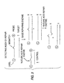
- the 5' region is so designed that it dissociates from the target polynucleotide when the mismatch is cleaved, as shown in side A of FIGURE 2.
- the 5' cleavage fragment then can be extended with polyA polymerase or terminal deoxynucleotidyl transferase (TdT), as shown in side A of FIGURE 2, and detected.
- the tails consist of polymers of adenosines
- the tails will hybridize to polymers of oligonucleotide composed of thymines. If the nucleotides used in this polyA polymerase or TdT reaction are labeled with a radionucleotide or with some other marker, such as a fluorescent label, then the amount of cleavage can be assayed, using these labeled nucleotides as the signal.
- the oligonucleotide probe described in the previous two aspects of the present embodiment can have another feature, a ligand type molecule at the 5' end of the probe oligonucleotide.
- the ligand can serve to label the probe.
- the ligand can be a biotin group, or a group extendable with poly (ADP) ribose polymerase. Additionally, the ligand can be DNP (dinitrophenol) or cholesterol moieties.
- molecules that bind to the ligands can be used to affinity purify the newly formed tails.
- the probes with a biotin ligand can be passed over an avidin or streptavidin column.
- the probes with a poly(ADP)ribose ligand can be passed over a antipoly(ADP)ribose column.
- the DNP and cholesterol moieties can be passed over column with antibody against DNP and cholesterol, respectively.
- Affinity purification techniques, including affinity chromatography are well known to those skilled in the art.
- Yet another embodiment of this invention provides for detecting, identifying, measuring, or localizing, inter alia, known sequences in a target polynucleotide in a biological sample.
- This aspect of the invention includes hybridizing a single-stranded oligonucleotide probe to a target polynucleotide to form a hybrid double-stranded polynucleotide.
- the probe is designed so that it includes a mismatched or repairable base sequence.
- the probe so designed is not complementary to the target polynucleotide, there is a mismatch between the probe containing a synthesized mutation or mismatch site and the wild-type target polynucleotide.
- the mismatch occurs at the site of a mismatched or repairable base sequence.
- the probe is cleaved at the point of mismatch with a nucleic acid repair enzyme as mentioned above.
- the cleaved polynucleotide fragments are then detected as mentioned above.
- the probe oligonucleotide can be synthesized so that it is complementary to the target polynucleotide sequence except at a single nucleotide, which is chosen to be near the middle of the probe.
- the wild-type probe contains a cytosine base in the middle of the probe that correctly hybridizes to a guanosine base on a wild-type target polynucleotide sequence.
- the mismatched probe can be designed by synthesizing an oligonucleotide probe wherein the cytosine base is replaced by an adenine base.
- the adenine base of the probe oligonucleotide mismatches to a guanosine base in the target molecule. But the rest of the strands of the probe and target are complementary.
- the probe When treated with mutY, the probe are cleaved if it has hybridized to a non-mutated wild-type target polynucleotide.
- the probe containing a mutation will not be cleaved. This is true because a mismatch will not have occurred, the probe adenine being complementary to the target thymine.
- Another aspect of the present embodiment includes two probes, used independently, which contain mismatches at slightly different sites. Cleavage occurring for both probes in separate reactions confirms the presence of the target polynucleotide.
- the present embodiment can utilize helix destabilizing molecules, as discussed above. Additionally, this embodiment can be performed using the oscillating and tailing methods already described. Moreover, this embodiment may employ the linking methods and use of a combination of glycosylases and either AP endocuncleases or AP lyases, as described above.
- the present embodiment is useful in DNA diagnostics and DNA profiling.
- repair index in this regard is defined as the ratio of the amount of cleavage in a sample mixed with base-repairing enzyme (shown in the right side of FIGURE 3) to the amount of cleavage in a sample of control, which contains no base-repairing enzymes (control shown in FIGURE 3 on left side).
- the repair index indicates the extent to which base-repairing enzymes have repaired a sample of mismatched or damaged oligonucleotide probes.
- a "base-repairing enzyme” is one that effects repair of a mismatched probe sequence that has hybridized to a target polynucleotide. The base-repairing enzyme replaces the mismatched base with a base that is complementary to the target polynucleotide. Examples of base-repairing enzymes are E . coli DNA purine transferase (E.C. 2.6.99.1), human O 6 -methylguanine-DNA methyltransferase (Koike et al , J . Biol . Chem . 265: 14754) and E . coli DNA photolyase (EMBL Data Library Accession Number S32737).
- the embodiment depicted in FIGURE 3 involves hybridizing a single-stranded oligonucleotide probe to a target polynucleotide to form a hybrid double-stranded polynucleotide.
- the probe is designed so that it includes a mismatched or a repairable base sequence.
- Such a probe can be designed according to the methods described above.
- probe so designed will not be complementary to the target polynucleotide, there will be a mismatch between the mutated probe and the wild-type target polynucleotide. The mismatch will occur at the site of the mismatched or repairable base sequence.
- the hybrid double-stranded polynucleotide will be exposed to a base-repairing enzyme, which is defined above.
- the base-repairing enzyme will repair a certain amount of the mismatched or repairable base sequences, depending on the type of base-repairing enzymes employed.
- the hybrid double-stranded polynucleotide then will be exposed to a nucleic acid repair enzyme, which cleaves non-repaired probe at the point of mismatch.
- the cleaved nucleotide fragments can then be detected by the methods described above.
- the amount of cleavage in a sample mixed with base-repairing enzyme is compared to the amount of cleavage in a sample of unrepaired control.
- Determining the repair index is useful in determining the potency of cancer therapeutic agents on an individual.
- an oligonucleotide is synthesized with a base that has been modified to contain a base adduct.
- Several base adducts are known to be the result of certain chemotherapeutic drugs. See Friedman et al ., supra , table 5-3.
- Base adducts cause a significant distortion of the DNA helix and are both repairable by base repair enzymes and cleaved by nucleic acid repair enzymes.
- the probe is hybridized with target polynucleotide in a medium containing the base-repairing enzymes of a particular individual, and then the duplex is exposed to nucleic acid repair enzyme. Accordingly, the index of repair indicates the extent to which the individual would repair mutations that would be induced by a particular cancer therapeutic agent.
- the present embodiment can employ . helix destabilizing molecules as described above. Additionally, this embodiment of the invention can be performed using the oscillating and tailing methods described above. Moreover, this embodiment may employ the linking methods and use of a combination of glycosylases, as described above.
- the present invention describe, but does not claim, the modification of a single base in a target polynucleotide.
- This includes hybridizing a single-stranded probe to a circular single-stranded target polynucleotide.
- the probe is constructed so that it contains a single base that will form a mismatch with the target polynucleotide at a predetermined site.
- the probe is designed to contain fewer base sequences than the target polynucleotide so that hybridization results in a partially double-stranded circular nucleic acid molecule (See fig. 5).
- a probe which can be either an oligonucleotide molecule or a peptide nucleic acid molecule.
- the partially double-stranded construct, described above is converted into a completely double-stranded construct by filling in the single-stranded region of the probe-target hybrid (see Fig. 5). This conversion can be accomplished using DNA Polymerase and Ligase according to the methods employed by M. Smith and S. Gilliam (1981), in 3 GENETIC ENGINEERING 1 (J.K. Setlow & A. Hollaender, eds.).
- the partially double-stranded construct is treated with T4 or T7 DNA polymerases and T7 or T4 DNA lygases.
- T4 or T7 DNA polymerases and T7 or T4 DNA lygases are superior because they lack 5' to 3' exonuclease activity. Additionally, these enzymes will not effect strand displacement of the oligonucleotide probe. Accordingly a completely double-stranded construct will be formed containing the original probe base sequences.
- This fully double-stranded molecule is then treated with a nucleic acid repair enzyme which cleaves the target polynucleotide strand at the point of mismatch, creating a gap in the target polynucleotide (see Fig. 5).
- a nucleic acid repair enzyme which cleaves the target polynucleotide strand at the point of mismatch, creating a gap in the target polynucleotide (see Fig. 5).
- the probe-target hybrid is transformed into a bacteria according to techniques well known to those skilled in the art. In the process of transformation, the gap created by cleavage is filled with base(s) complementary to exposed probe bases.
- PNA peptide nucleic acid molecule
- PNA's as contemplated in the instant invention are molecules containing neutral, peptide-like backbones and nucleobases. PNAs allow for the hybridization to RNA and DNA with higher affinity and specificity than conventional oligonucleotides. PNAs are commercially available, for example, from PerSpective Biosystems, Inc. (Framingham, MA), and are described in Wittung et al ., Nature 368, 561-563 (1994); Hanvey, et al ., Science 258: 1481-85 (1992).
- a partially double-stranded probe target construct is formed, as discussed above.
- a nucleic repair enzyme effects cleavage of the target polynucleotide at the point of mismatch. Because of its peptide-like backbone, the PNA probe is impervious to cleavage by a nucleic acid repair enzyme. Accordingly, the PNA probe is advantageous in that it assures that cleavage will not occur in the probe molecule at the point of mismatch. Instead, the nucleic repair enzyme will only be able to effect cleavage in the target polynucleotide.
- the gap resulting from this cleavage can then be filled in with DNA polymerase and ligase in combination with the desired dNTP's (see Fig. 10).
- T4 DNA polymerase, T7 DNA polymerase, T4 DNA ligase, and T7 DNA ligase are preferred enzymes for this reaction.
- other enzymes can be used to fill the gap in the target polynucleotide without causing strand displacement.
- Klenow fragment of DNA polymerase I may be used as described by Kunkel, T.A. Nucleic Acids Mol . Biol . 12: 124 (1988); Proc . Natl . Acad . Sci . U . S . A . 82: 488 (1985).
- the target polynucleotide strand can be separated from the PNA probe by heating the PNA-target hybrid to dissociate the two from each other.
- the temperature for disassociation is approximately 3°C per mer in the PNA probe molecule plus approximately 20°C as described in Wittung et al ., Nature 368, 561-563 (1994).
- the target polynucleotide can then be purified by column size exclusion chromatography (e.g. G-SO) to obtain target that is free of PNA probe molecules.
- the target polynucleotide may then be transformed into a bacteria as a single-stranded circular construct.
- the skilled artisan may obtain a double stranded construct by use of a primer, polymerase, ligase enzymes and dNTP's.
- a double-stranded construct may be advantageous because double-stranded circular polynucleotides generally transform with higher efficiency than single-stranded molecules.
- the enzyme mutY was purified by the method of Wu et al ., Proc . Nat'l Acad . Sci . USA 89: 8779-83 (1992), to a concentration of about 50 units• ⁇ l -1 from an overexpressed E. coli clone. (One unit is defined as the conversion of 1 fmol A/G mismatch synthetic oligonucleotide converted to nicked substrate per minute.) Two synthetic oligodeoxyribonucleotides were synthesized by standard phosphoramidite chemistries with the following sequences:
- the oligonucleotides were purified on 20% polyacrylamide/7M Urea 1XTBE gels to about 95% homogeneity. The fragment was detected by autoradiography and a band cut from the gel. It was eluted from the gel slice by electrophoresis onto NA-45 paper (Schleicher & Schuler, Inc) by band interception, followed by elution from the paper by heating at 65°C for 5 minutes in 2M TEAA (triethylammonium acetate). The sample was dried in a vacuum centrifuge until all solvent was removed.
- Oligonucleotide #1 (Oligo 1) was radiolabeled to high specific activity with about 60 pmols (0.1 mCi; 6000 Ci• mMol -1 ) 32P-gamma-ATP and 100 units of T4 polynucleotide kinase at 37°C for 1 hour. The radiolabeled fragment was further gel purified, as described above.
- oligonucleotide #2 (Oligo 2) was diluted from 1 pmol• ⁇ l -1 to 10 -9 pmol• ⁇ l -1 in sterile deionized water.
- a typical reaction mix was set up that contained about 0.2 pmol of radiolabeled oligo 1, mutY buffer (20 mM Tris-HCl, pH 7.6, 80 mM NaCl, 1 mM dithioerythritol, 1 mM EDTA, pH 8.0, 3% glycerol; final concentration), 50 units of mutY enzyme, some reactions contained a dilution of Oligo 2 from 1 to 10 -9 pmol. The final volume was 10 ⁇ l. The reaction proceeded at 37°C for 60 minutes.
- the reaction was stopped by the addition of 1 ⁇ l of loading buffer (98% Formamide, 10 mM EDTA, pH 8.0, 0.025% Xylene Cyanol FF, 0.025% Bromophenol Blue).
- loading buffer 98% Formamide, 10 mM EDTA, pH 8.0, 0.025% Xylene Cyanol FF, 0.025% Bromophenol Blue.
- the reaction was electrophoresed on a 20% polyacrylamide/7M Urea 1X TBE gel, then autoradiographed.
- Lane 1 contains no target molecule.
- Lanes 2 to 11 contain 10°, 10 -1 , 10 -2 , 10 -3 , 10 -4 , 10 -5 , 10 -6 , 10 -7 , 10 -8 , 10 -9 pmol of target.
- the cleavage from the reaction was detected by the generation of a smaller cleaved synthetic oligonucleotide fragment found in lanes 2 to 4. The truncated shorter molecule was due
- the nucleic acid repair enzyme mutY and the helix destabilization enzyme *I were used along with three synthetic oligonucleotides having the sequences: Probes 2 and 3 are complementary. Probes 1 and 3 are complementary with a single base mismatch. The three DNA synthetic oligonucleotides were used as is or were radiolabeled with 32 P- ⁇ -ATP via T4 polynucleotide kinase.
- Reactions were set up as indicated below with combinations of radiolabeled or cold oligonucleotides 1, 2 and 3, with or without mutY and or *I.
- Lanes 1 to 20 contained radiolabeled oligonucleotide 1.
- Lanes 3 and 4 contained radiolabeled oligonucleotide 2.
- Lanes 5 to 7 contained radiolabeled oligonucleotide 3.
- Lane 6 contained 10 pmols of cold oligonucleotide 1.
- Lane 7 contained 10 pmols of cold oligonucleotide 2.
- Lanes 2, 4, 9 and 16 contained 10 pmols of cold oligonucleotide 3.
- Lanes 2, 4,6 to 20 contained mutY enzyme.
- Lanes 15 to 20 contained *I. The reactions were setup with standard mutY reaction buffer and allowed to proceed for 1 hour at 37°C. The reactions were stopped, loading dye was added, and the material was run on a 20% polyacrylamide/7 M Urea gel (1XTBE).
- a synthetic oligonucleotide of length N nucleotides has a temperature of hybridization (melting temperature or Im, in °C) equal to 3N.
- a synthetic oligonucleotide of 20 nucleotides has a temperature of hybridization of about 60°C.
- a shorter synthetic oligonucleotide of 10 nucleotides has a temperature of hybridization of about 30°C.
- the long 20-mer synthetic oligonucleotide hybridizes but the shorter 10-mer synthetic oligonucleotide does not.
- the longer synthetic oligonucleotide hybridizes to a target sequence, then when it is cleaved at a specific place where a mismatch occurs two or more shorter synthetic oligonucleotides are be generated, which have lower thermodynamic stability. These shortened cleaved oligonucleotides dissociate from the target sequence, making it available for another hybridization event with the longer synthetic oligonucleotide.
- the hybridization kinetics favors better, faster hybridization. Further, this allows the detection of a hybridization event by the accumulation of the shortened oligonucleotide fragments.
- a helix destabilizing molecule can also be added to the system of Example 3.
- the operating temperature is 45°C and is kept at that temperature for this example.
- the longer 20-mer synthetic oligonucleotide hybridizes not at 60°C, but rather at a lower temperature, for example, 48°C.
- the hybridization temperature and the operating temperature are closer. This allows in one scenario the reduction of the operating temperature to a lower temperature, perhaps 37°C.
- mesophilic enzymes can be used in the reaction.
- the practice of this example at such a temperature is advantageous in that it does not require thermophilic enzymes (those that function at high temperature extremes, e.g. 60 - 70°C, and can withstand near boiling temperatures).
- in vitro mutagenesis For modifying and generating new genetic sequences, in vitro mutagenesis is used.
- the earliest method of in vitro mutagenesis is that of M. Smith and S. Gilliam (1981), in 3 GENETIC ENGINEERING 1 (J.K. Setlow & A. Hollaender, eds.).
- the nucleic acid repair enzymes can be used for oligonucleotide directed in vitro mutagenesis.
- the mutY is used as the example nucleic acid repair enzyme, however, other enzymes can also be used.
- Single stranded DNA containing the cloned gene sequence to be modified can be generated by the method of Zinder & Boeke, Gene 19: 1-10 (1982), using the M13 bacteriophage system.
- a synthetic oligonucleotide is designed, which is complementary to the region to be modified with a Guanine mismatch at a position where the adenine is to be changed to a Cytosine (figure 5).
- the oligonucleotide is hybridized to the single stranded DNA, treated with T4 DNA polymerase and T4 DNA ligase by methods used by Smith (1981).
- the newly synthesized double stranded molecule with a single mismatch is then treated with 50 units of mutY in mutY buffer (20 mM Tris-HCl, pH 7.6, 80 mM NaCl, 1 mM dithioerythritol, 1 mM EDTA, pH 8.0) for one hour at 37°C.
- the material that is cleaved at the mismatch site is transformed into E . coli using methods well known to those skilled in the art.
- the transformants then are enriched for sequences containing the conversion of the adenine to the cytosine base pair.
- a synthetic oligonucleotide complementary to a region of the p53 gene is synthesized using standard phosphoramidite chemistry.
- Purified mutant E . coli mutY mismatch enzyme (with associated AP endonuclease) is linked to the 5' end of the oligonucleotide with linker using methods already described (Corey et al ., 1987, Science, vol 238: 1401; Corey et al ., 1989, Journal of the American Chemical Society, vol 111:8523). This forms a covalent linkage of an oligonucleotide of specific sequence, with a linker region covalently linked to E . coli mutY mismatch enzyme.
- oligonucleotide is radiolabeled at the 3' end using poly (A) polymerase and 32 P-cordycepin.
- poly (A) polymerase poly (A) polymerase
- 32 P-cordycepin 32 P-cordycepin
- chromosomal DNA prepared from ML-1 myeloid leukemia cells
- dilutions of the target DNA from 10 -1 to 10 -8 pmol are setup.
- 10 pmols of radiolabeled probe 100 ng of purified E . coli mutY mismatch enzyme for the unlinked oligonucleotide and buffer are added. The reaction was allowed to precede for 30 minutes at 37°C.
- the reaction product is then electrophoresed on a 20% polyacrylamide/7M urea gel, then autoradiographed.
- the generation of shortened cleaved oligonucleotides indicates the presence of a DNA mismatch.
- the covalently linked enzyme is expected to show enhanced activity over unlinked enzyme, as expected from previous data (Bruice et al ., 1963, Journal of American Chemical Society, vol 85:1).
- a first synthetic oligonucleotide probe complementary to a region of the p53 gene is synthesized using standard phosphoramidite chemistry.
- Purified E . coli G/T mismatch enzyme is linked to the 5' end of the oligonucleotide with a linker using methods already described (Corey et al ., 1987, Science, vol 238:1401; Corey et al ., 1989, Journal of the American Chemical Society, vol 111:8523). This forms a covalent linkage of an oligonucleotide of specific sequence, with a linker region covalently linked to E . coli G/T mismatch enzyme.
- a second oligonucleotide is synthesized, where the sequence is near to the first oligonucleotide.
- This second oligonucleotide is synthesized as above with a linker which is attached to AP endonuclease (see FIGURE 6a).
- the first oligonucleotide is radiolabeled at the 3' end using poly (A) polymerase and 32 P-cordycepin.
- oligonucleotides without enzymes are prepared and treated alongside with the enzyme linked oligonucleotides.
- chromosomal DNA prepared from ML-1 myeloid leukemia cells
- dilutions of the target DNA from 10 -1 to 10 -8 pmol are setup.
- 10 pmols of radiolabeled probe 100 ng of purified E . coli G/T mismatch enzyme and 100 ng of AP endonuclease for the unlinked oligonucleotide and buffer are added. The reaction are allowed to proceed for 30 minutes at 37°C.
- the reaction product is then electrophoresed on a 20% polyacrylamide/7M urea gel, then autoradiographed.
- the generation of shortened cleaved oligonucleotides indicates the presence of a DNA mismatch.
- the covalently linked enzyme is expected to show enhanced activity over unlinked enzyme, as expected from previous data (Bruice et al ., 1963, Journal of American Chemical Society, vol 85:1).
- a synthetic oligonucleotide (first) complementary to a region of the p53 gene is synthesized using standard phosphoramidite chemistry.
- Purified E. coli G/T mismatch enzyme is linked to the 3' end of the first oligonucleotide with a linker using methods already described (Corey et al ., 1987 Science, vol 238:1401; Corey et al ., 1989, Journal of the American Chemical Society, vol 111:8523). This forms a covalent linkage of an oligonucleotide of specific sequence, with a linker region covalently linked to E . coli G/T mismatch enzyme.
- a second oligonucleotide is synthesized, where the sequence is near to the first oligonucleotide but allows a space at the 3' end where a mutation probe can hybridize.
- This second oligonucleotide is synthesized as above with a linker which is attached to AP endonuclease.
- a third mutation probe is synthesized and radiolabeled at the 5' end using polynucleotide kinase and 32 P- ⁇ -ATP (see FIGURE 7).
- oligonucleotides without enzymes attached are prepared and treated alongside with the enzyme linked oligonucleotides.
- chromosomal DNA prepared from ML-1 myeloid leukemia cells
- dilutions of the target DNA from 10 -1 to 10 -8 pmol are setup.
- 10 pmols of radiolabeled probe 100 ng of purified E . coli G/T mismatch enzyme and 100 ng of AP endonuclease for the unlinked oligonucleotide and buffer are added. The reaction is allowed to proceed for 30 minutes at 37°C.
- the reaction product is then electrophoresed on a 20% polyacrylamide/7M urea gel, then autoradiographed.
- the generation of shortened cleaved oligonucleotides indicates the presence of a DNA mismatch.
- the covalently linked enzyme is expected to show enhanced activity over unlinked enzyme, as expected from previous data (Bruice et al., 1963, Journal of American Chemical Society, vol 85:1).
- a synthetic oligonucleotide complementary to a region of the p53 gene is synthesized using standard phosphoramidite chemistry.
- Purified E . coli G/T mismatch enzyme is linked to the 5' end of the oligonucleotide with a linker using methods already described (Corey et al ., 1987, Science, vol 238:1401; Corey et al ., 1989, Journal of the American Chemical Society, vol 111:8523). This forms a covalent linkage of an oligonucleotide of specific sequence, with a linker region covalently linked to E . coli G/T mismatch enzyme.
- AP endonuclease (see FIGURE 8) is attached to the same oligonucleotide at the 3' end.
- oligonucleotides without enzymes are prepared and treated alongside with the enzyme linked oligonucleotides.
- chromosomal DNA prepared from ML-1 myeloid leukemia cells
- dilutions of the target DNA from 10 -1 to 10 -8 pmol are setup.
- 10 pmols of radiolabeled probe 100 ng of purified E . coli G/T mismatch enzyme and 100 ng of AP endonuclease for the unlinked oligonucleotide and buffer are added. The reaction is allowed to proceed for 30 minutes at 37°C.
- the reaction product is then electrophoresed on a 50 ⁇ m i.d. x 35.5 cm Supelco H-75 methyl coated borosilicate capillary containing TreviSolTM CE B (Trevigen, Inc.). 40 ⁇ l of the reaction product is applied electrokinetically to the capillary, then electrophoresed at -140 V/cm for about 12 minutes.
- the synthetic oligonucleotides are detected by UV detection.
- the generation of shortened cleaved oligonucleotides indicates the presence of a DNA mismatch.
- the covalently linked enzyme is expected to show enhanced activity over unlinked enzyme, as expected from previous data (Bruice et al ., 1963, Journal of American Chemical Society, vol 85:1).
- a synthetic oligonucleotide complementary to a region of the p53 gene is synthesized using standard phosphoramidite chemistry.
- Purified E . coli G/T mismatch enzyme is linked to the 5' end of the oligonucleotide with a linker using methods already described (Corey et al ., 1987, Science, vol 238: 1401; Corey et al ., 1989, Journal of the American Chemical Society, vol 111:8523). This forms a covalent linkage of an oligonucleotide of specific sequence, with a linker region covalently linked to E. coli G/T mismatch enzyme.
- oligonucleotide is radiolabeled at the 3' end using poly (A) polymerase and 32 P-cordycepin.
- poly (A) polymerase poly (A) polymerase
- 32 P-cordycepin 32 P-cordycepin
- chromosomal DNA prepared from ML-1 myeloid leukemia cells
- dilutions of the target DNA from 10 -1 to 10 -8 pmol are setup.
- 10 pmols of radiolabeled probe 100 ng of purified E . coli G/T mismatch enzyme for the unlinked oligonucleotide and buffer are added.
- the reaction is allowed to proceed for 30 with the temperature being cycled from about 30° to 42°C every two minutes to allow the probes with AP sites to fall off of the target molecule and new un-treated probes to hybridize to the target molecule.
- This oscillating process is known as the Mismatch Chain Reaction (MCR).
- MCR Mismatch Chain Reaction
- the AP cleavage is effected by treatment of the samples at high pH (adjusting the pH to a range of 8 to 14) with NaOH and higher temperatures (adjusting the temperature from a temperature range of 50°C to 95°C). This effects AP cleavage.
- the reaction product is then electrophoresed on a 20% polyacrylamide/7M urea gel, then autoradiographed.
- the generation of shortened cleaved oligonucleotides indicates the presence of a DNA mismatch.
- the covalently linked enzyme is expected to show enhanced activity over unlinked enzyme, as expected from previous data (Bruice et al ., 1963, Journal of American Chemical Society, vol 85:1).
- a synthetic oligonucleotide sequence complementary to a region of the p53 gene is synthesized using standard phosphoramidite chemistry. Different specific syntheses of this sequence are made with the attachment of different fluorophores (fluorescein (488 nm), rhodamine (546 nm), coumarin (365 nm)) at the 3' end of the molecule. At different wavelengths, these different fluorescent molecules can be differentiated from each other. With a linker intermediate, three different glycosylases are specifically added to three different oligonucleotide-fluorophore molecules, respectively.
- the three glycosylases are mutY (A/G and A/C mispairs), Thymine mismatch (also called G/T mismatch) and All type enzyme (all mispairs).
- the glycosylase/linker attachment is done using methods already described (Corey et al., 1987, Science, vol 238:1401; Corey et al., 1989, Journal of the American Chemical Society, vol 111:8523).
- chromosomal DNA prepared from HL-60 myeloid leukemia cells
- dilutions of the target DNA from 10 -1 to 10 -8 pmol are setup.
- 10 pmols of radiolabeled probe 100 ng of AP endonuclease are added. The reaction was allowed to proceed for 30 minutes at 37°C.
- reaction products are then electrophoresed on a 50 ⁇ m i.d. x 35.5 cm H-75 methyl coated borosilicate capillary containing TreviSolTM CE B (Trevigen, Inc.) capillary matrix.
- 40 ⁇ l of the reaction product is applied electrokinetically to the capillary, then electrophoresed at -140 V/cm for about 12 minutes.
- the synthetic oligonucleotides are detected by a UV/VIS detector.
- the generation of shortened cleaved oligonucleotides indicates the presence of DNA mismatch.
- the exact form of the mismatch is identified from the specific fluorophore indicating the respective glycosylase used in the reaction.
- both forms of DNA could be present. This technique would allow differentiating between the relative amount of each form of DNA present by measuring the amounts of both forms of the oligonucleotides.
- a synthetic oligonucleotide sequence complementary to a region of the p53 gene is synthesized using standard phosphoramidite chemistry.
- the mismatch or no mismatch
- Different specific syntheses of this sequence are made which have slightly longer lengths at the 3' end of the molecule and where the specific base with the mismatch is synthesized as a different base at that position (e.g. one oligo is A, another oligo is C, and the final oligo is G at one base in the oligonucleotide.
- the oligonucleotides are 30, 32 and 34 nucleotides in length.
- glycosylases are specifically added to the 5' end of three different oligonucleotide molecules of different lengths, respectively.
- the three glycosylases were mutY (A/G and A/C mispairs), Thymine mismatch (also called G/T mismatch) and All type enzyme (all mispairs).
- the glycosylase/linker attachment is done using methods already described (Corey et al., 1987, Science, vol 238:1401; Corey et al., 1989, Journal of the American Chemical Society, vol 111:8523).
- the oligonucleotides are labeled with 32 P-labelled cordycepin and poly (A) polymerase at the 3' end.
- chromosomal DNA prepared from HL-60 myeloid leukemia cells
- dilutions of the target DNA from 10 -1 to 10 -8 pmol are setup.
- 10 pmols of different radiolabeled probe are added to the target DNA, and 100 ng of AP endonuclease is added to each tube. The reaction is allowed to proceed for 30 minutes at 37°C.
- reaction products are then electrophoresed on a 20% polyacrylamide/7 M urea gel.
- the presence of cleaved fragments in the autoradiograph indicates the presence of a mismatch.
- the length of the cleaved molecule indicates which specific mismatch was present.
- both forms of DNA could be present. This technique would allow differentiating between the relative amount of each form of DNA present by measuring the amounts of both forms of the oligonucleotides.
- Escherichia coli JM109 is available from New England Biolabs of Beverly, MA and Escherichia coli BW415 is available form the laboratory of Dr. Richard P. Cunningham at the State University of New York, at Albany, Department of Biological Sciences.
- a similar strain suitable for this protocol is BW434 and it is available from the Coli Genetic Stock Center at Yale University School of Medicine, New Haven, CT.
- BW415 ⁇ DE3 was made with a ⁇ DE3 lysogenization kit from Novagen Inc. of Madison, WI. This integration allowed for the efficient expression of the T/G mismatch specific thymine-DNA glycosylase from a T7 RNA polymerase driven promoter in an endonuclease III deficient strain of Escherichia coli .
- the expression system was contained on plasmid pET14B from Novagen Inc.
- Plasmid pUV2 containing the orf 10 coding sequence is available from Dr. Jork Nolling, Wageningen Agricultrual University of the Netherlands, Department of Microbiology, Hesselink van Suchtelenweg 4, 6703 CT Wageningen, The Netherlands.
- the pUV2 plasmid contains a portion of pFV1, including the orf 10 coding sequence, cloned into pUC19.
- Plasmid pUC19 is available from Sigma Chemical Co., of St. Louis, MO.
- Plasmid DNA was prepared from JM109 by a modified alkaline lysis method.
- the orf 10 coding sequence from pUV2 was cloned into pUC19 via a DraI-EcoRI to HincII-EcoRI ligation using restriction endonucleases and ligase (from New England Bio Labs) according to suppliers' recommendations.
- the gene was PCR mutagenized using Taq Polymerase from Perkin Elmer Corp.
- the cell suspension was sonicated 5 X 3 minutes with a Branson sonifer, and then stirred on ice for one hour. The sonicate was centrifuged at 48,000X g for 20 minutes and the supernatant was retained. Five percent polyethelenimine was added to a final concentration of 0.1% and the suspension was stirred for 1 hour and centrifuged at 48,000X g to give a supernatant with a volume of 500 mls. The supernatant was dialyzed against 4 L of 50 mM KPO 4 pH 7.2 overnight.
- the crude extract was loaded onto an 80 ml SP Sepharose Fast Flow (Pharmacia Biotech Inc. of Piscataway, NJ) column, washed with 100 ml of 50mM KPO 4 pH 7.2 and eluted with a 1 L gradient from 0 to 1M NaCl at a flow rate of 10 ml/min.
- the protein eluted at 0.6M NaCl as detected by 31 kD band visualized by SDS-PAGE, and a characteristic yellow color in the fractions indicative of an Fe-S cluster of the protein.
- the fractions containing the protein were pooled and dialyzed against 50 mM KPO 4 pH 6.6, loaded onto a 5 ml DNA agarose (Pharmacia Biotech Inc.) column, washed with 10 ml of 50 mM KPO 4 and eluted with a 100ml gradient of 0-1 M NaCl at a rate of 1 ml/min. The protein eluted at 0.8M NaCl as determined by the criteria above. Fractions containing the protein were pooled and dialyzed overnight against 50 mM KPO 4 pH 6.6. The protein was loaded onto a 5ml SP high trap column (Pharmacia Biotech Inc.) and eluted with 1M NaCl to concentrate the protein.
- the extract was further concentrated to 1.5 mls with Centriprep 10 concentrators (Amicon Division, W.R. Grace & Co., of Danvers, MA). At this point the protein was a single band on an overloaded Coomassie stained gel and gave an A 410 /A 280 ratio of 0.295. This protein was active in our T-G mismatch assay. The pure protein was stored in 50% glycerol at -20 C.
- Oligonucleotide A was labeled with 32 P ATP from New England Nuclear of Boston, MA using T4 Polynucleotide kinase from New England Bio Labs. The two oligonucleotides were annealed at a three to one excess of B to A on ice for one hour to generate a duplex oligonucleotide with a unique T/G mismatch.
- Glycosylase activity was monitored by measuring DNA strand breakage.
- 32 P labeled substrate was incubated with T/G mismatch specific thymine-DNA glycosylase protein in 50 mM HEPES-KOH pH 7.5, 100 mM KCl and 1mM EDTA at 65°C for 45 minutes.
- the enzyme was inactivated with 5M urea at 100°C for 5 minutes and then the reaction mixture was incubated in 0.15 M NaOH for 15 minutes at 55°C to create strand breaks at the site of AP sites.
- Loading buffer was added to the mixture and then run on a 20% acrylamide gel containing 8M urea for 2 hours at 200 volts. The gels were dried and analyzed on X-ray film.
- the enzyme showed a glycosylase activity against both thymine and uracil mismatched with guanine.
- the activity on each substrate was at roughly the same level of magnitude, with a mared decrease in activity against thymines mismatched with cytosine.
- Temperature assays were run in the same manner using a labeled T-G mismatch, but incubation temperatures were 25,35,45,55,65, and 75°C. Peak activity was identified at 65°C (92.4% cleavage), with 85.9% cleavage seen at 75°C, 90.6% cleavage seen at 55°C, 84.5% cleavage seen at 45°C, 70.2% cleavage seen at 35°C and 36% cleavage seen at 25°C.
- the enzyme was active after being boiled in the absence of 5M urea.
- Oligonucleotide RO563 is complementary to both RO561 and RO562, however the underlined base G in RO562 does not complement the T in RO563, forming a mismatch.
- the length and composition of these synthetic oligonucleotides were chosen to have a hybridization temperature around 70°C The sequences were based on the human p53 gene, which has a mutation at the underlined G position found in the RO562 sequence.
- thermostable mismatch glycosylase made according to Example 14 in a volume of 10 ⁇ l.
- the reaction was allowed to proceed for about 3 hours at 60°C.
- the reaction was loaded onto a 20% acrylamide/7 M Urea/1X TBE gel and electrophoresed at about 300 V for about 3 hours. The gel was then electrophoresed for different lengths of time.
- thermostable enzyme at elevated temperatures resulted in a level of cleavage significantly higher than that found with a mesophilic enzyme. This increased level of cleavage results in a more sensitive assay than one carried out at lower temperatures. For instance, the autoradiograph of the above experiment after a brief 3 hour exposure showed faint cleavage at 0.000001. Longer autoradiographic exposure showed further sensitivity.
- Probe may be present in the assay in concentrations from 1 fmol to 100 pmol.
- the probe need not be radioactively labelled, and may be labelled by other means as are known to those of skill in the art.
- concentration of the thermostable enzyme may be present from approximately 1 to 100 ng, and finally, the buffer composition can have higher concentrations of KCl from 50 mM to 100 mM.
Landscapes
- Chemical & Material Sciences (AREA)
- Life Sciences & Earth Sciences (AREA)
- Organic Chemistry (AREA)
- Health & Medical Sciences (AREA)
- Engineering & Computer Science (AREA)
- Wood Science & Technology (AREA)
- Zoology (AREA)
- Proteomics, Peptides & Aminoacids (AREA)
- Genetics & Genomics (AREA)
- General Engineering & Computer Science (AREA)
- Biotechnology (AREA)
- Bioinformatics & Cheminformatics (AREA)
- General Health & Medical Sciences (AREA)
- Biophysics (AREA)
- Physics & Mathematics (AREA)
- Biochemistry (AREA)
- Molecular Biology (AREA)
- Microbiology (AREA)
- Immunology (AREA)
- Analytical Chemistry (AREA)
- Biomedical Technology (AREA)
- Crystallography & Structural Chemistry (AREA)
- Plant Pathology (AREA)
- Chemical Kinetics & Catalysis (AREA)
- Measuring Or Testing Involving Enzymes Or Micro-Organisms (AREA)
- Enzymes And Modification Thereof (AREA)
Abstract
Claims (16)
- Procédé de détection d'une mutation ponctuelle dans un polynucléotide cible, comprenant les étapes suivantes :(a) l'hybridation d'une sonde d'oligonucléotide à simple brin, dans des conditions strictes, sur ledit polynucléotide cible pour former un polynucléotide hybride à deux brins de sorte qu'un mésappariement se produise sur le site de ladite mutation ponctuelle, dans lequel ladite sonde est complémentaire d'une séquence non mutée dudit polynucléotide cible;(b) le clivage dudit brin de sonde dudit polynucléotide hybride audit point de mésappariement à une température prédéterminée avec une enzyme de réparation d'acide nucléique, produisant des fragments d'oligonucléotide qui se dissocient spontanément dudit polynucléotide cible à ladite température prédéterminée;(c) la reproduction des étapes (a) et (b);(d) la détection desdits fragments d'oligonucléotide; et, par suite,(e) la détection de ladite mutation ponctuelle.
- Procédé de détection d'une séquence dans un polynucléotide cible, comprenant les étapes suivantes :(a) l'hybridation d'une sonde d'oligonucléotide à simple brin, dans des conditions strictes, sur ledit polynucléotide cible pour former un polynucléotide hybride à deux brins, dans lequel ladite sonde contient une séquence de bases mésappariée ou réparable de sorte qu'un mésappariement se produise sur le site de ladite séquence de bases mésappariée ou réparable;(b) le clivage dudit brin de sonde dudit polynucléotide hybride audit point de mésappariement à une température prédéterminée avec une enzyme de réparation d'acide nucléique, produisant des fragments d'oligonucléotide qui se dissocient spontanément dudit polynucléotide cible à ladite température prédéterminée;(c) la reproduction des étapes (a) et (b);(d) la détection desdits fragments d'oligonucléotide produits par ledit clivage; et, par suite,(e) la détection de ladite séquence dans ledit polynucléotide cible.
- Procédé de détection d'une mutation ponctuelle dans un polynucléotide cible, comprenant les étapes suivantes :(a) l'hybridation d'au moins deux sondes d'oligonucléotides à simple brin, dans des conditions strictes, sur au moins deux polynucléotides cibles pour former au moins deux polynucléotides hybrides à double brin de sorte qu'un mésappariement se produise sur le site de ladite mutation ponctuelle, dans lequel lesdites sondes sont complémentaires d'une séquence non mutée desdits polynucléotides cibles, et dans lequel est fixée à chaque sonde une glycosylase différente, dont une seule effectuera le clivage;(b) la production de fragments de sonde en clivant l'un desdits brins de sonde audit point de mésappariement, dans lequel ledit clivage est effectué par une desdites glycosylases et une enzyme de clivage AP; et(c) la détection desdits fragments de sonde.
- Procédé selon la revendication 3, dans lequel un fluorogène différent est fixé à chacune desdites sondes.
- Procédé selon la revendication 3, dans lequel chacune desdites sondes est d'une longueur différente.
- Procédé selon la revendication 3, dans lequel, à l'étape (a), une sonde d'oligonucléotide avec une enzyme de clivage AP fixée s'hybride sur lesdits polynucléotides cibles sur un site adjacent auxdites sondes fixées aux glycolases.
- Procédé de détermination d'un indice de réparation pour une sonde d'oligonucléotide mésappariée ou endommagée, comprenant les étapes suivantes :(a) l'hybridation d'une sonde d'oligonucléotide de synthèse à simple brin, dans des conditions strictes, sur ledit polynucléotide cible pour former un polynucléotide hybride à double brin, dans lequel ladite sonde contient une séquence de bases mésappariée ou réparable de sorte qu'un mésappariement se produise sur le site de ladite séquence de bases mésappariée ou réparable; ensuite,(b) l'exposition dudit polynucléotide à double brin à une enzyme de réparation de bases, de sorte qu'une réparation moindre qu'une réparation complète de ladite séquence de bases mésappariée ou réparable soit effectuée, laissant une sonde réparée et une sonde non réparée;(c) le clivage de ladite sonde non réparée sur ledit site avec une enzyme de réparation d'acide nucléique;(d) la détection de fragments de polynucléotides produits par ledit clivage; et, par suite,(e) la détermination dudit indice de réparation.
- Procédé selon l'une quelconque des revendications 1, 2 et 3, dans lequel ledit clivage de l'étape (b) est effectué par une glycosylase thermostable.
- Procédé selon la revendication 8, dans lequel ladite glycosylase est codée par la séquence d'ADN de la Fig. 11.
- Procédé selon l'une quelconque des revendications 1, 2 et 6, dans lequel ledit clivage de l'étape (b) est effectué par une enzyme du groupe constitué de la mutY, de l'enzyme de césure spécifique au mésappariement T/G, d'enzymes humaines ou d'enzymes de levure de tous types et de la désoxyinosine 3'-endonucléase de E. coli.
- Procédé selon la revendication 10, dans lequel ladite enzyme est combinée avec une lyase d'ADN ou une endonucléase AP d'ADN.
- Procédé selon la revendication 1 ou 2, dans lequel ledit clivage de l'étape (b) est effectué par une glycolase fixée à ladite sonde et une enzyme de clivage AP.
- Procédé selon la revendication 12, dans lequel ladite enzyme de clivage AP est fixée à ladite sonde.
- Procédé selon la revendication 1 ou 2, dans lequel, à l'étape (a), une deuxième sonde d'oligonucléotide est hybridée sur ledit polynucléotide cible sur un site adjacent à ladite première sonde, et dans lequel ledit clivage de l'étape (b) est effectué par une glycosylase fixée à ladite première sonde et une enzyme de clivage AP fixée à ladite deuxième sonde.
- Procédé selon la revendication 1 ou 2, dans lequel :(i) à l'étape (a), une deuxième et une troisième sonde d'oligonucléotide à simple brin sont hybridées sur ladite polynucléotide cible sur un site adjacent à ladite première sonde et dans lequel une glycosylase est fixée sur ladite deuxième sonde à simple brin et une enzyme de clivage AP est fixée sur ladite troisième sonde à simple brin; et(ii) dans lequel ledit clivage de l'étape (b) est effectué par ladite glycosylase et lesdites enzymes de clivage AP.
- Procédé selon la revendication 1 ou 2, dans lequel ledit clivage de l'étape (b) est effectué par une glycosylase employée en combinaison avec des conditions de base et une température plus élevée.
Applications Claiming Priority (5)
| Application Number | Priority Date | Filing Date | Title |
|---|---|---|---|
| US483089 | 1995-06-07 | ||
| US08/483,089 US5656430A (en) | 1995-06-07 | 1995-06-07 | Oscillating signal amplifier for nucleic acid detection |
| US1295096P | 1996-03-06 | 1996-03-06 | |
| US12950P | 1996-03-06 | ||
| PCT/US1996/008694 WO1996040902A1 (fr) | 1995-06-07 | 1996-06-07 | Procede dans lequel une enzyme de reparation d'acide nucleique est utilisee pour la detection de mutation ponctuelle et une mutagenese in vitro |
Publications (2)
| Publication Number | Publication Date |
|---|---|
| EP0832210A1 EP0832210A1 (fr) | 1998-04-01 |
| EP0832210B1 true EP0832210B1 (fr) | 2004-10-27 |
Family
ID=26684228
Family Applications (1)
| Application Number | Title | Priority Date | Filing Date |
|---|---|---|---|
| EP96921269A Expired - Lifetime EP0832210B1 (fr) | 1995-06-07 | 1996-06-07 | Procede dans lequel une enzyme de reparation d'acide nucleique est utilisee pour la detection de mutation ponctuelle et une mutagenese in vitro |
Country Status (5)
| Country | Link |
|---|---|
| EP (1) | EP0832210B1 (fr) |
| JP (1) | JPH11506932A (fr) |
| AU (1) | AU6252796A (fr) |
| DE (1) | DE69633723T2 (fr) |
| WO (1) | WO1996040902A1 (fr) |
Families Citing this family (14)
| Publication number | Priority date | Publication date | Assignee | Title |
|---|---|---|---|---|
| WO1998046793A1 (fr) * | 1997-04-16 | 1998-10-22 | Trevigen, Inc. | Detection et cartographie de mutations ponctuelles au moyen d'une digestion partielle |
| US6969584B2 (en) | 1997-06-12 | 2005-11-29 | Rigel Pharmaceuticals, Inc. | Combinatorial enzymatic complexes |
| WO1999042595A1 (fr) * | 1998-02-19 | 1999-08-26 | Trevigen, Inc. | Enzymes de clivage incompatibles provenant de microorganismes extremement thermophiles et leurs utilisations |
| US6403309B1 (en) * | 1999-03-19 | 2002-06-11 | Valigen (Us), Inc. | Methods for detection of nucleic acid polymorphisms using peptide-labeled oligonucleotides and antibody arrays |
| WO2001036679A2 (fr) * | 1999-11-15 | 2001-05-25 | Hartwell John G | METHODES DE GENERATION DE FRAGMENTS D'ADNc SIMPLE BRIN |
| CA2332729A1 (fr) | 2000-02-17 | 2001-08-17 | Amsterdam Support Diagnostics B.V. | Reduction du signal de fond dans des reactions d'hybridation |
| EP1130119A1 (fr) * | 2000-02-17 | 2001-09-05 | Amsterdam Support Diagnostics B.V. | Réduction de signaux de fond dans des réactions d'hybridisation |
| US7582423B2 (en) | 2001-02-02 | 2009-09-01 | Novici Biotech Llc | Population of polynucleotide sequence variants |
| CA2436214C (fr) | 2001-02-02 | 2012-12-04 | Large Scale Biology Corporation | Procede destine a ameliorer la complementarite d'un heteroduplex |
| US7838219B2 (en) | 2001-02-02 | 2010-11-23 | Novici Biotech Llc | Method of increasing complementarity in a heteroduplex |
| US7078211B2 (en) | 2002-02-01 | 2006-07-18 | Large Scale Biology Corporation | Nucleic acid molecules encoding endonucleases and methods of use thereof |
| US20120076871A1 (en) | 2010-09-24 | 2012-03-29 | Genomic Vision Sa | Method for detecting, quantifying and mapping damage and/or repair of dna strands |
| JP6009649B2 (ja) * | 2013-03-14 | 2016-10-19 | タカラバイオ株式会社 | 耐熱性のミスマッチエンドヌクレアーゼの利用方法 |
| WO2023196499A1 (fr) * | 2022-04-07 | 2023-10-12 | Twist Bioscience Corporation | Clivage de substrat pour la synthèse d'acides nucléiques |
Family Cites Families (4)
| Publication number | Priority date | Publication date | Assignee | Title |
|---|---|---|---|---|
| US5556750A (en) * | 1989-05-12 | 1996-09-17 | Duke University | Methods and kits for fractionating a population of DNA molecules based on the presence or absence of a base-pair mismatch utilizing mismatch repair systems |
| AU3919293A (en) * | 1992-03-27 | 1993-11-08 | University Of Maryland At Baltimore | Detection of gene mutations with mismatch repair enzymes |
| FR2709761B1 (fr) * | 1993-09-10 | 1995-11-24 | Pasteur Institut | Procédé de détection de molécules contenant des mésappariements nucléotidiques et de localisation de ces mésappariements, et application à la détection de substitutions ou de délétions de bases . |
| WO1995014106A2 (fr) * | 1993-11-17 | 1995-05-26 | Id Biomedical Corporation | Detection cyclique par scission de sondes de sequences d'acides nucleiques |
-
1996
- 1996-06-07 DE DE69633723T patent/DE69633723T2/de not_active Expired - Fee Related
- 1996-06-07 AU AU62527/96A patent/AU6252796A/en not_active Abandoned
- 1996-06-07 JP JP9501202A patent/JPH11506932A/ja not_active Ceased
- 1996-06-07 EP EP96921269A patent/EP0832210B1/fr not_active Expired - Lifetime
- 1996-06-07 WO PCT/US1996/008694 patent/WO1996040902A1/fr not_active Ceased
Also Published As
| Publication number | Publication date |
|---|---|
| WO1996040902A1 (fr) | 1996-12-19 |
| AU6252796A (en) | 1996-12-30 |
| JPH11506932A (ja) | 1999-06-22 |
| DE69633723D1 (de) | 2004-12-02 |
| EP0832210A1 (fr) | 1998-04-01 |
| DE69633723T2 (de) | 2006-02-02 |
Similar Documents
| Publication | Publication Date | Title |
|---|---|---|
| US5763178A (en) | Oscillating signal amplifier for nucleic acid detection | |
| US5656430A (en) | Oscillating signal amplifier for nucleic acid detection | |
| CA2320666C (fr) | Nucleases 5' derivees d'adn polymerase thermostable | |
| US5614402A (en) | 5' nucleases derived from thermostable DNA polymerase | |
| US5660988A (en) | Cycling probe cleavage detection of nucleic acid sequences | |
| US5846717A (en) | Detection of nucleic acid sequences by invader-directed cleavage | |
| CA2284552C (fr) | Detection d'acides nucleique par clivages sequentiels invasifs multiples | |
| EP0850315B1 (fr) | Detection de mesappariements par clivage par une resolvase a l'aide d'un support magnetique | |
| EP0832210B1 (fr) | Procede dans lequel une enzyme de reparation d'acide nucleique est utilisee pour la detection de mutation ponctuelle et une mutagenese in vitro | |
| US6635425B2 (en) | 5′-thio phosphate directed ligation of oligonucleotides and use in detection of single nucleotide polymorphisms | |
| EP2055781A2 (fr) | Endonucléases FEN-1, mélanges et procédé de division | |
| US20040214174A1 (en) | Reactions on a solid surface | |
| AU703951B2 (en) | Detection of mismatches by resolvase cleavage on a solid support | |
| US6548247B1 (en) | Detection and mapping of point mutations using partial digestion | |
| JP2609238B2 (ja) | 標的ヌクレオチド配列の検定に有用な試薬複合体を作製するための方法および核酸構築物 | |
| Detection | Use of Uracil DNA Glycosylase in the Detection of Known DNA Mutations and Polymorphisms | |
| Hou et al. | A Method for HLA Genotyping Using the Specific Cleavage of DNA-rN1-DNA/DNA with RNase HII from Chlamydia pneumoniae | |
| Yan | JUNCTION PROBES-SEQUENCE SPECIFIC DETECTION OF NUCLEIC ACIDS VIA TEMPLATE ENHANCED HYBRIDIZATION PROCESSES | |
| EP1656448A1 (fr) | Ligases de reparation d'adn prokaryote | |
| AU2002239809A1 (en) | 5'-thio phosphate directed ligation of oligonucleotides and use in detection of single nucleotide polymorphisms | |
| HK1163158B (en) | Fen-1 endonucleases, mixtures and cleavage methods | |
| HK1130837B (en) | Fen-1 endonucleases, mixtures and cleavage methods | |
| HK1158701A1 (en) | Invasive cleavage of nucleic acids | |
| HK1158701B (en) | Invasive cleavage of nucleic acids |
Legal Events
| Date | Code | Title | Description |
|---|---|---|---|
| PUAI | Public reference made under article 153(3) epc to a published international application that has entered the european phase |
Free format text: ORIGINAL CODE: 0009012 |
|
| 17P | Request for examination filed |
Effective date: 19980105 |
|
| AK | Designated contracting states |
Kind code of ref document: A1 Designated state(s): BE CH DE FR GB IT LI NL SE |
|
| 17Q | First examination report despatched |
Effective date: 20030812 |
|
| GRAP | Despatch of communication of intention to grant a patent |
Free format text: ORIGINAL CODE: EPIDOSNIGR1 |
|
| GRAS | Grant fee paid |
Free format text: ORIGINAL CODE: EPIDOSNIGR3 |
|
| GRAA | (expected) grant |
Free format text: ORIGINAL CODE: 0009210 |
|
| AK | Designated contracting states |
Kind code of ref document: B1 Designated state(s): BE CH DE FR GB IT LI NL SE |
|
| REG | Reference to a national code |
Ref country code: GB Ref legal event code: FG4D |
|
| REG | Reference to a national code |
Ref country code: CH Ref legal event code: EP |
|
| REF | Corresponds to: |
Ref document number: 69633723 Country of ref document: DE Date of ref document: 20041202 Kind code of ref document: P |
|
| REG | Reference to a national code |
Ref country code: SE Ref legal event code: TRGR |
|
| REG | Reference to a national code |
Ref country code: CH Ref legal event code: NV Representative=s name: ISLER & PEDRAZZINI AG |
|
| PGFP | Annual fee paid to national office [announced via postgrant information from national office to epo] |
Ref country code: GB Payment date: 20050606 Year of fee payment: 10 |
|
| PGFP | Annual fee paid to national office [announced via postgrant information from national office to epo] |
Ref country code: FR Payment date: 20050610 Year of fee payment: 10 |
|
| PGFP | Annual fee paid to national office [announced via postgrant information from national office to epo] |
Ref country code: DE Payment date: 20050613 Year of fee payment: 10 |
|
| PGFP | Annual fee paid to national office [announced via postgrant information from national office to epo] |
Ref country code: SE Payment date: 20050614 Year of fee payment: 10 Ref country code: CH Payment date: 20050614 Year of fee payment: 10 |
|
| PGFP | Annual fee paid to national office [announced via postgrant information from national office to epo] |
Ref country code: NL Payment date: 20050615 Year of fee payment: 10 |
|
| PGFP | Annual fee paid to national office [announced via postgrant information from national office to epo] |
Ref country code: BE Payment date: 20050704 Year of fee payment: 10 |
|
| PLBE | No opposition filed within time limit |
Free format text: ORIGINAL CODE: 0009261 |
|
| STAA | Information on the status of an ep patent application or granted ep patent |
Free format text: STATUS: NO OPPOSITION FILED WITHIN TIME LIMIT |
|
| ET | Fr: translation filed | ||
| 26N | No opposition filed |
Effective date: 20050728 |
|
| PG25 | Lapsed in a contracting state [announced via postgrant information from national office to epo] |
Ref country code: GB Free format text: LAPSE BECAUSE OF NON-PAYMENT OF DUE FEES Effective date: 20060607 |
|
| PG25 | Lapsed in a contracting state [announced via postgrant information from national office to epo] |
Ref country code: SE Free format text: LAPSE BECAUSE OF NON-PAYMENT OF DUE FEES Effective date: 20060608 |
|
| PG25 | Lapsed in a contracting state [announced via postgrant information from national office to epo] |
Ref country code: LI Free format text: LAPSE BECAUSE OF NON-PAYMENT OF DUE FEES Effective date: 20060630 Ref country code: CH Free format text: LAPSE BECAUSE OF NON-PAYMENT OF DUE FEES Effective date: 20060630 Ref country code: BE Free format text: LAPSE BECAUSE OF NON-PAYMENT OF DUE FEES Effective date: 20060630 |
|
| PGFP | Annual fee paid to national office [announced via postgrant information from national office to epo] |
Ref country code: IT Payment date: 20060630 Year of fee payment: 11 |
|
| PG25 | Lapsed in a contracting state [announced via postgrant information from national office to epo] |
Ref country code: NL Free format text: LAPSE BECAUSE OF NON-PAYMENT OF DUE FEES Effective date: 20070101 |
|
| PG25 | Lapsed in a contracting state [announced via postgrant information from national office to epo] |
Ref country code: DE Free format text: LAPSE BECAUSE OF NON-PAYMENT OF DUE FEES Effective date: 20070103 |
|
| REG | Reference to a national code |
Ref country code: CH Ref legal event code: PL |
|
| EUG | Se: european patent has lapsed | ||
| GBPC | Gb: european patent ceased through non-payment of renewal fee |
Effective date: 20060607 |
|
| NLV4 | Nl: lapsed or anulled due to non-payment of the annual fee |
Effective date: 20070101 |
|
| REG | Reference to a national code |
Ref country code: FR Ref legal event code: ST Effective date: 20070228 |
|
| BERE | Be: lapsed |
Owner name: *TREVIGEN INC. Effective date: 20060630 |
|
| PG25 | Lapsed in a contracting state [announced via postgrant information from national office to epo] |
Ref country code: FR Free format text: LAPSE BECAUSE OF NON-PAYMENT OF DUE FEES Effective date: 20060630 |
|
| PG25 | Lapsed in a contracting state [announced via postgrant information from national office to epo] |
Ref country code: IT Free format text: LAPSE BECAUSE OF NON-PAYMENT OF DUE FEES Effective date: 20070607 |