EP0832604A1 - Verfahren und Vorrichtung zum Messen der Elastizität einer Arterie mittels Ultraschall-Echographie - Google Patents
Verfahren und Vorrichtung zum Messen der Elastizität einer Arterie mittels Ultraschall-Echographie Download PDFInfo
- Publication number
- EP0832604A1 EP0832604A1 EP96402081A EP96402081A EP0832604A1 EP 0832604 A1 EP0832604 A1 EP 0832604A1 EP 96402081 A EP96402081 A EP 96402081A EP 96402081 A EP96402081 A EP 96402081A EP 0832604 A1 EP0832604 A1 EP 0832604A1
- Authority
- EP
- European Patent Office
- Prior art keywords
- artery
- arterial
- elasticity
- instantaneous
- blood
- Prior art date
- Legal status (The legal status is an assumption and is not a legal conclusion. Google has not performed a legal analysis and makes no representation as to the accuracy of the status listed.)
- Withdrawn
Links
- 210000001367 artery Anatomy 0.000 title claims abstract description 33
- 238000000034 method Methods 0.000 title claims abstract description 29
- 230000017531 blood circulation Effects 0.000 claims abstract description 13
- 230000000747 cardiac effect Effects 0.000 claims abstract description 11
- 230000036772 blood pressure Effects 0.000 claims abstract description 6
- 230000000694 effects Effects 0.000 claims abstract description 6
- 230000005284 excitation Effects 0.000 claims abstract description 5
- 239000008280 blood Substances 0.000 claims description 12
- 210000004369 blood Anatomy 0.000 claims description 12
- 230000010339 dilation Effects 0.000 claims description 11
- 238000005259 measurement Methods 0.000 description 9
- 230000014509 gene expression Effects 0.000 description 6
- 238000004458 analytical method Methods 0.000 description 4
- 230000004872 arterial blood pressure Effects 0.000 description 4
- 238000001727 in vivo Methods 0.000 description 4
- 230000008602 contraction Effects 0.000 description 3
- 238000000338 in vitro Methods 0.000 description 3
- 230000010354 integration Effects 0.000 description 3
- 238000002604 ultrasonography Methods 0.000 description 3
- 238000010200 validation analysis Methods 0.000 description 3
- 230000002792 vascular Effects 0.000 description 3
- 206010018910 Haemolysis Diseases 0.000 description 2
- 238000012512 characterization method Methods 0.000 description 2
- 125000004122 cyclic group Chemical group 0.000 description 2
- 238000006073 displacement reaction Methods 0.000 description 2
- 238000002474 experimental method Methods 0.000 description 2
- 230000008588 hemolysis Effects 0.000 description 2
- 230000004962 physiological condition Effects 0.000 description 2
- 230000003068 static effect Effects 0.000 description 2
- 230000001225 therapeutic effect Effects 0.000 description 2
- 208000024172 Cardiovascular disease Diseases 0.000 description 1
- HTTJABKRGRZYRN-UHFFFAOYSA-N Heparin Chemical compound OC1C(NC(=O)C)C(O)OC(COS(O)(=O)=O)C1OC1C(OS(O)(=O)=O)C(O)C(OC2C(C(OS(O)(=O)=O)C(OC3C(C(O)C(O)C(O3)C(O)=O)OS(O)(=O)=O)C(CO)O2)NS(O)(=O)=O)C(C(O)=O)O1 HTTJABKRGRZYRN-UHFFFAOYSA-N 0.000 description 1
- 238000012404 In vitro experiment Methods 0.000 description 1
- 210000000702 aorta abdominal Anatomy 0.000 description 1
- 230000004219 arterial function Effects 0.000 description 1
- 238000010009 beating Methods 0.000 description 1
- 210000002302 brachial artery Anatomy 0.000 description 1
- 210000001715 carotid artery Anatomy 0.000 description 1
- 210000001168 carotid artery common Anatomy 0.000 description 1
- 230000004087 circulation Effects 0.000 description 1
- 230000015271 coagulation Effects 0.000 description 1
- 238000005345 coagulation Methods 0.000 description 1
- 230000008094 contradictory effect Effects 0.000 description 1
- 230000001419 dependent effect Effects 0.000 description 1
- 238000001514 detection method Methods 0.000 description 1
- 238000003745 diagnosis Methods 0.000 description 1
- 238000002592 echocardiography Methods 0.000 description 1
- 238000000605 extraction Methods 0.000 description 1
- 238000001595 flow curve Methods 0.000 description 1
- 238000005534 hematocrit Methods 0.000 description 1
- 230000000004 hemodynamic effect Effects 0.000 description 1
- 229960002897 heparin Drugs 0.000 description 1
- 229920000669 heparin Polymers 0.000 description 1
- 230000001939 inductive effect Effects 0.000 description 1
- 230000003902 lesion Effects 0.000 description 1
- 238000012886 linear function Methods 0.000 description 1
- 238000000691 measurement method Methods 0.000 description 1
- 230000001936 parietal effect Effects 0.000 description 1
- 230000002572 peristaltic effect Effects 0.000 description 1
- 238000012545 processing Methods 0.000 description 1
- 230000001105 regulatory effect Effects 0.000 description 1
- 230000001360 synchronised effect Effects 0.000 description 1
- 238000012360 testing method Methods 0.000 description 1
- 210000000115 thoracic cavity Anatomy 0.000 description 1
- 238000012285 ultrasound imaging Methods 0.000 description 1
- 208000019553 vascular disease Diseases 0.000 description 1
Images

Classifications
-
- A—HUMAN NECESSITIES
- A61—MEDICAL OR VETERINARY SCIENCE; HYGIENE
- A61B—DIAGNOSIS; SURGERY; IDENTIFICATION
- A61B5/00—Measuring for diagnostic purposes; Identification of persons
- A61B5/02—Detecting, measuring or recording for evaluating the cardiovascular system, e.g. pulse, heart rate, blood pressure or blood flow
- A61B5/02007—Evaluating blood vessel condition, e.g. elasticity, compliance
-
- A—HUMAN NECESSITIES
- A61—MEDICAL OR VETERINARY SCIENCE; HYGIENE
- A61B—DIAGNOSIS; SURGERY; IDENTIFICATION
- A61B8/00—Diagnosis using ultrasonic, sonic or infrasonic waves
- A61B8/06—Measuring blood flow
-
- G—PHYSICS
- G01—MEASURING; TESTING
- G01S—RADIO DIRECTION-FINDING; RADIO NAVIGATION; DETERMINING DISTANCE OR VELOCITY BY USE OF RADIO WAVES; LOCATING OR PRESENCE-DETECTING BY USE OF THE REFLECTION OR RERADIATION OF RADIO WAVES; ANALOGOUS ARRANGEMENTS USING OTHER WAVES
- G01S15/00—Systems using the reflection or reradiation of acoustic waves, e.g. sonar systems
- G01S15/88—Sonar systems specially adapted for specific applications
- G01S15/89—Sonar systems specially adapted for specific applications for mapping or imaging
- G01S15/8906—Short-range imaging systems; Acoustic microscope systems using pulse-echo techniques
- G01S15/8979—Combined Doppler and pulse-echo imaging systems
-
- G—PHYSICS
- G01—MEASURING; TESTING
- G01S—RADIO DIRECTION-FINDING; RADIO NAVIGATION; DETERMINING DISTANCE OR VELOCITY BY USE OF RADIO WAVES; LOCATING OR PRESENCE-DETECTING BY USE OF THE REFLECTION OR RERADIATION OF RADIO WAVES; ANALOGOUS ARRANGEMENTS USING OTHER WAVES
- G01S7/00—Details of systems according to groups G01S13/00, G01S15/00, G01S17/00
- G01S7/52—Details of systems according to groups G01S13/00, G01S15/00, G01S17/00 of systems according to group G01S15/00
- G01S7/52017—Details of systems according to groups G01S13/00, G01S15/00, G01S17/00 of systems according to group G01S15/00 particularly adapted to short-range imaging
- G01S7/52023—Details of receivers
- G01S7/52036—Details of receivers using analysis of echo signal for target characterisation
- G01S7/52042—Details of receivers using analysis of echo signal for target characterisation determining elastic properties of the propagation medium or of the reflective target
Definitions
- the present invention relates to a method for measuring locally the elasticity ⁇ of an artery under the effect of the blood pressure P by means of an ultrasonic echograph suitable to determine the instantaneous blood flow rates (Q A (t), Q B (t)), the instantaneous radius variations ( ⁇ r A (t), ⁇ r B (t)) and the mean arterialy radius r 0 for two neighbouring, parallel excitation lines A and B traversing said artery according to one of its meridian planes, said method comprising the following steps :
- the invention also relates to a corresponding device for measuring locally the elasticity ⁇ of an artery, allowing to carry out this method.
- Diagnosis of vascular diseases and therapeutic choices are nowadays based on the analysis of the arterial lesions morphology and on the analysis of blood flows. This information is achieved with ultrasound imaging and Doppler examinations. These tools, although widely clinically validated, only get round the problem which can be formulated in the following manner : does the examined artery fulfil efficiently its function of pressure wave guide, conveying the kinetic power generated by the cardiac pump while adapting the wave form to the downstream arterial system ? To answer this question, the echographic tool must detect the beating artery and the flowing blood in order to reveal the mechanical working of the artery. The analysis of these observations, in the light of known blood flow and arterial strain models, must allow the characterization of the arterial mechanical properties. The a priori knowledge imbedded in these models is of course the cornerstone of this principle.
- a measuring method as defined in the preamble of this text is for instance described in the document EP-0 603 967. This method already enables to deduce from some knowledge of the behaviour of a point of an artery the elasticity of said artery at this point.
- the object of the invention is however to propose an improved measuring method, said improvement being based on a better knowledge of the mechanical behaviour of arterial walls, which is then formulated through a parametric stress-strain relation linking pressure and dilation variations.
- the invention relates to a method as described in the preamble, wherein said method comprises the following additional step :
- the invention relates to a device for measuring locally the elasticity ⁇ of an artery under the effect of the blood pressure and comprising, in an ultrasonic echograph used in profilometer mode (M mode) and provided with transmitting and receiving means including a device for forming channels in the receiving mode, a first measuring sub-assembly for the determination of the instantaneous blood flow rates (Q A (t), Q B (t)), the instantaneous radius variations ( ⁇ r A (t), ⁇ r B (t)) and the artery mean radius r 0 for two neighbouring, parallel excitation lines A and B, said lines traversing said artery according to one of its meridian planes and being situated at a given distance e from each other taken in the direction of the axis of the artery and comprised between some tenths of a millimeter and some millimeters, a second storing sub-assembly for storing at the output
- the variations of the artery size with time allow local blood storage followed by restitution of blood volume.
- the storage takes place during systole (cardiac ejection) while the volume flow is restituted during diastole (no cardiac outflow).
- This regulation of flow describes the basic arterial function. It closely depends on the arterial ability to be dilated with the pressure wave propagation.
- the measurement methods are time domain correlation techniques applied to ultrasonic signals, which track the echoes with time and allow the estimation of the displacements of the corresponding biological structures [2].
- the flow curve Q(t) is derived by integration of the velocity profiles measured along the acoustic beam axis.
- the arterial diameters r(x,t) are determined by locally measuring displacements of arterial front and posterior walls and combining them to finally obtain the arterial dilation [3].
- the arterial compliance is a non linear function of the pressure. Besides, this non linearity gives its genuine flow regulation ability to the artery.
- the inversion of equation (3) is tremendously complicated since it is necessary to introduce this non linarity in the left term. It is now convenient to discuss the model considered for the arterial compliance, i.e. the elastic and viscoelastic models of arterial walls.
- the identification method mentioned in the above paragraph consists in tuning the parameters K, ⁇ , ⁇ , in order to verify at best identity (3), by minimizing the resulting quadratic error.
- the Languewouters law obtained in static pressure conditions, corresponds to the hypothesis of pure elastic behaviour of the arterial wall. It is validated in vivo by Y. Tardy et al [5] in the particular cases of humeral and brachial arteries, which are relatively little distendable. In vivo experiments carried out on the carotid artery, using this pure elastic model and undertaking the measurements of volume flow and arterial dilations previously described, have never enabled to determine R, L, C with a satisfactory confidence level. This contradictory results re-open the question of the initial pure elastic hypothesis and lead finally to the introduction of a viscous component in the parietal behaviour, that neither Languewouters in vitro experiments nor in vivo Tardy ones could evidence.
- the stress (pressure P) depends on both strain (r) and strain speed ( ⁇ r/ ⁇ t).
- the graphic representation P(r) shows an hysteresis characteristic of viscous phenomenons.
- it is chosen to parametrize separately the two phases of the cardiac cycle (systole, diastole), while keeping the general relations proposed by Languewouters.
- two sets of parameters K, ⁇ , ⁇ corresponding respectively to the arterial expansion and its return to the initial state, allow to completely describe the viscoelastic behaviour of the arterial wall.
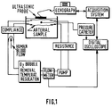
- Figure 1 shows a schematic representation of this arterial set-up and of the related acquisition and control instrumentation.
- An arterial sample freshly taken from a cadaver is placed in a temperature regulated conservation bath. It is perfused with total human blood supplied by a blood transfusion center. The hematocrit is controlled during the experiment to verify the hemolysis degree and the blood viscosity. Heparin is used to prevent any coagulation of the system.
- the hemodynamic set-up is made up of an extracorporal peristaltic pump interfaced with a frequency generator allowing pulsed flow generation, and of a dedicated conduction equipment ensuring a low hemolysis. A compliance and a resistance allow to adjust the flow and pressure curves.
- a heat exchanger and an extracorporal circulation module are place in parallel to ensure a constant temperature, to oxygenate and eliminate bubbles.
- the instrumentation allows on the one hand to control flow conditions, using an external flow-meter and manometer, and on the other hand to acquire data.
- a pressure catheter provides the intra luminal pressure, and an echograph allows the recording of acoustic signals.
- the pressure acquisition is performed using a digital oscilloscope, the ultrasound signals are stored in a dedicated acquisition system, and these two recordings are synchronized to avoid an artificial delay which would hide any viscous behaviour.
- the arterial diameter variations are derived by applying the ultrasound signal processing techniques previously described.
- dp dx ( ⁇ p ⁇ x )r 0 + ⁇ p ⁇ r . ⁇ r ⁇ x
- the first term is here considered as a constant term, responsible for the continuous component of the flow RQ 0 .
- the time varying pressure gradient dp/dx is then modelized according to two different hypotheses :
- variation range of ⁇ and ⁇ is the following : - for ⁇ : - ⁇ /2 ⁇ ⁇ ⁇ ⁇ /2 - for ⁇ : 0 ⁇ ⁇ ⁇ (( ⁇ /2)- ⁇ )/r max
Landscapes
- Health & Medical Sciences (AREA)
- Engineering & Computer Science (AREA)
- Physics & Mathematics (AREA)
- Life Sciences & Earth Sciences (AREA)
- Remote Sensing (AREA)
- Radar, Positioning & Navigation (AREA)
- Acoustics & Sound (AREA)
- Veterinary Medicine (AREA)
- Pathology (AREA)
- Biomedical Technology (AREA)
- Heart & Thoracic Surgery (AREA)
- Medical Informatics (AREA)
- Molecular Biology (AREA)
- Surgery (AREA)
- Animal Behavior & Ethology (AREA)
- General Health & Medical Sciences (AREA)
- Public Health (AREA)
- General Physics & Mathematics (AREA)
- Computer Networks & Wireless Communication (AREA)
- Biophysics (AREA)
- Physiology (AREA)
- Nuclear Medicine, Radiotherapy & Molecular Imaging (AREA)
- Cardiology (AREA)
- Hematology (AREA)
- Vascular Medicine (AREA)
- Radiology & Medical Imaging (AREA)
- Ultra Sonic Daignosis Equipment (AREA)
- Measuring Pulse, Heart Rate, Blood Pressure Or Blood Flow (AREA)
Priority Applications (7)
| Application Number | Priority Date | Filing Date | Title |
|---|---|---|---|
| EP96402081A EP0832604A1 (de) | 1996-09-30 | 1996-09-30 | Verfahren und Vorrichtung zum Messen der Elastizität einer Arterie mittels Ultraschall-Echographie |
| US09/077,414 US6113543A (en) | 1996-09-30 | 1997-09-27 | Method and device for determining the compliance and the blood pressure of an artery by ultrasonic echography |
| JP10516344A JP2000501327A (ja) | 1996-09-30 | 1997-09-29 | 超音波エコーグラフィーによって動脈のコンプライアンス及び血圧を決定する方法及び装置 |
| PCT/IB1997/001180 WO1998014119A1 (en) | 1996-09-30 | 1997-09-29 | Method and device for determining the compliance and the blood pressure of an artery by ultrasonic echography |
| AT97941136T ATE283665T1 (de) | 1996-09-30 | 1997-09-29 | Verfahren und vorrichtung zum bestimmen der nachgiebigkeit und des blutdrucks einer arterie mit hilfe einer ultraschall echographie |
| DE69731817T DE69731817T2 (de) | 1996-09-30 | 1997-09-29 | Verfahren und vorrichtung zum bestimmen der nachgiebigkeit und des blutdrucks einer arterie mit hilfe einer ultraschall echographie |
| EP97941136A EP0876127B1 (de) | 1996-09-30 | 1997-09-29 | Verfahren und vorrichtung zum bestimmen der nachgiebigkeit und des blutdrucks einer arterie mit hilfe einer ultraschall echographie |
Applications Claiming Priority (1)
| Application Number | Priority Date | Filing Date | Title |
|---|---|---|---|
| EP96402081A EP0832604A1 (de) | 1996-09-30 | 1996-09-30 | Verfahren und Vorrichtung zum Messen der Elastizität einer Arterie mittels Ultraschall-Echographie |
Publications (1)
| Publication Number | Publication Date |
|---|---|
| EP0832604A1 true EP0832604A1 (de) | 1998-04-01 |
Family
ID=8225292
Family Applications (2)
| Application Number | Title | Priority Date | Filing Date |
|---|---|---|---|
| EP96402081A Withdrawn EP0832604A1 (de) | 1996-09-30 | 1996-09-30 | Verfahren und Vorrichtung zum Messen der Elastizität einer Arterie mittels Ultraschall-Echographie |
| EP97941136A Expired - Lifetime EP0876127B1 (de) | 1996-09-30 | 1997-09-29 | Verfahren und vorrichtung zum bestimmen der nachgiebigkeit und des blutdrucks einer arterie mit hilfe einer ultraschall echographie |
Family Applications After (1)
| Application Number | Title | Priority Date | Filing Date |
|---|---|---|---|
| EP97941136A Expired - Lifetime EP0876127B1 (de) | 1996-09-30 | 1997-09-29 | Verfahren und vorrichtung zum bestimmen der nachgiebigkeit und des blutdrucks einer arterie mit hilfe einer ultraschall echographie |
Country Status (6)
| Country | Link |
|---|---|
| US (1) | US6113543A (de) |
| EP (2) | EP0832604A1 (de) |
| JP (1) | JP2000501327A (de) |
| AT (1) | ATE283665T1 (de) |
| DE (1) | DE69731817T2 (de) |
| WO (1) | WO1998014119A1 (de) |
Cited By (2)
| Publication number | Priority date | Publication date | Assignee | Title |
|---|---|---|---|---|
| FR2830430A1 (fr) * | 2001-10-08 | 2003-04-11 | Cong Hoan Nguyen | Procede et dispositif pour determiner la loi de comportement d'une artere a partir des mesures non invasives de diametre et epaisseur en fonction de la pression sanguine |
| US8092655B2 (en) | 2000-02-24 | 2012-01-10 | Basf Aktiengesellschaft | Dividing wall column for fractionation of a multicomponent mixture |
Families Citing this family (19)
| Publication number | Priority date | Publication date | Assignee | Title |
|---|---|---|---|---|
| US6510337B1 (en) * | 1999-11-26 | 2003-01-21 | Koninklijke Philips Electronics, N.V. | Multi-phase cardiac imager |
| EP1157285A1 (de) * | 1999-12-21 | 2001-11-28 | Koninklijke Philips Electronics N.V. | Verfahren zur ultraschallbildgebung und untersuchungssystem zur darstellung einer zusammengesetzten bildsequenz einer arterie |
| US7374538B2 (en) * | 2000-04-05 | 2008-05-20 | Duke University | Methods, systems, and computer program products for ultrasound measurements using receive mode parallel processing |
| JP5076203B2 (ja) | 2001-06-21 | 2012-11-21 | 学校法人日本大学 | 血管疾患検査装置およびバイパス血管診断装置 |
| JP3898047B2 (ja) | 2001-07-09 | 2007-03-28 | セイコーインスツル株式会社 | 血液レオロジー測定装置 |
| JP4206218B2 (ja) * | 2002-04-03 | 2009-01-07 | セイコーインスツル株式会社 | 循環動態測定装置 |
| US7052463B2 (en) * | 2002-09-25 | 2006-05-30 | Koninklijke Philips Electronics, N.V. | Method and apparatus for cooling a contacting surface of an ultrasound probe |
| JP4269623B2 (ja) * | 2002-10-07 | 2009-05-27 | 株式会社 東北テクノアーチ | 血流可視化診断装置 |
| US20070219447A1 (en) * | 2003-12-10 | 2007-09-20 | Hiroshi Kanai | Ultrasonograph and ultrasonography |
| US7125383B2 (en) * | 2003-12-30 | 2006-10-24 | General Electric Company | Method and apparatus for ultrasonic continuous, non-invasive blood pressure monitoring |
| FR2899336B1 (fr) | 2006-03-29 | 2008-07-04 | Super Sonic Imagine | Procede et dispositif pour l'imagerie d'un milieu viscoelastique |
| EP2146640B1 (de) | 2007-05-16 | 2018-05-23 | Super Sonic Imagine | Verfahren und vorrichtung zur messung eines mittelwerts der viskoelastizität einer interessierenden region |
| JP5426101B2 (ja) * | 2008-02-25 | 2014-02-26 | 株式会社東芝 | 超音波診断装置及、超音波画像処理装置及び超音波画像処理プログラム |
| JP5474986B2 (ja) | 2009-09-09 | 2014-04-16 | 株式会社ユネクス | 血管機能検査装置 |
| US10405757B2 (en) | 2014-02-25 | 2019-09-10 | Icu Medical, Inc. | Patient monitoring system with gatekeeper signal |
| US11622730B2 (en) * | 2014-11-17 | 2023-04-11 | Rochester Institute Of Technology | Pulse wave velocity, arterial compliance, and blood pressure |
| JP6674553B2 (ja) | 2015-10-19 | 2020-04-01 | アイシーユー・メディカル・インコーポレーテッド | 着脱可能ディスプレイユニットを備える血行動態監視システム |
| US10438355B2 (en) | 2015-11-10 | 2019-10-08 | General Electric Company | System and method for estimating arterial pulse wave velocity |
| EP3569155B1 (de) * | 2018-05-16 | 2022-12-14 | Esaote S.p.A. | Verfahren und ultraschallsystem zur scherwellenelastizitätsbildgebung |
Citations (3)
| Publication number | Priority date | Publication date | Assignee | Title |
|---|---|---|---|---|
| GB2156985A (en) * | 1984-04-02 | 1985-10-16 | Teltec Electronic Equip | Apparatus for measuring movable part-structures, eg blood vessels, within a living body |
| US5099852A (en) * | 1989-03-08 | 1992-03-31 | Asulab S.A. | Method for determining the arterial blood pressure in a non-invasive manner |
| EP0603967A1 (de) * | 1992-12-22 | 1994-06-29 | Laboratoires D'electronique Philips S.A.S. | Gerät und Verfahren zur Elastizitätsmessung einer Arterie mittels Ultraschall-Echographie |
Family Cites Families (5)
| Publication number | Priority date | Publication date | Assignee | Title |
|---|---|---|---|---|
| US5054493A (en) * | 1986-01-31 | 1991-10-08 | Regents Of The University Of Minnesota | Method for diagnosing, monitoring and treating hypertension |
| CH678691A5 (de) * | 1989-03-08 | 1991-10-31 | Asulab Sa | |
| FR2662348A1 (fr) * | 1990-05-22 | 1991-11-29 | Philips Electronique Lab | Dispositif de mesure et de visualisation par echographie ultrasonore de debit d'un ecoulement sanguin et de dilatation du vaisseau associe. |
| US5211177A (en) * | 1990-12-28 | 1993-05-18 | Regents Of The University Of Minnesota | Vascular impedance measurement instrument |
| US5590649A (en) * | 1994-04-15 | 1997-01-07 | Vital Insite, Inc. | Apparatus and method for measuring an induced perturbation to determine blood pressure |
-
1996
- 1996-09-30 EP EP96402081A patent/EP0832604A1/de not_active Withdrawn
-
1997
- 1997-09-27 US US09/077,414 patent/US6113543A/en not_active Expired - Lifetime
- 1997-09-29 DE DE69731817T patent/DE69731817T2/de not_active Expired - Fee Related
- 1997-09-29 JP JP10516344A patent/JP2000501327A/ja not_active Withdrawn
- 1997-09-29 AT AT97941136T patent/ATE283665T1/de not_active IP Right Cessation
- 1997-09-29 WO PCT/IB1997/001180 patent/WO1998014119A1/en not_active Ceased
- 1997-09-29 EP EP97941136A patent/EP0876127B1/de not_active Expired - Lifetime
Patent Citations (3)
| Publication number | Priority date | Publication date | Assignee | Title |
|---|---|---|---|---|
| GB2156985A (en) * | 1984-04-02 | 1985-10-16 | Teltec Electronic Equip | Apparatus for measuring movable part-structures, eg blood vessels, within a living body |
| US5099852A (en) * | 1989-03-08 | 1992-03-31 | Asulab S.A. | Method for determining the arterial blood pressure in a non-invasive manner |
| EP0603967A1 (de) * | 1992-12-22 | 1994-06-29 | Laboratoires D'electronique Philips S.A.S. | Gerät und Verfahren zur Elastizitätsmessung einer Arterie mittels Ultraschall-Echographie |
Non-Patent Citations (1)
| Title |
|---|
| P.J. DHAWALE ET AL.: "In Vivo Estimation of Arteries with Intracoronary Ultrasound", PROC. OF THE ANNUAL INTERNATIONAL CONF. OF THE IEEE ENGINEERING IN MEDICINE & BIOLOGY SOCIETY, vol. 15, no. 1, 28 October 1993 (1993-10-28) - 31 October 1993 (1993-10-31), SAN DIEGO (US), pages 204 - 205, XP000436720 * |
Cited By (2)
| Publication number | Priority date | Publication date | Assignee | Title |
|---|---|---|---|---|
| US8092655B2 (en) | 2000-02-24 | 2012-01-10 | Basf Aktiengesellschaft | Dividing wall column for fractionation of a multicomponent mixture |
| FR2830430A1 (fr) * | 2001-10-08 | 2003-04-11 | Cong Hoan Nguyen | Procede et dispositif pour determiner la loi de comportement d'une artere a partir des mesures non invasives de diametre et epaisseur en fonction de la pression sanguine |
Also Published As
| Publication number | Publication date |
|---|---|
| EP0876127A1 (de) | 1998-11-11 |
| JP2000501327A (ja) | 2000-02-08 |
| DE69731817D1 (de) | 2005-01-05 |
| DE69731817T2 (de) | 2005-12-15 |
| WO1998014119A1 (en) | 1998-04-09 |
| EP0876127B1 (de) | 2004-12-01 |
| ATE283665T1 (de) | 2004-12-15 |
| US6113543A (en) | 2000-09-05 |
Similar Documents
| Publication | Publication Date | Title |
|---|---|---|
| EP0832604A1 (de) | Verfahren und Vorrichtung zum Messen der Elastizität einer Arterie mittels Ultraschall-Echographie | |
| Chirinos | Arterial stiffness: basic concepts and measurement techniques | |
| Beulen et al. | Toward noninvasive blood pressure assessment in arteries by using ultrasound | |
| Reuderink et al. | Linear and nonlinear one-dimensional models of pulse wave transmission at high Womersley numbers | |
| US20220160328A1 (en) | Fluid Flow Analysis | |
| US8784327B2 (en) | Method and system for obtaining dimension related information for a flow channel | |
| US20030109785A1 (en) | Method and apparatus for measuring volume flow and area for a dynamic orifice | |
| Warriner et al. | A viscoelastic model of arterial wall motion in pulsatile flow: implications for Doppler ultrasound clutter assessment | |
| Lillie et al. | Pulse wave velocity prediction and compliance assessment in elastic arterial segments | |
| Olesen et al. | Noninvasive estimation of pressure changes using 2-D vector velocity ultrasound: an experimental study with in vivo examples | |
| Migliavacca et al. | Calculating blood flow from Doppler measurements in the systemic-to-pulmonary artery shunt after the Norwood operation: a method based on computational fluid dynamics | |
| WO2000055579A2 (en) | A system and method for detection and characterization of stenosis, blood vessels flow and vessel walls properties using vessel geometrical measurements | |
| US12144674B2 (en) | Fluid flow analysis | |
| Gudmundsson et al. | Factors affecting color Doppler energy ultrasound recordings in an in-vitro model | |
| EP3922173A1 (de) | Systeme und verfahren zur erlangung einer pulswellengeschwindigkeitsmessung | |
| Jenni et al. | In vitro validation of volumetric blood flow measurement using Doppler flow wire | |
| Jenni et al. | A novel in vivo procedure for volumetric flow measurements | |
| Urban et al. | Understanding Arterial Biomechanics with Ultrasound and Waveguide–Models | |
| EP3831307A1 (de) | Vorrichtung zur bestimmung eines physiologischen parameters in bezug auf ein gefäss | |
| Kanai et al. | Transcutaneous measurement of frequency dispersion in the regional pulse wave velocity | |
| Kara et al. | Spectral broadening of lower extremity venous Doppler signals using STFT and AR modeling | |
| Banerjee et al. | Flow-pressure drop measurement and calculation in a tapered femoral artery of a dog | |
| Bassil | The mechanics of cuff-based blood pressure measurement | |
| Mai et al. | Vascular compliance using elasticity imaging | |
| Cikirikcioglu et al. | Pre-clinical validation of a new intra-operative" dual beam Doppler" blood flowmeter in an artificial circuit |
Legal Events
| Date | Code | Title | Description |
|---|---|---|---|
| PUAI | Public reference made under article 153(3) epc to a published international application that has entered the european phase |
Free format text: ORIGINAL CODE: 0009012 |
|
| AK | Designated contracting states |
Kind code of ref document: A1 Designated state(s): AT BE CH DE DK ES FI FR GB GR IE IT LI LU MC NL PT SE |
|
| AKX | Designation fees paid | ||
| RBV | Designated contracting states (corrected) | ||
| STAA | Information on the status of an ep patent application or granted ep patent |
Free format text: STATUS: THE APPLICATION IS DEEMED TO BE WITHDRAWN |
|
| 18D | Application deemed to be withdrawn |
Effective date: 19981002 |