EP0834556A1 - Procédés de culture de cellules - Google Patents
Procédés de culture de cellules Download PDFInfo
- Publication number
- EP0834556A1 EP0834556A1 EP96202686A EP96202686A EP0834556A1 EP 0834556 A1 EP0834556 A1 EP 0834556A1 EP 96202686 A EP96202686 A EP 96202686A EP 96202686 A EP96202686 A EP 96202686A EP 0834556 A1 EP0834556 A1 EP 0834556A1
- Authority
- EP
- European Patent Office
- Prior art keywords
- cells
- growth
- serum
- hpc
- elastase
- Prior art date
- Legal status (The legal status is an assumption and is not a legal conclusion. Google has not performed a legal analysis and makes no representation as to the accuracy of the status listed.)
- Withdrawn
Links
- 238000012258 culturing Methods 0.000 title claims abstract description 16
- 238000000034 method Methods 0.000 title claims abstract description 15
- 210000004027 cell Anatomy 0.000 claims abstract description 99
- 210000003958 hematopoietic stem cell Anatomy 0.000 claims abstract description 65
- 102000035195 Peptidases Human genes 0.000 claims abstract description 16
- 108091005804 Peptidases Proteins 0.000 claims abstract description 16
- 239000004365 Protease Substances 0.000 claims abstract description 7
- 239000000137 peptide hydrolase inhibitor Substances 0.000 claims description 21
- 239000003001 serine protease inhibitor Substances 0.000 claims description 21
- 102000016387 Pancreatic elastase Human genes 0.000 claims description 14
- 108010067372 Pancreatic elastase Proteins 0.000 claims description 14
- 239000003102 growth factor Substances 0.000 claims description 14
- 239000001963 growth medium Substances 0.000 claims description 14
- 239000002609 medium Substances 0.000 claims description 11
- 239000005557 antagonist Substances 0.000 claims description 10
- 101710170876 Antileukoproteinase Proteins 0.000 claims description 9
- 102100021253 Antileukoproteinase Human genes 0.000 claims description 9
- 229940124158 Protease/peptidase inhibitor Drugs 0.000 claims description 9
- LKDMKWNDBAVNQZ-UHFFFAOYSA-N 4-[[1-[[1-[2-[[1-(4-nitroanilino)-1-oxo-3-phenylpropan-2-yl]carbamoyl]pyrrolidin-1-yl]-1-oxopropan-2-yl]amino]-1-oxopropan-2-yl]amino]-4-oxobutanoic acid Chemical compound OC(=O)CCC(=O)NC(C)C(=O)NC(C)C(=O)N1CCCC1C(=O)NC(C(=O)NC=1C=CC(=CC=1)[N+]([O-])=O)CC1=CC=CC=C1 LKDMKWNDBAVNQZ-UHFFFAOYSA-N 0.000 claims description 8
- 108090000617 Cathepsin G Proteins 0.000 claims description 8
- 102000004173 Cathepsin G Human genes 0.000 claims description 8
- 102000008847 Serpin Human genes 0.000 claims description 7
- 108050000761 Serpin Proteins 0.000 claims description 7
- 238000004519 manufacturing process Methods 0.000 claims description 6
- 239000012634 fragment Substances 0.000 claims description 5
- 208000025113 myeloid leukemia Diseases 0.000 claims description 4
- 101000851058 Homo sapiens Neutrophil elastase Proteins 0.000 claims description 3
- 239000003112 inhibitor Substances 0.000 claims description 3
- 210000000777 hematopoietic system Anatomy 0.000 claims description 2
- 102000052502 human ELANE Human genes 0.000 claims description 2
- 210000000130 stem cell Anatomy 0.000 abstract description 12
- 230000005764 inhibitory process Effects 0.000 abstract description 4
- 230000012010 growth Effects 0.000 description 86
- 210000002966 serum Anatomy 0.000 description 71
- 102100031573 Hematopoietic progenitor cell antigen CD34 Human genes 0.000 description 36
- 101000777663 Homo sapiens Hematopoietic progenitor cell antigen CD34 Proteins 0.000 description 36
- 102000004169 proteins and genes Human genes 0.000 description 36
- 108090000623 proteins and genes Proteins 0.000 description 36
- 235000018102 proteins Nutrition 0.000 description 35
- 239000003636 conditioned culture medium Substances 0.000 description 33
- 108010082545 Secretory Leukocyte Peptidase Inhibitor Proteins 0.000 description 30
- 102000004002 Secretory Leukocyte Peptidase Inhibitor Human genes 0.000 description 30
- 210000002960 bfu-e Anatomy 0.000 description 25
- 210000001744 T-lymphocyte Anatomy 0.000 description 20
- 241000282414 Homo sapiens Species 0.000 description 19
- 210000001185 bone marrow Anatomy 0.000 description 18
- 230000000694 effects Effects 0.000 description 17
- HVYWMOMLDIMFJA-DPAQBDIFSA-N cholesterol Chemical compound C1C=C2C[C@@H](O)CC[C@]2(C)[C@@H]2[C@@H]1[C@@H]1CC[C@H]([C@H](C)CCCC(C)C)[C@@]1(C)CC2 HVYWMOMLDIMFJA-DPAQBDIFSA-N 0.000 description 16
- 238000000338 in vitro Methods 0.000 description 16
- NOESYZHRGYRDHS-UHFFFAOYSA-N insulin Chemical compound N1C(=O)C(NC(=O)C(CCC(N)=O)NC(=O)C(CCC(O)=O)NC(=O)C(C(C)C)NC(=O)C(NC(=O)CN)C(C)CC)CSSCC(C(NC(CO)C(=O)NC(CC(C)C)C(=O)NC(CC=2C=CC(O)=CC=2)C(=O)NC(CCC(N)=O)C(=O)NC(CC(C)C)C(=O)NC(CCC(O)=O)C(=O)NC(CC(N)=O)C(=O)NC(CC=2C=CC(O)=CC=2)C(=O)NC(CSSCC(NC(=O)C(C(C)C)NC(=O)C(CC(C)C)NC(=O)C(CC=2C=CC(O)=CC=2)NC(=O)C(CC(C)C)NC(=O)C(C)NC(=O)C(CCC(O)=O)NC(=O)C(C(C)C)NC(=O)C(CC(C)C)NC(=O)C(CC=2NC=NC=2)NC(=O)C(CO)NC(=O)CNC2=O)C(=O)NCC(=O)NC(CCC(O)=O)C(=O)NC(CCCNC(N)=N)C(=O)NCC(=O)NC(CC=3C=CC=CC=3)C(=O)NC(CC=3C=CC=CC=3)C(=O)NC(CC=3C=CC(O)=CC=3)C(=O)NC(C(C)O)C(=O)N3C(CCC3)C(=O)NC(CCCCN)C(=O)NC(C)C(O)=O)C(=O)NC(CC(N)=O)C(O)=O)=O)NC(=O)C(C(C)CC)NC(=O)C(CO)NC(=O)C(C(C)O)NC(=O)C1CSSCC2NC(=O)C(CC(C)C)NC(=O)C(NC(=O)C(CCC(N)=O)NC(=O)C(CC(N)=O)NC(=O)C(NC(=O)C(N)CC=1C=CC=CC=1)C(C)C)CC1=CN=CN1 NOESYZHRGYRDHS-UHFFFAOYSA-N 0.000 description 16
- 108091003079 Bovine Serum Albumin Proteins 0.000 description 14
- 210000002798 bone marrow cell Anatomy 0.000 description 14
- 229940098773 bovine serum albumin Drugs 0.000 description 13
- 238000004007 reversed phase HPLC Methods 0.000 description 9
- 206010005003 Bladder cancer Diseases 0.000 description 8
- 101000615334 Homo sapiens Antileukoproteinase Proteins 0.000 description 8
- 102000004877 Insulin Human genes 0.000 description 8
- 201000001531 bladder carcinoma Diseases 0.000 description 8
- 235000012000 cholesterol Nutrition 0.000 description 8
- 229940125396 insulin Drugs 0.000 description 8
- 235000019833 protease Nutrition 0.000 description 8
- 230000003319 supportive effect Effects 0.000 description 8
- 208000010570 urinary bladder carcinoma Diseases 0.000 description 8
- 102000004127 Cytokines Human genes 0.000 description 7
- 108090000695 Cytokines Proteins 0.000 description 7
- 108091006905 Human Serum Albumin Proteins 0.000 description 7
- 102000008100 Human Serum Albumin Human genes 0.000 description 7
- 108090001061 Insulin Proteins 0.000 description 7
- 238000002474 experimental method Methods 0.000 description 7
- 230000003394 haemopoietic effect Effects 0.000 description 7
- 230000002401 inhibitory effect Effects 0.000 description 7
- 230000035755 proliferation Effects 0.000 description 7
- 238000000746 purification Methods 0.000 description 7
- 239000000126 substance Substances 0.000 description 7
- 108010028275 Leukocyte Elastase Proteins 0.000 description 6
- 102000016799 Leukocyte elastase Human genes 0.000 description 6
- DTQVDTLACAAQTR-UHFFFAOYSA-N Trifluoroacetic acid Chemical compound OC(=O)C(F)(F)F DTQVDTLACAAQTR-UHFFFAOYSA-N 0.000 description 6
- 229940019748 antifibrinolytic proteinase inhibitors Drugs 0.000 description 6
- 210000005087 mononuclear cell Anatomy 0.000 description 6
- 230000001629 suppression Effects 0.000 description 6
- 108010017080 Granulocyte Colony-Stimulating Factor Proteins 0.000 description 5
- 102000004269 Granulocyte Colony-Stimulating Factor Human genes 0.000 description 5
- 101000897494 Homo sapiens C-C motif chemokine 27 Proteins 0.000 description 5
- 102000004889 Interleukin-6 Human genes 0.000 description 5
- 108090001005 Interleukin-6 Proteins 0.000 description 5
- 101710187074 Serine proteinase inhibitor Proteins 0.000 description 5
- 150000001413 amino acids Chemical class 0.000 description 5
- 238000004113 cell culture Methods 0.000 description 5
- 238000002523 gelfiltration Methods 0.000 description 5
- 230000001965 increasing effect Effects 0.000 description 5
- 239000007787 solid Substances 0.000 description 5
- UZOVYGYOLBIAJR-UHFFFAOYSA-N 4-isocyanato-4'-methyldiphenylmethane Chemical compound C1=CC(C)=CC=C1CC1=CC=C(N=C=O)C=C1 UZOVYGYOLBIAJR-UHFFFAOYSA-N 0.000 description 4
- 102000009027 Albumins Human genes 0.000 description 4
- 108010088751 Albumins Proteins 0.000 description 4
- DHMQDGOQFOQNFH-UHFFFAOYSA-N Glycine Chemical compound NCC(O)=O DHMQDGOQFOQNFH-UHFFFAOYSA-N 0.000 description 4
- 108090000723 Insulin-Like Growth Factor I Proteins 0.000 description 4
- 108010002386 Interleukin-3 Proteins 0.000 description 4
- 102100039064 Interleukin-3 Human genes 0.000 description 4
- 102000016943 Muramidase Human genes 0.000 description 4
- 108010014251 Muramidase Proteins 0.000 description 4
- 108010062010 N-Acetylmuramoyl-L-alanine Amidase Proteins 0.000 description 4
- 238000012300 Sequence Analysis Methods 0.000 description 4
- 229940122055 Serine protease inhibitor Drugs 0.000 description 4
- 101710102218 Serine protease inhibitor Proteins 0.000 description 4
- 239000005289 controlled pore glass Substances 0.000 description 4
- 230000000925 erythroid effect Effects 0.000 description 4
- 238000004128 high performance liquid chromatography Methods 0.000 description 4
- 238000011534 incubation Methods 0.000 description 4
- 229960000274 lysozyme Drugs 0.000 description 4
- 235000010335 lysozyme Nutrition 0.000 description 4
- 239000004325 lysozyme Substances 0.000 description 4
- BDAGIHXWWSANSR-UHFFFAOYSA-N methanoic acid Natural products OC=O BDAGIHXWWSANSR-UHFFFAOYSA-N 0.000 description 4
- 238000006386 neutralization reaction Methods 0.000 description 4
- 230000003472 neutralizing effect Effects 0.000 description 4
- 239000006228 supernatant Substances 0.000 description 4
- 230000008093 supporting effect Effects 0.000 description 4
- LYCAIKOWRPUZTN-UHFFFAOYSA-N Ethylene glycol Chemical compound OCCO LYCAIKOWRPUZTN-UHFFFAOYSA-N 0.000 description 3
- 102000003974 Fibroblast growth factor 2 Human genes 0.000 description 3
- 108090000379 Fibroblast growth factor 2 Proteins 0.000 description 3
- 102000015696 Interleukins Human genes 0.000 description 3
- 108010063738 Interleukins Proteins 0.000 description 3
- 108010046938 Macrophage Colony-Stimulating Factor Proteins 0.000 description 3
- 102000007651 Macrophage Colony-Stimulating Factor Human genes 0.000 description 3
- 108010058846 Ovalbumin Proteins 0.000 description 3
- 239000012980 RPMI-1640 medium Substances 0.000 description 3
- 235000001014 amino acid Nutrition 0.000 description 3
- 238000003556 assay Methods 0.000 description 3
- 239000011324 bead Substances 0.000 description 3
- 230000004071 biological effect Effects 0.000 description 3
- 230000015556 catabolic process Effects 0.000 description 3
- 238000012512 characterization method Methods 0.000 description 3
- 238000006731 degradation reaction Methods 0.000 description 3
- 230000004069 differentiation Effects 0.000 description 3
- LOKCTEFSRHRXRJ-UHFFFAOYSA-I dipotassium trisodium dihydrogen phosphate hydrogen phosphate dichloride Chemical class P(=O)(O)(O)[O-].[K+].P(=O)(O)([O-])[O-].[Na+].[Na+].[Cl-].[K+].[Cl-].[Na+] LOKCTEFSRHRXRJ-UHFFFAOYSA-I 0.000 description 3
- 230000007246 mechanism Effects 0.000 description 3
- 239000000203 mixture Substances 0.000 description 3
- 210000000066 myeloid cell Anatomy 0.000 description 3
- 235000015097 nutrients Nutrition 0.000 description 3
- 229940092253 ovalbumin Drugs 0.000 description 3
- 230000003248 secreting effect Effects 0.000 description 3
- 230000001225 therapeutic effect Effects 0.000 description 3
- OSWFIVFLDKOXQC-UHFFFAOYSA-N 4-(3-methoxyphenyl)aniline Chemical compound COC1=CC=CC(C=2C=CC(N)=CC=2)=C1 OSWFIVFLDKOXQC-UHFFFAOYSA-N 0.000 description 2
- QFVHZQCOUORWEI-UHFFFAOYSA-N 4-[(4-anilino-5-sulfonaphthalen-1-yl)diazenyl]-5-hydroxynaphthalene-2,7-disulfonic acid Chemical compound C=12C(O)=CC(S(O)(=O)=O)=CC2=CC(S(O)(=O)=O)=CC=1N=NC(C1=CC=CC(=C11)S(O)(=O)=O)=CC=C1NC1=CC=CC=C1 QFVHZQCOUORWEI-UHFFFAOYSA-N 0.000 description 2
- IJGRMHOSHXDMSA-UHFFFAOYSA-N Atomic nitrogen Chemical compound N#N IJGRMHOSHXDMSA-UHFFFAOYSA-N 0.000 description 2
- 241000894006 Bacteria Species 0.000 description 2
- 241000283690 Bos taurus Species 0.000 description 2
- 108010038061 Chymotrypsinogen Proteins 0.000 description 2
- -1 GM-CSF Proteins 0.000 description 2
- 101000993347 Gallus gallus Ciliary neurotrophic factor Proteins 0.000 description 2
- 239000004471 Glycine Substances 0.000 description 2
- 101000766306 Homo sapiens Serotransferrin Proteins 0.000 description 2
- 102000004218 Insulin-Like Growth Factor I Human genes 0.000 description 2
- 102000048143 Insulin-Like Growth Factor II Human genes 0.000 description 2
- 108090001117 Insulin-Like Growth Factor II Proteins 0.000 description 2
- 108010002352 Interleukin-1 Proteins 0.000 description 2
- 102000000589 Interleukin-1 Human genes 0.000 description 2
- 241000283973 Oryctolagus cuniculus Species 0.000 description 2
- 102000010780 Platelet-Derived Growth Factor Human genes 0.000 description 2
- 108010038512 Platelet-Derived Growth Factor Proteins 0.000 description 2
- 229920001213 Polysorbate 20 Polymers 0.000 description 2
- 239000012979 RPMI medium Substances 0.000 description 2
- BQCADISMDOOEFD-UHFFFAOYSA-N Silver Chemical compound [Ag] BQCADISMDOOEFD-UHFFFAOYSA-N 0.000 description 2
- FAPWRFPIFSIZLT-UHFFFAOYSA-M Sodium chloride Chemical compound [Na+].[Cl-] FAPWRFPIFSIZLT-UHFFFAOYSA-M 0.000 description 2
- 102000013275 Somatomedins Human genes 0.000 description 2
- 108090000631 Trypsin Proteins 0.000 description 2
- 102000004142 Trypsin Human genes 0.000 description 2
- 238000004458 analytical method Methods 0.000 description 2
- 230000003305 autocrine Effects 0.000 description 2
- 230000010261 cell growth Effects 0.000 description 2
- 239000012141 concentrate Substances 0.000 description 2
- 238000010828 elution Methods 0.000 description 2
- 239000012091 fetal bovine serum Substances 0.000 description 2
- 210000002950 fibroblast Anatomy 0.000 description 2
- 239000012530 fluid Substances 0.000 description 2
- 235000019253 formic acid Nutrition 0.000 description 2
- 238000005194 fractionation Methods 0.000 description 2
- 230000011132 hemopoiesis Effects 0.000 description 2
- 230000000977 initiatory effect Effects 0.000 description 2
- 238000002955 isolation Methods 0.000 description 2
- 210000001616 monocyte Anatomy 0.000 description 2
- 239000002953 phosphate buffered saline Substances 0.000 description 2
- 229920002401 polyacrylamide Polymers 0.000 description 2
- 229920001223 polyethylene glycol Polymers 0.000 description 2
- 239000000256 polyoxyethylene sorbitan monolaurate Substances 0.000 description 2
- 235000010486 polyoxyethylene sorbitan monolaurate Nutrition 0.000 description 2
- 230000003389 potentiating effect Effects 0.000 description 2
- 238000002360 preparation method Methods 0.000 description 2
- 239000000047 product Substances 0.000 description 2
- 238000000159 protein binding assay Methods 0.000 description 2
- 230000002797 proteolythic effect Effects 0.000 description 2
- XNSAINXGIQZQOO-SRVKXCTJSA-N protirelin Chemical compound NC(=O)[C@@H]1CCCN1C(=O)[C@@H](NC(=O)[C@H]1NC(=O)CC1)CC1=CN=CN1 XNSAINXGIQZQOO-SRVKXCTJSA-N 0.000 description 2
- 230000001105 regulatory effect Effects 0.000 description 2
- 229920006395 saturated elastomer Polymers 0.000 description 2
- 238000000926 separation method Methods 0.000 description 2
- 239000012679 serum free medium Substances 0.000 description 2
- 229910052709 silver Inorganic materials 0.000 description 2
- 239000004332 silver Substances 0.000 description 2
- 238000001179 sorption measurement Methods 0.000 description 2
- 230000004936 stimulating effect Effects 0.000 description 2
- 239000012588 trypsin Substances 0.000 description 2
- VBEQCZHXXJYVRD-GACYYNSASA-N uroanthelone Chemical compound C([C@@H](C(=O)N[C@H](C(=O)N[C@@H](CS)C(=O)N[C@@H](CC(N)=O)C(=O)N[C@@H](CS)C(=O)N[C@H](C(=O)N[C@@H]([C@@H](C)CC)C(=O)NCC(=O)N[C@@H](CC=1C=CC(O)=CC=1)C(=O)N[C@@H](CO)C(=O)NCC(=O)N[C@@H](CC(O)=O)C(=O)N[C@@H](CCCNC(N)=N)C(=O)N[C@@H](CS)C(=O)N[C@@H](CCC(N)=O)C(=O)N[C@@H]([C@@H](C)O)C(=O)N[C@@H](CCCNC(N)=N)C(=O)N[C@@H](CC(O)=O)C(=O)N[C@@H](CC(C)C)C(=O)N[C@@H](CCCNC(N)=N)C(=O)N[C@@H](CC=1C2=CC=CC=C2NC=1)C(=O)N[C@@H](CC=1C2=CC=CC=C2NC=1)C(=O)N[C@@H](CCC(O)=O)C(=O)N[C@@H](CC(C)C)C(=O)N[C@@H](CCCNC(N)=N)C(O)=O)C(C)C)[C@@H](C)O)NC(=O)[C@H](CO)NC(=O)[C@H](CC(O)=O)NC(=O)[C@H](CC(C)C)NC(=O)[C@H](CO)NC(=O)[C@H](CCC(O)=O)NC(=O)[C@@H](NC(=O)[C@H](CC=1NC=NC=1)NC(=O)[C@H](CCSC)NC(=O)[C@H](CS)NC(=O)[C@@H](NC(=O)CNC(=O)CNC(=O)[C@H](CC(N)=O)NC(=O)[C@H](CC(C)C)NC(=O)[C@H](CS)NC(=O)[C@H](CC=1C=CC(O)=CC=1)NC(=O)CNC(=O)[C@H](CC(O)=O)NC(=O)[C@H](CC=1C=CC(O)=CC=1)NC(=O)[C@H](CO)NC(=O)[C@H](CO)NC(=O)[C@H]1N(CCC1)C(=O)[C@H](CS)NC(=O)CNC(=O)[C@H]1N(CCC1)C(=O)[C@H](CC=1C=CC(O)=CC=1)NC(=O)[C@H](CO)NC(=O)[C@@H](N)CC(N)=O)C(C)C)[C@@H](C)CC)C1=CC=C(O)C=C1 VBEQCZHXXJYVRD-GACYYNSASA-N 0.000 description 2
- DGVVWUTYPXICAM-UHFFFAOYSA-N β‐Mercaptoethanol Chemical compound OCCS DGVVWUTYPXICAM-UHFFFAOYSA-N 0.000 description 2
- PQMRRAQXKWFYQN-UHFFFAOYSA-N 1-phenyl-2-sulfanylideneimidazolidin-4-one Chemical compound S=C1NC(=O)CN1C1=CC=CC=C1 PQMRRAQXKWFYQN-UHFFFAOYSA-N 0.000 description 1
- WEVYAHXRMPXWCK-UHFFFAOYSA-N Acetonitrile Chemical compound CC#N WEVYAHXRMPXWCK-UHFFFAOYSA-N 0.000 description 1
- 102100033312 Alpha-2-macroglobulin Human genes 0.000 description 1
- 108010039627 Aprotinin Proteins 0.000 description 1
- 102000004506 Blood Proteins Human genes 0.000 description 1
- 108010017384 Blood Proteins Proteins 0.000 description 1
- 102000003846 Carbonic anhydrases Human genes 0.000 description 1
- 108090000209 Carbonic anhydrases Proteins 0.000 description 1
- 108010012236 Chemokines Proteins 0.000 description 1
- 102000019034 Chemokines Human genes 0.000 description 1
- 108090000317 Chymotrypsin Proteins 0.000 description 1
- 238000002965 ELISA Methods 0.000 description 1
- 241000196324 Embryophyta Species 0.000 description 1
- 241000283073 Equus caballus Species 0.000 description 1
- 102000018233 Fibroblast Growth Factor Human genes 0.000 description 1
- 108050007372 Fibroblast Growth Factor Proteins 0.000 description 1
- 229920001917 Ficoll Polymers 0.000 description 1
- 241000233866 Fungi Species 0.000 description 1
- 244000068988 Glycine max Species 0.000 description 1
- 235000010469 Glycine max Nutrition 0.000 description 1
- 108010054017 Granulocyte Colony-Stimulating Factor Receptors Proteins 0.000 description 1
- 102100039622 Granulocyte colony-stimulating factor receptor Human genes 0.000 description 1
- 108010009202 Growth Factor Receptors Proteins 0.000 description 1
- 102000009465 Growth Factor Receptors Human genes 0.000 description 1
- 241000238631 Hexapoda Species 0.000 description 1
- 241000836430 Hilda Species 0.000 description 1
- 241000282412 Homo Species 0.000 description 1
- 101000976075 Homo sapiens Insulin Proteins 0.000 description 1
- 108090000144 Human Proteins Proteins 0.000 description 1
- 102000003839 Human Proteins Human genes 0.000 description 1
- 102000014150 Interferons Human genes 0.000 description 1
- 108010050904 Interferons Proteins 0.000 description 1
- 108090000177 Interleukin-11 Proteins 0.000 description 1
- 108010002350 Interleukin-2 Proteins 0.000 description 1
- 108090000978 Interleukin-4 Proteins 0.000 description 1
- 108010002616 Interleukin-5 Proteins 0.000 description 1
- 108010002586 Interleukin-7 Proteins 0.000 description 1
- 108090001007 Interleukin-8 Proteins 0.000 description 1
- 108010002335 Interleukin-9 Proteins 0.000 description 1
- 239000007760 Iscove's Modified Dulbecco's Medium Substances 0.000 description 1
- 241001529936 Murinae Species 0.000 description 1
- 101001076414 Mus musculus Interleukin-6 Proteins 0.000 description 1
- 206010028980 Neoplasm Diseases 0.000 description 1
- 102100033174 Neutrophil elastase Human genes 0.000 description 1
- 229910019142 PO4 Inorganic materials 0.000 description 1
- 241001494479 Pecora Species 0.000 description 1
- 108010065081 Phosphorylase b Proteins 0.000 description 1
- 239000002202 Polyethylene glycol Substances 0.000 description 1
- 108010015078 Pregnancy-Associated alpha 2-Macroglobulins Proteins 0.000 description 1
- 206010036790 Productive cough Diseases 0.000 description 1
- 240000004808 Saccharomyces cerevisiae Species 0.000 description 1
- 108010071390 Serum Albumin Proteins 0.000 description 1
- 102000046299 Transforming Growth Factor beta1 Human genes 0.000 description 1
- 101800002279 Transforming growth factor beta-1 Proteins 0.000 description 1
- 229940122618 Trypsin inhibitor Drugs 0.000 description 1
- 101710162629 Trypsin inhibitor Proteins 0.000 description 1
- 108060008682 Tumor Necrosis Factor Proteins 0.000 description 1
- 102000000852 Tumor Necrosis Factor-alpha Human genes 0.000 description 1
- USDJGQLNFPZEON-UHFFFAOYSA-N [[4,6-bis(hydroxymethylamino)-1,3,5-triazin-2-yl]amino]methanol Chemical compound OCNC1=NC(NCO)=NC(NCO)=N1 USDJGQLNFPZEON-UHFFFAOYSA-N 0.000 description 1
- 238000010521 absorption reaction Methods 0.000 description 1
- 230000009471 action Effects 0.000 description 1
- 230000006978 adaptation Effects 0.000 description 1
- 239000000654 additive Substances 0.000 description 1
- 230000000996 additive effect Effects 0.000 description 1
- 230000001464 adherent effect Effects 0.000 description 1
- 108010050122 alpha 1-Antitrypsin Proteins 0.000 description 1
- 102000015395 alpha 1-Antitrypsin Human genes 0.000 description 1
- 125000000539 amino acid group Chemical group 0.000 description 1
- 238000003277 amino acid sequence analysis Methods 0.000 description 1
- 230000001093 anti-cancer Effects 0.000 description 1
- 229960004405 aprotinin Drugs 0.000 description 1
- 244000052616 bacterial pathogen Species 0.000 description 1
- 210000004369 blood Anatomy 0.000 description 1
- 239000008280 blood Substances 0.000 description 1
- 238000010322 bone marrow transplantation Methods 0.000 description 1
- 239000000872 buffer Substances 0.000 description 1
- 201000011510 cancer Diseases 0.000 description 1
- 210000000170 cell membrane Anatomy 0.000 description 1
- 230000001413 cellular effect Effects 0.000 description 1
- 210000003679 cervix uteri Anatomy 0.000 description 1
- 238000004182 chemical digestion Methods 0.000 description 1
- 239000003795 chemical substances by application Substances 0.000 description 1
- 230000003399 chemotactic effect Effects 0.000 description 1
- 229960002376 chymotrypsin Drugs 0.000 description 1
- 238000009643 clonogenic assay Methods 0.000 description 1
- 231100000096 clonogenic assay Toxicity 0.000 description 1
- 230000005757 colony formation Effects 0.000 description 1
- 239000012228 culture supernatant Substances 0.000 description 1
- 125000000151 cysteine group Chemical group N[C@@H](CS)C(=O)* 0.000 description 1
- 230000016396 cytokine production Effects 0.000 description 1
- 230000000593 degrading effect Effects 0.000 description 1
- 230000001419 dependent effect Effects 0.000 description 1
- 238000001514 detection method Methods 0.000 description 1
- 238000000502 dialysis Methods 0.000 description 1
- 229940042399 direct acting antivirals protease inhibitors Drugs 0.000 description 1
- 238000009826 distribution Methods 0.000 description 1
- 231100000673 dose–response relationship Toxicity 0.000 description 1
- 239000003602 elastase inhibitor Substances 0.000 description 1
- 230000003511 endothelial effect Effects 0.000 description 1
- 238000005516 engineering process Methods 0.000 description 1
- 230000002708 enhancing effect Effects 0.000 description 1
- 230000008378 epithelial damage Effects 0.000 description 1
- 210000000267 erythroid cell Anatomy 0.000 description 1
- 230000008713 feedback mechanism Effects 0.000 description 1
- 229940126864 fibroblast growth factor Drugs 0.000 description 1
- MHMNJMPURVTYEJ-UHFFFAOYSA-N fluorescein-5-isothiocyanate Chemical compound O1C(=O)C2=CC(N=C=S)=CC=C2C21C1=CC=C(O)C=C1OC1=CC(O)=CC=C21 MHMNJMPURVTYEJ-UHFFFAOYSA-N 0.000 description 1
- 238000001641 gel filtration chromatography Methods 0.000 description 1
- 210000003714 granulocyte Anatomy 0.000 description 1
- 102000051410 human SLPI Human genes 0.000 description 1
- 210000005260 human cell Anatomy 0.000 description 1
- 210000000987 immune system Anatomy 0.000 description 1
- 230000006872 improvement Effects 0.000 description 1
- 239000000411 inducer Substances 0.000 description 1
- ZPNFWUPYTFPOJU-LPYSRVMUSA-N iniprol Chemical compound C([C@H]1C(=O)NCC(=O)NCC(=O)N[C@H]2CSSC[C@H]3C(=O)N[C@@H](CCCCN)C(=O)N[C@@H](C)C(=O)N[C@@H](CCCNC(N)=N)C(=O)N[C@H](C(N[C@H](C(=O)N[C@@H](CCCNC(N)=N)C(=O)N[C@@H](CC=4C=CC(O)=CC=4)C(=O)N[C@@H](CC=4C=CC=CC=4)C(=O)N[C@@H](CC=4C=CC(O)=CC=4)C(=O)N[C@@H](CC(N)=O)C(=O)N[C@@H](C)C(=O)N[C@@H](CCCCN)C(=O)N[C@@H](C)C(=O)NCC(=O)N[C@@H](CC(C)C)C(=O)N[C@@H](CSSC[C@H](NC(=O)[C@H](CC(O)=O)NC(=O)[C@H](CCC(O)=O)NC(=O)[C@H](C)NC(=O)[C@H](CO)NC(=O)[C@H](CCCCN)NC(=O)[C@H](CC=4C=CC=CC=4)NC(=O)[C@H](CC(N)=O)NC(=O)[C@H](CC(N)=O)NC(=O)[C@H](CCCNC(N)=N)NC(=O)[C@H](CCCCN)NC(=O)[C@H](C)NC(=O)[C@H](CCCNC(N)=N)NC2=O)C(=O)N[C@@H](CCSC)C(=O)N[C@@H](CCCNC(N)=N)C(=O)N[C@@H]([C@@H](C)O)C(=O)N[C@@H](CSSC[C@H](NC(=O)[C@H](CC=2C=CC=CC=2)NC(=O)[C@H](CC(O)=O)NC(=O)[C@H]2N(CCC2)C(=O)[C@@H](N)CCCNC(N)=N)C(=O)N[C@@H](CC(C)C)C(=O)N[C@@H](CCC(O)=O)C(=O)N2[C@@H](CCC2)C(=O)N2[C@@H](CCC2)C(=O)N[C@@H](CC=2C=CC(O)=CC=2)C(=O)N[C@@H]([C@@H](C)O)C(=O)NCC(=O)N2[C@@H](CCC2)C(=O)N3)C(=O)NCC(=O)NCC(=O)N[C@@H](C)C(O)=O)C(=O)N[C@@H](CCC(N)=O)C(=O)N[C@H](C(=O)N[C@@H](CC=2C=CC=CC=2)C(=O)N[C@H](C(=O)N1)C(C)C)[C@@H](C)O)[C@@H](C)CC)=O)[C@@H](C)CC)C1=CC=C(O)C=C1 ZPNFWUPYTFPOJU-LPYSRVMUSA-N 0.000 description 1
- 229940079322 interferon Drugs 0.000 description 1
- 229940100601 interleukin-6 Drugs 0.000 description 1
- 229940047122 interleukins Drugs 0.000 description 1
- 208000032839 leukemia Diseases 0.000 description 1
- 210000000265 leukocyte Anatomy 0.000 description 1
- 239000007788 liquid Substances 0.000 description 1
- 210000002751 lymph Anatomy 0.000 description 1
- 210000004698 lymphocyte Anatomy 0.000 description 1
- 210000004962 mammalian cell Anatomy 0.000 description 1
- 239000003550 marker Substances 0.000 description 1
- 239000000463 material Substances 0.000 description 1
- 238000005259 measurement Methods 0.000 description 1
- 108020004999 messenger RNA Proteins 0.000 description 1
- 229920000609 methyl cellulose Polymers 0.000 description 1
- YLGXILFCIXHCMC-JHGZEJCSSA-N methyl cellulose Chemical compound COC1C(OC)C(OC)C(COC)O[C@H]1O[C@H]1C(OC)C(OC)C(OC)OC1COC YLGXILFCIXHCMC-JHGZEJCSSA-N 0.000 description 1
- 239000001923 methylcellulose Substances 0.000 description 1
- GGGDNPWHMNJRFN-UHFFFAOYSA-N metrizoic acid Chemical compound CC(=O)N(C)C1=C(I)C(NC(C)=O)=C(I)C(C(O)=O)=C1I GGGDNPWHMNJRFN-UHFFFAOYSA-N 0.000 description 1
- 244000005700 microbiome Species 0.000 description 1
- ZAHQPTJLOCWVPG-UHFFFAOYSA-N mitoxantrone dihydrochloride Chemical compound Cl.Cl.O=C1C2=C(O)C=CC(O)=C2C(=O)C2=C1C(NCCNCCO)=CC=C2NCCNCCO ZAHQPTJLOCWVPG-UHFFFAOYSA-N 0.000 description 1
- 238000010369 molecular cloning Methods 0.000 description 1
- 210000003097 mucus Anatomy 0.000 description 1
- 229910052757 nitrogen Inorganic materials 0.000 description 1
- 230000003076 paracrine Effects 0.000 description 1
- 230000007310 pathophysiology Effects 0.000 description 1
- 239000000816 peptidomimetic Substances 0.000 description 1
- NBIIXXVUZAFLBC-UHFFFAOYSA-K phosphate Chemical compound [O-]P([O-])([O-])=O NBIIXXVUZAFLBC-UHFFFAOYSA-K 0.000 description 1
- 239000010452 phosphate Substances 0.000 description 1
- 230000008569 process Effects 0.000 description 1
- 235000019419 proteases Nutrition 0.000 description 1
- 229940024999 proteolytic enzymes for treatment of wounds and ulcers Drugs 0.000 description 1
- 230000002685 pulmonary effect Effects 0.000 description 1
- 108020003175 receptors Proteins 0.000 description 1
- 102000005962 receptors Human genes 0.000 description 1
- 238000009738 saturating Methods 0.000 description 1
- 230000028327 secretion Effects 0.000 description 1
- 239000004017 serum-free culture medium Substances 0.000 description 1
- 239000011780 sodium chloride Substances 0.000 description 1
- 210000003802 sputum Anatomy 0.000 description 1
- 208000024794 sputum Diseases 0.000 description 1
- 230000006641 stabilisation Effects 0.000 description 1
- 238000010186 staining Methods 0.000 description 1
- 230000000638 stimulation Effects 0.000 description 1
- 238000003756 stirring Methods 0.000 description 1
- 239000013589 supplement Substances 0.000 description 1
- 238000002560 therapeutic procedure Methods 0.000 description 1
- 210000001519 tissue Anatomy 0.000 description 1
- 229940099456 transforming growth factor beta 1 Drugs 0.000 description 1
- 239000002753 trypsin inhibitor Substances 0.000 description 1
- 210000004291 uterus Anatomy 0.000 description 1
Images



Classifications
-
- A—HUMAN NECESSITIES
- A61—MEDICAL OR VETERINARY SCIENCE; HYGIENE
- A61K—PREPARATIONS FOR MEDICAL, DENTAL OR TOILETRY PURPOSES
- A61K38/00—Medicinal preparations containing peptides
- A61K38/16—Peptides having more than 20 amino acids; Gastrins; Somatostatins; Melanotropins; Derivatives thereof
- A61K38/55—Protease inhibitors
- A61K38/57—Protease inhibitors from animals; from humans
-
- A—HUMAN NECESSITIES
- A61—MEDICAL OR VETERINARY SCIENCE; HYGIENE
- A61P—SPECIFIC THERAPEUTIC ACTIVITY OF CHEMICAL COMPOUNDS OR MEDICINAL PREPARATIONS
- A61P35/00—Antineoplastic agents
- A61P35/02—Antineoplastic agents specific for leukemia
-
- A—HUMAN NECESSITIES
- A61—MEDICAL OR VETERINARY SCIENCE; HYGIENE
- A61P—SPECIFIC THERAPEUTIC ACTIVITY OF CHEMICAL COMPOUNDS OR MEDICINAL PREPARATIONS
- A61P43/00—Drugs for specific purposes, not provided for in groups A61P1/00-A61P41/00
-
- C—CHEMISTRY; METALLURGY
- C07—ORGANIC CHEMISTRY
- C07K—PEPTIDES
- C07K14/00—Peptides having more than 20 amino acids; Gastrins; Somatostatins; Melanotropins; Derivatives thereof
- C07K14/81—Protease inhibitors
- C07K14/8107—Endopeptidase (E.C. 3.4.21-99) inhibitors
- C07K14/811—Serine protease (E.C. 3.4.21) inhibitors
-
- C—CHEMISTRY; METALLURGY
- C12—BIOCHEMISTRY; BEER; SPIRITS; WINE; VINEGAR; MICROBIOLOGY; ENZYMOLOGY; MUTATION OR GENETIC ENGINEERING
- C12N—MICROORGANISMS OR ENZYMES; COMPOSITIONS THEREOF; PROPAGATING, PRESERVING, OR MAINTAINING MICROORGANISMS; MUTATION OR GENETIC ENGINEERING; CULTURE MEDIA
- C12N5/00—Undifferentiated human, animal or plant cells, e.g. cell lines; Tissues; Cultivation or maintenance thereof; Culture media therefor
- C12N5/06—Animal cells or tissues; Human cells or tissues
- C12N5/0602—Vertebrate cells
- C12N5/0634—Cells from the blood or the immune system
- C12N5/0647—Haematopoietic stem cells; Uncommitted or multipotent progenitors
-
- A—HUMAN NECESSITIES
- A61—MEDICAL OR VETERINARY SCIENCE; HYGIENE
- A61K—PREPARATIONS FOR MEDICAL, DENTAL OR TOILETRY PURPOSES
- A61K35/00—Medicinal preparations containing materials or reaction products thereof with undetermined constitution
- A61K35/12—Materials from mammals; Compositions comprising non-specified tissues or cells; Compositions comprising non-embryonic stem cells; Genetically modified cells
- A61K2035/124—Materials from mammals; Compositions comprising non-specified tissues or cells; Compositions comprising non-embryonic stem cells; Genetically modified cells the cells being hematopoietic, bone marrow derived or blood cells
-
- C—CHEMISTRY; METALLURGY
- C12—BIOCHEMISTRY; BEER; SPIRITS; WINE; VINEGAR; MICROBIOLOGY; ENZYMOLOGY; MUTATION OR GENETIC ENGINEERING
- C12N—MICROORGANISMS OR ENZYMES; COMPOSITIONS THEREOF; PROPAGATING, PRESERVING, OR MAINTAINING MICROORGANISMS; MUTATION OR GENETIC ENGINEERING; CULTURE MEDIA
- C12N2500/00—Specific components of cell culture medium
- C12N2500/05—Inorganic components
- C12N2500/10—Metals; Metal chelators
- C12N2500/20—Transition metals
- C12N2500/24—Iron; Fe chelators; Transferrin
- C12N2500/25—Insulin-transferrin; Insulin-transferrin-selenium
-
- C—CHEMISTRY; METALLURGY
- C12—BIOCHEMISTRY; BEER; SPIRITS; WINE; VINEGAR; MICROBIOLOGY; ENZYMOLOGY; MUTATION OR GENETIC ENGINEERING
- C12N—MICROORGANISMS OR ENZYMES; COMPOSITIONS THEREOF; PROPAGATING, PRESERVING, OR MAINTAINING MICROORGANISMS; MUTATION OR GENETIC ENGINEERING; CULTURE MEDIA
- C12N2500/00—Specific components of cell culture medium
- C12N2500/90—Serum-free medium, which may still contain naturally-sourced components
-
- C—CHEMISTRY; METALLURGY
- C12—BIOCHEMISTRY; BEER; SPIRITS; WINE; VINEGAR; MICROBIOLOGY; ENZYMOLOGY; MUTATION OR GENETIC ENGINEERING
- C12N—MICROORGANISMS OR ENZYMES; COMPOSITIONS THEREOF; PROPAGATING, PRESERVING, OR MAINTAINING MICROORGANISMS; MUTATION OR GENETIC ENGINEERING; CULTURE MEDIA
- C12N2501/00—Active agents used in cell culture processes, e.g. differentation
- C12N2501/70—Enzymes
-
- C—CHEMISTRY; METALLURGY
- C12—BIOCHEMISTRY; BEER; SPIRITS; WINE; VINEGAR; MICROBIOLOGY; ENZYMOLOGY; MUTATION OR GENETIC ENGINEERING
- C12N—MICROORGANISMS OR ENZYMES; COMPOSITIONS THEREOF; PROPAGATING, PRESERVING, OR MAINTAINING MICROORGANISMS; MUTATION OR GENETIC ENGINEERING; CULTURE MEDIA
- C12N2502/00—Coculture with; Conditioned medium produced by
- C12N2502/30—Coculture with; Conditioned medium produced by tumour cells
Definitions
- the present invention relates to the field of biotechnology, in particular cell biology and specifically to cell cultures.
- Cells taken from an organism can, under certain conditions, be kept alive and multiplied by culturing.
- Culturing means that the cells are brought into a container, usually a flask, a bottle, a tank or the like, together with a fluid which is physiologically acceptable for said cells and which contains nutrients and factors allowing the cells to grow (expand).
- This so called medium is usually synthetic and therefore well defined except for the growth factors which are often at least partially supplied in the form of mammalian serum (be it of bovine, horse or human origin).
- mammalian serum be it of bovine, horse or human origin.
- Many different kinds of cells can and have been cultured, ranging from plant cells to insect cells and from amphibian to mammalian cells.
- micro-organisms such as bacteria, bacilli and other prokaryotes as well as yeast and filamentous fungi can and have also been cultured.
- Cells can be divided into two groups for culturing; those that need to adhere in order to be able to be cultured and those that grow suspended in the medium.
- Cells are cultured for many different reasons. Among these many reasons are the production of (clinically) useful proteins which are made and preferably excreted by the cells in question, obtaining a larger number of cells which themselves can be used (therapeutically), etc.
- Serum contains many undefined proteins and other substances that may be harmful for the recipient of the therapy. However, many cells need this undefined mixture containing many factors and nutrients in order to survive and expand.
- the present invention now provides a factor which obviates the use of serum in certain cell cultures.
- the present invention thus provides a method for culturing cells which can be inhibited by the presence of proteases comprising bringing the cells into a container suitable for culturing together with a culture medium and growth factors, whereby a protease inhibitor is added to said culture medium.
- a protease inhibitor is added to said culture medium.
- Purified hematopoietic progenitor cells c.q. stem cells can be cultured in the presence of multiple hematopoietic growth factors in the presence as well as in the absence of serum. In the presence of bone marrow accessory cells (in particular mononuclear cells) growth of hematopoietic progenitor cells is suppressed in the absence of serum. This suppression can be neutralized by the presence of serum, or conditioned medium from 5637 bladder carcinoma cells. Purification from this conditioned medium showed that the neutralizing capacity was due to the presence of a serine protease inhibitor antileukoprotease (ALP).
- ALP serine protease inhibitor antileukoprotease
- Purified as well as recombinant ALP were capable of replacing both serum and 5637 illustrating that the suppressive effect of bone marrow mononuclear cells can be neutralized by serum or 5637, probably due to the presence of a serine protease inhibitor in these fluids.
- the main serine protease inhibitor in serum is ⁇ 1-PI. It was demonstrated that ⁇ 1-PI was similar to ALP capable of neutralizing the suppressive effect of bone marrow mononuclear cells.
- the invention is explained in more detail relating to culturing hematopoietic cells, together with a serine protease inhibitor (antileukoproteinase).
- serine protease inhibitors serine protease inhibitors
- the invention can be applied to other types of cells which are protease sensitive in the sense that small amounts inhibit cell growth. It is also clear that only a functional part of the protease inhibitor needs to be present, meaning that only the proteinase inhibiting activity is necessary.
- This may be achieved by a derivative or a fragment of the original serpin or protease inhibitor. It may even be achieved by anti-idiotypic antibodies or peptidomimetics.
- the method is of course preferred for processes leading to clinical applications or products and is in the case of cells thus preferred to be used with human cells and in the case of therapeutic substances to be derived from the cells to be used for human substances or substances compatible with the human system.
- a particular application of interest is in bone marrow transplantation wherein stem cells and/or progenitor cells are expanded in vitro before they are introduced to repopulate a human hematopoietic and immune system.
- HPC hematopoietic progenitor cells
- the T cell line CTLL-2 either in wild type configuration, or transfected with a G-CSF receptor, could be inhibited in the presence of the relevant growth factors by elastase or cathepsin-G.
- myelocytic leukaemia cells produce signifantly increased amounts of elastase (found complexed with ⁇ 1 -PI (26). It is also known that in patients suffering from such leukemias the normal production of healthy bone marrow cells is suppressed. Knowing now, because of the present invention that elastases inhibit the outgrowth of stem cells and/or progenitor cells, it is clear that a clinical application of serpins or other antagonists of these elastases is within reach. The normal production of healthy bone marrow cells may be restored, or at least be less inhibited by administration of elastase inhibitors to patients suffering from myeloid leukemias.
- Serum contains many growth factors and nutrients that stimulate colony formation of hematopoietic progenitor cells (HPC) in semisolid cultures.
- HPC hematopoietic progenitor cells
- no proliferation of HPC could be obtained in semi-solid medium cultures of partially purified bone marrow cells in the presence of multiple hematopoietic growth factors, insulin, cholesterol and purified clinical grade human albumin. This appeared to be due to a suppressive activity induced by monocyte- and T lymphocyte-depleted accessory cells on CD34 positive HPC.
- Serum-free conditioned medium from the bladder carcinoma cell-line 5637 could replace serum to support the growth of HPC in these cultures.
- HPC hematopoietic progenitor cells
- HPC can be studied using semi-solid medium cultures, and their potential for proliferation and differentiation can be characterized by their ability to form colonies of hematopoietic cells in these cultures.
- the growth and differentiation of HPC in colony assays depends on the presence of hematopoietic growth factors (HGF) in the cultures (1).
- HGF hematopoietic growth factors
- HGF may be added to the culture medium or produced by accessory cells or HPC as paracrine or autocrine growth factors, respectively.
- factors that are present in serum including insulin, cholesterol or albumin are critical for the in vitro growth of HPC (2-7).
- Bovine serum albumin (BSA) frequently used as a source of albumin, is usually not more than 95-99 % pure because of its strong protein binding properties (8,9).
- bone marrow samples were obtained from healthy donors. The cells were centrifuged on Ficoll Isopaque (density 1.077 g/cm 3 , 20 min., 1,000 g) and mononuclear cells were depleted of monocytes and T lymphocytes as described previously (10). These enriched bone marrow samples were cryopreserved and stored in liquid nitrogen.
- the cells were thawed rapidly in a waterbath of 37°C, washed once in cold (O°C) RPMI-medium (Hyclone, Logan, UT) supplemented with 20 % (v/v) Fetal Bovine Serum (FBS, Boehringer, Mannheim, Germany), and twice with cold RPMI-medium, supplemented with 0.1 % (w/v) clinical grade Human Serum Albumin (HSA, Central Laboratory of the Bloodtransfusion Services, Amsterdam, The Netherlands).
- the cells were resuspended in Iscove's Modified Dulbecco's Medium (IMDM, Hyclone), supplemented with 0.1 % (w/v) HSA.
- IMDM Iscove's Modified Dulbecco's Medium
- CD34 positive cells were isolated from the monocyte- and T lymphocyte-depleted bone marrow cells.
- the CD34 positive cells were either selected after staining with FITC conjugated CD34 specific monoclonal antibodies (HPCA-2, Becton Dickinson, Mountain View, CA), and sorting with a FACStar PLUS (Becton Dickinson) or isolated after incubation with magnetic Dynabeads M-450 (Dynal Inc., Lake Success, NY) coated with a primary antibody specific for the CD34 Ag. After magnetizing the CD34 positive cells the non-binding population was harvested as the CD34 negative. accessory cells. The CD34 positive cells were recovered after incubation with a sheep polyclonal antibody anti CD34 Fab IgG (Detachabead CD34, Dynal Inc) resulting in > 80 % pure CD34 positive cells.
- cytokines used in this study were recombinant human proteins and used at plateau concentration to induce optimal colony growth.
- the cytokines and final concentrations used were: Stem Cell Factor (SCF) (50 ng/ml), granulocyte-colony stimulating factor (GCSF) (10 ng/ml), basic fibroblast growth factor (bFGF) (10 ng/ml), and epidermal growth factor (EGF) (10 ng/ml) provided by Amgen (Thousand Oaks, CA); IL-3 (50 ng/ml), IL-11 (10 ng/ml), Monocyte-CSF (M-CSF) (1,000 U/ml) and granulocyte/monocyte-CSF (GM-CSF) (10 ng/ml) were provided by Genetics Institute (Cambridge, MA); IL-l ⁇ (10 ng/ml), IL-5 (10 ng/ml), IL-12 (10 ng/ml), were gifts from Hoffman la Roche (Nutley, NJ); IL-4 (500
- IL-9 1,000 U/ml was provided by Ludwig Institute for Cancer (Brussel, Belgium)
- IL-10 (10 ng/ml) was obtained from Schering-Plough Research Institute (Kenilworth, NJ) and IL-2 (300 U/ml) was a gift from Roussel Uclaf (Paris, France).
- Recombinant human antileukoproteinase (rALP) was a generous gift from Dr. R.C. Thompson (Synergen Inc., Boulder, CO).
- the separated NH 2 -terminal and the COOH-terminal domains of ALP designated ALP-D-1 and ALP-D-2 respectively, were kindly donated by Dr. G.
- ⁇ -I proteinase inhibitor used at a final concentration of 0.2 to 20 ⁇ g/ml, was obtained from Cutter Biologic (Hartford, Connecticut).
- a neutralizing anti-huALP antibody was prepared in rabbits.
- the antibody has been found to have a high titer and specificity for human antileukoproteinase (11).
- Semi solid medium cultures were performed in sixfold in flatbottomed 96 well microtiterplates (Greiner, Alphen a/d Rijn, the Netherlands) in aliquots of 0.1 ml per well, containing 10 3 monocyte- and T lymphocyte-depleted mononuclear bone marrow cells or 150 purified CD34 positive cells per well.
- the culture medium consisted of IMDM, supplemented with 0.6 % (w/v) HSA, 20 ⁇ g/ml Cholesterol (Sigma C-7402), 10 ⁇ g/ml Insulin (Sigma I-4011), 5xlO -5 M ⁇ -Mercaptoethanol, 0.47 mg/ml Human Transferrin (Behringwerke AG, Marburg, Germany) saturated with FeCl 3 .6H 2 0, Methylcellulose (Methocel 4000 cps, Fluka, Freiburg, Germany) at a final concentration of 1.1 % (w/v) and a mixture of cytokines: GM-CSF (10 ng/ml), G-CSF (10 ng/ml), IL-3 (50 ng/ml), SCF (50 ng/ml) and EPO (1 IU/ml).
- cytokines were present at saturating concentrations. This culture medium was supplemented either with control medium, human AB serum from healthy blooddonors (10 % (v/v)), prescreened for alloantibodies and inactivated for 30 min. at 56°C, or a conditioned medium (CM), produced by the human bladder carcinoma cell-line 5637 in the absence of serum, or (partially) purified fractions from this CM or proteinase inhibitors. Purified CD34 positive cells were cultured in the absence or presence of increasing numbers of monocyte- and T lymphocyte-depleted bone marrow accessory cells. In some experiments, to neutralize the biologic activity of these supplements a 2 h incubation at 37°C and 5 % C0 2 in the presence of neutralizing concentrations of anti-huALP antibodies was performed before initiation of the BM culture.
- control medium human AB serum from healthy blooddonors (10 % (v/v)
- CM conditioned medium
- Purified CD34 positive cells were cultured in the absence or presence
- CFU-GW Granulocyte-macrophage colonies
- BFU-E erythroid colonies
- the human bladder carcinoma cell-line 5637 (12,13) was used to produce a serum-free conditioned medium (CM).
- Confluent adherent cells were washed three times with RPMI-1640 and incubated for 24 h in RPMI-1640 serum-free medium, supplemented with 0.6% HSA. Then, the cells were washed three times with RPMI-1640 and cultured in IMDM, supplemented with 5xlO -5 M ⁇ -Mercaptoethanol and 0.47 mg/ml human Transferrin, saturated with FeClO 3 .6H 2 0. After 48 h. the culture supernatant was harvested. The crude supernatant was centrifuged (15 min., 500 g, 20 ° C), irradiated (50 Gy) and frozen at -20 ° C after addition of 0.1 % (w/v) HSA for stabilisation.
- CPG Controlled Pore Glass
- PBS Dulbecco's modified phosphate buffered saline
- PEG 20.000 polyethylene glycol
- the protein concentration was analyzed by a Coomassie blue/G-250-binding assay, using the Bio-Rad (Richmond, CA) commercial kit and bovine serum albumin as a standard. Fractions from the Ultrogel column, that could support colony growth of HPC, were supplemented with Tween 20 (0.01% v/v), concentrated on a centricon-3 filter (Amicon Inc., Beverly, MA) and further fractionated by Reverse Phase HPLC (RP-HPLC). The concentrate was injected on a C8-Aquapore RP-300 (2.1 x 220 mm) column (Applied Biosystems Inc. Foster City, CA), equilibrated with 0.1 % trifluoroacetic acid (TFA).
- Tween 20 0.01% v/v
- RP-HPLC Reverse Phase HPLC
- Proteins were eluted at a flow rate of 0.4 ml/min. with a linear gradient of, acetonitril (0-80% in 80 min.) in 0.1% TFA. Fractions of 400 ⁇ l were collected in tubes, containing 0.01 % (final concentration) Tween 20 and the absorption at 220 nm was measured.
- Active RP-HPLC fractions containing low protein concentrations were analyzed for purity by SDS/PAGE.
- Samples (20 ⁇ l) were loaded on to a polyacrylamide gel (reducing conditions), run simultaneously with molecular mass markers and proteins were silver stained.
- the electrophoretically pure fractions were finally analyzed on a 477A protein sequencer with on-line detection of the phenylthiohydantoin AA in a 120A analyzer (Applied Biosystems) (17). Chemical digestion of protein was obtained with 75% formic acid at 37 ° C for 50 h (17).
- Figure 1 shows that addition of monocyte- and T lymphocyte-depleted CD34 negative bone marrow cells to purified CD34 positive cells resulted in a dose-dependent inhibition of HPC growth in the absence, but not in the presence of AB serum, illustrating that serum contains factors capable of supporting HPC growth in the presence of bone marrow accessory cells.
- IL-1 through IL-12 Interleukins
- growth factors platelet derived growth factor (PDGF), basic fibroblast growth factor (FGF), epidermal growth factor (EGF), insulin like growth factors (IGF-I, IGF-II) or macrophage-colony stimulating factor (M-CSF)
- PDGF platelet derived growth factor
- FGF basic fibroblast growth factor
- EGF epidermal growth factor
- IGF-I, IGF-II insulin like growth factors
- M-CSF macrophage-colony stimulating factor
- addition of 3-10% (v/v) conditioned medium (CM) produced by 5637 cells in the absence of serum could support the colony growth up to levels similar to the growth observed in the presence of 10% AB serum.
- the protein fraction from the 5637 CM concentrated by adsorption to and elution from the CPG beads, contained the colony growth supportive factor with a 5-fold higher concentration of activity compared to the crude 5637 supernatant (data not shown).
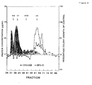
- the major protein peaks were recovered from fractions 30 to 40 (> 45 kD).
- addition of 0.3% (v/v) of these high M r protein containing fractions to the culture medium did not result in an enhancement of colony growth.
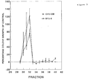
- fractions 30, 31 and 32 were chosen for further purification by RP-HPLC technique. After HPLC separation of fraction 62 the enhancing activity for both GM-colony growth and BFU-E growth was found in fractions 30, 31 and 32 (figure 3). Fraction 32 supported CFU-GM and BFU-E growth upto 75 ⁇ 12% and 125 ⁇ 20% of control cultures, respectively.
- HPLC fractions which contained the highest growth supportive activity, were electrophoretically analyzed for purity by means of SDS/PAGE and the molecular mass was defined as shown in figure 4.
- the HPLC fractions 30, 31 and 32 from gel filtration fraction 62 showed a distinct protein band of 15 kD with the highest concentration in fraction 32(f-32).
- ALP serine proteinase inhibitor Antileukoproteinase
- the second peak of activity from gel filtration was purified by BPLC, yielding again a predominant 15 kD protein (fraction 28), which was also identified as ALP by NH 2 -terminal sequence analysis (45 residues) (figure 5).
- purified recombinant human ALP rALP
- polyclonal anti-huALP antibodies could completely neutralize the biologic activity of 5637 conditioned medium (10% v/v), HPLC fraction-32 (0.1% v/v), and rALP (10 ng/ml).
- Monocyte- and T lymphocyte-depleted bone marrow cells were separated with Dynabeads into CD34 negative cells and a population, containing 80% CD34 positive cells.
- Figure 8 shows the colony growth of these purified CD34 positive cells in the absence or presence of increasing numbers of CD34 negative cells.
- rALP 100 ng/ml
- CM 10% v/v
- the proteinase inhibitor ⁇ -1 PI (2 ⁇ g/ml), present in serum at concentrations of 1-2 mg/ml, could also neutralize the suppression of both CFU-GM and BFU-E growth by CD34 negative cells in the absence of serum.
- Serum-free conditioned medium from the bladder carcinoma cell line 5637 could replace the need for serum or BSA in the semi-solid medium cultures to support the growth of HPC from normal bone marrow.
- Activity-based purification of this conditioned medium led to the isolation of a protein with a molecular mass of 15 kD (SDS/PAGE) which was identified by NH 2 -terminal sequence analysis as Antileukoproteinase, a potent inhibitor of leukocyte elastase.
- SDS/PAGE SDS/PAGE
- Both purified natural and recombinant ALP supported 100% of erythroid colony growth and more than 60% of the myeloid colony growth as compared to the control culture in the presence of 10% AB serum.
- ALP is a 107 amino acid cationic protein consisting of two domains of similar architecture but with different inhibitory activities (18,19).
- the NH 2 -terminal domain ALP-D-1 that is assumed to bind trypsin, could not support growth of HPC in the absence of serum.
- the COOH-terminal domain ALP-D-2 containing a strong inhibitory activity against chymotrypsin, neutrophil elastase and trypsin restored the colony growth indicating that the proteinase inhibitory activity of ALP was indeed responsible for the support of the HPC growth in vitro .
- ALP also known as secretary leukocyte proteinase inhibitor (SLPI)
- SLPI secretary leukocyte proteinase inhibitor
- Serum contains various serine proteinase inhibitors, of which ⁇ -1 proteinase inhibitor, and ⁇ 2-macroglobulin are present at relatively high concentrations of 1.3 mg/ml and 1.7 mg/ml respectively (22); ALP has been found to circulate at a concentration of 50 ng/ml (21).
- ⁇ -1 proteinase inhibitor the main plasma proteinase inhibitor of neutrophil elastase (22)
- the growth support was comparable to rALP.
- the myeloid cells appeared to be the mediator cells for this effect. Probably, these myeloid cells secrete proteolytic proteinases like neutrophil elastase, that are capable of degrading cytokines, growth factor receptors or other proteins essential for the proliferation of HPC (23-25). ALP or other proteinase inhibitors present in serum apparently neutralized these proteolytic enzymes.
- the mechanisms by which ALP supported the proliferation of HPC in vitro may be at the cell membrane level by protecting cellular receptors from degradation, or by protection from degradation of proteins in the culture medium that are essential for HPC growth.
- Colony growth of 150 CD34 positive cells plated per well, supplemented with increasing numbers of CD34 negative cells at ratio's up to 16:1, is expressed as percentage growth of control culture, obtained in the presence of 10% (v/v) AB serum in the absence of CD34 negative accessory cells. Absolute numbers of colonies in the control culture were 8 ⁇ l CFU-GM and 11 ⁇ 1 BFU-E (mean ⁇ SE per 150 cells plated).
- CFU-GM x-x
- bovine serum albumin bovine serum albumin (BSA, M r 67,000), ovalbumin (OV, M r 45,000), chymotrypsinogen (CHYM, M r 25,000) and lysozyme (LYS, M r 14,300).
- BSA bovine serum albumin
- OV ovalbumin
- CHYM chymotrypsinogen
- LYS lysozyme
- Absolute numbers of colonies in the control culture were 12 ⁇ 3 CFU-GM and 16 ⁇ 4 BFU-E (mean ⁇ SE per 10 3 cells plated) ,Fraction f-32 contained the highest level of supportive activity for both the myeloid and erythroid colony growth, 75 ⁇ 12% and 121 ⁇ 20% (mean percentage growth ⁇ SE) respectively.
- Molecular mass markers (Bio-Rad. Richmond, CA) were phosphorylase b (M, 92,500), bovine serum albumin (M, 66,200), ovalbumin (M r 45,000), carbonic anhydrase (M r 31,000), soybean trypsin inhibitor (M r 21,500, lysozyme (M r 14,400) and the low M r marker (Pierce Chemical Company, Rockford, IL) aprotinin (M r 6,500).
- Amino acid sequences were determined by Edman degradation with an on-line Applied Biosystems 477A/120A sequencer. Since cysteine residues cannot be detected unless modified, their presence was deducted from the absence of any detectable amino acid signal at such position.
- AB serum (AB, 1% v/v), 5637 CM (CM, 10 % v/v), purified natural ALP (f-32, 0.1 % v/v), rALP (10 ng/ml) were incubated at 37 ° C during 2 h in the presence of control medium or 40 ⁇ g of polyclonal IgG anti-huALP antibodies, sufficient to neutralize 400 ng rALP.
- Colony growth was expressed as percentage growth of control culture, obtained in the presence of 10% (v/v) AB serum.
- the number of CFU-GM and BFU-E in the control culture was respectively 27 and 28 per 10 3 cells plated respectively.
- In the presence of anti-huALP antibodies colony growth supported by 5637 CM, fraction f-32 or rALP, but not by 1 % (v/v) AB serum, was completely neutralized.
- CD34 negative accessory cells Inhibition of the suppressive effect of CD34 negative accessory cells on HPC growth of purified CD34 positive cells in the absence of serum by proteinase inhibitors.
- the CD34 positive cells were cultured with increasing numbers of CD34 negative cells up to a ratio 1:16 supplemented with IMDM, rALP (100 ng/ml), CM (10 % v/v), AB serum (10% v/v) or ⁇ -1 PI (2 ⁇ g/ml).
- the growth of CFU-GM and BFU-E is expressed as percentage growth of control culture, obtained in the presence of 10% (V/V) AB serum in the absence of CD34 negative cells.
- the results of two experiments (A and B) are shown.
- HGF GM-CSF 10 ng/ml, G-CSF 10 ng/ml, IL-3 50 ng/ml, SCF 50 ng/ml
- EPO 1 IU/ml EPO 1 IU/ml
Landscapes
- Health & Medical Sciences (AREA)
- Life Sciences & Earth Sciences (AREA)
- Chemical & Material Sciences (AREA)
- Organic Chemistry (AREA)
- Engineering & Computer Science (AREA)
- General Health & Medical Sciences (AREA)
- Medicinal Chemistry (AREA)
- Bioinformatics & Cheminformatics (AREA)
- Hematology (AREA)
- Pharmacology & Pharmacy (AREA)
- Genetics & Genomics (AREA)
- Biomedical Technology (AREA)
- Animal Behavior & Ethology (AREA)
- Zoology (AREA)
- Public Health (AREA)
- Veterinary Medicine (AREA)
- Chemical Kinetics & Catalysis (AREA)
- Immunology (AREA)
- Nuclear Medicine, Radiotherapy & Molecular Imaging (AREA)
- Wood Science & Technology (AREA)
- Biochemistry (AREA)
- General Chemical & Material Sciences (AREA)
- Proteomics, Peptides & Aminoacids (AREA)
- Biotechnology (AREA)
- Gastroenterology & Hepatology (AREA)
- Biophysics (AREA)
- Developmental Biology & Embryology (AREA)
- Molecular Biology (AREA)
- Cell Biology (AREA)
- Microbiology (AREA)
- Epidemiology (AREA)
- General Engineering & Computer Science (AREA)
- Oncology (AREA)
- Micro-Organisms Or Cultivation Processes Thereof (AREA)
- Medicines That Contain Protein Lipid Enzymes And Other Medicines (AREA)
Priority Applications (6)
| Application Number | Priority Date | Filing Date | Title |
|---|---|---|---|
| EP96202686A EP0834556A1 (fr) | 1996-09-25 | 1996-09-25 | Procédés de culture de cellules |
| EP97940491A EP0928328A1 (fr) | 1996-09-25 | 1997-09-24 | Procedes destines a la culture de cellules |
| CA002267332A CA2267332A1 (fr) | 1996-09-25 | 1997-09-24 | Procedes destines a la culture de cellules |
| AU42257/97A AU4225797A (en) | 1996-09-25 | 1997-09-24 | Methods for culturing cells |
| JP10515523A JP2001501818A (ja) | 1996-09-25 | 1997-09-24 | 細胞を培養する方法 |
| PCT/NL1997/000532 WO1998013475A1 (fr) | 1996-09-25 | 1997-09-24 | Procedes destines a la culture de cellules |
Applications Claiming Priority (1)
| Application Number | Priority Date | Filing Date | Title |
|---|---|---|---|
| EP96202686A EP0834556A1 (fr) | 1996-09-25 | 1996-09-25 | Procédés de culture de cellules |
Publications (1)
| Publication Number | Publication Date |
|---|---|
| EP0834556A1 true EP0834556A1 (fr) | 1998-04-08 |
Family
ID=8224426
Family Applications (2)
| Application Number | Title | Priority Date | Filing Date |
|---|---|---|---|
| EP96202686A Withdrawn EP0834556A1 (fr) | 1996-09-25 | 1996-09-25 | Procédés de culture de cellules |
| EP97940491A Withdrawn EP0928328A1 (fr) | 1996-09-25 | 1997-09-24 | Procedes destines a la culture de cellules |
Family Applications After (1)
| Application Number | Title | Priority Date | Filing Date |
|---|---|---|---|
| EP97940491A Withdrawn EP0928328A1 (fr) | 1996-09-25 | 1997-09-24 | Procedes destines a la culture de cellules |
Country Status (5)
| Country | Link |
|---|---|
| EP (2) | EP0834556A1 (fr) |
| JP (1) | JP2001501818A (fr) |
| AU (1) | AU4225797A (fr) |
| CA (1) | CA2267332A1 (fr) |
| WO (1) | WO1998013475A1 (fr) |
Cited By (2)
| Publication number | Priority date | Publication date | Assignee | Title |
|---|---|---|---|---|
| US6027937A (en) * | 1998-08-18 | 2000-02-22 | Harper; William | Method for propagation of insecta germ cells |
| FR2794473A1 (fr) * | 1999-06-03 | 2000-12-08 | Centre Nat Rech Scient | Procede de multiplication de cellules souches |
Families Citing this family (1)
| Publication number | Priority date | Publication date | Assignee | Title |
|---|---|---|---|---|
| ES2608937T3 (es) * | 2010-02-22 | 2017-04-17 | Université Pierre Et Marie Curie (Paris 6) | Medio de cultivo celular para el crecimiento y diferenciación de células del linaje hematopoyético |
Citations (8)
| Publication number | Priority date | Publication date | Assignee | Title |
|---|---|---|---|---|
| WO1986003497A1 (fr) * | 1984-12-06 | 1986-06-19 | Synergen Biologicals, Inc. | Inhibiteurs de protease de serine et procedes d'isolation de ceux-ci |
| WO1990002175A1 (fr) * | 1988-08-16 | 1990-03-08 | Novo Nordisk A/S | Procede de production de polypeptides par culture de cellules eukaryotiques en presence d'inhibiteurs de protease |
| EP0402068A1 (fr) * | 1989-06-09 | 1990-12-12 | Zeneca Limited | Polypeptides et analogues de polypeptides ayant une activité inhibitrice vis-à-vis de l'élastase humaine |
| WO1992006706A1 (fr) * | 1990-10-16 | 1992-04-30 | John Lezdey | Traitement d'inflammations |
| EP0525973A2 (fr) * | 1991-06-25 | 1993-02-03 | Merck & Co. Inc. | Azetidinones substituées comme agents antiinflammatoires et antidégénératifs |
| EP0550760A1 (fr) * | 1991-07-18 | 1993-07-14 | Fuji Yakuhin Kogyo Kabushiki Kaisha | Milieu de culture tissulaire sans serum, contenant un inhibiteur tissulaire de metalloproteinase, et procede de croissance cellulaire |
| WO1994003055A1 (fr) * | 1992-07-31 | 1994-02-17 | The Government Of The United States Of America, Asrepresented By The Secretary Of The Department Of Health And Human Services | Production de quantites elevees de cellules hematopoietiques par administration d'inhibiteurs de la dipeptidyle peptidase iv |
| WO1996040866A1 (fr) * | 1995-06-07 | 1996-12-19 | Novartis Ag | Milieux sans serum pour des cellules hematopoietiques primaires et procedes d'utilisation |
Family Cites Families (1)
| Publication number | Priority date | Publication date | Assignee | Title |
|---|---|---|---|---|
| GB2266525A (en) * | 1992-03-17 | 1993-11-03 | Merck & Co Inc | Substituted cephalosporin sulfone compositions useful in the treatment of leukemia |
-
1996
- 1996-09-25 EP EP96202686A patent/EP0834556A1/fr not_active Withdrawn
-
1997
- 1997-09-24 AU AU42257/97A patent/AU4225797A/en not_active Abandoned
- 1997-09-24 CA CA002267332A patent/CA2267332A1/fr not_active Abandoned
- 1997-09-24 JP JP10515523A patent/JP2001501818A/ja active Pending
- 1997-09-24 EP EP97940491A patent/EP0928328A1/fr not_active Withdrawn
- 1997-09-24 WO PCT/NL1997/000532 patent/WO1998013475A1/fr not_active Ceased
Patent Citations (8)
| Publication number | Priority date | Publication date | Assignee | Title |
|---|---|---|---|---|
| WO1986003497A1 (fr) * | 1984-12-06 | 1986-06-19 | Synergen Biologicals, Inc. | Inhibiteurs de protease de serine et procedes d'isolation de ceux-ci |
| WO1990002175A1 (fr) * | 1988-08-16 | 1990-03-08 | Novo Nordisk A/S | Procede de production de polypeptides par culture de cellules eukaryotiques en presence d'inhibiteurs de protease |
| EP0402068A1 (fr) * | 1989-06-09 | 1990-12-12 | Zeneca Limited | Polypeptides et analogues de polypeptides ayant une activité inhibitrice vis-à-vis de l'élastase humaine |
| WO1992006706A1 (fr) * | 1990-10-16 | 1992-04-30 | John Lezdey | Traitement d'inflammations |
| EP0525973A2 (fr) * | 1991-06-25 | 1993-02-03 | Merck & Co. Inc. | Azetidinones substituées comme agents antiinflammatoires et antidégénératifs |
| EP0550760A1 (fr) * | 1991-07-18 | 1993-07-14 | Fuji Yakuhin Kogyo Kabushiki Kaisha | Milieu de culture tissulaire sans serum, contenant un inhibiteur tissulaire de metalloproteinase, et procede de croissance cellulaire |
| WO1994003055A1 (fr) * | 1992-07-31 | 1994-02-17 | The Government Of The United States Of America, Asrepresented By The Secretary Of The Department Of Health And Human Services | Production de quantites elevees de cellules hematopoietiques par administration d'inhibiteurs de la dipeptidyle peptidase iv |
| WO1996040866A1 (fr) * | 1995-06-07 | 1996-12-19 | Novartis Ag | Milieux sans serum pour des cellules hematopoietiques primaires et procedes d'utilisation |
Non-Patent Citations (2)
| Title |
|---|
| GOSELINK H.M. ET AL: "Colony growth of human hematopoietic progenitor cells in the absence of serum is supported by a proteinase inhibitor identified as antileukoproteinase", THE JOURNAL OF EXPERIMENTAL MEDICINE, vol. 184, October 1996 (1996-10-01), pages 1305 - 1312, XP000645063 * |
| R. IAN FRESHNEY: "CULTURE OF ANIMAL CELLS A MANUAL OF BASIC TECHNIQUE", 1988, ALAN R. LISS INC., NEW YORK, XP002026643 * |
Cited By (4)
| Publication number | Priority date | Publication date | Assignee | Title |
|---|---|---|---|---|
| US6027937A (en) * | 1998-08-18 | 2000-02-22 | Harper; William | Method for propagation of insecta germ cells |
| FR2794473A1 (fr) * | 1999-06-03 | 2000-12-08 | Centre Nat Rech Scient | Procede de multiplication de cellules souches |
| WO2000075290A1 (fr) * | 1999-06-03 | 2000-12-14 | Centre National De La Recherche Scientifique | Procede de multiplication de cellules souches |
| US7601534B1 (en) | 1999-06-03 | 2009-10-13 | Centre National De La Recherche Scientifique (Cnrs) | Method for multiplying stem cells |
Also Published As
| Publication number | Publication date |
|---|---|
| EP0928328A1 (fr) | 1999-07-14 |
| JP2001501818A (ja) | 2001-02-13 |
| CA2267332A1 (fr) | 1998-04-02 |
| AU4225797A (en) | 1998-04-17 |
| WO1998013475A1 (fr) | 1998-04-02 |
Similar Documents
| Publication | Publication Date | Title |
|---|---|---|
| Zucali et al. | Recombinant human tumor necrosis factors alpha and beta stimulate fibroblasts to produce hemopoietic growth factors in vitro. | |
| Onozaki et al. | Role of interleukin 1 in promoting human monocyte-mediated tumor cytotoxicity. | |
| Platzer et al. | Biological activities of a human pluripotent hemopoietic colony stimulating factor on normal and leukemic cells. | |
| Jirik et al. | Bacterial lipopolysaccharide and inflammatory mediators augment IL-6 secretion by human endothelial cells. | |
| Murphy et al. | Gamma interferon and lymphotoxin, released by activated T cells, synergize to inhibit granulocyte/monocyte colony formation. | |
| Fibbe et al. | Human fibroblasts produce granulocyte-CSF, macrophage-CSF, and granulocyte-macrophage-CSF following stimulation by interleukin-1 and poly (rI). poly (rC) | |
| Brown et al. | Purification and characterization of cytostatic lymphokines produced by activated human T lymphocytes. Synergistic antiproliferative activity of transforming growth factor beta 1, interferon-gamma, and oncostatin M for human melanoma cells. | |
| Offner et al. | Transforming growth factor-beta synthesis by human peritoneal mesothelial cells. Induction by interleukin-1 | |
| Lanfrancone et al. | Human peritoneal mesothelial cells produce many cytokines (granulocyte colony-stimulating factor [CSF], granulocyte-monocyte-CSF, macrophage-CSF, interleukin-1 [IL-1], and IL-6) and are activated and stimulated to grow by IL-1 | |
| Olsson et al. | Isolation and characterization of a T lymphocyte-derived differentiation inducing factor for the myeloid leukemic cell line HL-60 | |
| Otsuka et al. | Metachromatic cell progenitors and specific growth and differentiation factors in human nasal mucosa and polyps | |
| US6419918B1 (en) | Human pluripotent hematopoietic colony stimulating factor, method of use | |
| Bennicelli et al. | Production of interleukin 1 activity by cultured human melanoma cells | |
| JPH1028595A (ja) | オンコスタチンmを使用したヒト内皮細胞におけるインターロイキン6の合成を誘導する方法 | |
| Holzinger et al. | Effects of interleukin-1,-2,-4,-6, interferon-gamma and granulocyte/macrophage colony stimulating factor on human vascular endothelial cells | |
| Goldfarb et al. | Production of plasminogen activator by human natural killer cells. Large granular lymphocytes. | |
| CA2087170A1 (fr) | Promotion de la maturation de cellules hematopoietiques parentes | |
| US5895646A (en) | Isolated native primate GM-CSF protein | |
| Mochizuki et al. | Biochemical and biologic characterization of lymphocyte regulatory molecules. IV. Purification of Interleukin 2 from a murine T cell lymphoma. | |
| Dohlman et al. | Generation of a unique fibroblast-activating factor by human monocytes | |
| Goselink et al. | Colony growth of human hematopoietic progenitor cells in the absence of serum is supported by a proteinase inhibitor identified as antileukoproteinase. | |
| Caldwell et al. | IL‐1α and TNFα act synergistically to stimulate production of myeloid colony‐stimulating factors by cultured human bone marrow stromal cells and cloned stromal cell strains | |
| Wetzler et al. | Leukemia inhibitory factor in long-term adherent layer cultures: increased levels of bioactive protein in leukemia and modulation by IL-4, IL-1β, and TNF-α | |
| Hori et al. | Regulation of eosinophil migration by adult T cell leukemia-derived factor. | |
| EP0834556A1 (fr) | Procédés de culture de cellules |
Legal Events
| Date | Code | Title | Description |
|---|---|---|---|
| PUAI | Public reference made under article 153(3) epc to a published international application that has entered the european phase |
Free format text: ORIGINAL CODE: 0009012 |
|
| AK | Designated contracting states |
Kind code of ref document: A1 Designated state(s): AT BE CH DE DK ES FI FR GB GR IE IT LI LU MC NL PT SE |
|
| AX | Request for extension of the european patent |
Free format text: LT;LV;SI |
|
| AKX | Designation fees paid | ||
| RBV | Designated contracting states (corrected) | ||
| STAA | Information on the status of an ep patent application or granted ep patent |
Free format text: STATUS: THE APPLICATION IS DEEMED TO BE WITHDRAWN |
|
| 18D | Application deemed to be withdrawn |
Effective date: 19981009 |