EP0843005A2 - Human colon carcinoma cell line with stable HSP72 expression - Google Patents
Human colon carcinoma cell line with stable HSP72 expression Download PDFInfo
- Publication number
- EP0843005A2 EP0843005A2 EP97119900A EP97119900A EP0843005A2 EP 0843005 A2 EP0843005 A2 EP 0843005A2 EP 97119900 A EP97119900 A EP 97119900A EP 97119900 A EP97119900 A EP 97119900A EP 0843005 A2 EP0843005 A2 EP 0843005A2
- Authority
- EP
- European Patent Office
- Prior art keywords
- hsp72
- cell
- colon carcinoma
- cells
- expression
- Prior art date
- Legal status (The legal status is an assumption and is not a legal conclusion. Google has not performed a legal analysis and makes no representation as to the accuracy of the status listed.)
- Withdrawn
Links
Images


Classifications
-
- C—CHEMISTRY; METALLURGY
- C12—BIOCHEMISTRY; BEER; SPIRITS; WINE; VINEGAR; MICROBIOLOGY; ENZYMOLOGY; MUTATION OR GENETIC ENGINEERING
- C12N—MICROORGANISMS OR ENZYMES; COMPOSITIONS THEREOF; PROPAGATING, PRESERVING, OR MAINTAINING MICROORGANISMS; MUTATION OR GENETIC ENGINEERING; CULTURE MEDIA
- C12N5/00—Undifferentiated human, animal or plant cells, e.g. cell lines; Tissues; Cultivation or maintenance thereof; Culture media therefor
- C12N5/06—Animal cells or tissues; Human cells or tissues
- C12N5/0602—Vertebrate cells
- C12N5/0693—Tumour cells; Cancer cells
Definitions
- the invention relates to human colon carcinoma cell lines with a stable HSP72 expression.
- HSP Heat shock proteins
- HSP70 both in the cell nucleus (3), in the cytosol and on the cell surface certain tumor cells (4, 5) is localized.
- Their intracellular function as chaperones appears to be the members of the HSP70 family to stimulate the immune system to be involved, i.e. in the development of certain autoimmune diseases (6, 7), inflammatory processes involving of pathogens (8, 9) and in the cellular anti-tumor immune response in vivo and in vitro (10).
- the invention is illustrated below using the example of the colon carcinoma cell line CX2 explained in more detail. However, she is not on this specific cell line limited. According to the invention, too other colon carcinoma cell lines are used to make up the posed To solve the task.
- Any human colon carcinoma cell lines can be used according to the invention can be used, which are already known per se to the person skilled in the art or the inventive step without further ado by known per se Techniques can be established.
- the present invention was carried out using the example of two human colon carcinoma cell lines, namely the CX2 and HT29 cell lines.
- the CX2 parent cell line is from the German Cancer Research Center in Heidelberg available.
- a tumor profile of this cell line with important genotypic parameters are attached. same for for the HT29 cell line.
- a deposit according to the Budapest Contract is not necessary because the invention based on the existing literature can be reworked repeatedly.
- Farther can, as already mentioned above, any other Human colon carcinoma cell lines are used to make sublines get a stable HSP72 expression of> 80% or ⁇ 20% and a substantially identical cell surface protein expression pattern of MHC and cell adhesion molecules.
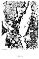
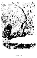
- HSP72 Cryo-ultramicrotome imaging of labeled HSP72 on CX2 human colon carcinoma cells.
- the frozen cuts were made with anti-HSP72 mAb (RPN1197, Amersham) and subsequently with ultra small Gold treated with silver.
- the mark of HSP72 is not due to the subcellular parts in the cytoplasm and limited to the core; HSP72 can also be on the plasma membrane can be demonstrated, as A and B show.
- C shows one Control mark with silver reinforcement without primary antibody.
- the arrow shows the accumulation of HSP72 on the Processes of the plasma membrane; N shows the nucleus (x 43,000).
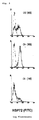
- the results are logarithmic applied, namely the intensity of green fluorescence against the relative cell number.
- the dashed lines show the negative control; the solid line HSP72 mAb.
- the colon carcinoma mother cell line CX2 in to separate a cell line that expresses HSP72 in high quantity (CX +: Expression> 80%, especially> 90%) and one Cell line expressing HSP72 in a small amount (CX-: Expression ⁇ 20%, especially ⁇ 10%).
- CX + Expression> 80%, especially> 90%
- CX- Expression ⁇ 20%, especially ⁇ 10%.
- These sublines were sorted by cells due to the expression of HSP72 on the cell surface obtained with the help of a suitable antibody.
- the previously available human tumor cell lines the disadvantage that they have heat shock proteins over a expressed for a longer period of time, i.e. after a certain time the HSP expression decreased and again a heat shock was necessary to induce expression.
- human colon carcinoma cell lines after sorting the starting cell line with the help of a monoclonal antibody directed against HSP72 in the Are able to stably express HSP72, surprisingly was possible by sorting HSP72 in a lot of> 80% and stably expressing in an amount of ⁇ 20% To get cell lines.
- Such cell lines normally have different characteristics Cell surface protein expression pattern.
- the invention could surprisingly be shown that by cell sorting cell lines obtained from human colon carcinoma cell lines an identical cell surface protein expression pattern from MHC and show cell adhesion molecules.
- the typical Cell adhesion molecules ICAM intercellular cellular adhesion molecule
- NCAM neural cellular adhesion molecule
- VCAM vascular cellular adhesion molecule
- CX2 the cell lines CX + and CX- from a homogeneous, defined mother cell line (CX2) is derived from the HLA background (all HLA alleles) identical; it is an autologous HLA tumor cell system.
- cell lines were thus provided which excellent for examining the function of heat shock proteins the HSP70 family.
- the human colon carcinoma subcell lines obtained by cell separation offer for example the Possibility of the importance of HSP (HLA independent) for the To investigate immunogenicity of tumor cells.
- HSP72 is expressed on the cell surface of colon carcinoma cells and that these cell lines can be split into sublines by cell sorting, which stably express HSP72 in an amount> 80% and ⁇ 20% over more than 20 cell passages.
- Cell surface expression and subcellular distribution of HSP72 was performed by electron microscopic studies using ultra-small gold particles and silver staining.
- the immunoelectron micrograph of Figure 1A shows that HSP72 (black dots) is associated with the plasma membrane of CX2 colon carcinoma cells. As the membrane was permeable before antibody incubation, HSP72 is also detectable in the cytoplasm and in the cell nucleus.
- FIG. 1B The results of the HSP72 distribution on a different section of two colon carcinoma cells are shown in FIG. 1B: The cell in the upper part of the electron micrograph shows an accumulation of HSP72 molecules on extensions of the plasma membrane and in vesicular structures.
- FIG. 1C shows a background staining of three colon carcinoma cells without incubation with the first antibody.
- HSP72 cell surface expression was up CX2 tumor cells by selective biotinylation of the cell surface shown.
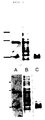
- a comparison of the HSP72 expressing biotinylated Cell surface with recombinant HSP70 shows an intact HSP72 molecule from the cell surface is immunoprecipitable from CX2 cells (FIG. 2, lane B).
- the Figure 2, lane C shows a 45 kDA band with mAB W6 / 32 represents immunoprecipitated class I MHC molecules.
- a Quantification of cell surface expression by HSP72 CX2 cells were identified by indirect immunofluorescence studies using mAB RPN1197 and 3A3 specific for HSP72 and subsequent FACScan analysis. The HSP72 molecules were under physiological conditions constantly expressed on about 60% of the cells (see FIG. 3).
- the CX2 colon carcinoma cell line was sorted by cells in the manner described in material and methods in a more than 80%, preferably more than 90% expressing HSP72 Subline and a less than 20%, preferably less than 10%, HSP72 expressing subline separated.
- Representative Histograms of the phenotypic characterization of HSP72 cell surface expression on CX2, CX + and CX cell lines are in shown in Figure 4.
- adhesion molecules and HSP expression could are shown that the CX2 starting cell line, the CX + and CX sublines only in their HSP72 expression ability on the Differentiate cell surface, but not in the other examined Cell surface markers (see Table 1).
- the CX2, CX + and CX- cells showed cell densities a comparable ICAM-1 expression. This cell surface expression pattern proved over 30 cell passages stable.
- HSP72 Human colon carcinoma cell lines express cell surface expression of HSP72 of about 60% of the cells already under physiological Have conditions.
- This unexpected HSP72 cell surface expression on colon carcinoma cells formed the basis for the splitting according to the invention into two CX2 colon carcinoma sublines, the HSP72 at least more than 80% (CX +) and express less than 20% (CX-) of the cells.
- the Separation into sublines was due to the expression of HSP72 on the cell surface of the colon carcinoma cell lines Cell sorting performed using appropriate antibodies.
- HSP72 expression instructed the cells at comparable cell densities comparable and constant expression pattern of MHC and Adhesion molecules.
- the “autologous" tumor cell system provided according to the invention thus differs only in the expression of an antigen (HSP72), while the expression of MHC and cell adhesion molecules is identical.
- a system is thus available for the first time posed to the role of HSP72 as tumor-specific To investigate the recognition structure in an autologous tumor cell system.
- HSP72 The total length of HSP72 is approximately 700 amino acids. Amino acids 1-450 are located at the N-terminus and amino acids 450-650 at the C-terminus (see FIG. 6). The amino acid sequence of HSP72 is known per se and is published, for example, in Hunt + Morimoto, Proc.Natl.Acad.Sci.USA, 82 (1985), 6455-6459.
- Antibody blocking tests enabled that by NK cells mediated lysis sensitivity correlated with the amount of HSP72 that is expressed on the cell surface. Under use of the monoclonal antibody specific for HSP72 the strongest inhibition was found in CX + cells, one medium lysis inhibition in the original CX2 tumor cell line and no lysis inhibition on the CX cell subline.
- a IgG1 isotype control antibody and an MHC class I specific Antibody showed no inhibitory effect on the lysis pattern in the three tumor cell lines named CX2, CX + and CX-.
- the "autologous" tumor cell system differs only in the expression of a single antigen; thus the investigation of the role of HSP72 enabled as a tumor-specific recognition structure.
- literature (16) shows that members of the HSP70 family a cellular immune response both in vivo and in vitro cause. Therefore, the lysis sensitivity according to the invention the "autologous" colon carcinoma sublines according to the invention, which are only in their HSP72 expression on the cell surface distinguish, compared.
- an NK-enriched effector cell population was used for cytotoxicity studies in these colon carcinoma sublines.
- the heat-independent cell surface expression of HSP72 on CX2 and CX + colon carcinoma cells correlated closely with that Lysis sensitivity mediated by NK cells.
- HSP72 is therefore an additional factor which determines the sensitivity of tumor target cells for lysis mediated by NK cells.
- the human colon carcinoma cell line CX2 (tumor bank DKFZ Heidelberg) and the subclones CX + and CX- were in RPMI 1640 medium (Gibco, Eggenstein), supplemented with 10% heat-inactivated fetal calf serum (FCS, Gibco, Eggenstein) and antibiotics, cultured.
- the cells to be analyzed were washed twice with cold PBS / 10% FCS medium.
- the cell pellet (5 ⁇ 10 6 cells) was resuspended and incubated for 30 minutes on ice with 5 ⁇ l RPN1197 (anti-HSP72 mAb; Amersham; CODE RPN 1197).
- RPN1197 anti-HSP72 mAb; Amersham; CODE RPN 1197.
- the cells were washed again twice with cold PBS / 10% FCS medium.
- FITC fluorescein isothiocyanate
- DAKO fluorescein isothiocyanate
- FITC antibody was removed by washing twice, then the cells were resuspended in 2 ml of cold PBS / 10% FCS medium and stored on ice in the dark until separated. To distinguish living and dead cells, 10 ⁇ l propidium iodide (PI; 50 ⁇ g / ml) was added to the samples. With the help of the FACS-Star sorter, the "mother cell line" CX2 was separated into two daughter lines due to the fluorescent labeling: a CX line that expresses less than 20% HSP72 on the cell surface or a CX + line that more than 80 % HSP72 expressed.
- PI propidium iodide
- Exponentially growing CX2 cells are used in the non-lethal Temperature of 41.8 ° C for two hours in a temperature controlled Water bath (Haake E3, Düsseldorf) treated and then incubated at 37 ° C for twelve hours.
- the survival rate of the heat treated cells were always over 95% as determined by Trypan blue exclusion and PI staining.
- the flow cytometric studies were carried out with exponentially growing cells and at comparable cell densities. Viable cells (1x10 6 ) were incubated with one of the antibodies shown above to a final concentration of 1-5 ⁇ g / 0.5-1x10 6 cells for 30 minutes at 4 ° C.
- the antibodies used for FACScan analysis and cell sorting contained 0.1% sodium azide; the antibodies used for the antibody blocking studies did not contain sodium azide.
- the cells were washed twice with PBS / 10% FCS solution and with a second, fluorescein isothiocyanate-conjugated (FITC) rabbit anti-mouse Ig antibody (FITC, DAKO, Hamburg) for a further 30 Incubated for minutes at 4 ° C.
- the percentage of positively stained cells was defined as the difference in the number of specifically stained cells minus the number of cells stained with control antibodies matched to the isotype.
- the values obtained from the FACScan analysis represent mean values from at least 4 independent tests.
- HSP72 mAB RPN1197 (Amersham) antibody
- the antibody selectively detects HSP72 and does not cross-react with HSP73.
- HSP70 cytoplasmic expression was detectable with the following antibodies: RPN1197 (Amersham), 3A3 (S. Fox), C92F3A-5 (StressGen) and 5G10 (Pharmagen).
- Cell surface staining and cell sorting of the tumor cells was performed with mAb RPN1197 (Amersham).
- the HSP72 high (CX +) and low (CX-) expressing colon carcinoma sublines were cultivated separately after the cell sorting.
- the cell sorting of the effector cells was via CD3 and CD14 carried out.
- the negatively separated CD3 effector cell population was stimulated with 100 IU rIL-2 and for functional examinations used.
- the phenotypic characterization of this Cells were grown using CD3 / CD16 + 56 and CD14 / CD45 double stained antibodies.
- CX2 tumor cells washed with PBS were fixed in 8% paraformaldehyde in Hepes (250 mM) buffer for 1 hour. After two washes, free aldehyde groups in 50 mM NH 4 Cl were deleted for 10 minutes.
- the cell pellets were kept in a volume of 2.1 M sucrose in polyvinylpyrrolidone (17%) at 20 ° C for 30 minutes in suspended form for cryoprotection.
- the cell pellets were then frozen in liquid nitrogen and ultra-thin sections (70 nm) were cut at -100 ° C on an ultracut microtome (Reichert-Jung FC4E) using a glass knife and attached to 150 mesh grids made of parlodione-coated nickel.
- the immuno-gold labeling of HSP72 was carried out using ultra-small gold samples and subsequent silver enhancement according to the method described by Danscher (12).
- viable cells (2x10 7 ) were biotinylated with biotin ester (RPN2202, Amersham) for 30 minutes at 4 ° C. After washing, the cells were kept in ice-cold lysis buffer (0.1% NP40; 250 mM NaCl; 25 mM Tris-HCl, pH 7.5; 5 mM EDTA, 2 ⁇ g / ml aprotinin, 100 ⁇ g / ml PMSF) for 20 minutes. lysed.
- biotin ester RPN2202, Amersham
- the extracts were purified by microcentrifugation at 4 ° C for 15 minutes and with HSP72 (10 ⁇ l) or with W6 / 32 nAb (2 ⁇ g) and 50 ⁇ l gamma-bind plus protein G-Sepharose beads for 3 hours at 4 ° C immunoprecipitated.
- the conjugated, antigen-loaded Sepharose beads were then washed three times in ice-cold lysis buffer and resuspended in 75 ⁇ l sample buffer (200 mM DTT; 20% glycerol; 0.12 M Tris; bromophenol blue).
- the proteins from the Sepharose beads were separated on 10% SDS-PAGE, blotted on a PVDF membrane and detected by ECL after incubation with a secondary peroxidase-conjugated streptavidin antibody.
- Equal amounts of protein (10 ⁇ g) from cell lysates and 50 ng rHSP70 (StressGen, Victoria, Canada) were on 10% SDS-PAGE as in (5), according to the method of Laemmli (13) one Subjected to electrophoresis.
- the proteins on Immobilon PVDF membrane (Millipore Corp., Bedford, MA) transmitted according to a standard protocol (14).
- the non-specific Immobilon PVDF membrane was bound with 5% skim milk blocked in PBS.
- the blots were made with the primary HSP72 antibody (Amersham) and the secondary antibody (goat anti-mouse IgG peroxidase conjugated, BioRad) 1 hour each incubated for a long time.
- the inhibition tests were carried out by pre-incubating the target cells with the following antibodies at a final concentration of 5 ⁇ g / 1x10 6 cells for each antibody: RPN1197 (anti-HSP72), an isotype-matched IgG1 control antibody and W6 / 32 (15) . After incubation, the cells were washed twice with RPMI1640 / 10% FCS and the cytotoxicity test was performed as described above.
- the results obtained for the CX2 cell line were also satisfactory confirm HT29 for the human colon carcinoma cell lines.
- the cell line HT29 is commercially available from AMERICAN TISSUE TYPE CULTURE COLLECTION available under the number ATCC HTB38.
- the genotypic Description is from the ATCC catalog and the one listed there Literature evident.
- FIGS. 9A to C show the results of the HT29 cell sorting. The attempt was made according to the Cell sorting of the CX2 cell line performed.
- the HT29 origin cell line expresses on its cell surface HSP72 in an amount of 30 to 40% of the cells.
- an HT29 + cell line could be obtained which was based on their cell surface expressed HSP72 in 87% of the cells, and an HT29 cell line that expresses HSP72 in 15% of the cells.
- the HT29 sublines obtained show stable HSP72 expression over at least 20 cell passages as well as an identical MHC and Cell adhesion molecule cell surface expression pattern.
Landscapes
- Health & Medical Sciences (AREA)
- Engineering & Computer Science (AREA)
- Life Sciences & Earth Sciences (AREA)
- Biomedical Technology (AREA)
- Wood Science & Technology (AREA)
- Zoology (AREA)
- Organic Chemistry (AREA)
- Bioinformatics & Cheminformatics (AREA)
- Genetics & Genomics (AREA)
- Biotechnology (AREA)
- Chemical & Material Sciences (AREA)
- Biochemistry (AREA)
- Oncology (AREA)
- Microbiology (AREA)
- General Engineering & Computer Science (AREA)
- General Health & Medical Sciences (AREA)
- Cell Biology (AREA)
- Micro-Organisms Or Cultivation Processes Thereof (AREA)
- Peptides Or Proteins (AREA)
- Medicines Containing Antibodies Or Antigens For Use As Internal Diagnostic Agents (AREA)
- Medicines Containing Material From Animals Or Micro-Organisms (AREA)
- Medicines That Contain Protein Lipid Enzymes And Other Medicines (AREA)
Abstract
Description
Die Erfindung betrifft Human-Coloncarcinom-Zellinien mit einer stabilen HSP72-Expression.The invention relates to human colon carcinoma cell lines with a stable HSP72 expression.
Chaperone sind für eine Anzahl fundamentaler Prozesse notwendig, insbesondere, um dem Zellstreß entgegenzuwirken. Die am besten untersuchte Klasse von Chaperonen ist die Gruppe der Hitzeschock-Proteine (HSP) mit einem Molekulargewicht von 70 kDa. Sie binden intrazellulär an nicht native Konformationen anderer Proteine, stabilisieren sie und inhibieren auf diese Weise die Aggregation ungefalteter Proteine oder ermöglichen eine Transmembran-Translokation (1, 2). Es wurde beschrieben, daß HSP70 sowohl im Zellkern (3), im Cytosol und an der Zelloberfläche bestimmter Tumorzellen (4, 5) lokalisiert ist. Neben ihrer intrazellulären Aufgabe als Chaperone scheinen die Mitglieder der HSP70-Familie an der Stimulierung des Immunsystems beteiligt zu sein, d.h. bei der Entstehung bestimmter Autoimmunerkrankungen (6, 7), entzündlicher Prozesse unter Beteiligung von Pathogenen (8, 9) und bei der zellulären anti-Tumorimmunantwort in vivo und in vitro (10).Chaperones are necessary for a number of fundamental processes, especially to counteract cell stress. The most The best studied class of chaperones is the group of Heat shock proteins (HSP) with a molecular weight of 70 kDa. They bind intracellularly to non-native conformations other proteins, stabilize them and inhibit them Way to aggregate unfolded proteins or enable a transmembrane translocation (1, 2). It has been described that HSP70 both in the cell nucleus (3), in the cytosol and on the cell surface certain tumor cells (4, 5) is localized. Next Their intracellular function as chaperones appears to be the members of the HSP70 family to stimulate the immune system to be involved, i.e. in the development of certain autoimmune diseases (6, 7), inflammatory processes involving of pathogens (8, 9) and in the cellular anti-tumor immune response in vivo and in vitro (10).
Die Gründe, weshalb hochkonservierte Proteine wie die Hitzeschockproteine immundominant sind, sind weiterhin unbekannt. Eine Zusammenfassung der zur Zeit diskutierten Hypothesen ist (11) zu finden.The reasons why highly conserved proteins like heat shock proteins are immunodominant, are still unknown. A summary of the hypotheses currently under discussion is (11) to find.
Für alle immunstimulatorischen Funktionen scheint es notwendig zu sein, daß das HSP für immunkompetente Effektorzellen zugänglich ist. Untersuchungen zeigen, daß eine hitzeinduzierte Membran-Expression von HSP72 auf Human-Sarkomzellen mit einer erhöhten Lysesensitivität korrelliert, die durch natürliche Killerzellen (NK-Zellen) bewirkt wird. Die Tatsache, daS HSP72 nur auf der Zelloberfläche von Tumoren exprimiert wird, nicht dagegen auf normalen Zellen, zeigt, daß HSP als tumorspezifische Targetstrukturen für immunkompetente Effektorzellen wirken können.It seems necessary for all immunostimulatory functions to be the HSP accessible to immunocompetent effector cells is. Studies show that heat-induced membrane expression of HSP72 on human sarcoma cells with an increased Lysis sensitivity correlated by natural killer cells (NK cells) is effected. The fact that HSP72 only is expressed on the cell surface of tumors, but not against it on normal cells, shows that HSP is tumor-specific Target structures for immunocompetent effector cells can act.
Es ist eine Aufgabe der vorliegenden Erfindung, HSP72 exprimierende Zellinien bereitzustellen, die die Möglichkeit bieten, vergleichende Untersuchungen, beispielsweise über die Bedeutung von HSP72 für die Immunogenität von Tumorzellen, durchzuführen.It is an object of the present invention to express HSP72 To provide cell lines that offer the possibility of comparative studies, for example on the meaning of HSP72 for the immunogenicity of tumor cells.
Diese Aufgabe wird erfindungsgemäß durch die im Anspruch 1 näher
gekennzeichneten Merkmale gelöst. Bevorzugte Ausführungsformen
der Erfindung ergeben sich aus den Unteransprüchen.This object is achieved by the invention in
Zwei bevorzugte Zellinien wurden bei der DSM am 29. Oktober 1996 hinterlegt:Two preferred cell lines were used at the DSM on October 29th 1996 deposited:
Eine Human-Coloncarcinom-Zellinie CX+ mit einer stabilen Expression von HSP72 an der Zelloberfläche von > 80%, bevorzugt > 90%, mit der Zugangsnummer DSM ACC 2287. Eine Human-Coloncarcinom-Zellinie CX- mit einer stabilen Expression von HSP72 an der Zelloberfläche von < 20%, bevorzugt < 10%, mit der Zugangsnummer DSM ACC 2288.A human colon carcinoma cell line CX + with a stable expression of HSP72 on the cell surface of> 80%, preferably> 90%, with the accession number DSM ACC 2287. A human colon carcinoma cell line CX - with a stable expression of HSP72 on the cell surface of <20%, preferably <10%, with the access number DSM ACC 2288.
Die Erfindung wird nachfolgend am Beispiel der Coloncarcinom-Zellinie CX2 näher erläutert. Sie ist jedoch nicht auf diese spezifische Zellinie beschränkt. Erfindungsgemäß können auch andere Coloncarcinom-Zellinien benutzt werden, um die gestellte Aufgabe zu lösen.The invention is illustrated below using the example of the colon carcinoma cell line CX2 explained in more detail. However, she is not on this specific cell line limited. According to the invention, too other colon carcinoma cell lines are used to make up the posed To solve the task.
Erfindungsgemäß können beliebige Human-Coloncarcinom-Zellinien verwendet werden, die dem Fachmann bereits an sich bekannt sind oder die ohne weiteres erfinderisches Zutun durch an sich bekannte Techniken etablierbar sind. Die vorliegende Erfindung wurde am Beispiel zweier Human-Coloncarcinom-Zellinien durchgeführt, nämlich der CX2- und der HT29-Zellinien. Die CX2-Ausgangszellinie ist vom Deutschen Krebsforschungszentrum in Heidelberg erhältlich. Ein Tumorsteckbrief dieser Zellinie mit wichtigen genotypischen Parametern ist beigefügt. Gleiches gilt für die Zellinie HT29. Eine Hinterlegung gemäß dem Budapester Vertrag ist deshalb nicht erforderlich, da die Erfindung anhand der vorliegenden Literatur wiederholbar nacharbeitbar ist. Weiterhin können, wie bereits oben angeführt, beliebige andere Human-Coloncarcinom-Zellinien verwendet werden, um Sublinien zu erhalten, die eine stabile HSP72-Expression von > 80% oder < 20% und ein im wesentlichen identisches Zelloberflächen-Protein-Expressionsmuster von MHC und Zelladhäsionsmolekülen besitzen.Any human colon carcinoma cell lines can be used according to the invention can be used, which are already known per se to the person skilled in the art or the inventive step without further ado by known per se Techniques can be established. The present invention was carried out using the example of two human colon carcinoma cell lines, namely the CX2 and HT29 cell lines. The CX2 parent cell line is from the German Cancer Research Center in Heidelberg available. A tumor profile of this cell line with important genotypic parameters are attached. same for for the HT29 cell line. A deposit according to the Budapest Contract is not necessary because the invention based on the existing literature can be reworked repeatedly. Farther can, as already mentioned above, any other Human colon carcinoma cell lines are used to make sublines get a stable HSP72 expression of> 80% or <20% and a substantially identical cell surface protein expression pattern of MHC and cell adhesion molecules.
Nachfolgend wird die Erfindung unter Bezugnahme auf die beiliegenden Figuren näher erläutert. Die Figuren zeigen:The invention will now be described with reference to the accompanying drawings Figures explained in more detail. The figures show:
Kryo-Ultramikrotom-Abbildung von markiertem HSP72 auf CX2-Human-Coloncarcinomzellen. Die gefrorenen Schnitte wurden mit anti-HSP72 mAb (RPN1197, Amersham) und nachfolgend mit ultrakleinem Gold, das mit Silber verstärkt war, behandelt. Die Markierung von HSP72 ist nicht auf die subzellulären Teile im Zytoplasma und dem Kern beschränkt; HSP72 kann auch auf der Plasmamembran nachgewiesen werden, wie A und B zeigen. C zeigt eine Kontroll-Markierung mit Silberverstärkung ohne primären Antikörper. Der Pfeil zeigt die Anreicherung von HSP72 auf den Fortsätzen der Plasmamembran; N zeigt den Kern (x 43.000).Cryo-ultramicrotome imaging of labeled HSP72 on CX2 human colon carcinoma cells. The frozen cuts were made with anti-HSP72 mAb (RPN1197, Amersham) and subsequently with ultra small Gold treated with silver. The mark of HSP72 is not due to the subcellular parts in the cytoplasm and limited to the core; HSP72 can also be on the plasma membrane can be demonstrated, as A and B show. C shows one Control mark with silver reinforcement without primary antibody. The arrow shows the accumulation of HSP72 on the Processes of the plasma membrane; N shows the nucleus (x 43,000).
SDS-PAGE-Analyse von HSP72, das von der Zelloberfläche biotinylierter
CX2-Tumorzellen immunpräzipitiert wurde.
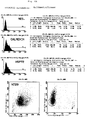
HSP72-Zelloberflächenexpression auf CX2-Coloncarcinomzellen nach durchflußzytometrischer Zellsortierung unter Verwendung eines HSP72 spezifischen mAb. Die Ergebnisse sind logarithmisch aufgetragen, und zwar die Intensität der grünen Fluoreszenz gegen die relative Zellzahl. Die gestrichelten Linien zeigen die Negativ-Kontrolle; die durchgezogene Linie HSP72 mAb.HSP72 cell surface expression on CX2 colon carcinoma cells after flow cytometric cell sorting using of a HSP72 specific mAb. The results are logarithmic applied, namely the intensity of green fluorescence against the relative cell number. The dashed lines show the negative control; the solid line HSP72 mAb.
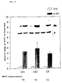
HSP72-Immunblot von Zytoplasmalysaten von CX+, CX2 und CX- Carcinomzellen
unter physiologischen Bedingungen (offene Balken)
und nach einem milden Hitzestreß bei 41,8°C, 2 Stunden und
nachfolgender Erholungsphase bei 37°C für 2 Stunden (schraffierte
Balken). HSP72-Proteinmengen (mit einem Pfeil markiert)
wurden auf Aktin (mit "a" bezeichnet) bezogen. Gleiche Proteinmengen
(10 µg der Zellysate) wurden auf einer 10% SDS-PAGE unter
reduzierenden Bedingungen elektrophoretisch getrennt und
auf Nitrozellulose übertragen. Die 72 kDa-Proteinbande wurde
mit dem HSP72 spezifischen mAb dargestellt und mit dem ECL-System
nachgewiesen. Die Immunblots wurden mit Laser-Densitometrie
quantifiziert. Die Ergebnisse zeigen den Mittelwert aus
zumindest zwei unabhängigen Versuchen.
Beispiel einer HSP72-Sortierung mit Einstellungsparametern.Example of an HSP72 sorting with setting parameters.
Erfindungsgemäß konnte die Coloncarcinom-Mutter-Zellinie CX2 in eine Zellinie getrennt werden, die HSP72 in hoher Menge exprimiert (CX+: Expression > 80%, insbesondere > 90%) und eine HSP72 in geringer Menge exprimierende Zellinie (CX-: Expression < 20%, insbesondere < 10%). Diese Sublinien wurden durch Zellsortierung aufgrund der Expression von HSP72 an der Zelloberfläche mit Hilfe eines geeigneten Antikörpers gewonnen. Die bisher zur Verfügung stehenden menschlichen Tumorzellinien wiesen den Nachteil auf, daß sie Hitzeschockproteine über einen längeren Zeitraum nicht stabil exprimierten, d.h. nach einer bestimmten Zeit die HSP-Expression abnahm und erneut ein Hitzeschock notwendig war, um die Expression zu induzieren.According to the invention, the colon carcinoma mother cell line CX2 in to separate a cell line that expresses HSP72 in high quantity (CX +: Expression> 80%, especially> 90%) and one Cell line expressing HSP72 in a small amount (CX-: Expression <20%, especially <10%). These sublines were sorted by cells due to the expression of HSP72 on the cell surface obtained with the help of a suitable antibody. The previously available human tumor cell lines the disadvantage that they have heat shock proteins over a expressed for a longer period of time, i.e. after a certain time the HSP expression decreased and again a heat shock was necessary to induce expression.
Erfindungsgemäß konnte gezeigt werden, daß Human-Coloncarcinom-Zellinien nach Zellsortierung der Ausgangszellinie mit Hilfe eines gegen HSP72 gerichteten monoklonalen Antikörpers in der Lage sind, HSP72 stabil zu exprimieren, wobei es überraschenderweise möglich war, durch Zellsortierung HSP72 in einer Menge von > 80% und in einer Menge von < 20% stabil exprimierende Zellinien zu erhalten.According to the invention it could be shown that human colon carcinoma cell lines after sorting the starting cell line with the help of a monoclonal antibody directed against HSP72 in the Are able to stably express HSP72, surprisingly was possible by sorting HSP72 in a lot of> 80% and stably expressing in an amount of <20% To get cell lines.
Dieses Ergebnis war deshalb so unerwartet, da bei der Mutterzellinie ursprünglich eine über mehr als 20 getestete Zellpassagen stabile HSP72-Zelloberflächenexpression von > 60% nachweisbar war. Dieser Befund legt den Schluß nahe, daß diese 60 %-ige Expressionsrate beispielsweise für das Überleben, für den Zellzyklus, für die Zelladhäsion, usw. unverzichtbar ist.This result was so unexpected because of the mother cell line originally one of more than 20 cell passages tested stable HSP72 cell surface expression of> 60% detectable was. This finding suggests that this 60th % expression rate for survival, for example Cell cycle for which cell adhesion, etc. is essential.
Es war deshalb zu erwarten, daß nach Zellseparation von CX2-Zellen in eine HSP72 in hoher Menge exprimierende Sublinie und in eine HSP72 in geringer Menge exprimierende Sublinie sich beide Subzellinien nach wenigen Zellpassagen wieder bei einer ca. 60 %-igen HSP72-Zelloberflächenexpressionsrate einpendeln. Überraschenderweise war dies jedoch nicht der Fall. Beide Zellinien, CX+ und CX-, wiesen ein über mehr als 20 Zellpassagen stabiles Expressionsmuster der HSP72-Zelloberflächenexpression auf: CX+ > 80%, CX- < 20%.It was therefore expected that after cell separation of CX2 cells into a high-level subline expressing HSP72 and into a subline expressing HSP72 in small amounts after a few cell passages, both subcell lines again at one Level out approx. 60% HSP72 cell surface expression rate. Surprisingly, however, this was not the case. Both cell lines, CX + and CX- had over 20 cell passages stable expression pattern of HSP72 cell surface expression on: CX +> 80%, CX- <20%.
Normalerweise weisen derartige Zellinien ein unterschiedliches Zelloberflächenproteinexpressionsmuster auf. Erfindungsgemäß konnte überraschenderweise gezeigt werden, daß durch Zellsortierung von Human-Coloncarcinom-Zellinien erhaltene Zellinien ein identisches Zelloberflächenproteinexpressionsmuster von MHC und Zelladhäsionsmolekülen zeigen. Hierbei wurden die typischen Zelladhäsionsmoleküle ICAM (intercellular cellular adhesion molecule), NCAM (neural cellular adhesion molecule) und VCAM (vascular cellular adhesion molecule) sowie MHC Klasse I und Klasse II auf ihre Expression untersucht.Such cell lines normally have different characteristics Cell surface protein expression pattern. According to the invention could surprisingly be shown that by cell sorting cell lines obtained from human colon carcinoma cell lines an identical cell surface protein expression pattern from MHC and show cell adhesion molecules. Here the typical Cell adhesion molecules ICAM (intercellular cellular adhesion molecule), NCAM (neural cellular adhesion molecule) and VCAM (vascular cellular adhesion molecule) as well as MHC class I and Class II examined for their expression.
Die erfindungsgemäßen Coloncarcinom-Zellinien unterscheiden sich somit nur in ihrem HSP72-Zelloberflächenexpressionsmuster, während sie in ihrem untersuchten MHC- und Zelladhäsionsmolekül-Zelloberflächenexpressionsmuster identisch sind.Differentiate the colon carcinoma cell lines according to the invention thus only in their HSP72 cell surface expression pattern, while in their examined MHC and cell adhesion molecule cell surface expression patterns are identical.
Da die vorliegend beispielhaft bereitgestellten Zellinien CX+ und CX- aus einer homogenen, definierten Mutterzellinie (CX2) abgeleitet wurden, ist der HLA-Hintergrund (alle HLA Allele) identisch; es handelt sich also um ein autologes HLA-Tumorzellsystem. Since the cell lines CX + and CX- from a homogeneous, defined mother cell line (CX2) is derived from the HLA background (all HLA alleles) identical; it is an autologous HLA tumor cell system.
Erfindungsgemäß wurden somit Zellinien bereitgestellt, die sich hervorragend zur Untersuchung der Funktion von Hitzeschockproteinen der HSP70-Familie eignen.According to the invention, cell lines were thus provided which excellent for examining the function of heat shock proteins the HSP70 family.
Die durch Zellseparation gewonnenen Human-Coloncarcinom-Subzellinien, beispielsweise CX+ und CX-, bieten beispielsweise die Möglichkeit, die Bedeutung von HSP (HLA unabhängig) für die Immunogenität von Tumorzellen zu untersuchen. Weiterhin können die erfindungsgemäß bereitgestellten Zellinien, beispielsweise CX+ und CX-, als Feederzellen für NK-Klonierungen und zur Selektion der geeigneten Zellen verwendet werden (NK-Klone mit HSP72-Spezifität). Mit diesen Klonen könnten Rezeptoranalysen durchgeführt werden, um herauszufinden, ob auf NK-Zellen ein HSP72-spezifischer Rezeptor existiert.The human colon carcinoma subcell lines obtained by cell separation, for example CX + and CX-, offer for example the Possibility of the importance of HSP (HLA independent) for the To investigate immunogenicity of tumor cells. Can continue the cell lines provided according to the invention, for example CX + and CX-, as feeder cells for NK cloning and for selection of the appropriate cells (NK clones with HSP72 specificity). With these clones, receptor analysis could be done be done to find out if there is any on NK cells HSP72-specific receptor exists.
Erfindungsgemäß konnte somit gezeigt werden, daß HSP72 auf der
Zelloberfläche von Coloncarcinomzellen exprimiert wird und diese
Zellinien durch Zellsortierung in Sublinien aufspaltbar
sind, die HSP72 in einer Menge > 80% und < 20% stabil über mehr
als 20 Zellpassagen exprimieren. Die Zelloberflächenexpression
und die subzelluläre Verteilung von HSP72 wurde durch elektronenmikroskopische
Untersuchungen unter Verwendung von ultrakleinen
Goldteilchen und Silberfärbung durchgeführt. Die immunelektronenmikroskopische
Aufnahme der Figur 1A zeigt, daß HSP72
(schwarze Punkte) mit der Plasamembran von CX2-Coloncarcinomzellen
assoziiert ist. Da die Membran vor Antikörperinkubation
permeabel war, ist HSP72 auch im Zytoplasma und im Zellkern
nachweisbar. Die Ergebnisse der HSP72-Verteilung auf einem unterschiedlichen
Abschnitt von zwei Coloncarcinomzellen ist in
der Figur 1B dargestellt:
Die Zelle im oberen Teil der elektronenmikroskopischen Aufnahme
zeigt eine Anreicherung von HSP72-Molekülen auf Fortsätzen der
Plasmamembran und in vesikulären Strukturen. Die Figur 1C zeigt
eine Hintergrundsfärbung von drei Coloncarcinomzellen ohne Inkubation
mit dem ersten Antikörper.According to the invention, it was thus possible to show that HSP72 is expressed on the cell surface of colon carcinoma cells and that these cell lines can be split into sublines by cell sorting, which stably express HSP72 in an amount> 80% and <20% over more than 20 cell passages. Cell surface expression and subcellular distribution of HSP72 was performed by electron microscopic studies using ultra-small gold particles and silver staining. The immunoelectron micrograph of Figure 1A shows that HSP72 (black dots) is associated with the plasma membrane of CX2 colon carcinoma cells. As the membrane was permeable before antibody incubation, HSP72 is also detectable in the cytoplasm and in the cell nucleus. The results of the HSP72 distribution on a different section of two colon carcinoma cells are shown in FIG. 1B:
The cell in the upper part of the electron micrograph shows an accumulation of HSP72 molecules on extensions of the plasma membrane and in vesicular structures. FIG. 1C shows a background staining of three colon carcinoma cells without incubation with the first antibody.
Weiterhin wurde die Zelloberflächenexpression von HSP72 auf CX2-Tumorzellen durch selektive Biotinylierung der Zelloberfläche gezeigt. Ein Vergleich der HSP72 exprimierenden biotinylierten Zelloberfläche mit rekombinantem HSP70 (Fig. 2, Spur A) zeigt, daß ein intaktes HSP72-Molekül von der Zelloberfläche von CX2-Zellen immunpräzipitierbar ist (Fig. 2, Spur B). Die Figur 2, Spur C zeigt eine 45 kDA große Bande, die mit mAB W6/32 immunpräzipitierte Klasse I-MHC-Moleküle darstellt. Eine Quantifizierung der Zelloberflächenexpression von HSP72 auf CX2-Zellen wurde durch indirekte Immunfluoreszenzuntersuchungen unter Verwendung von mAB RPN1197 und 3A3, die für HSP72 spezifisch sind, und nachfolgender FACScan-Analyse durchgeführt. Unter physiologischen Bedingungen wurden die HSP72-Moleküle konstant auf etwa 60% der Zellen exprimiert (vgl. Fig. 3).Furthermore, the cell surface expression of HSP72 was up CX2 tumor cells by selective biotinylation of the cell surface shown. A comparison of the HSP72 expressing biotinylated Cell surface with recombinant HSP70 (Fig. 2, lane A) shows an intact HSP72 molecule from the cell surface is immunoprecipitable from CX2 cells (FIG. 2, lane B). The Figure 2, lane C shows a 45 kDA band with mAB W6 / 32 represents immunoprecipitated class I MHC molecules. A Quantification of cell surface expression by HSP72 CX2 cells were identified by indirect immunofluorescence studies using mAB RPN1197 and 3A3 specific for HSP72 and subsequent FACScan analysis. The HSP72 molecules were under physiological conditions constantly expressed on about 60% of the cells (see FIG. 3).
Erfindungsgemäß wurde die CX2-Coloncarcinomzellinie durch Zellsortierung in der in Material und Methoden beschriebenen Weise in eine mehr als 80%, bevorzugt mehr als 90%, HSP72 exprimierende Sublinie und eine weniger als 20%, bevorzugt weniger als 10%, HSP72 exprimierende Sublinie aufgetrennt. Repräsentative Histogramme der phänotypischen Charakterisierung der HSP72-Zelloberflächenexpression auf CX2, CX+ und CX- Zellinien sind in der Figur 4 dargestellt. Durch vergleichende flußzytometrische Analyse von MHC-, Adhäsionsmolekülen und HSP-Expression konnte gezeigt werden, daß sich die CX2-Ausgangszellinie, die CX+ und CX- Sublinien nur in ihrer HSP72-Expressionsfähigkeit auf der Zelloberfläche unterscheiden, nicht dagegen in den anderen untersuchten Zelloberflächenmarkern (vgl. Tabelle 1). Bei vergleichbaren Zelldichten zeigten die CX2, CX+ und CX- Zellen eine vergleichbare ICAM-1-Expression. Dieses Zelloberflächenexpressionsmuster erwies sich über mehr als 30 Zellpassagen stabil.According to the invention, the CX2 colon carcinoma cell line was sorted by cells in the manner described in material and methods in a more than 80%, preferably more than 90% expressing HSP72 Subline and a less than 20%, preferably less than 10%, HSP72 expressing subline separated. Representative Histograms of the phenotypic characterization of HSP72 cell surface expression on CX2, CX + and CX cell lines are in shown in Figure 4. By comparative flow cytometric Analysis of MHC, adhesion molecules and HSP expression could are shown that the CX2 starting cell line, the CX + and CX sublines only in their HSP72 expression ability on the Differentiate cell surface, but not in the other examined Cell surface markers (see Table 1). With comparable The CX2, CX + and CX- cells showed cell densities a comparable ICAM-1 expression. This cell surface expression pattern proved over 30 cell passages stable.
Im Gegensatz zu den deutlichen Unterschieden in der Menge der Expression von HSP72 auf der Zelloberfläche konnte durch Flußzytometrie und Elektronenmikroskopie gezeigt werden, daß der zytoplasmatische Anteil von HSP72 in der CX2-Ausgangszellinie und den Sublinien CX+ und CX- unter physiologischen Bedingungen und nach einer milden Hitzebehandlung (41,8°C, 2 Stunden) vergleichbar war. Wie die Figur 5 zeigt, ergibt ein milder Hitzeschock in allen drei Zellinien eine vergleichbare Induktionsrate von HSP72 (etwa 13-fach).Contrary to the clear differences in the amount of Expression of HSP72 on the cell surface could be determined by flow cytometry and electron microscopy show that the cytoplasmic fraction of HSP72 in the CX2 starting cell line and the sublines CX + and CX- under physiological conditions and comparable after a mild heat treatment (41.8 ° C, 2 hours) was. As FIG. 5 shows, a mild heat shock results a comparable induction rate in all three cell lines of HSP72 (about 13 times).
Erfindungsgemäß konnte somit überraschend gezeigt werden, daß Human-Coloncarcinom-Zellinien eine Zelloberflächenexpression von HSP72 von etwa 60% der Zellen bereits unter physiologischen Bedingungen aufweisen. Diese unerwartete HSP72-Zelloberflächenexpression auf Coloncarcinom-Zellen bildete die Grundlage für die erfindungsgemäß durchgeführte Aufsplittung in zwei CX2-Coloncarcinom-Sublinien, die HSP72 zu mindestens mehr als 80% (CX+) und zu weniger als 20% (CX-) der Zellen exprimieren. Die Auftrennung in Sublinien wurde aufgrund der Expression von HSP72 auf der Zelloberfläche der Coloncarcinom-Zellinien durch Zellsortierung unter Verwendung geeigneter Antikörper durchgeführt. Überraschenderweise zeigte sich bei der durchgeführten Flußzytometrieanalyse, daß das Zelloberflächenexpressionsmuster von HSP72 in der CX2-Ausgangszellinie und den zwei Sublinien CX+ und CX- über mehr als 30 Zellpassagen stabil blieb. Auch ein Hitzestreß beeinflußte das Oberflächenexpressionsmuster nicht. Trotz dieser deutlichen Unterschiede in der HSP72-Expression wiesen die Zellen bei vergleichbaren Zelldichten ein vergleichbares und konstantes Expressionsmuster von MHC- und Adhäsionsmolekülen auf.According to the invention, it was surprisingly shown that Human colon carcinoma cell lines express cell surface expression of HSP72 of about 60% of the cells already under physiological Have conditions. This unexpected HSP72 cell surface expression on colon carcinoma cells formed the basis for the splitting according to the invention into two CX2 colon carcinoma sublines, the HSP72 at least more than 80% (CX +) and express less than 20% (CX-) of the cells. The Separation into sublines was due to the expression of HSP72 on the cell surface of the colon carcinoma cell lines Cell sorting performed using appropriate antibodies. Surprisingly, it was shown in the Flow cytometric analysis that the cell surface expression pattern of HSP72 in the CX2 parent cell line and the two sublines CX + and CX- remained stable over more than 30 cell passages. Also heat stress affected the surface expression pattern Not. Despite these clear differences in HSP72 expression instructed the cells at comparable cell densities comparable and constant expression pattern of MHC and Adhesion molecules.
Das erfindungsgemäß bereitgestellte "autologe" Tumorzellsystem unterscheidet sich somit-nur in der Expression eines Antigens (HSP72), während die Expression von MHC und Zelladhäsionsmolekülen identisch ist.The "autologous" tumor cell system provided according to the invention thus differs only in the expression of an antigen (HSP72), while the expression of MHC and cell adhesion molecules is identical.
In der Expressionsrate und in der Menge des exprimierten HSP72 konnten auf der zytoplasmatischen Ebene keine Unterschiede festgestellt werden. Nach Hitzeexposition konnte die Expression von HSP72 im Zytoplasma in beiden Zellinien gleichermaßen induziert werden. Auch bezüglich der Wachstumsparameter und der Morphologie der Zellinien bestehen keine signifikanten Unterschiede. Die Verdopplungszeit bei beiden Zellinien beträgt ca. 25 Stunden. Die Überlebensfähigkeit nach Hitzeexposition oder nach Einwirkung anderer Streßfaktoren ist für beide Zellinien vergleichbar. Ebenfalls wurde eine vergleichbare Thermotoleranz gegenüber Temperaturen im Bereich von 40°C - 44°C festgestellt.In the expression rate and in the amount of HSP72 expressed could see no differences at the cytoplasmic level be determined. After exposure to heat, the expression induced equally by HSP72 in the cytoplasm in both cell lines will. Also regarding the growth parameters and the There are no significant differences in the morphology of the cell lines. The doubling time for both cell lines is approx. 25 hours. The survivability after heat exposure or after exposure to other stress factors for both cell lines comparable. There was also a comparable thermal tolerance against temperatures in the range of 40 ° C - 44 ° C.
Erfindungsgemäß wird somit zum ersten Mal ein System zur Verfügung gestellt, um die Rolle von HSP72 als tumorspezifische Erkennungsstruktur in einem autologen Tumorzellsystem zu untersuchen.According to the invention, a system is thus available for the first time posed to the role of HSP72 as tumor-specific To investigate the recognition structure in an autologous tumor cell system.
Es konnte weiterhin gezeigt werden, daß die hitzeunabhängige Expression von HSP72 auf der Zelloberfläche von CX2 und CX+-Coloncarcinomzellen eng mit der Sensitivität dieser Zellen zur Lyse durch NK-Zellen korreliert. Dies zeigt, daß neben HSP72 kein weiterer hitzeinduzierbarer Faktor zur Erkennung durch die NK-Zellen notwendig ist.It could also be shown that the heat independent Expression of HSP72 on the cell surface of CX2 and CX + colon carcinoma cells closely related to the sensitivity of these cells Lysis correlated by NK cells. This shows that in addition to HSP72 no further heat-inducible factor for detection by the NK cells is necessary.
Die gesamte Länge von HSP72 beträgt etwa 700 Aminosäuren. Am N-Terminus liegen die Aminosäuren 1 - 450, am C-Terminus die Aminosäuren 450 - 650 (vgl. Fig. 6). Die Aminosäure-Sequenz von HSP72 ist an sich bekannt und ist beispielsweise in Hunt + Morimoto, Proc.Natl.Acad.Sci.USA, 82 (1985), 6455-6459, veröffentlicht.The total length of HSP72 is approximately 700 amino acids. Amino acids 1-450 are located at the N-terminus and amino acids 450-650 at the C-terminus (see FIG. 6). The amino acid sequence of HSP72 is known per se and is published, for example, in Hunt + Morimoto, Proc.Natl.Acad.Sci.USA, 82 (1985), 6455-6459.
Erfindungsgemäß wurde weiterhin die Rolle der differentiellen Expression des Zelloberflächenproteins HSP72 in diesem "autologen" Colon-Carcinom-Zellsystem in Bezug auf seine Sensitivität gegen durch NK-Zellen vermittelte Lyse untersucht. Es konnte gezeigt werden, daß die Lyse durch NK-Zellen in der HSP72 in hoher Menge exprimierenden CX+ Sublinie um mehr als das Zweifache erhöht war im Vergleich zu der HSP72 in geringer Menge exprimierenden CX- Sublinie. Die Lyse-Sensitivität der Ausgangs-Tumor-Zellinie CX2 lag in etwa zwischen den CX+ und CX- Zellen. Weiterhin konnte gezeigt werden, daß ein Hitzeschock weder eine erhöhte Zelloberflächenexpression von HSP72 noch eine erhöhte Sensitivität gegen Lyse durch NK-Zellen bewirkt. According to the invention, the role of the differential continued Expression of the cell surface protein HSP72 in this "autologous" Colon carcinoma cell system in terms of its sensitivity against lysis mediated by NK cells. It could shown that lysis by NK cells in HSP72 in high amount expressing CX + subline by more than twice was increased compared to expressing the HSP72 in small amounts CX subline. The lysis sensitivity of the original tumor cell line CX2 was roughly between the CX + and CX cells. Furthermore, it could be shown that a heat shock is neither increased cell surface expression of HSP72 still increased Sensitivity to lysis caused by NK cells.
Durch Antikörper-Blockierungstests konnte die durch NK-Zellen vermittelte Lyse-Sensitivität mit der HSP72-Menge korreliert werden, die an der Zelloberfläche exprimiert wird. Unter Verwendung des für HSP72 spezifischen monoklonalen Antikörpers wurde die stärkste Inhibierung bei CX+ Zellen gefunden, eine mittlere Lyse-Inhibierung bei der ursprünglichen CX2-Tumorzellinie und keine Lyse-Inhibierung bei der CX- Zellsublinie. Ein Kontrollantikörper vom IgG1-Isotyp und ein MHC-Klasse-I-spezifischer Antikörper zeigte keinen Inhibierungseffekt auf das Lysemuster bei den drei genannten Tumorzellinien CX2, CX+ und CX-.Antibody blocking tests enabled that by NK cells mediated lysis sensitivity correlated with the amount of HSP72 that is expressed on the cell surface. Under use of the monoclonal antibody specific for HSP72 the strongest inhibition was found in CX + cells, one medium lysis inhibition in the original CX2 tumor cell line and no lysis inhibition on the CX cell subline. A IgG1 isotype control antibody and an MHC class I specific Antibody showed no inhibitory effect on the lysis pattern in the three tumor cell lines named CX2, CX + and CX-.
Wie bereits ausgeführt, unterscheidet sich das "autologe" Tumorzellsystem nur in der Expression eines einzigen Antigens; damit wird erfindungsgemäß die Untersuchung der Rolle von HSP72 als tumorspezifische Erkennungsstruktur ermöglicht. Es konnte in der Literatur (16) gezeigt werden, daß Mitglieder der HSP70-Familie sowohl in vivo als auch in vitro eine zelluläre Immunantwort hervorrufen. Deshalb wurde erfindungsgemäß die Lyse-Sensitivität der erfindungsgemäßen "autologen" Coloncarcinom-Sublinien, die sich nur in ihrer HSP72-Expression auf der Zelloberfläche unterscheiden, miteinander verglichen. Für die Zytotoxizitäts-Untersuchungen bei diesen Coloncarcinom-Sublinien wurde eine mit NK angereicherte Effektor-Zellpopulation verwendet. Die hitzeunabhängige Zelloberflächenexpression von HSP72 auf CX2 und CX+ Coloncarcinomzellen korrelierte eng mit der durch NK-Zellen vermittelten Lyse-Sensitivität. Diese Daten zeigen, daß neben HSP72 kein zusätzlicher hitzeinduzierbarer Faktor für den durch NK-Zellen vermittelten Erkennungsmechanismus notwendig ist.As already stated, the "autologous" tumor cell system differs only in the expression of a single antigen; thus the investigation of the role of HSP72 enabled as a tumor-specific recognition structure. It could literature (16) shows that members of the HSP70 family a cellular immune response both in vivo and in vitro cause. Therefore, the lysis sensitivity according to the invention the "autologous" colon carcinoma sublines according to the invention, which are only in their HSP72 expression on the cell surface distinguish, compared. For cytotoxicity studies in these colon carcinoma sublines an NK-enriched effector cell population was used. The heat-independent cell surface expression of HSP72 on CX2 and CX + colon carcinoma cells correlated closely with that Lysis sensitivity mediated by NK cells. These dates show that besides HSP72 no additional heat inducible Factor for the recognition mechanism mediated by NK cells necessary is.
Die Expression von HSP72 ist damit ein zusätzlicher Faktor, der
die Sensitivität von Tumor-Targetzellen für eine durch NK-Zellen
vermittelte Lyse bestimmt. Die Expression von HSP72 könnte
somit bei der Tumortherapie verwendet werden, um NK-Zellen zu
stimulieren.
++: > 50%
+ : > 20%
- : < 20%
++:> 50%
+:> 20%
-: <20%
Die Human-Coloncarcinom-Zellinie CX2 (Tumorbank DKFZ Heidelberg) und die Subklone CX+ und CX- wurden in RPMI 1640 Medium (Gibco, Eggenstein), supplementiert mit 10% hitze-inaktiviertem fötalen Kälberserum (FCS, Gibco, Eggenstein) und Antibiotika, kultiviert.The human colon carcinoma cell line CX2 (tumor bank DKFZ Heidelberg) and the subclones CX + and CX- were in RPMI 1640 medium (Gibco, Eggenstein), supplemented with 10% heat-inactivated fetal calf serum (FCS, Gibco, Eggenstein) and antibiotics, cultured.
Zunächst wurden die zu analysierenden Zellen zweimal mit kaltem PBS/10% FKS-Medium gewaschen. Das Zellpellet (5 x 10⁶ Zellen) wurde resuspendiert und 30 Minuten auf Eis mit 5 µl RPN1197 (anti-HSP72 mAk; Amersham; CODE RPN 1197) inkubiert. Um die nach dieser Inkubationsphase nicht an die Zellen gebundenen Antikörper zu entfernen, wurden die Zellen erneut zweimal mit kaltem PBS/10% FKS-Medium gewaschen. Daraufhin wurden diese Zellen mit 5 µl eines Fluoreszeinisothiocyanat- (FITC)-konjugierten Kaninchen-anti-Maus-Antikörpers (DAKO; CODE No. F0232) 30 Minuten bei Dunkelheit auf Eis inkubiert. Überschüssiger FITC-Antikörper wurde durch zweimaliges Waschen entfernt, anschließend die Zellen in 2 ml kaltem PBS/10% FKS-Medium resuspendiert und bis zur Separation im Dunkeln auf Eis gelagert. Zur Unterscheidung lebender und toter Zellen wurde den Proben 10 µl Propidiumiodid (PI; 50 µg/ml) zugegeben. Mit Hilfe des FACS-Star-Sorters wurde die "Mutterzell-Linie" CX2 aufgrund der Fluoreszenzmarkierung in 2 Tochterlinien aufgetrennt: eine CX-Linie, die weniger als 20% HSP72 auf der Zelloberfläche exprimiert bzw. eine CX+Linie, die mehr als 80% HSP72 exprimiert. First, the cells to be analyzed were washed twice with cold PBS / 10% FCS medium. The cell pellet (5 × 10 6 cells) was resuspended and incubated for 30 minutes on ice with 5 μl RPN1197 (anti-HSP72 mAb; Amersham; CODE RPN 1197). In order to remove the antibodies not bound to the cells after this incubation phase, the cells were washed again twice with cold PBS / 10% FCS medium. These cells were then incubated with 5 μl of a fluorescein isothiocyanate (FITC) -conjugated rabbit anti-mouse antibody (DAKO; CODE No. F0232) for 30 minutes in the dark on ice. Excess FITC antibody was removed by washing twice, then the cells were resuspended in 2 ml of cold PBS / 10% FCS medium and stored on ice in the dark until separated. To distinguish living and dead cells, 10 μl propidium iodide (PI; 50 μg / ml) was added to the samples. With the help of the FACS-Star sorter, the "mother cell line" CX2 was separated into two daughter lines due to the fluorescent labeling: a CX line that expresses less than 20% HSP72 on the cell surface or a CX + line that more than 80 % HSP72 expressed.
Exponentiell wachsende CX2-Zellen werden bei der nicht-letalen Temperatur von 41,8°C zwei Stunden lang in einem temperaturkontrollierten Wasserbad (Haake E3, Karlsruhe) behandelt und dann bei 37°C zwölf Stunden lang inkubiert. Die Überlebensrate der hitzebehandelten Zellen betrug immer über 95%, bestimmt durch Trypan-Blau-Ausschluß und PI-Färbung.Exponentially growing CX2 cells are used in the non-lethal Temperature of 41.8 ° C for two hours in a temperature controlled Water bath (Haake E3, Karlsruhe) treated and then incubated at 37 ° C for twelve hours. The survival rate of the heat treated cells were always over 95% as determined by Trypan blue exclusion and PI staining.
Die nachfolgenden Antikörper wurden zur phänotypischen Charakterisierung
von Effektor- und Tumorzellen auf einem FACScan-Instrument
(Becton Dickinson, Heidelberg) zur Zellsortierung*
auf einen FACS-Stab-Plus-Instrument (Becton Dickinson, Heidelberg),
für Antikörper-Blockierungs-Untersuchungen** und zur
Immunpräzipitation*** verwendet (Methoden wie unten beschrieben)
Die durchflußzytometrischen Untersuchungen wurden mit exponentiell wachsenden Zellen und bei vergleichbaren Zelldichten durchgeführt. Lebensfähige Zellen (1x10⁶) wurden mit einem der oben gezeigten Antikörper in eine Endkonzentration von 1-5 µg/0,5-1x106 Zellen 30 Minuten lang bei 4°C inkubiert. Die zur FACScan-Analyse und zum Zellsortieren verwendeten Antikörper enthielten 0,1% Natriumazid; die für die Antikörper-Blockierungs-Untersuchungen verwendeten Antikörper enthielten kein Natriumazid. Nach Inkubation mit den primären, ungefärbten Antikörpern wurden die Zellen zweimal mit PBS/10% FKS-Lösung gewaschen und mit einem zweiten, Fluoreszeinisothiocyanat-konjugierten (FITC) Kaninchen-anti-Maus Ig-Antikörper (FITC, DAKO, Hamburg) für weitere 30 Minuten bei 4°C inkubiert. Der Prozentsatz an positiv gefärbten Zellen wurde als Differenz der Anzahl spezifisch gefärbter Zellen minus der Anzahl der Zellen, die mit an den Isotyp angepaßten Kontrollantikörpern gefärbt wurden, definiert. Die aus der FACScan-Analyse erhaltenen Werte repräsentieren Mittelwerte aus zumindest 4 unabhängigen Versuchen.The flow cytometric studies were carried out with exponentially growing cells and at comparable cell densities. Viable cells (1x10 6 ) were incubated with one of the antibodies shown above to a final concentration of 1-5 µg / 0.5-1x10 6 cells for 30 minutes at 4 ° C. The antibodies used for FACScan analysis and cell sorting contained 0.1% sodium azide; the antibodies used for the antibody blocking studies did not contain sodium azide. After incubation with the primary, unstained antibodies, the cells were washed twice with PBS / 10% FCS solution and with a second, fluorescein isothiocyanate-conjugated (FITC) rabbit anti-mouse Ig antibody (FITC, DAKO, Hamburg) for a further 30 Incubated for minutes at 4 ° C. The percentage of positively stained cells was defined as the difference in the number of specifically stained cells minus the number of cells stained with control antibodies matched to the isotype. The values obtained from the FACScan analysis represent mean values from at least 4 independent tests.
Die Spezifität des HSP72 mAB RPN1197 (Amersham) Antikörpers, der bei den vorliegenden Untersuchungen verwendet wurde, wurde bereits früher (5) durch Western-Blotting nach 2D und 1D SDS-PAGE gezeigt: Der Antikörper weist selektiv HSP72 nach und kreuzreagiert nicht mit HSP73. HSP70-Zytoplasmaexpression war mit den nachfolgenden Antikörpern nachweisbar: RPN1197 (Amersham), 3A3 (S. Fox), C92F3A-5 (StressGen) und 5G10 (Pharmagen). Eine Zelloberflächenfärbung und Zellsortierung der Tumorzellen wurde mit mAb RPN1197 (Amersham) durchgeführt. Die HSP72 hoch (CX+) und gering (CX-) exprimierenden Coloncarcinom-Sublinien wurden nach der Zellsortierung getrennt kultiviert.The specificity of the HSP72 mAB RPN1197 (Amersham) antibody, which was used in the present investigations earlier (5) by Western blotting according to 2D and 1D SDS-PAGE shown: The antibody selectively detects HSP72 and does not cross-react with HSP73. HSP70 cytoplasmic expression was detectable with the following antibodies: RPN1197 (Amersham), 3A3 (S. Fox), C92F3A-5 (StressGen) and 5G10 (Pharmagen). Cell surface staining and cell sorting of the tumor cells was performed with mAb RPN1197 (Amersham). The HSP72 high (CX +) and low (CX-) expressing colon carcinoma sublines were cultivated separately after the cell sorting.
Die Zellsortierung der Effektorzellen wurde über CD3 und CD14 durchgeführt. Die negativ aufgetrennte CD3-Effektorzell-Population wurde mit 100 IU rIL-2 stimuliert und für Funktionsuntersuchungen verwendet. Die phänotypische Charakterisierung dieser Zellen wurde unter Verwendung von CD3/CD16+56 und CD14/CD45 doppelt gefärbten Antikörpern durchgeführt.The cell sorting of the effector cells was via CD3 and CD14 carried out. The negatively separated CD3 effector cell population was stimulated with 100 IU rIL-2 and for functional examinations used. The phenotypic characterization of this Cells were grown using CD3 / CD16 + 56 and CD14 / CD45 double stained antibodies.
Mit PBS gewaschene CX2-Tumorzellen wurden in 8% Paraformaldehyd in Hepes(250 mM)-Puffer 1 Stunde lang fixiert. Nach zwei Waschungen wurden freie Aldehydgruppen in 50mM NH4Cl für 10 Minuten gelöscht. Die Zellpellets wurden in einem Volumen von 2,1 M Saccharose in Polyvinylpyrrolidon (17%) bei 20°C 30 Minuten lang zum Kryoschutz in suspendierter Form aufbewahrt. Dann wurden die Zellpellets in flüssigem Stickstoff eingefroren, und ultradünne Schnitte (70 nm) wurden bei -100°C auf einem Ultracut-Mikrotom (Reichert-Jung FC4E) unter Verwendung eines Glasmessers abgeschnitten und auf 150-mesh-Gittern aus Parlodionbeschichtetem Nickel befestigt. Die Immun-Gold-Markierung von HSP72 wurde unter Verwendung von ultrakleinen Goldproben und nachfolgender Silberverstärkung nach dem von Danscher beschriebenen Verfahren (12) durchgeführt.CX2 tumor cells washed with PBS were fixed in 8% paraformaldehyde in Hepes (250 mM) buffer for 1 hour. After two washes, free aldehyde groups in 50 mM NH 4 Cl were deleted for 10 minutes. The cell pellets were kept in a volume of 2.1 M sucrose in polyvinylpyrrolidone (17%) at 20 ° C for 30 minutes in suspended form for cryoprotection. The cell pellets were then frozen in liquid nitrogen and ultra-thin sections (70 nm) were cut at -100 ° C on an ultracut microtome (Reichert-Jung FC4E) using a glass knife and attached to 150 mesh grids made of parlodione-coated nickel. The immuno-gold labeling of HSP72 was carried out using ultra-small gold samples and subsequent silver enhancement according to the method described by Danscher (12).
Zum Nachweis der Zelloberflächenexpression von HSP72 wurden lebensfähige Zellen (2x10⁷) mit Biotinester (RPN2202, Amersham) 30 Minuten lang bei 4°C biotinyliert. Nach dem Waschen wurden die Zellen 20 Minuten lang in eiskaltem Lysepuffer (0,1% NP40; 250 mM NaCl; 25 mM Tris-HCl, pH 7,5; 5 mM EDTA, 2 µg/ml Aprotinin, 100 µg/ml PMSF) lysiert. Die Extrakte wurden durch Mikrozentrifugation 15 Minuten lang bei 4°C gereinigt und mit HSP72 (10 µl) oder mit W6/32 nAb (2 µg) und 50 µl gamma-Bind plus Protein G-Sepharose-Kügelchen 3 Stunden lang bei 4°C immunpräzipitiert. Dann wurden die konjugierten, mit dem Antigen beladenen Sepharosekügelchen dreimal im eiskalten Lysepuffer gewaschen und in 75 µl Probenpuffer (200 mM DTT; 20% Glycerin; 0,12 M Tris; Bromphenol-Blau) resuspendiert. Die Proteine aus den Sepharosekügelchen wurden auf 10% SDS-PAGE aufgetrennt, auf eine PVDF-Membran geblottet und durch ECL nach Inkubation mit einem sekundären Peroxidase-konjugierten Streptavidin-Antikörper nachgewiesen.To detect the cell surface expression of HSP72, viable cells (2x10 7 ) were biotinylated with biotin ester (RPN2202, Amersham) for 30 minutes at 4 ° C. After washing, the cells were kept in ice-cold lysis buffer (0.1% NP40; 250 mM NaCl; 25 mM Tris-HCl, pH 7.5; 5 mM EDTA, 2 µg / ml aprotinin, 100 µg / ml PMSF) for 20 minutes. lysed. The extracts were purified by microcentrifugation at 4 ° C for 15 minutes and with HSP72 (10 µl) or with W6 / 32 nAb (2 µg) and 50 µl gamma-bind plus protein G-Sepharose beads for 3 hours at 4 ° C immunoprecipitated. The conjugated, antigen-loaded Sepharose beads were then washed three times in ice-cold lysis buffer and resuspended in 75 μl sample buffer (200 mM DTT; 20% glycerol; 0.12 M Tris; bromophenol blue). The proteins from the Sepharose beads were separated on 10% SDS-PAGE, blotted on a PVDF membrane and detected by ECL after incubation with a secondary peroxidase-conjugated streptavidin antibody.
Gleiche Proteinmengen (10 µg) von Zellysaten und 50 ng rHSP70 (StressGen, Victoria, Canada) wurden auf 10% SDS-PAGE, wie in (5) beschrieben, gemäß dem Verfahren von Laemmli (13) einer Elektrophorese unterzogen. Nach der SDS-PAGE wurden die Proteine auf Immobilon-PVDF-Membrane (Millipore Corp., Bedford, MA) gemäß einem Standardprotokoll (14) übertragen. Die unspezifische Bindung der Immobilon-PVDF-Membrane wurde mit 5% Magermilch in PBS blockiert. Die Blots wurden mit dem primären HSP72-Antikörper (Amersham) und dem sekundären Antikörper (Ziege-anti-Maus IgG Peroxidase konjugiert, BioRad) je 1 Stunde lang inkubiert. Unter Verwendung des ECL Western-Blotting-Nachweissystems (Amersham, Braunschweig) wurden Immunkomplexe nachgewiesen. Für biotinylierte Proben wurden 20 µl der Proben einer Elektrophorese unterzogen und auf Immobilon PVDF-Membrane (Millipore Corp., Bedford, MA) übertragen. Die Membranen wurden 1 Stunde lang bei Raumtemperatur in 5% Blockierungsmittel (Amersham, NIF833) und 1% Tween-20 (Sigma) enthaltendem PBS blockiert, dreimal für 10 Minuten je in 1% Tween-20 (PBS-T) enthaltendem PBS gewaschen und 1 Stunde lang mit Meerrettichperoxidase-konjugiertem Streptavidin (Amersham), verdünnt 1:1500 in PBS-T, markiert. Die biotinylierten Proteine wurden durch das ECL-System nachgewiesen.Equal amounts of protein (10 µg) from cell lysates and 50 ng rHSP70 (StressGen, Victoria, Canada) were on 10% SDS-PAGE as in (5), according to the method of Laemmli (13) one Subjected to electrophoresis. After the SDS-PAGE, the proteins on Immobilon PVDF membrane (Millipore Corp., Bedford, MA) transmitted according to a standard protocol (14). The non-specific Immobilon PVDF membrane was bound with 5% skim milk blocked in PBS. The blots were made with the primary HSP72 antibody (Amersham) and the secondary antibody (goat anti-mouse IgG peroxidase conjugated, BioRad) 1 hour each incubated for a long time. Using the ECL Western blotting detection system (Amersham, Braunschweig) immune complexes have been detected. For biotinylated samples, 20 ul of the samples were one Subjected to electrophoresis and on Immobilon PVDF membrane (Millipore Corp., Bedford, MA). The membranes were For 1 hour at room temperature in 5% blocking agent (Amersham, NIF833) and PBS containing 1% Tween-20 (Sigma) blocked, three times for 10 minutes each in 1% Tween-20 (PBS-T) containing PBS and washed with horseradish peroxidase-conjugated for 1 hour Streptavidin (Amersham), diluted 1: 1500 in PBS-T, labeled. The biotinylated proteins were proven by the ECL system.
Die Inhibierungstests wurden durch Vorinkubation der Targetzellen mit den nachfolgenden Antikörpern in einer Endkonzentration von 5 µg/1x106 Zellen für jeden Antikörper durchgeführt: RPN1197 (anti-HSP72), ein an den Isotyp angepaßter IgG1-Kontroll-Antikörper und W6/32 (15). Nach Inkubation wurden die Zellen zweimal mit RPMI1640/10% FKS gewaschen, und der Zytotoxizitätstest wurde, wie oben beschrieben, durchgeführt.The inhibition tests were carried out by pre-incubating the target cells with the following antibodies at a final concentration of 5 µg / 1x10 6 cells for each antibody: RPN1197 (anti-HSP72), an isotype-matched IgG1 control antibody and W6 / 32 (15) . After incubation, the cells were washed twice with RPMI1640 / 10% FCS and the cytotoxicity test was performed as described above.
Die für die Zellinie CX2 erhaltenen Ergebnisse ließen sich auch für die Human-Coloncarcinom-Zellinien HT29 bestätigen. Die Zellinie HT29 ist kommerziell bei der AMERICAN TISSUE TYPE CULTURE COLLECTION unter der Nummer ATCC HTB38 erhältlich. Die genotypische Beschreibung ist aus dem ATCC-Katalog und der dort angeführten Literatur ersichtlich.The results obtained for the CX2 cell line were also satisfactory confirm HT29 for the human colon carcinoma cell lines. The cell line HT29 is commercially available from AMERICAN TISSUE TYPE CULTURE COLLECTION available under the number ATCC HTB38. The genotypic Description is from the ATCC catalog and the one listed there Literature evident.
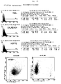
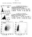
Die beiliegenden Figuren 9A bis C zeigen die Ergebnisse der Zellsortierung von HT29. Der Versuch wurde entsprechend der Zellsortierung der CX2-Zellinie durchgeführt.The accompanying FIGS. 9A to C show the results of the HT29 cell sorting. The attempt was made according to the Cell sorting of the CX2 cell line performed.
Die HT29-Ursprungszellinie exprimiert auf ihrer Zelloberfläche HSP72 in einer Menge von 30 bis 40% der Zellen. Durch Zellsortierung konnte eine HT29+ Zellinie erhalten werden, die auf ihrer Zelloberfläche HSP72 in 87% der Zellen exprimiert, und eine HT29- Zellinie, die HSP72 in 15% der Zellen exprimiert. Die erhaltenen HT29-Sublinien zeigen eine stabile HSP72-Expression über zumindest 20 Zellpassagen sowie ein identisches MHC- und Zelladhäsionsmolekül-Zelloberflächenexpressionsmuster.The HT29 origin cell line expresses on its cell surface HSP72 in an amount of 30 to 40% of the cells. By cell sorting an HT29 + cell line could be obtained which was based on their cell surface expressed HSP72 in 87% of the cells, and an HT29 cell line that expresses HSP72 in 15% of the cells. The HT29 sublines obtained show stable HSP72 expression over at least 20 cell passages as well as an identical MHC and Cell adhesion molecule cell surface expression pattern.
Aufgrund dieser Untersuchungen konnte gezeigt werden, daß es sich nicht um einen Spezialfall bei CX2-Zellinien handelt, sondern daß eine Verallgemeinerung auf Human-Coloncarcinom-Zellinien in ihrer Gesamtheit möglich ist. Based on these investigations it could be shown that is not a special case of CX2 cell lines, but that a generalization to human colon carcinoma cell lines is possible in its entirety.
Claims (8)
dadurch gekennzeichnet, daß
eine Human-Coloncarcinom-Zellinie, die HSP72 auf ihrer Zelloberfläche exprimiert, durch Zellsortierung in zwei Sublinien aufgetrennt wird, wobei die eine Sublinie HSP72 zu mehr als 80% und die andere Sublinie HSP72 zu weniger als 20% exprimiert.Process for the production of human colon carcinoma cell lines according to one or more of the preceding claims,
characterized in that
a human colon carcinoma cell line expressing HSP72 on its cell surface is separated into two sublines by cell sorting, one subline expressing HSP72 by more than 80% and the other subline expressing HSP72 by less than 20%.
Applications Claiming Priority (2)
| Application Number | Priority Date | Filing Date | Title |
|---|---|---|---|
| DE19647426 | 1996-11-15 | ||
| DE19647426A DE19647426C1 (en) | 1996-11-15 | 1996-11-15 | Human colon carcinoma cell lines with stable HSP72 expression |
Publications (2)
| Publication Number | Publication Date |
|---|---|
| EP0843005A2 true EP0843005A2 (en) | 1998-05-20 |
| EP0843005A3 EP0843005A3 (en) | 1999-03-31 |
Family
ID=7811854
Family Applications (1)
| Application Number | Title | Priority Date | Filing Date |
|---|---|---|---|
| EP97119900A Withdrawn EP0843005A3 (en) | 1996-11-15 | 1997-11-13 | Human colon carcinoma cell line with stable HSP72 expression |
Country Status (4)
| Country | Link |
|---|---|
| US (1) | US5932478A (en) |
| EP (1) | EP0843005A3 (en) |
| JP (1) | JPH10179146A (en) |
| DE (1) | DE19647426C1 (en) |
Cited By (2)
| Publication number | Priority date | Publication date | Assignee | Title |
|---|---|---|---|---|
| WO1999049881A3 (en) * | 1998-03-27 | 1999-12-23 | Gabriele Multhoff | Application of hsp70 proteins |
| CN102286429A (en) * | 2011-07-20 | 2011-12-21 | 中国人民解放军第二军医大学 | A kind of Han nationality human renal sarcoma-like cancer cell line and its preparation method |
Families Citing this family (3)
| Publication number | Priority date | Publication date | Assignee | Title |
|---|---|---|---|---|
| US7361730B1 (en) | 1999-03-15 | 2008-04-22 | University Of Maryland | Surface localized colligin/Hsp47 in carcinoma cells |
| US20030086929A1 (en) * | 2001-10-11 | 2003-05-08 | Tso J. Yun | Treatment of prostate cancer by inhibitors of ATP synthase |
| MX2009003661A (en) * | 2006-10-06 | 2009-04-22 | Baxter Int | Microencapsules containing surface-modified microparticles and methods of forming and using the same. |
-
1996
- 1996-11-15 DE DE19647426A patent/DE19647426C1/en not_active Expired - Fee Related
-
1997
- 1997-11-13 EP EP97119900A patent/EP0843005A3/en not_active Withdrawn
- 1997-11-14 US US08/970,699 patent/US5932478A/en not_active Expired - Fee Related
- 1997-11-14 JP JP9313858A patent/JPH10179146A/en active Pending
Non-Patent Citations (5)
Cited By (3)
| Publication number | Priority date | Publication date | Assignee | Title |
|---|---|---|---|---|
| WO1999049881A3 (en) * | 1998-03-27 | 1999-12-23 | Gabriele Multhoff | Application of hsp70 proteins |
| US7396681B1 (en) | 1998-03-27 | 2008-07-08 | Gabriele Multhoff | Application of Hsp70 proteins |
| CN102286429A (en) * | 2011-07-20 | 2011-12-21 | 中国人民解放军第二军医大学 | A kind of Han nationality human renal sarcoma-like cancer cell line and its preparation method |
Also Published As
| Publication number | Publication date |
|---|---|
| JPH10179146A (en) | 1998-07-07 |
| US5932478A (en) | 1999-08-03 |
| DE19647426C1 (en) | 1998-06-25 |
| EP0843005A3 (en) | 1999-03-31 |
Similar Documents
| Publication | Publication Date | Title |
|---|---|---|
| DE69531607T2 (en) | FUSION GENES FOR THE INHIBITOR OF THE TERMINAL COMPLEMENT AND PROTEINS | |
| DE69128253T2 (en) | SPECIFIC ANTIBODIES, METHOD FOR THEIR PRODUCTION AND THEIR USE | |
| DE69426743T2 (en) | Bispecific trigger molecules that recognize the CD2 lymphocyte antigen and tumor antigens | |
| DE69125735T2 (en) | Method for activating the cytolytic effect of lymphocytes using anti-CD28 antibody | |
| DE19725586C2 (en) | Process for the preparation of cell preparations for immunization by means of heterologous intact bispecific and / or trispecific antibodies | |
| DE69131331T2 (en) | Permanent transplantation and development of human hematopoietic cell lines in normal mammals | |
| DE4447484C2 (en) | Apoptosis inhibitor | |
| DE69231250T2 (en) | COMBINED CELLULAR AND IMMUNE SUPPRESSIVE THERAPIES | |
| DE19821060A1 (en) | T cell co-stimulating polypeptide, monoclonal antibodies, and the production and use thereof | |
| EP0438102A1 (en) | Process for the production of antibodies | |
| EP0093436A2 (en) | Process for preparing permanent animal and human cell lines, and their use | |
| EP0952213A2 (en) | Process for inducing an immune response mediated by NK-cells | |
| EP0531300A1 (en) | VARIANT CD44 SURFACE PROTEINS, THESE ENCODING DNA SEQUENCES, ANTIBODIES AGAINST THESE PROTEINS AND THEIR USE IN DIAGNOSTICS AND THERAPY. | |
| DE19708713C2 (en) | Use of preparations containing anti-CD44 antibodies for the treatment of certain tumors and for the suppression of immune reactions | |
| DE69522689T2 (en) | CELL CYCLE-INDEPENDENT, GLIOMA-SURFACE-SPECIFIC, HUMAN MONOCLONAL ANTIBODIES | |
| EP3615073A1 (en) | Biological binding molecule | |
| DE3249567C2 (en) | Hybrid cell line, process for its production and its use | |
| DE69123121T2 (en) | SYNTHETIC POLY-IG RECEPTOR, RECEPTOR-ANTIBODY COMPLEXES, PRODUCTION AND USE | |
| WO1994023014A1 (en) | Tumoricide t lymphocytes | |
| DE69929909T2 (en) | Method for making choroid plexus immortalized cell lines | |
| DE19647426C1 (en) | Human colon carcinoma cell lines with stable HSP72 expression | |
| DE69230762T2 (en) | ICAM'S CONSTANT DOMAIN WATER-SOLUBLE FUSION PROTEINS, IMMUNOGLOBULIN | |
| DE3786673T2 (en) | METHOD FOR REMOVING UNWANTED CELLS FROM HUMAN LYMPHOCYTE POPULATIONS, APPLICATION OF THE METHOD FOR PRODUCING MONOCLONAL ANTIBODIES AND A KIT SUITABLE FOR THIS. | |
| DE69434024T2 (en) | SURFACE PROTEINS EXPRESSED ON HUMAN CORTICAL THYMOCYTES AND THEIR USE | |
| DE69020914T2 (en) | Monoclonal antibodies from rats to human antigens and methods for their production. |
Legal Events
| Date | Code | Title | Description |
|---|---|---|---|
| PUAI | Public reference made under article 153(3) epc to a published international application that has entered the european phase |
Free format text: ORIGINAL CODE: 0009012 |
|
| AK | Designated contracting states |
Kind code of ref document: A2 Designated state(s): AT BE CH DE DK ES FI FR GB GR IE IT LI LU NL SE |
|
| AX | Request for extension of the european patent |
Free format text: AL;LT;LV;MK;RO;SI |
|
| PUAL | Search report despatched |
Free format text: ORIGINAL CODE: 0009013 |
|
| AK | Designated contracting states |
Kind code of ref document: A3 Designated state(s): AT BE CH DE DK ES FI FR GB GR IE IT LI LU MC NL PT SE |
|
| AX | Request for extension of the european patent |
Free format text: AL;LT;LV;MK;RO;SI |
|
| 17P | Request for examination filed |
Effective date: 19990928 |
|
| AKX | Designation fees paid |
Free format text: AT BE CH DE DK ES FI LI |
|
| RBV | Designated contracting states (corrected) |
Designated state(s): AT BE CH DE DK ES FI FR GB GR IE IT LI LU NL SE |
|
| 17Q | First examination report despatched |
Effective date: 20021111 |
|
| RAP1 | Party data changed (applicant data changed or rights of an application transferred) |
Owner name: MULTHOFF, GABRIELE |
|
| RIN1 | Information on inventor provided before grant (corrected) |
Inventor name: MULTHOFF, GABRIELE, PROF. DR. |
|
| RIC1 | Information provided on ipc code assigned before grant |
Ipc: 7C 12Q 1/02 A |
|
| STAA | Information on the status of an ep patent application or granted ep patent |
Free format text: STATUS: THE APPLICATION IS DEEMED TO BE WITHDRAWN |
|
| 18D | Application deemed to be withdrawn |
Effective date: 20050207 |