EP0854192A2 - Verfahren zur Herstellung von menschlichen Cathepsin und dessen Verwendung - Google Patents
Verfahren zur Herstellung von menschlichen Cathepsin und dessen Verwendung Download PDFInfo
- Publication number
- EP0854192A2 EP0854192A2 EP98100280A EP98100280A EP0854192A2 EP 0854192 A2 EP0854192 A2 EP 0854192A2 EP 98100280 A EP98100280 A EP 98100280A EP 98100280 A EP98100280 A EP 98100280A EP 0854192 A2 EP0854192 A2 EP 0854192A2
- Authority
- EP
- European Patent Office
- Prior art keywords
- cathepsin
- human
- human cathepsin
- gene
- cells
- Prior art date
- Legal status (The legal status is an assumption and is not a legal conclusion. Google has not performed a legal analysis and makes no representation as to the accuracy of the status listed.)
- Withdrawn
Links
Images





Classifications
-
- C—CHEMISTRY; METALLURGY
- C12—BIOCHEMISTRY; BEER; SPIRITS; WINE; VINEGAR; MICROBIOLOGY; ENZYMOLOGY; MUTATION OR GENETIC ENGINEERING
- C12N—MICROORGANISMS OR ENZYMES; COMPOSITIONS THEREOF; PROPAGATING, PRESERVING, OR MAINTAINING MICROORGANISMS; MUTATION OR GENETIC ENGINEERING; CULTURE MEDIA
- C12N9/00—Enzymes; Proenzymes; Compositions thereof; Processes for preparing, activating, inhibiting, separating or purifying enzymes
- C12N9/14—Hydrolases (3)
- C12N9/48—Hydrolases (3) acting on peptide bonds (3.4)
- C12N9/50—Proteinases, e.g. Endopeptidases (3.4.21-3.4.25)
- C12N9/64—Proteinases, e.g. Endopeptidases (3.4.21-3.4.25) derived from animal tissue
- C12N9/6421—Proteinases, e.g. Endopeptidases (3.4.21-3.4.25) derived from animal tissue from mammals
- C12N9/6472—Cysteine endopeptidases (3.4.22)
-
- C—CHEMISTRY; METALLURGY
- C07—ORGANIC CHEMISTRY
- C07K—PEPTIDES
- C07K16/00—Immunoglobulins [IG], e.g. monoclonal or polyclonal antibodies
- C07K16/40—Immunoglobulins [IG], e.g. monoclonal or polyclonal antibodies against enzymes
-
- A—HUMAN NECESSITIES
- A61—MEDICAL OR VETERINARY SCIENCE; HYGIENE
- A61K—PREPARATIONS FOR MEDICAL, DENTAL OR TOILETRY PURPOSES
- A61K38/00—Medicinal preparations containing peptides
-
- F—MECHANICAL ENGINEERING; LIGHTING; HEATING; WEAPONS; BLASTING
- F02—COMBUSTION ENGINES; HOT-GAS OR COMBUSTION-PRODUCT ENGINE PLANTS
- F02B—INTERNAL-COMBUSTION PISTON ENGINES; COMBUSTION ENGINES IN GENERAL
- F02B2275/00—Other engines, components or details, not provided for in other groups of this subclass
- F02B2275/14—Direct injection into combustion chamber
-
- Y—GENERAL TAGGING OF NEW TECHNOLOGICAL DEVELOPMENTS; GENERAL TAGGING OF CROSS-SECTIONAL TECHNOLOGIES SPANNING OVER SEVERAL SECTIONS OF THE IPC; TECHNICAL SUBJECTS COVERED BY FORMER USPC CROSS-REFERENCE ART COLLECTIONS [XRACs] AND DIGESTS
- Y02—TECHNOLOGIES OR APPLICATIONS FOR MITIGATION OR ADAPTATION AGAINST CLIMATE CHANGE
- Y02T—CLIMATE CHANGE MITIGATION TECHNOLOGIES RELATED TO TRANSPORTATION
- Y02T10/00—Road transport of goods or passengers
- Y02T10/10—Internal combustion engine [ICE] based vehicles
- Y02T10/12—Improving ICE efficiencies
Definitions
- the present invention relates to a method of producing human cathepsin and a therapeutic composition for intervertebral disk hernia containing said enzyme.
- Intervertebral disk hernia a disease likely to develop in the lower lumbar vertebrae and the cervical vertebrae, is accompanied by sciatic neuralgia, upper limb neuralgia and other symptoms. With high compression on intervertebral disks after initial degeneration and loss of fibrous ring elasticity, the vertebral pulp is pushed out from fibrous ring cracks. This condition, called intervertebral disk hernia, results in pain and other symptoms caused by inflammation in and around the nerve root, when the herniated intervertebral disks have compressed the equine caudal spine and the nerve root branched out from the spine.
- intervertebral disk hernia has been treated by various methods of pain relief: by administration of anti-inflammatory sedatives, muscular relaxants and other drugs in the acute phase attended by severe pain; and, in the chronic phase, by traction, thermotherapy and kinesitherapy.
- surgical treatment is often adopted in cases where the more conservative therapy fails.
- a conservative therapy is supplemented with an oral anti-inflammatory sedative and rest, as there is severe pain. Even after mitigation of the severe pain and inflammation in the acute phase, the hernia does not disappear.
- Other conservative therapies include traction, thermotherapy in which painful regions in the lumbar and feet are warmed to relax stiff muscles, and lumbar pain exercise for strengthening abdominal and dorsal muscles. In cases where the conservative therapy fails, intervertebral disks are excised by surgery, as stated above.
- chymopapain a protease of plant origin
- This aims at mitigating compression on the nerve tissue by chemically lysing intervertebral disks and reducing the internal pressure thereof.
- chymopapain as a foreign protein poses the problem of side effects such as anaphylactic shock. To solve this problem, there is a need for the development of a more effective drug with a lower prevalence of toxicity.
- the only available method of producing human cathepsin is expression of a cDNA encoding human cathepsin L in Escherichia coli , there being no production method employing any other host such as an animal cell.
- human cathepsin has a therapeutic effect on intervertebral disk hernia.
- the inventors demonstrated that human cathepsin L or S produced in animal cells by gene engineering and cathepsin L or S derived from human cell lines are suitable for treatment of intervertebral disk hernia, and developed the present invention.
- the present invention provides a therapeutic composition for intervertebral disk hernia containing human cathepsin.
- the invention also provides a method of producing human cathepsin, wherein animal cells carrying a DNA encoding human cathepsin are cultured to produce and accumulate said enzyme in the culture, which is then harvested.
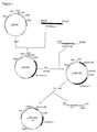
- Figure 1 shows a construction scheme for the plasmids obtained in Examples 1 and 4.
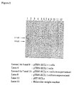
- Figure 2 shows production of human cathepsin L, by a COS-7 cell transformant, observed in Example 2.
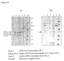
- Figure 3 shows production of human cathepsin L, by an Sp2/0 cell transformant, observed in Example 5.
- panels A and B respectively show the results of western blotting and CBB staining.
- Figure 4 is a toluidine blue stained image obtained after a dose of 1600milliunits cathepsin L in Example 7.
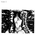
- Figure 5 is a toluidine blue stained image obtained after a dose of 800milliunits cathepsin L in Example 7.
- Figure 6 is a toluidine blue stained image obtained after a dose of 400milliunits cathepsin L in Example 7.
- Figure 7 is a toluidine blue stained image obtained after a dose of 200milliunits cathepsin L in Example 7
- Figure 8 is a hematoxylin-eosin stained image obtained after a dose of 1600milliunits cathepsin L in Example 7.
- Figure 9 is a hematoxylin-eosin stained image obtained after a dose of 800milliunits cathepsin L in Example 7.
- Figure 10 is a hematoxylin-eosin stained image obtained after a dose of 400milliunits cathepsin L in Example 7.
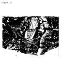
- Figure 11 is a hematoxylin-eosin stained image obtained after a dose of 200milliunits cathepsin L in Example 7.
- Figure 12 is a macroscopic view of a normal rabbit intervertebral disk.
- Figure 13 is a macroscopic view of a rabbit intervertebral disk after a dose of 800milliunits cathepsin L in Example 7.
- Figure 14 shows a construction scheme for the plasmid obtained in Example 9.
- Human cathepsins used in the present invention include cathepsins A, B, C, D, E, F, G, H, L, S and T. These human cathepsins may be of a natural type or a mutein. Among them, the use of those having a molecular weight of about 20,000 to about 40,000 is preferable. In the preparation of the present invention, preference was given to cathepsin L or S.
- the human cathepsin L or S may be a product of either gene engineering technology or a human cell line.
- Muteins of human cathepsin include those resulting from mutation in the amino acid sequence of the original protein except all the eight cystein residues and the 163rd histidine residue produced by amino acid addition, constituent amino acid deletion, or replacement with another amino acid.
- Amino acid addition may be such that a mutein results from the addition of at least one amino acid to human cathepsin, excluding methionine or signal peptides from the initiation codon used to express the peptide, as long as the human cathepsin activity is maintained.
- Deletion of constituent amino acids may be such that a mutein results from a lack of at least one constituent amino acid of human cathepsin, as long as the activity of human cathepsin is retained.
- Replacement with another amino acid may be such that a mutein results from replacement of at least one human cathepsin constituent amino acid with another amino acid, as long as the activity of human cathepsin is retained.
- Said mutein may involve a combination of two or more of addition, deletion and replacement. Such deletion or replacement is generally carried out in position(s) of 1 to 3 amino acid residue(s) of the original protein.
- Such mutein is preferably a mutein of human cathepsin L or S.
- the human cathepsin and mutein thereof in the present invention can be derived from a human cell line producing human cathepsin selected by established human cell line screening. They can also be produced by gene engineering technology using a cDNA encoding human cathepsin. For example, it is possible to use a reported cDNA encoding human cathepsin [S. Gal and M. M. Gottesman, Biochem. J., 253 , 303 (1988)]. In the production method of the present invention, human cathepsin can be obtained by expressing said cDNA in cultured animal cells.
- the present invention comprises (1) synthesis of a PCR primer to amplify a reported nucleotide sequence of cathepsin of human renal origin, (2) preparation of a recombinant DNA to express the amplified human cathepsin gene using cultured animal cells, (3) establishment of an animal cell line transformed with the recombinant DNA described in (2) above, and (4) separation and purification of human cathepsin from a culture of the animal cell line described in (3) above.
- a polymerase chain reaction can be conducted in accordance with the known method, such as the instructions for a kit (e.g., Cetus/Perkin-Elmer).
- the amplified cDNA can be separated by a commonly known method such as agarose electrophoresis and then recovered from the gel.
- the nucleotide sequence of this cDNA can be determined by the dideoxynucleotide synthetic chain termination method [T. Messing et al., Nucl. Acids Res., 9 , 309 (1981)].
- the plasmid harboring the cloned cDNA can be used as it is or after being cut out with an appropriate restriction enzyme and then inserted into another vector as necessary.
- any vector can be used, as long as it is replicable in the host.
- the host is a bacterium of the genus Escherichia (e.g., Escherichia coli )
- such vectors include plasmids derived from Escherichia coli such as pBR322 [F. Bolivar et al., Gene 2 , 95(1979)], pBR325, pUC12 and pUC13.
- plasmids derived from Staphylococcus such as pUB110 [T. J. Gryczan and D. Dubnau, Proc. Natl. Acad. Sci.
- such vectors include plasmids derived from yeast such as pSH19 [S. Harashima et al., Mol. Cell. Biol., 4 , 771 (1984)] and pSH19-1 [European Patent Publication EP-A-0235430].
- such vectors include pSV2-X, resulting from insertion of SV40 ori into pBR322 [R. C. Mulligan and P. Berg, Proc. Natl. Acad. Sci. USA 78 , 2072 (1981)] and pcD-X [H. Okayama and P. Berg, Mol. Cell. Biol., 3 , 280 (1983)].
- the cloned cDNA may have a translational initiation codon (ATG) at its 5'-terminal and a translational termination codon (TAG, TGA or TAA) at its 3'-terminal.
- ATG translational initiation codon
- TAG translational termination codon
- TAA translational termination codon
- a promoter sequence, promoter-pre-pro sequence or promoter-signal sequence is ligated upstream.
- any promoter can be used for the present invention, as long as it is appropriate for the host used to express the gene.
- promoters are: when the host is Escherichia coli , the trp promoter, lac promoter, ⁇ PL promoter, T7 promoter, tac promoter, lpp promoter and recA promoter, with preference given to the T7 promoter; when the host is a bacterium of the genus Bacillus , the SPO1 promoter, SPO2 promoter, penP promoter and MWP promoter, with preference given to the MWP promoter; when the host is a yeast, the GAPDH promoter, PGK promoter, PHO5 promoter, ADH promoter and PHO81 promoter, with preference given to the GAPDH promoter; and when the host is an animal cell, the SV40-derived promoter, retrovirus promoter and human cytomegalovirus promoter.
- the promoter can be prepared from the corresponding gene or chemically synthesized by known methods.
- the signal sequence and pre-pro sequence may be any one, as long as they function in the host.
- Escherichia coli they include the ⁇ -lactamase gene signal sequence, enterotoxin gene signal sequence, alkaline phosphatase gene signal sequence and OmpA gene signal sequence.
- a bacterium of the genus Bacillus they include the ⁇ -amylase gene signal sequence, neutral protease gene signal sequence and MWP gene signal sequence.
- a yeast include the egg white lysozyme gene signal sequence, human lysozyme gene signal sequence, invertase gene signal sequence and ⁇ -factor gene pre-pro sequence.
- interleukin-2 gene signal sequence In the case of an animal cell, they include the interleukin-2 gene signal sequence.
- the DNA-harboring vector thus constructed is used to produce a transformant.
- the host include bacteria of the genus Escherichia , bacteria of the genus Bacillus , yeasts and animal cells, with preference given to animal cells.
- bacteria of the genus Escherichia include Escherichia coli K12 DH1 [B. Low, Proc. Natil. Acad. Sci. USA, 60 , 160 (1968)], C600 [R. K. Appleyard, Genetics, 39 , 440 (1954)], MM294 [K. Backman et al., Proceedings of the Natl. Acad. Sci. USA, 73 , 4174 (1976)] and N4830 [M. E.
- Bacillus subtilis MI114 Bacillus subtilis MI114 [K. Yoshimura et al., Gene 24 , 255 (1983)], 207-21 [K. Ohmura et al., J. Biochem., 95 , 87 (1984)] and Bacillus brevis 47 [S. Udaka, Agric. Biol. Chem., 40 , 523 (1976)].
- yeasts include Saccharomyces cerevisiae AH22R - [A. Miyanohara et al., Proc. Natl. Acad. Sci.
- animal cells examples include simian COS-7 cells, simian Vero cells, Chinese hamster (CHO) cells, mouse L cells, human FL cells, all of which are adherent cells, and mouse myeloma cells (e.g., Sp2/0), mouse YAC-1 cells, mouse MethA cells, mouse P388 cells and mouse EL-4 cells, all of which are suspending cells.
- simian COS-7 cells simian Vero cells
- Chinese hamster (CHO) cells mouse L cells
- human FL cells all of which are adherent cells
- mouse myeloma cells e.g., Sp2/0
- mouse YAC-1 cells mouse MethA cells
- mouse P388 cells mouse EL-4 cells
- Bacteria of the genus Escherichia can be transformed in accordance with the method described by T. Maniatis et al. in Molecular Cloning, Cold Spring Harbor Laboratory, page 249 (1982) and other publications, for instance. Bacteria of the genus Bacillus can be transformed in accordance with the method described by S. Chang and S. N. Cohen in Molecular and General Genetics, 168 , 111 (1979) and other publications, for instance. Yeasts can be transformed in accordance with the method described by A. Hinnen et al. in the Proceedings of the National Academy of Science, USA, 75 , 1929 (1978), for instance. Animal cells can be transformed in accordance with the method described by M. Wigler et al. in Cell, 14 , 725 (1978), for instance.
- the transformant thus obtained is cultured by a commonly known method.
- media preferably used to cultivate a transformant whose host is an Escherichia bacteria include the M9 medium containing glucose and casamino acid [J. H. Miller, Experiments in Molecular Genetics, page 431, Cold Spring Harbor Laboratory, (1972)].
- a chemical agent such as isopropylthiogalactoside (IPTG) or indolyl 3-acrylate may be added. Cultivation is normally carried out at about 15 to 43°C for about 3 to 24 hours, with aeration and/or stirring as necessary.
- Examples of media used to cultivate a transformant whose host is a Bacillus bacteria include the L-broth medium and T2 medium [S. Udaka, Agric. Biol. Chem., 40 , 523 (1976)]. Cultivation is normally carried out at about 15 to 37°C for about 6 to 96 hours, with aeration and/or stirring as necessary.
- Examples of media for cultivating a transformant whose host is a yeast include Burkholder's minimal medium [K. L. Bostian et al., Proc. Natl. Acad. Sci. USA 77 , 4504 (1980)]. It is preferable to adjust the medium to a pH level of about 5 to 8. Cultivation is normally carried out at about 20 to 35°C for about 24 to 72 hours, with aeration and/or stirring as necessary.
- Examples of media for cultivating a transformant whose host is an animal cell include MEM medium containing about 5 to 20% fetal bovine serum [H. Eagle, Science, 130 , 432 (1959)], DMEM medium [R. Dulbecco and G. Freeman, Virology, 8 , 396 (1959)], RPMI-1640 medium [G. E. More et al., J. Am. Med. Assoc., 199 , 519 (1967)], 199 medium [J. F. Morgan et al., Proc. Soc. Exp. Biol. Med. 73 , 1 (1950)] and ASF104 medium [Y. Minamoto, Cell Technology, 7 , 42 (1988)]. Cultivation is normally carried out at about 30 to 40°C for about 15 to 60 hours, with aeration and/or stirring as necessary.
- Such commonly known methods of separation and purification include those based on solubility differences such as salting-out and solvent precipitation, those based mainly on molecular weight differences such as dialysis, ultrafiltration, gel filtration and SDS-polyacrylamide gel electrophoresis (SDS-PAGE), those based on charge differences such as ion exchange chromatography, those based on specific affinity such as affinity chromatography, those based on hydrophobicity differences such as reverse-phase high performance liquid chromatography, and those based on isoelectric point differences such as isoelectric focusing.
- solubility differences such as salting-out and solvent precipitation
- SDS-PAGE SDS-polyacrylamide gel electrophoresis
- charge differences such as ion exchange chromatography
- affinity chromatography those based on specific affinity such as affinity chromatography
- hydrophobicity differences such as reverse-phase high performance liquid chromatography
- isoelectric point differences such as isoelectric focusing.
- the present invention has made it possible to express human cathepsin by gene engineering using animal cells, enabling production of human cathepsin of high purity in large amounts.
- human cathepsin can be administered alone or in a pharmaceutical composition with pharmacologically acceptable carriers, excipients and diluents to mammals (e.g., dogs, cats, bovines, horses, humans).
- mammals e.g., dogs, cats, bovines, horses, humans.
- the most suitable route of administered for intervertebral disk hernia is direct injection to the intervertebral pulp via a posterior lateral approach. This promotes the safety of treatment.
- the pharmaceutical composition of the present invention which uses the cathepsin derived from human, is particularly advantageous because of its low antigencity.
- Preferred dosage forms include injections, solutions for injection, frozen products and freeze-dried products.
- composition In preparing a pharmaceutical composition, pharmaceutically acceptable additives, diluents, excipients, etc. are used as necessary, in accordance with commonly accepted pharmaceutical production methods.
- an aqueous solution for injection is prepared by a conventional method using a solvent such as an aqueous solvent (e.g., distilled water), a water-soluble solvent (e.g., physiological saline, Ringer's solution) or an oily solvent (e.g., sesame oil, olive oil), and if desired, additives such as dissolution aids (e.g., sodium salicylate, sodium acetate), buffers (e.g., sodium citrate, glycerol), isotonizing agents (e.g., glucose, invert sugar), stabilizers (e.g., human serum albumin, polyethylene glycol), preservatives (e.g., benzyl alcohol, phenol) and soothing agents (e.g. benzalkonium chloride, procaine hydrochloride).
- a solvent such as an aqueous solvent (e.g., distilled water), a water-soluble solvent (e.g., physiological saline, Ringer's solution)
- a composition for injection is prepared in a solid form of tablet, powder or granule powder by conventional methods such as a mixing process of powdered cathepsin and pharmaceutically acceptable adjuvant or freeze-drying of solution containing cathepsin and pharmaceutically acceptable adjuvant.
- the pharmaceutically acceptable adjuvant includes excipients (e.g., carboxymethyl cellulose (CMC), sodium alginate), preservatives (e.g., benzyl alcohol, benzalkonium chloride, phenol) and soothing agents (e.g., glucose, calcium gluconate, procaine hydrochloride).
- this solid composition When used this solid composition is dissolved or suspended in an aqueous solution e.g. distilled water, physiological saline, glucose solution.
- an aqueous solution e.g. distilled water, physiological saline, glucose solution.
- monosaccharides such as glucose, amino acids, various salts, human serum albumin etc. may be added, and isotonizing agents, pH regulators, soothing agents, antiseptics and other additives may be added for a safe and effective preparation of human cathepsin L or a mutein thereof.
- the preparation dose of the present invention for injection into intervertebral pulp can be determined as appropriate to the intervertebral disk hernia symptoms, it is preferable to administer it at 100 to 500,000milliunits, and more preferably about 1,000 to 100,000milliunits, per administration.
- one unit corresponds to 1 mg of substrate degarded per minute at 37°C (Method in Enzmology 80 , 535 (1981)).
- Administration frequency may be once a day or at longer intervals, once a week, for instance.
- composition containing human cathepsin of the present invention appropriately lyses mammalian intervertebral pulp and can therefore be used to treat intervertebral disk hernia.
- the composition of the present invention is useful as a therapeutic agent for intervertebral disk hernia.
- the PCR product was separated by electrophoresis on 1.2% agarose gel; an amplified DNA fragment was observed at a position corresponding to the size (1132 bp) expected from the nucleotide sequence of cathepsin L of human renal origin.
- This DNA fragment was recovered from the gel and subcloned to the plasmid vector pBluescript R II SK + (produced by STRATAGENE).
- the nucleotide sequence of the cDNA portion was determined by the dideoxynucleotide synthetic chain termination method [J. Messing et al., Nucleic Acids Res., 9 , 309 (1981)] and proved identical with the reported sequence.
- the plasmid containing this cDNA fragment was named pHCL-5.
- the cDNA obtained in Reference Example 1 was cleaved with restriction enzyme EcoRI and a 798 bp fragment (which encodes a part of the of human pro-cathepsin L and the whole mature region) was recovered. To both ends of this fragment was ligated a BamHI linker (5'-pCCCGGATCCGGG-3'; sequence ID No. 5), and the ligation product was inserted into the plasmid vector pET-3c for expression in Escherichia coli [Methods in Enzymology, ed. D. V. Goeddel, 185 , 68, Academic Press (1990)]. The plasmid thus constructed was designated pET-HCL ⁇ .
- Escherichia coli MM294(DE3) was transformed with pET-HCL ⁇ to express human cathepsin L in the presence of the T7 promoter [Methods in Enzymology, 185 , 60 (1990)].
- the transformant was cultured and cells were disrupted by sonication and subjected to SDS-PAGE; a unique band appeared near 30 kDal, corresponding to human cathepsin L. Since the expressed product formed an inclusion body, human cathepsin L was partially purified from the precipitated fraction of the ultrasonically disrupted transformant.
- the partially purified recombinant human cathepsin L obtained in Reference Example 2 was mixed with an equal amount of Freund's complete adjuvant and a rabbit was inoculated with about 1 ml. Later, a mixture of a partially purified human cathepsin L and an equal amount of Freund's incomplete adjuvant was injected thrice at 10-day intervals, and blood was collected seven days after the final injection. The blood obtained was kept standing at 37°C for 30 minutes and then at 4°C overnight, after which it was centrifuged to yield an antiserum to human cathepsin L.
- a 5 ⁇ l solution of the human placental cDNA library ⁇ gt11 (CLONTECH Laboratories, Inc.) and 45 ⁇ l of distilled water were mixed. After incubation at 95°C for 5 minutes, the mixture was immediately ice cooled. Two primers (Nos. 5 and 6 above, 50 pmol of each) were added, and PCR carried out using 1 unit of Vent R DNA Polymerase in the presence of 20 mM MgSO 4 , in which a series of reactions at 94°C for 1 minute, 55°C for 1 minute and 72°C for 1 minute 10 seconds was repeated in 50 cycles. To the reaction mixture were added two other primers (Nos. 6 and 7 above, 50 pmol of each), and PCR was carried out in the same manner as above.
- the PCR product was separated by electrophoresis on 1.2% agarose gel; an amplified DNA fragment was seen at a position corresponding to the size (1076 bp) expected from the nucleotide sequence of cathepsin S of human placental origin.
- This DNA fragment was recovered from the gel and subcloned to the plasmid vector pT7Blue(R)T-Vector (produced by Novagen Company).
- the nucleotide sequence of the cDNA portion was determined by the dideoxynucleotide synthetic chain termination method [J. Messing et al., Nucleic Acids Res., 9 , 309 (1981)].
- the plasmid containing this cDNA fragment was named pHCS-2.
- Example 1 Preparation (1) of recombinant DNA for expression of human cathepsin L gene in animal cells
- Example 2 Expression (1) of human cathepsin L gene in animal cells
- Example 2 To 20 ⁇ g of the plasmid described in Example 1 (pTBN-HCL), 50 ⁇ l of a 2.5 M calcium chloride solution (sterilized) was added, and sterile water was added for a final volume of 500 ⁇ l. This solution was added to 500 ⁇ l of a 2 ⁇ HBS solution (280 mM sodium chloride, 1.5 mM disodium monohydrogen phosphate, 50 mM HEPES, pH 7.05) to form a DNA-calcium phosphate precipitate. Two hours after the medium replacement, 500 ⁇ l of this precipitate suspension was added to cells drop by drop, and this mixture was cultured at 37°C in the presence of 5% carbon dioxide.
- a 2 ⁇ HBS solution 280 mM sodium chloride, 1.5 mM disodium monohydrogen phosphate, 50 mM HEPES, pH 7.05
- the medium was removed, and 1.5 ml of a 15% glycerol/1 ⁇ HBS solution was added. After the mixture was kept standing for 30 seconds, the glycerol/1 ⁇ HBS solution was removed, and the cells adhering to the petri dish were washed with DMEM medium (serum-free). After 4 ml of the complete medium was added, cultivation was continued under the above conditions, and 24 hours later, the medium was replaced with serum-free ASF104 medium (produced by Ajinomoto Co., Inc.), followed by two more days of cultivation.
- serum-free ASF104 medium produced by Ajinomoto Co., Inc.
- the culture supernatant was subjected to western blot analysis using the antiserum to human cathepsin L prepared in Reference Example 3; in response to the antiserum, unique bands appeared, which had molecular weights of about 40,000 to 30,000 and lower molecular weights. These were identified as human pro-cathepsin L and decomposed products thereof, estimated from these molecular weights ( Figure 2).
- cathepsin L enzyme activity according to the method of A. J. Barrett and H. Kirschke [Methods in Enzymology 80 , 535 (1981)].
- To 25 ⁇ l of this concentrated culture supernatant were added 25 ⁇ l of 1 M sodium formate buffer (pH 3.0) and 10 ⁇ l of 0.1 M DTT, and water was added for a final volume of 250 ⁇ l, followed by incubation at 37°C for 30 seconds.
- activating buffer 340 mM sodium acetate, 60 mM acetic acid, 4 mM EDTA disodium, pH 5.5
- activating buffer 340 mM sodium acetate, 60 mM acetic acid, 4 mM EDTA disodium, pH 5.5
- the enzyme reaction was carried out at 30°C for 10 minutes in the presence of 125 ⁇ l of a synthetic substrate solution [20 ⁇ M Z-Phe-Arg-NMec (Z: benzyloxycarbonyl-, NMec: -7-(4-methyl)coumarylamide)], and stopped by the addition of 500 ⁇ l of a reaction stopper (100 mM sodium monochloroacetate, 30 mM sodium acetate, 70 mM acetic acid, pH 4.3).
- a culture supernatant of COS-7 cells not expressing cathepsin L was used as an enzyme solution.
- FCA produced by Baxter
- the enzyme inhibitors used were 10 ⁇ M leupeptin (cysteine protease inhibitor), 10 ⁇ M (p-amidinophenyl)methanesulfonyl fluoride hydrochloride (APMSF: serine protease inhibitor), 1 ⁇ M Z-Phe-Phe-CHN 2 (cathepsin L inhibitor, -CHN 2 : -diazomethane) and 1 ⁇ M pepstatin A (aspartic acid protease inhibitor).
- the assay detected cathepsin L activity in the culture supernatant of the COS-7 cells transformed with human cathepsin L gene.
- the enzyme activities obtained in the presence of enzyme inhibitors leupeptin, APMSF, Z-Phe-Phe-CHN 2 and pepstatin A were 3.4%, 79%, 6.3% and 99%, respectively, in comparison with the inhibitor-free control.
- Example 4 Preparation (2) of recombinant DNA for expression of human cathepsin L gene in animal cells
- the drug resistance marker neo gene was inserted into the vector pTBN-HCL described in Example 1. Namely, a fragment comprising the SV40 early promoter and the neo gene was inserted between the restriction enzyme ClaI and SalI sites of the plasmid pTBN-HCL to yield the plasmid pTBN-HCL neo ( Figure 1).
- mouse myeloma Sp2/0 cells were transformed as follows: Sp2/0 cells, cultivated in an ASF104 medium supplemented with 5% FCS (5% FCS/ASF), were suspended in PBS(-) [the same as Dullbecco's PBS but CaCl 2 and MgCl 2 are removed], so that the concentration of the medium became 1 ⁇ 10 7 cells/ml. 500 ⁇ l of this cell suspension was injected into a cuvette, 10 ⁇ g of said plasmid DNA was added, and the mixture was kept standing on ice for 5 minutes.
- PBS(-) the same as Dullbecco's PBS but CaCl 2 and MgCl 2 are removed
- This liquid was pulsated at 125 ⁇ F and 300 V using a gene pulsar (produced by Bio-Rad Laboratories) and then kept standing on ice again for 10 minutes. The liquid was then transferred to 10 ml of 5% FCS/ASF104 medium and cultured at 37°C in the presence of 5% carbon dioxide. 48 hours later the culture was transferred to selection medium (5% FCS/ASF104 medium containing 200 ⁇ g/ml G418) and cultured on a 24-well plate for 2 weeks.
- selection medium 5% FCS/ASF104 medium containing 200 ⁇ g/ml G418)
- the strain obtained in Example 5, showing high expression of cathepsin L was cultured in 20 ml of ASF104 medium supplemented with 10% FCS and 200 ⁇ g/ml G418, after which it was transferred to 50 ml of serum-free selection medium (ASF104 medium supplemented with 200 ⁇ g/ml G418) and cultured for 5 days.
- CM-Sephadex C-50 25 ⁇ 4.4 cm
- the column was washed with buffer A (20 mM sodium acetate, 1 mM EDTA, pH 5.5), followed by elution on a sodium chloride (NaCl) density gradient from 0 to 1 M, to elute human cathepsin L near an NaCl concentration of about 0.4 M.
- This fraction was applied to a Mono S column (HR5/5) of an FPLC system (Pharmacia), followed by column washing and human cathepsin L elution in the same manner as above.
- the human cathepsin L fraction eluted near an NaCl concentration of about 0.36 M, was concentrated to yield a purified preparation.
- Example 7 Action of human cathepsin L on intervertebral pulp
- Aqueous solution compositions containing human cathepsin L at 200, 400,800 or 1600milliunits were prepared.
- 0.01 ml of the composition was injected in the lumbar intervertebral pulp of rabbits (weighing 2.5 to 3 kg) via a posterior lateral approach under radiographic observation.
- One week later radiography revealed stenosis in lumbar intervertebral disks in the rabbits receiving cathepsin L at doses of 800milliunits or more.
- the rabbits were sacrificed by exsanguination under anesthesia, and the lumbar vertebrae were collected from the site of cathepsin L injection.
- the bone tissue was fixed in formalin and decalcified with EDTA, after which it was trimmed to prepare for longitudinal sections and embedded in paraffin.
- 5-micron thin sections were prepared using a microtome and stained with toluidine blue (which selectively stains cartilage, Figures 4 through 7) and hematoxylin-eosin ( Figures 8 through 11), after which these sectional specimens were observed using a light microscope.
- CM-Sepharose Fast Flow 3.0 ⁇ 30 cm, Pharmacia LKB Biotechnology.
- human cathepsin L was eluted with buffer A containing 0.4 M NaCl.
- the eluate was concentrated using a Centricut U-10 (produced by Kurabo Industries, Ltd.) and applied to a column of Sephacryl S-200 HR (2.5 ⁇ 100 cm, Pharmacia LKB Biotechnology), equilibrated with buffer A containing 0.9% NaCl, followed by gel filtration.
- the fraction containing human cathepsin L was recovered and dialysed against buffer A overnight.
- the dialysate was applied to a column of CM-Sephadex C-50 (1.6 ⁇ 20 cm, Pharmacia LKB Biotechnology). After column washing with buffer A in an amount three times the bed volume, human cathepsin L was eluted on a linear sodium chloride density gradient (final concentration 300 mM). The human cathepsin L fraction eluted at a concentration of about 160 mM was concentrated using a Centricut U-10 and then subjected to the same procedure of gel filtration chromatography as above.
- the human cathepsin L fraction was recovered, and 50% saturated with ammonium sulfate, after which it was applied to a HiLoad Phenyl-Sepharose High Performance column (16/10, Pharmacia LKB Biotechnology), equilibrated with buffer A containing 50% saturated ammonium sulfate. After column washing with buffer A containing 50% saturated ammonium sulfate, the column was eluted with buffer A containing 50% (v/v) ethylene glycol to yield 0.67 mg of a purified human cathepsin L. A 17 ⁇ g sample of this preparation was subjected to polyacrylamide gel electrophoresis. A band of a 33 kD mature region of cathepsin L alone appeared.
- the human cathepsin L was secreted in the form of an about 41 kD precursor in the culture supernatant, it was processed into the about 33 kD highly active mature protein as a result of removal of the FCS-derived albumin in the medium during the purification process.
- Example 9 Preparation of recombinant DNA for expression of human cathepsin S gene in animal cells
- mouse myeloma Sp2/0 cells were transformed as follows: Sp2/0 cells, cultivated in an ASF104 medium supplemented with 5% FCS (5% FCS/ASF), were suspended in PBS(-) [the same as Dullbecco's PBS but CaCl 2 and MgCl 2 were removed] to 1 ⁇ 10 7 cells/ml. 500 ⁇ l of this cell suspension was injected to a cuvette, 10 ⁇ g of said plasmid DNA was added, and the mixture kept standing on ice for 5 minutes.
- PBS(-) the same as Dullbecco's PBS but CaCl 2 and MgCl 2 were removed
- This liquid was pulsated at 125 ⁇ F and 300 V using a gene pulsar (produced by Bio-Rad Laboratories) and kept standing on ice for a further 10 minutes.
- This liquid was transferred to 10 ml of 5% FCS/ASF104 medium and cultured at 37°C in the presence of 5% carbon dioxide. 48 hours later the culture was transferred to selection medium (5% FCS/ASF104 medium containing 200 ⁇ g/ml G418) and cultured on a 24-well plate for 2 weeks.
- a selection medium ASP104 medium supplemented with 3% FCS and 600 ⁇ g/ml G418
- Example 12 Action of human cathepsin S on intervertebral pulp
- Human cathepsin S was injected in the rabbit lumbar intervertebral pulp as directed in Example 7. As in the case of human cathepsin L, intervertebral disk anastomosis was noted.
Landscapes
- Chemical & Material Sciences (AREA)
- Health & Medical Sciences (AREA)
- Organic Chemistry (AREA)
- Life Sciences & Earth Sciences (AREA)
- Genetics & Genomics (AREA)
- Engineering & Computer Science (AREA)
- Zoology (AREA)
- General Health & Medical Sciences (AREA)
- Biomedical Technology (AREA)
- Molecular Biology (AREA)
- Bioinformatics & Cheminformatics (AREA)
- Wood Science & Technology (AREA)
- Medicinal Chemistry (AREA)
- Biochemistry (AREA)
- General Engineering & Computer Science (AREA)
- Microbiology (AREA)
- Biotechnology (AREA)
- Immunology (AREA)
- Biophysics (AREA)
- Proteomics, Peptides & Aminoacids (AREA)
- Micro-Organisms Or Cultivation Processes Thereof (AREA)
- Medicines That Contain Protein Lipid Enzymes And Other Medicines (AREA)
- Enzymes And Modification Thereof (AREA)
- Preparation Of Compounds By Using Micro-Organisms (AREA)
- Medicines Containing Material From Animals Or Micro-Organisms (AREA)
Applications Claiming Priority (5)
| Application Number | Priority Date | Filing Date | Title |
|---|---|---|---|
| JP185947/92 | 1992-06-19 | ||
| JP18594792 | 1992-06-19 | ||
| JP15782793 | 1993-06-02 | ||
| JP157827/93 | 1993-06-02 | ||
| EP93109879A EP0576953A3 (de) | 1992-06-19 | 1993-06-21 | Verfahren zur Herstellung von menschlichen Cathepsin und dessen Verwendung. |
Related Parent Applications (1)
| Application Number | Title | Priority Date | Filing Date |
|---|---|---|---|
| EP93109879A Division EP0576953A3 (de) | 1992-06-19 | 1993-06-21 | Verfahren zur Herstellung von menschlichen Cathepsin und dessen Verwendung. |
Publications (2)
| Publication Number | Publication Date |
|---|---|
| EP0854192A2 true EP0854192A2 (de) | 1998-07-22 |
| EP0854192A3 EP0854192A3 (de) | 1999-05-19 |
Family
ID=26485140
Family Applications (2)
| Application Number | Title | Priority Date | Filing Date |
|---|---|---|---|
| EP93109879A Ceased EP0576953A3 (de) | 1992-06-19 | 1993-06-21 | Verfahren zur Herstellung von menschlichen Cathepsin und dessen Verwendung. |
| EP98100280A Withdrawn EP0854192A3 (de) | 1992-06-19 | 1993-06-21 | Verfahren zur Herstellung von menschlichen Cathepsin und dessen Verwendung |
Family Applications Before (1)
| Application Number | Title | Priority Date | Filing Date |
|---|---|---|---|
| EP93109879A Ceased EP0576953A3 (de) | 1992-06-19 | 1993-06-21 | Verfahren zur Herstellung von menschlichen Cathepsin und dessen Verwendung. |
Country Status (2)
| Country | Link |
|---|---|
| EP (2) | EP0576953A3 (de) |
| CA (1) | CA2098777A1 (de) |
Families Citing this family (1)
| Publication number | Priority date | Publication date | Assignee | Title |
|---|---|---|---|---|
| TWI395593B (zh) | 2008-03-06 | 2013-05-11 | Halozyme Inc | 可活化的基質降解酵素之活體內暫時性控制 |
Family Cites Families (1)
| Publication number | Priority date | Publication date | Assignee | Title |
|---|---|---|---|---|
| US3320131A (en) * | 1964-10-23 | 1967-05-16 | Baxter Laboratories Inc | Method for the treatment of herniation of intervertebral discs |
-
1993
- 1993-06-18 CA CA002098777A patent/CA2098777A1/en not_active Abandoned
- 1993-06-21 EP EP93109879A patent/EP0576953A3/de not_active Ceased
- 1993-06-21 EP EP98100280A patent/EP0854192A3/de not_active Withdrawn
Non-Patent Citations (6)
| Title |
|---|
| BERND WIEDERANDERS ET AL.: "Phylogenetic conservation of Cysteine Proteinases. Cloning and expression of a cDNA coding for human Cathepsin S" JOURNAL OF BIOLOGICAL CHEMISTRY (MICROFILMS), vol. 267, no. 19, 5 July 1992, pages 13708-13713, XP002097577 MD US * |
| BRUCE R. TROEN ET AL.: "Sequence and expression of the cDNA for MEP (major excreted protein), a transformation-regulated secreted cathepsin" THE BIOCHEMICAL JOURNAL, vol. 246, no. 3, 15 September 1987, pages 731-735, XP002097576 * |
| CHAUHAN S S ET AL: "Cloning, genomic organization, and chromosomal localization of human cathepsin L" JOURNAL OF BIOLOGICAL CHEMISTRY (MICROFILMS), vol. 268, no. 2, 15 January 1993, pages 1039-1045, XP002079135 * |
| NOZOMI YAMAGUCHI ET AL.: "Caractérisation de l'enzyme analogue à la cathepsine L produite par des cellules tumorales" COMPTES RENDUS DES SÉANCES DE LA SOCIÉTÉ DE BIOLOGIE ET DE SES FILIALES, vol. 183, no. 1, 1989, pages 79-84, XP002097575 * |
| SUSAN E. KANE ET AL.: "Use of a cloned multidrug resistance gene for coamplification and overproduction of Major Excreted Protein, a transformation-regulated secreted acid protease" MOLECULAR AND CELLULAR BIOLOGY, vol. 8, no. 8, August 1988, pages 3316-3321, XP004119401 * |
| SUSANNAH GAL ET AL.: "Isolation and sequence of a cDNA for human pro-(cathepsin L)" THE BIOCHEMICAL JOURNAL, vol. 253, no. 1, 1 July 1988, pages 303-306, XP002067218 * |
Also Published As
| Publication number | Publication date |
|---|---|
| CA2098777A1 (en) | 1993-12-20 |
| EP0576953A2 (de) | 1994-01-05 |
| EP0854192A3 (de) | 1999-05-19 |
| EP0576953A3 (de) | 1995-02-22 |
Similar Documents
| Publication | Publication Date | Title |
|---|---|---|
| CN101010338B (zh) | 成纤维细胞生长因子21的突变蛋白 | |
| EP1417227B1 (de) | Peptide für die behandlung von tumoren und anderen zuständen, die das entfernen oder zerstören von zellen erfordern | |
| CA2216741C (en) | A novel protein and process for preparing the same | |
| US7030223B2 (en) | Megakaryocyte stimulating factors | |
| US5461034A (en) | Osteogenic growth polypeptides identified from regenerating bone marrow | |
| US5028420A (en) | Muteins of tumor necrosis factor | |
| JPH0720431B2 (ja) | ミュレル管抑制物質の活性を示すポリペプチドをコードするdna,組換dnaおよび形質転換宿主 | |
| EP0268561A2 (de) | Herstellung und Verwendung eines T-Zellen-Suppressorfaktors | |
| EP0384731B1 (de) | Osteogenische Wachstumfaktoren, die aus sich regenerierendem Knochenmark identifiziert werden | |
| US5874536A (en) | Proteins with Oncostatin M activity and process for their preparation | |
| EP0854192A2 (de) | Verfahren zur Herstellung von menschlichen Cathepsin und dessen Verwendung | |
| US20240150432A1 (en) | Fusion protein of tnfr2 and april baff receptor | |
| JPH0775573A (ja) | ヒトカテプシンの製造法および該酵素含有製剤 | |
| AU651330B2 (en) | Novel proteins with oncostatin M activity and process for their preparation | |
| JPH05213771A (ja) | ヘパリン結合性神経突起の成長プロモーター因子 | |
| EP0475719A2 (de) | Von Blutplättchen abstammendes, das Wachstum regulierendes Peptid | |
| HK1075258A (en) | Megakaryocyte stimulating factors | |
| IE904732A1 (en) | Novel proteins with oncostatin m activity and process for¹their preparation |
Legal Events
| Date | Code | Title | Description |
|---|---|---|---|
| PUAI | Public reference made under article 153(3) epc to a published international application that has entered the european phase |
Free format text: ORIGINAL CODE: 0009012 |
|
| AC | Divisional application: reference to earlier application |
Ref document number: 576953 Country of ref document: EP |
|
| AK | Designated contracting states |
Kind code of ref document: A2 Designated state(s): AT BE CH DE DK ES FR GB GR IE IT LI LU NL PT SE |
|
| PUAL | Search report despatched |
Free format text: ORIGINAL CODE: 0009013 |
|
| AK | Designated contracting states |
Kind code of ref document: A3 Designated state(s): AT BE CH DE DK ES FR GB GR IE IT LI LU NL PT SE |
|
| STAA | Information on the status of an ep patent application or granted ep patent |
Free format text: STATUS: THE APPLICATION IS DEEMED TO BE WITHDRAWN |
|
| 18D | Application deemed to be withdrawn |
Effective date: 19991120 |