EP0896546B1 - T1-zeitgewichteten kernspintomogram der res-orgäne - Google Patents
T1-zeitgewichteten kernspintomogram der res-orgäne Download PDFInfo
- Publication number
- EP0896546B1 EP0896546B1 EP97914482A EP97914482A EP0896546B1 EP 0896546 B1 EP0896546 B1 EP 0896546B1 EP 97914482 A EP97914482 A EP 97914482A EP 97914482 A EP97914482 A EP 97914482A EP 0896546 B1 EP0896546 B1 EP 0896546B1
- Authority
- EP
- European Patent Office
- Prior art keywords
- particles
- magnetic particles
- magnetic
- composition
- contrast
- Prior art date
- Legal status (The legal status is an assumption and is not a legal conclusion. Google has not performed a legal analysis and makes no representation as to the accuracy of the status listed.)
- Expired - Lifetime
Links
- 238000000034 method Methods 0.000 title claims abstract description 42
- 210000000056 organ Anatomy 0.000 title claims abstract description 27
- 238000002595 magnetic resonance imaging Methods 0.000 title claims description 3
- 239000006249 magnetic particle Substances 0.000 claims abstract description 65
- 239000000203 mixture Substances 0.000 claims abstract description 39
- 230000005291 magnetic effect Effects 0.000 claims abstract description 36
- 239000002872 contrast media Substances 0.000 claims abstract description 28
- 210000000865 mononuclear phagocyte system Anatomy 0.000 claims abstract description 27
- 210000005166 vasculature Anatomy 0.000 claims abstract description 18
- 238000002583 angiography Methods 0.000 claims abstract description 8
- 239000002245 particle Substances 0.000 claims description 93
- 239000008280 blood Substances 0.000 claims description 55
- 210000004369 blood Anatomy 0.000 claims description 55
- 239000000463 material Substances 0.000 claims description 37
- 239000013078 crystal Substances 0.000 claims description 24
- 210000004185 liver Anatomy 0.000 claims description 17
- 229920000642 polymer Polymers 0.000 claims description 14
- 229920002472 Starch Polymers 0.000 claims description 12
- 239000008107 starch Substances 0.000 claims description 12
- 235000019698 starch Nutrition 0.000 claims description 12
- 239000011246 composite particle Substances 0.000 claims description 9
- 239000000696 magnetic material Substances 0.000 claims description 9
- 239000003112 inhibitor Substances 0.000 claims description 7
- 230000014207 opsonization Effects 0.000 claims description 7
- 229920000233 poly(alkylene oxides) Polymers 0.000 claims description 7
- 239000000546 pharmaceutical excipient Substances 0.000 claims description 5
- 210000001165 lymph node Anatomy 0.000 claims description 4
- 238000004519 manufacturing process Methods 0.000 claims description 4
- 150000004676 glycans Chemical class 0.000 claims description 3
- 229920001282 polysaccharide Polymers 0.000 claims description 3
- 239000005017 polysaccharide Substances 0.000 claims description 3
- 229920001542 oligosaccharide Polymers 0.000 claims 1
- XEEYBQQBJWHFJM-UHFFFAOYSA-N iron Substances [Fe] XEEYBQQBJWHFJM-UHFFFAOYSA-N 0.000 description 27
- 238000003384 imaging method Methods 0.000 description 20
- 239000011248 coating agent Substances 0.000 description 15
- 238000000576 coating method Methods 0.000 description 13
- UQSXHKLRYXJYBZ-UHFFFAOYSA-N Iron oxide Chemical compound [Fe]=O UQSXHKLRYXJYBZ-UHFFFAOYSA-N 0.000 description 12
- 239000003795 chemical substances by application Substances 0.000 description 10
- 239000000243 solution Substances 0.000 description 10
- 210000004204 blood vessel Anatomy 0.000 description 9
- 230000000694 effects Effects 0.000 description 9
- XLYOFNOQVPJJNP-UHFFFAOYSA-N water Chemical compound O XLYOFNOQVPJJNP-UHFFFAOYSA-N 0.000 description 8
- 229910015189 FeOx Inorganic materials 0.000 description 7
- 229910052742 iron Inorganic materials 0.000 description 7
- 239000000725 suspension Substances 0.000 description 7
- 235000013980 iron oxide Nutrition 0.000 description 6
- 238000002360 preparation method Methods 0.000 description 6
- 230000001419 dependent effect Effects 0.000 description 5
- 210000001519 tissue Anatomy 0.000 description 5
- WQZGKKKJIJFFOK-GASJEMHNSA-N Glucose Natural products OC[C@H]1OC(O)[C@H](O)[C@@H](O)[C@@H]1O WQZGKKKJIJFFOK-GASJEMHNSA-N 0.000 description 4
- XSQUKJJJFZCRTK-UHFFFAOYSA-N Urea Chemical compound NC(N)=O XSQUKJJJFZCRTK-UHFFFAOYSA-N 0.000 description 4
- 239000002253 acid Substances 0.000 description 4
- 150000001720 carbohydrates Chemical class 0.000 description 4
- 238000009826 distribution Methods 0.000 description 4
- 229960005191 ferric oxide Drugs 0.000 description 4
- 238000010438 heat treatment Methods 0.000 description 4
- WQYVRQLZKVEZGA-UHFFFAOYSA-N hypochlorite Chemical compound Cl[O-] WQYVRQLZKVEZGA-UHFFFAOYSA-N 0.000 description 4
- 229910052747 lanthanoid Inorganic materials 0.000 description 4
- 229910052751 metal Inorganic materials 0.000 description 4
- 239000002184 metal Substances 0.000 description 4
- 229920001223 polyethylene glycol Polymers 0.000 description 4
- MYLBTCQBKAKUTJ-UHFFFAOYSA-N 7-methyl-6,8-bis(methylsulfanyl)pyrrolo[1,2-a]pyrazine Chemical compound C1=CN=CC2=C(SC)C(C)=C(SC)N21 MYLBTCQBKAKUTJ-UHFFFAOYSA-N 0.000 description 3
- VHUUQVKOLVNVRT-UHFFFAOYSA-N Ammonium hydroxide Chemical compound [NH4+].[OH-] VHUUQVKOLVNVRT-UHFFFAOYSA-N 0.000 description 3
- PEDCQBHIVMGVHV-UHFFFAOYSA-N Glycerine Chemical compound OCC(O)CO PEDCQBHIVMGVHV-UHFFFAOYSA-N 0.000 description 3
- 239000002202 Polyethylene glycol Substances 0.000 description 3
- DNIAPMSPPWPWGF-UHFFFAOYSA-N Propylene glycol Chemical compound CC(O)CO DNIAPMSPPWPWGF-UHFFFAOYSA-N 0.000 description 3
- HEMHJVSKTPXQMS-UHFFFAOYSA-M Sodium hydroxide Chemical compound [OH-].[Na+] HEMHJVSKTPXQMS-UHFFFAOYSA-M 0.000 description 3
- 238000004458 analytical method Methods 0.000 description 3
- 230000037396 body weight Effects 0.000 description 3
- 235000014633 carbohydrates Nutrition 0.000 description 3
- 239000002131 composite material Substances 0.000 description 3
- 239000011162 core material Substances 0.000 description 3
- 239000008367 deionised water Substances 0.000 description 3
- 229910021641 deionized water Inorganic materials 0.000 description 3
- 238000002296 dynamic light scattering Methods 0.000 description 3
- 238000010579 first pass effect Methods 0.000 description 3
- 230000001965 increasing effect Effects 0.000 description 3
- 238000002347 injection Methods 0.000 description 3
- 239000007924 injection Substances 0.000 description 3
- WTFXARWRTYJXII-UHFFFAOYSA-N iron(2+);iron(3+);oxygen(2-) Chemical compound [O-2].[O-2].[O-2].[O-2].[Fe+2].[Fe+3].[Fe+3] WTFXARWRTYJXII-UHFFFAOYSA-N 0.000 description 3
- VBMVTYDPPZVILR-UHFFFAOYSA-N iron(2+);oxygen(2-) Chemical class [O-2].[Fe+2] VBMVTYDPPZVILR-UHFFFAOYSA-N 0.000 description 3
- SZVJSHCCFOBDDC-UHFFFAOYSA-N iron(II,III) oxide Inorganic materials O=[Fe]O[Fe]O[Fe]=O SZVJSHCCFOBDDC-UHFFFAOYSA-N 0.000 description 3
- JEIPFZHSYJVQDO-UHFFFAOYSA-N iron(III) oxide Inorganic materials O=[Fe]O[Fe]=O JEIPFZHSYJVQDO-UHFFFAOYSA-N 0.000 description 3
- 150000002602 lanthanoids Chemical class 0.000 description 3
- 229920001308 poly(aminoacid) Polymers 0.000 description 3
- 230000002035 prolonged effect Effects 0.000 description 3
- 239000000126 substance Substances 0.000 description 3
- CIWBSHSKHKDKBQ-JLAZNSOCSA-N Ascorbic acid Chemical compound OC[C@H](O)[C@H]1OC(=O)C(O)=C1O CIWBSHSKHKDKBQ-JLAZNSOCSA-N 0.000 description 2
- VEXZGXHMUGYJMC-UHFFFAOYSA-N Hydrochloric acid Chemical compound Cl VEXZGXHMUGYJMC-UHFFFAOYSA-N 0.000 description 2
- 241001465754 Metazoa Species 0.000 description 2
- 241000699670 Mus sp. Species 0.000 description 2
- 238000005481 NMR spectroscopy Methods 0.000 description 2
- 229910019142 PO4 Inorganic materials 0.000 description 2
- FAPWRFPIFSIZLT-UHFFFAOYSA-M Sodium chloride Chemical compound [Na+].[Cl-] FAPWRFPIFSIZLT-UHFFFAOYSA-M 0.000 description 2
- 239000005708 Sodium hypochlorite Substances 0.000 description 2
- 241000399119 Spio Species 0.000 description 2
- 210000001015 abdomen Anatomy 0.000 description 2
- 239000000908 ammonium hydroxide Substances 0.000 description 2
- 239000012736 aqueous medium Substances 0.000 description 2
- 238000009835 boiling Methods 0.000 description 2
- 238000005282 brightening Methods 0.000 description 2
- AIYUHDOJVYHVIT-UHFFFAOYSA-M caesium chloride Chemical compound [Cl-].[Cs+] AIYUHDOJVYHVIT-UHFFFAOYSA-M 0.000 description 2
- 239000004202 carbamide Substances 0.000 description 2
- 238000000975 co-precipitation Methods 0.000 description 2
- 229940039231 contrast media Drugs 0.000 description 2
- 230000002708 enhancing effect Effects 0.000 description 2
- 238000011835 investigation Methods 0.000 description 2
- -1 lanthanide metals Chemical class 0.000 description 2
- 230000005415 magnetization Effects 0.000 description 2
- 229910052748 manganese Inorganic materials 0.000 description 2
- 229910021645 metal ion Inorganic materials 0.000 description 2
- 238000006386 neutralization reaction Methods 0.000 description 2
- 230000001151 other effect Effects 0.000 description 2
- 239000010452 phosphate Substances 0.000 description 2
- 229920001983 poloxamer Polymers 0.000 description 2
- 229920001987 poloxamine Polymers 0.000 description 2
- 238000001556 precipitation Methods 0.000 description 2
- SUKJFIGYRHOWBL-UHFFFAOYSA-N sodium hypochlorite Chemical compound [Na+].Cl[O-] SUKJFIGYRHOWBL-UHFFFAOYSA-N 0.000 description 2
- 210000000952 spleen Anatomy 0.000 description 2
- 229920001059 synthetic polymer Polymers 0.000 description 2
- 238000012800 visualization Methods 0.000 description 2
- 229910006297 γ-Fe2O3 Inorganic materials 0.000 description 2
- JNYAEWCLZODPBN-JGWLITMVSA-N (2r,3r,4s)-2-[(1r)-1,2-dihydroxyethyl]oxolane-3,4-diol Chemical class OC[C@@H](O)[C@H]1OC[C@H](O)[C@H]1O JNYAEWCLZODPBN-JGWLITMVSA-N 0.000 description 1
- IIZPXYDJLKNOIY-JXPKJXOSSA-N 1-palmitoyl-2-arachidonoyl-sn-glycero-3-phosphocholine Chemical compound CCCCCCCCCCCCCCCC(=O)OC[C@H](COP([O-])(=O)OCC[N+](C)(C)C)OC(=O)CCC\C=C/C\C=C/C\C=C/C\C=C/CCCCC IIZPXYDJLKNOIY-JXPKJXOSSA-N 0.000 description 1
- OWEGMIWEEQEYGQ-UHFFFAOYSA-N 100676-05-9 Natural products OC1C(O)C(O)C(CO)OC1OCC1C(O)C(O)C(O)C(OC2C(OC(O)C(O)C2O)CO)O1 OWEGMIWEEQEYGQ-UHFFFAOYSA-N 0.000 description 1
- QTBSBXVTEAMEQO-UHFFFAOYSA-M Acetate Chemical compound CC([O-])=O QTBSBXVTEAMEQO-UHFFFAOYSA-M 0.000 description 1
- GUBGYTABKSRVRQ-XLOQQCSPSA-N Alpha-Lactose Chemical compound O[C@@H]1[C@@H](O)[C@@H](O)[C@@H](CO)O[C@H]1O[C@@H]1[C@@H](CO)O[C@H](O)[C@H](O)[C@H]1O GUBGYTABKSRVRQ-XLOQQCSPSA-N 0.000 description 1
- 241000271566 Aves Species 0.000 description 1
- BTBUEUYNUDRHOZ-UHFFFAOYSA-N Borate Chemical compound [O-]B([O-])[O-] BTBUEUYNUDRHOZ-UHFFFAOYSA-N 0.000 description 1
- OKTJSMMVPCPJKN-UHFFFAOYSA-N Carbon Chemical compound [C] OKTJSMMVPCPJKN-UHFFFAOYSA-N 0.000 description 1
- ZAMOUSCENKQFHK-UHFFFAOYSA-N Chlorine atom Chemical compound [Cl] ZAMOUSCENKQFHK-UHFFFAOYSA-N 0.000 description 1
- KRKNYBCHXYNGOX-UHFFFAOYSA-K Citrate Chemical compound [O-]C(=O)CC(O)(CC([O-])=O)C([O-])=O KRKNYBCHXYNGOX-UHFFFAOYSA-K 0.000 description 1
- FBPFZTCFMRRESA-KVTDHHQDSA-N D-Mannitol Chemical compound OC[C@@H](O)[C@@H](O)[C@H](O)[C@H](O)CO FBPFZTCFMRRESA-KVTDHHQDSA-N 0.000 description 1
- RGHNJXZEOKUKBD-SQOUGZDYSA-M D-gluconate Chemical compound OC[C@@H](O)[C@@H](O)[C@H](O)[C@@H](O)C([O-])=O RGHNJXZEOKUKBD-SQOUGZDYSA-M 0.000 description 1
- FEWJPZIEWOKRBE-JCYAYHJZSA-N Dextrotartaric acid Chemical compound OC(=O)[C@H](O)[C@@H](O)C(O)=O FEWJPZIEWOKRBE-JCYAYHJZSA-N 0.000 description 1
- CWYNVVGOOAEACU-UHFFFAOYSA-N Fe2+ Chemical compound [Fe+2] CWYNVVGOOAEACU-UHFFFAOYSA-N 0.000 description 1
- VTLYFUHAOXGGBS-UHFFFAOYSA-N Fe3+ Chemical compound [Fe+3] VTLYFUHAOXGGBS-UHFFFAOYSA-N 0.000 description 1
- 229910002547 FeII Inorganic materials 0.000 description 1
- 229910002553 FeIII Inorganic materials 0.000 description 1
- 229920001499 Heparinoid Polymers 0.000 description 1
- 206010019695 Hepatic neoplasm Diseases 0.000 description 1
- GUBGYTABKSRVRQ-QKKXKWKRSA-N Lactose Natural products OC[C@H]1O[C@@H](O[C@H]2[C@H](O)[C@@H](O)C(O)O[C@@H]2CO)[C@H](O)[C@@H](O)[C@H]1O GUBGYTABKSRVRQ-QKKXKWKRSA-N 0.000 description 1
- GUBGYTABKSRVRQ-PICCSMPSSA-N Maltose Natural products O[C@@H]1[C@@H](O)[C@H](O)[C@@H](CO)O[C@@H]1O[C@@H]1[C@@H](CO)OC(O)[C@H](O)[C@H]1O GUBGYTABKSRVRQ-PICCSMPSSA-N 0.000 description 1
- 229930195725 Mannitol Natural products 0.000 description 1
- 238000004813 Moessbauer spectroscopy Methods 0.000 description 1
- 108091034117 Oligonucleotide Proteins 0.000 description 1
- 241000283973 Oryctolagus cuniculus Species 0.000 description 1
- OAICVXFJPJFONN-UHFFFAOYSA-N Phosphorus Chemical compound [P] OAICVXFJPJFONN-UHFFFAOYSA-N 0.000 description 1
- 244000082204 Phyllostachys viridis Species 0.000 description 1
- RVGRUAULSDPKGF-UHFFFAOYSA-N Poloxamer Chemical compound C1CO1.CC1CO1 RVGRUAULSDPKGF-UHFFFAOYSA-N 0.000 description 1
- 150000007513 acids Chemical class 0.000 description 1
- 125000002947 alkylene group Chemical group 0.000 description 1
- 239000003963 antioxidant agent Substances 0.000 description 1
- 235000006708 antioxidants Nutrition 0.000 description 1
- 239000007900 aqueous suspension Substances 0.000 description 1
- 239000008135 aqueous vehicle Substances 0.000 description 1
- 235000010323 ascorbic acid Nutrition 0.000 description 1
- 229960005070 ascorbic acid Drugs 0.000 description 1
- 239000011668 ascorbic acid Substances 0.000 description 1
- 229910052788 barium Inorganic materials 0.000 description 1
- 230000009286 beneficial effect Effects 0.000 description 1
- 229910052790 beryllium Inorganic materials 0.000 description 1
- GUBGYTABKSRVRQ-QUYVBRFLSA-N beta-maltose Chemical compound OC[C@H]1O[C@H](O[C@H]2[C@H](O)[C@@H](O)[C@H](O)O[C@@H]2CO)[C@H](O)[C@@H](O)[C@@H]1O GUBGYTABKSRVRQ-QUYVBRFLSA-N 0.000 description 1
- 238000006065 biodegradation reaction Methods 0.000 description 1
- 229920001400 block copolymer Polymers 0.000 description 1
- 230000017531 blood circulation Effects 0.000 description 1
- 210000001185 bone marrow Anatomy 0.000 description 1
- 239000000872 buffer Substances 0.000 description 1
- 150000001719 carbohydrate derivatives Chemical class 0.000 description 1
- 229910052799 carbon Inorganic materials 0.000 description 1
- 210000004027 cell Anatomy 0.000 description 1
- 239000000460 chlorine Substances 0.000 description 1
- 229910052801 chlorine Inorganic materials 0.000 description 1
- 229910052804 chromium Inorganic materials 0.000 description 1
- 230000004087 circulation Effects 0.000 description 1
- 150000001875 compounds Chemical class 0.000 description 1
- 238000001816 cooling Methods 0.000 description 1
- 238000011161 development Methods 0.000 description 1
- 239000008121 dextrose Substances 0.000 description 1
- 238000011026 diafiltration Methods 0.000 description 1
- 238000003745 diagnosis Methods 0.000 description 1
- 238000002059 diagnostic imaging Methods 0.000 description 1
- 230000004069 differentiation Effects 0.000 description 1
- 239000003814 drug Substances 0.000 description 1
- 229940079593 drug Drugs 0.000 description 1
- 239000000839 emulsion Substances 0.000 description 1
- 230000005293 ferrimagnetic effect Effects 0.000 description 1
- 230000005294 ferromagnetic effect Effects 0.000 description 1
- 125000000524 functional group Chemical group 0.000 description 1
- 229940050410 gluconate Drugs 0.000 description 1
- 239000008103 glucose Substances 0.000 description 1
- 239000002554 heparinoid Substances 0.000 description 1
- XLYOFNOQVPJJNP-UHFFFAOYSA-M hydroxide Chemical compound [OH-] XLYOFNOQVPJJNP-UHFFFAOYSA-M 0.000 description 1
- 210000003692 ilium Anatomy 0.000 description 1
- 239000004615 ingredient Substances 0.000 description 1
- FBAFATDZDUQKNH-UHFFFAOYSA-M iron chloride Chemical compound [Cl-].[Fe] FBAFATDZDUQKNH-UHFFFAOYSA-M 0.000 description 1
- WSSMOXHYUFMBLS-UHFFFAOYSA-L iron dichloride tetrahydrate Chemical compound O.O.O.O.[Cl-].[Cl-].[Fe+2] WSSMOXHYUFMBLS-UHFFFAOYSA-L 0.000 description 1
- 235000014413 iron hydroxide Nutrition 0.000 description 1
- NQXWGWZJXJUMQB-UHFFFAOYSA-K iron trichloride hexahydrate Chemical compound O.O.O.O.O.O.[Cl-].Cl[Fe+]Cl NQXWGWZJXJUMQB-UHFFFAOYSA-K 0.000 description 1
- 239000008101 lactose Substances 0.000 description 1
- 239000000787 lecithin Substances 0.000 description 1
- 229940067606 lecithin Drugs 0.000 description 1
- 235000010445 lecithin Nutrition 0.000 description 1
- 239000007791 liquid phase Substances 0.000 description 1
- 210000005228 liver tissue Anatomy 0.000 description 1
- 229920001427 mPEG Polymers 0.000 description 1
- 229910052749 magnesium Inorganic materials 0.000 description 1
- 239000000594 mannitol Substances 0.000 description 1
- 235000010355 mannitol Nutrition 0.000 description 1
- 238000005259 measurement Methods 0.000 description 1
- 239000002609 medium Substances 0.000 description 1
- 239000012528 membrane Substances 0.000 description 1
- 150000002736 metal compounds Chemical class 0.000 description 1
- 229910044991 metal oxide Inorganic materials 0.000 description 1
- 150000004706 metal oxides Chemical class 0.000 description 1
- 239000003607 modifier Substances 0.000 description 1
- 239000002159 nanocrystal Substances 0.000 description 1
- 239000002736 nonionic surfactant Substances 0.000 description 1
- 238000001208 nuclear magnetic resonance pulse sequence Methods 0.000 description 1
- 150000002482 oligosaccharides Polymers 0.000 description 1
- 238000007248 oxidative elimination reaction Methods 0.000 description 1
- 230000005298 paramagnetic effect Effects 0.000 description 1
- 239000011236 particulate material Substances 0.000 description 1
- NBIIXXVUZAFLBC-UHFFFAOYSA-K phosphate Chemical compound [O-]P([O-])([O-])=O NBIIXXVUZAFLBC-UHFFFAOYSA-K 0.000 description 1
- 239000011574 phosphorus Substances 0.000 description 1
- 229910052698 phosphorus Inorganic materials 0.000 description 1
- 229960000502 poloxamer Drugs 0.000 description 1
- 108091033319 polynucleotide Proteins 0.000 description 1
- 102000040430 polynucleotide Human genes 0.000 description 1
- 239000002157 polynucleotide Substances 0.000 description 1
- 229920000136 polysorbate Polymers 0.000 description 1
- 229940068965 polysorbates Drugs 0.000 description 1
- 229920001592 potato starch Polymers 0.000 description 1
- 239000000843 powder Substances 0.000 description 1
- 239000000047 product Substances 0.000 description 1
- 229960004063 propylene glycol Drugs 0.000 description 1
- 235000013772 propylene glycol Nutrition 0.000 description 1
- 102000004169 proteins and genes Human genes 0.000 description 1
- 108090000623 proteins and genes Proteins 0.000 description 1
- 238000005086 pumping Methods 0.000 description 1
- 239000011541 reaction mixture Substances 0.000 description 1
- 150000003839 salts Chemical class 0.000 description 1
- 238000004062 sedimentation Methods 0.000 description 1
- 239000011780 sodium chloride Substances 0.000 description 1
- HRZFUMHJMZEROT-UHFFFAOYSA-L sodium disulfite Chemical compound [Na+].[Na+].[O-]S(=O)S([O-])(=O)=O HRZFUMHJMZEROT-UHFFFAOYSA-L 0.000 description 1
- 235000010262 sodium metabisulphite Nutrition 0.000 description 1
- 241000894007 species Species 0.000 description 1
- 239000003381 stabilizer Substances 0.000 description 1
- 229910052712 strontium Inorganic materials 0.000 description 1
- 235000000346 sugar Nutrition 0.000 description 1
- 150000008163 sugars Chemical class 0.000 description 1
- 239000006228 supernatant Substances 0.000 description 1
- 239000004094 surface-active agent Substances 0.000 description 1
- 230000008685 targeting Effects 0.000 description 1
- 229940095064 tartrate Drugs 0.000 description 1
- 238000012360 testing method Methods 0.000 description 1
- 238000004448 titration Methods 0.000 description 1
- 230000007704 transition Effects 0.000 description 1
- 238000002604 ultrasonography Methods 0.000 description 1
- 210000003462 vein Anatomy 0.000 description 1
- 239000004034 viscosity adjusting agent Substances 0.000 description 1
- 238000005406 washing Methods 0.000 description 1
- 239000008215 water for injection Substances 0.000 description 1
- 229910052727 yttrium Inorganic materials 0.000 description 1
- 229910052725 zinc Inorganic materials 0.000 description 1
Images
Classifications
-
- A—HUMAN NECESSITIES
- A61—MEDICAL OR VETERINARY SCIENCE; HYGIENE
- A61K—PREPARATIONS FOR MEDICAL, DENTAL OR TOILETRY PURPOSES
- A61K49/00—Preparations for testing in vivo
-
- A—HUMAN NECESSITIES
- A61—MEDICAL OR VETERINARY SCIENCE; HYGIENE
- A61K—PREPARATIONS FOR MEDICAL, DENTAL OR TOILETRY PURPOSES
- A61K49/00—Preparations for testing in vivo
- A61K49/06—Nuclear magnetic resonance [NMR] contrast preparations; Magnetic resonance imaging [MRI] contrast preparations
- A61K49/18—Nuclear magnetic resonance [NMR] contrast preparations; Magnetic resonance imaging [MRI] contrast preparations characterised by a special physical form, e.g. emulsions, microcapsules, liposomes
- A61K49/1818—Nuclear magnetic resonance [NMR] contrast preparations; Magnetic resonance imaging [MRI] contrast preparations characterised by a special physical form, e.g. emulsions, microcapsules, liposomes particles, e.g. uncoated or non-functionalised microparticles or nanoparticles
- A61K49/1821—Nuclear magnetic resonance [NMR] contrast preparations; Magnetic resonance imaging [MRI] contrast preparations characterised by a special physical form, e.g. emulsions, microcapsules, liposomes particles, e.g. uncoated or non-functionalised microparticles or nanoparticles coated or functionalised microparticles or nanoparticles
- A61K49/1824—Nuclear magnetic resonance [NMR] contrast preparations; Magnetic resonance imaging [MRI] contrast preparations characterised by a special physical form, e.g. emulsions, microcapsules, liposomes particles, e.g. uncoated or non-functionalised microparticles or nanoparticles coated or functionalised microparticles or nanoparticles coated or functionalised nanoparticles
- A61K49/1827—Nuclear magnetic resonance [NMR] contrast preparations; Magnetic resonance imaging [MRI] contrast preparations characterised by a special physical form, e.g. emulsions, microcapsules, liposomes particles, e.g. uncoated or non-functionalised microparticles or nanoparticles coated or functionalised microparticles or nanoparticles coated or functionalised nanoparticles having a (super)(para)magnetic core, being a solid MRI-active material, e.g. magnetite, or composed of a plurality of MRI-active, organic agents, e.g. Gd-chelates, or nuclei, e.g. Eu3+, encapsulated or entrapped in the core of the coated or functionalised nanoparticle
- A61K49/1851—Nuclear magnetic resonance [NMR] contrast preparations; Magnetic resonance imaging [MRI] contrast preparations characterised by a special physical form, e.g. emulsions, microcapsules, liposomes particles, e.g. uncoated or non-functionalised microparticles or nanoparticles coated or functionalised microparticles or nanoparticles coated or functionalised nanoparticles having a (super)(para)magnetic core, being a solid MRI-active material, e.g. magnetite, or composed of a plurality of MRI-active, organic agents, e.g. Gd-chelates, or nuclei, e.g. Eu3+, encapsulated or entrapped in the core of the coated or functionalised nanoparticle having a (super)(para)magnetic core coated or functionalised with an organic macromolecular compound, i.e. oligomeric, polymeric, dendrimeric organic molecule
- A61K49/1857—Nuclear magnetic resonance [NMR] contrast preparations; Magnetic resonance imaging [MRI] contrast preparations characterised by a special physical form, e.g. emulsions, microcapsules, liposomes particles, e.g. uncoated or non-functionalised microparticles or nanoparticles coated or functionalised microparticles or nanoparticles coated or functionalised nanoparticles having a (super)(para)magnetic core, being a solid MRI-active material, e.g. magnetite, or composed of a plurality of MRI-active, organic agents, e.g. Gd-chelates, or nuclei, e.g. Eu3+, encapsulated or entrapped in the core of the coated or functionalised nanoparticle having a (super)(para)magnetic core coated or functionalised with an organic macromolecular compound, i.e. oligomeric, polymeric, dendrimeric organic molecule the organic macromolecular compound being obtained otherwise than by reactions only involving carbon-to-carbon unsaturated bonds, e.g. PLGA
- A61K49/186—Nuclear magnetic resonance [NMR] contrast preparations; Magnetic resonance imaging [MRI] contrast preparations characterised by a special physical form, e.g. emulsions, microcapsules, liposomes particles, e.g. uncoated or non-functionalised microparticles or nanoparticles coated or functionalised microparticles or nanoparticles coated or functionalised nanoparticles having a (super)(para)magnetic core, being a solid MRI-active material, e.g. magnetite, or composed of a plurality of MRI-active, organic agents, e.g. Gd-chelates, or nuclei, e.g. Eu3+, encapsulated or entrapped in the core of the coated or functionalised nanoparticle having a (super)(para)magnetic core coated or functionalised with an organic macromolecular compound, i.e. oligomeric, polymeric, dendrimeric organic molecule the organic macromolecular compound being obtained otherwise than by reactions only involving carbon-to-carbon unsaturated bonds, e.g. PLGA the organic macromolecular compound being polyethyleneglycol [PEG]
-
- A—HUMAN NECESSITIES
- A61—MEDICAL OR VETERINARY SCIENCE; HYGIENE
- A61K—PREPARATIONS FOR MEDICAL, DENTAL OR TOILETRY PURPOSES
- A61K49/00—Preparations for testing in vivo
- A61K49/06—Nuclear magnetic resonance [NMR] contrast preparations; Magnetic resonance imaging [MRI] contrast preparations
- A61K49/18—Nuclear magnetic resonance [NMR] contrast preparations; Magnetic resonance imaging [MRI] contrast preparations characterised by a special physical form, e.g. emulsions, microcapsules, liposomes
- A61K49/1818—Nuclear magnetic resonance [NMR] contrast preparations; Magnetic resonance imaging [MRI] contrast preparations characterised by a special physical form, e.g. emulsions, microcapsules, liposomes particles, e.g. uncoated or non-functionalised microparticles or nanoparticles
- A61K49/1821—Nuclear magnetic resonance [NMR] contrast preparations; Magnetic resonance imaging [MRI] contrast preparations characterised by a special physical form, e.g. emulsions, microcapsules, liposomes particles, e.g. uncoated or non-functionalised microparticles or nanoparticles coated or functionalised microparticles or nanoparticles
- A61K49/1824—Nuclear magnetic resonance [NMR] contrast preparations; Magnetic resonance imaging [MRI] contrast preparations characterised by a special physical form, e.g. emulsions, microcapsules, liposomes particles, e.g. uncoated or non-functionalised microparticles or nanoparticles coated or functionalised microparticles or nanoparticles coated or functionalised nanoparticles
- A61K49/1827—Nuclear magnetic resonance [NMR] contrast preparations; Magnetic resonance imaging [MRI] contrast preparations characterised by a special physical form, e.g. emulsions, microcapsules, liposomes particles, e.g. uncoated or non-functionalised microparticles or nanoparticles coated or functionalised microparticles or nanoparticles coated or functionalised nanoparticles having a (super)(para)magnetic core, being a solid MRI-active material, e.g. magnetite, or composed of a plurality of MRI-active, organic agents, e.g. Gd-chelates, or nuclei, e.g. Eu3+, encapsulated or entrapped in the core of the coated or functionalised nanoparticle
- A61K49/1851—Nuclear magnetic resonance [NMR] contrast preparations; Magnetic resonance imaging [MRI] contrast preparations characterised by a special physical form, e.g. emulsions, microcapsules, liposomes particles, e.g. uncoated or non-functionalised microparticles or nanoparticles coated or functionalised microparticles or nanoparticles coated or functionalised nanoparticles having a (super)(para)magnetic core, being a solid MRI-active material, e.g. magnetite, or composed of a plurality of MRI-active, organic agents, e.g. Gd-chelates, or nuclei, e.g. Eu3+, encapsulated or entrapped in the core of the coated or functionalised nanoparticle having a (super)(para)magnetic core coated or functionalised with an organic macromolecular compound, i.e. oligomeric, polymeric, dendrimeric organic molecule
- A61K49/1863—Nuclear magnetic resonance [NMR] contrast preparations; Magnetic resonance imaging [MRI] contrast preparations characterised by a special physical form, e.g. emulsions, microcapsules, liposomes particles, e.g. uncoated or non-functionalised microparticles or nanoparticles coated or functionalised microparticles or nanoparticles coated or functionalised nanoparticles having a (super)(para)magnetic core, being a solid MRI-active material, e.g. magnetite, or composed of a plurality of MRI-active, organic agents, e.g. Gd-chelates, or nuclei, e.g. Eu3+, encapsulated or entrapped in the core of the coated or functionalised nanoparticle having a (super)(para)magnetic core coated or functionalised with an organic macromolecular compound, i.e. oligomeric, polymeric, dendrimeric organic molecule the organic macromolecular compound being a polysaccharide or derivative thereof, e.g. chitosan, chitin, cellulose, pectin, starch
Definitions
- This invention relates to a method of generating enhanced images in magnetic resonance (MR) imaging procedures, in particular MR angiography, and to the use of magnetic particles as contrast agents in contrast media for such procedures.
- MR magnetic resonance
- contrast agents to enhance image contrast, e.g. between different organs or tissues or between healthy and unhealthy tissue, is a well established technique.
- the contrast agents used generally have a contrast generating effect due to their effects on the characteristic relaxation times T 1 (spin-lattice) or T 2 (spin-spin) of the imaging nuclei which are responsible for the MR signals which are detected and manipulated to yield images.
- the function for signal intensity is dependent on T 1 in such a way that, absent other effects, a decrease in T 1 would lead to an increase in signal intensity.
- the signal intensity function is dependent on T 2 in such a way that, again absent other effects, a decrease in T 2 (or T 2 *) would lead to a decrease in signal intensity.
- T 2 and T 2 dependency shall where appropriate cover T 2 * and T 2 * dependency as well.
- MR contrast agents may reduce both T 1 and T 2 , for a given imaging sequence one effect may dominate, and where the T 1 decrease dominates the agent may be referred to as a T 1 or positive contrast agent while where the T 2 decrease dominates the agent may be called a T 2 or negative contrast agent.
- the first parenteral MR contrast agents available commercially were low molecular weight lanthanide chelates, such as GdDTPA and GdDTPA-BMA, which distribute extracellularly and generally have an MR signal intensity increasing effect in the zones into which they distribute. Such contrast agents are thus commonly used in T 1 -weighted imaging sequences in which their image brightening effect is optimal.
- T 2 or negative agents for investigation of the liver was proposed in view of the ability of the organs of the reticuloendothelial system (RES) to abstract particulate materials from the blood.
- RES reticuloendothelial system
- T 2 -weighted imaging of the liver and spleen was proposed in US-A-4859210 (Widder). Widder taught that the particle should be administered and allowed to accumulate at the liver before a T 2 -weighted imaging sequence is used to generate images in which the particles' localized image darkening effect serves to enhance contrast, for example between healthy liver parenchyma and liver tumour tissue.
- MR contrast agents which assist visualization of the vasculature, i.e. so-called blood pool or angiographic contrast agents, by enhancing contrast between blood vessels and surrounding tissue or organs.
- the utility of an MR angiographic contrast agent depends largely upon its relaxivity profile and pharmacokinetic behaviour.
- the agent should remain in the intravascular space for a period of time sufficient to allow image generation, i.e. the blood half life should be sufficient to provide an adequate imaging window.
- the low molecular weight chelates are inadequate because of their rapid distribution to the whole extracellular volume.
- Macromolecular paramagnetic agents e.g. lanthanide chelates of polychelants
- Macromolecular paramagnetic agents have been found to be operable as MR angiographic contrast agents but only have the contrast enhancing effect of brightening blood vessels.
- certain magnetic particles having low r 2 /r 1 ratios may be used particularly effectively in liver angiography, the study of the vasculature within the liver in RES angiography (or the study of other phagocytosing organs which have appropriate vasculature), by virtue of their opposed contrast generating effects in the two environments of blood vessel (before RES uptake) and liver tissue (after RES uptake).
- a T 1 -weighted imaging sequence a positive signal enhancement of the vessels containing the magnetic particles is achieved whereas in the organs of the reticuloendothelial system, once the particles accumulate, there is a negative contrast effect.
- the vessel to liver contrast is increased significantly.
- the magnetic particles used as the contrast agent should comprise particles which, unlike those of Widder (supra) or of other commercially available magnetic particle MR contrast media, have a low r 2 /r 1 ratio and a prolonged blood residence half-life and that the images generated should comprise (i.e. be or include) T 1 -weighted images.
- the invention provides a method of contrast enhanced MR angiography of the human or non-human (preferably mammalian, avian or reptilian) animal body in which the vasculature thereof has been previously administered with a contrast effective amount of a contrast agent composition comprising magnetic particles having a r 2 /r 1 ratio of no more than 5, measured at 0.5T and 37°C, where said method comprises generating a T 1 -weighted image magnetic resonance image of at least an organ of the reticuloendothelial system at a time (preferably within 50 minutes of administration of the r 2 /r 1 ⁇ 5 particles, and especially preferably within 40 minutes of administration) when sufficient of said magnetic particles remain in the vasculature to provide positive contrast enhancement thereof in a T 1 -weighted image while sufficient magnetic particles have been taken up into said organ to provide negative contrast enhancement thereof in said T 1 -weighted image.
- a contrast agent composition comprising magnetic particles having a r 2 /r 1 ratio of no more than 5, measured at
- a magnetic resonance imaging contrast medium for use in a method of diagnosis which includes administering said medium into the vasculature of a human or non-human animal body, and, at a time when sufficient of said magnetic particles remain in the vasculature to provide positive contrast enhancement thereof in a T 1 -weighted image while sufficient magnetic particles have been taken up into an organ of the reticuloendothelial system to provide negative contrast enhancement thereof in said T 1 -weighted image, generating a T 1 -weighted image magnetic resonance image of at least said organ.
- the r 2 /r 1 threshold required by the method of the invention is important as conventional high r 2 /r 1 ratio magnetic particles do not appear to function adequately as positive contrast agents in T 1 -weighted angiographic imaging.
- the rate of magnetic particle uptake by the RES is highly dependent on the size and surface characteristics of the particles and may also be dosage dependant.
- particles with a high first pass liver clearance administered at a dosage below the RES saturation limit, may largely be cleared from the vasculature in a single passage through the liver.
- Such particles administered at higher dosages may show prolonged blood residence times (due for example to RES saturation) with successive drops in blood concentration as the clearance function returns.
- Particles coupled to opsonization inhibitors or blood lifetime prolonging agents may instead show a more constant blood clearance rate.
- the required length of the imaging window will of course depend upon the image generation technique (e.g. the selected pulse sequence) and the MR imaging apparatus used.
- magnetic particle is meant a particle, either a composite of magnetic and non-magnetic materials or being of magnetic material alone.
- magnetic material is meant a material which exhibits ferromagnetic, ferrimagnetic or, more preferably, superparamagnetic behaviour.
- a magnetic particle may thus contain a single magnetic crystal, either free or coupled to or coated with a non-magnetic (i.e. non ferro, ferri or superparamagnetic) material, or a plurality of magnetic crystals, either freely aggregated or coupled to, coated with, embedded in or otherwise associated with a non-magnetic material.
- the magnetic particles are preferably composite particles majoritatively or predominantly (e.g. substantially exclusively) comprising a single magnetic crystal coupled to a non-magnetic material.
- the size and size distribution of the magnetic crystals and particles and the chemical nature of the surface of the overall particle are of great importance in determining the contrast generation efficacy, the blood half-life, and the biodistribution and biodegradation of the contrast agent.
- the magnetic crystal size is within the single domain size range (such that the particles are superparamagnetic and thus have no hysteresis and a reduced tendency to aggregate) and the overall particle size distribution is narrow so that the particles have uniform biodistribution and bioelimination and uniform contrast effects in like environments.
- the magnetic particles should be coupled to or provided with a surface coating of a material which modifies particle biodistribution, e.g. by prolonging blood half-life, or by increasing stability, or which acts as a targeting vector causing active or passive distribution to a target site, such as for example the RES.
- the particles should be coupled to or provided with a coating of a blood half-life prolonging material.
- the magnetic particles which meet both the r 2 /r 1 ratio and sufficient blood residence time criteria set out above one may also administer into the vasculature of the subject under study further magnetic particles (the second particles) having different r 2 /r 1 ratios and/or blood half-lifes from the first particles.
- the second particles one may utilize particles having a shorter blood half-life than the first particles and optionally having a larger r 2 /r 1 ratio.
- one may use two sets of particles having similar magnetic properties but with one set (the first particles) coupled to or coated with a blood half-life prolonging material and the other set (the second particles) not.
- the first and second particles may be administered together or separately, but preferably together.
- the second set of particles is then more rapidly extracted from the blood by the RES and as a result a greater proportion of the imaging window of the first particles for blood pool visualization can be used for liver vessel imaging angiography.
- the invention provides a diagnostic composition comprising magnetic particles and a physiologically tolerable carrier or excipient, characterised in that said composition contains a first plurality of magnetic particles having an r 2 /r 1 ratio of no more than 5 and a blood half-life of up to 24 hours, e.g. up to 250 minutes, and a second plurality of magnetic particles having a blood half-life lower, preferably at least 50% lower, than that of said first plurality.
- the second particles should be particles having a high first pass effect and especially preferably the first particles should incorporate an opsonization inhibitor or other blood lifetime prolonging material.
- the invention also provides a diagnostic composition pack comprising a first composition comprising a first plurality of magnetic particles having an r 2 /r 1 ratio of no more than 5 and a blood half-life of up to 24 hours, e.g. up to 250 minutes, together with a physiologically tolerable carrier or excipient, and separately a second composition comprising a second plurality of magnetic particle together with a physiologically tolerable carrier, said second plurality of magnetic particles having a blood half-life lower than that of said first plurality for simultaneous, separate or sequential use in a method of MR imaging.
- second particles may but need not comply with the r 2 /r 1 ratio requirement placed on the first particles.
- SPIOs conventional iron oxide agents
- Such SPIOs have been widely described in the literature (see for example Hagspiel et al. Radiology 196 : 471-478 (1995) and Laniado et al. Radiologe, Suppl. 2:35, S266-S270 (1995)) and are available for example as Endorem from Guerbet SA.
- USPIOs ultra small iron oxide agents
- USPIOs have also been widely described in the literature (see Weissleder et al. Adv. Drug Rev. 16 : 321-334 (1995) and Benderbous et al. Radiologe, Suppl 2:35, S248-S252 (1995)) and are under trial for example as AMI227 (Sinerem) by Guerbet SA.
- the magnetic crystals in the magnetic particles used according to the invention may be particles of any material capable of showing ferri, ferro or superparamagnetic behaviour.
- the magnetic crystals will preferably be of any precipitable magnetic metal oxide or oxide hydroxide, including mixed metal compounds, for example compounds as discussed in US-A-4827945 (Groman), EP-A-525189 (Meito Sangyo), EP-A-580878 (BASF) and WO-A-96/09840 (Nycomed) or by US-A-5160725 (Pilgrimm) or WO94/21240 (Pilgrimm).
- mixed metal compounds for example compounds as discussed in US-A-4827945 (Groman), EP-A-525189 (Meito Sangyo), EP-A-580878 (BASF) and WO-A-96/09840 (Nycomed) or by US-A-5160725 (Pilgrimm) or WO94/21240 (Pilgrimm).
- M II and M III are transition or lanthanide metals in the II or III valence state, at least one of which is Fe, and n is zero or a positive number, or more particularly of formula (M II O) n Fe 2 O 3 (M III 2 O 3 ) m where M II is a divalent metal such as Fe, Mg, Be, Mn, Zn, Co, Ba, Sr, and Cu, M III is a trivalent metal such as Al, Yb, Y, Mn, Cr or a lanthanide, and n and m are each zero or a positive number.
- the magnetic crystals are iron oxides of formula (FeO) n Fe 2 O 3 where n is in the range 0 to 1, typified by maghemite ( ⁇ -Fe 2 O 3 ) and magnetite (Fe 3 O 4 ) or are mixtures of such magnetic iron oxides.
- Mean crystal sizes i.e. of the magnetic core material, should generally be in the range 1 to 50 nm, preferably 1 to 20 nm and especially preferably 2 to 15 nm and, for use as blood pool agents, the mean overall particle size including any coating material should preferably be below 250 nm, especially preferably below 100 nm, more especially below 30 nm.
- the magnetic crystals may be produced by liquid phase precipitation, generally in a solution of a polymeric coating agent (e.g. using a co-precipitation technique such as that described by Molday in US-A-4452773).
- the co-precipitation technique described in WO-A-97/25073 will be used, especially for the preparation of particles containing a blood lifetime prolonging material.
- This technique involves precipitation in a branched polymer containing aqueous medium and subsequently cleaving the polymer to release composite particles comprising magnetic crystals and a cleaved polymer coating.
- the composite particles may simultaneously or subsequently be coupled to a blood lifetime prolonging material, such as polyethylene glycol (PEG).
- PEG polyethylene glycol
- the non-magnetic material in composite magnetic particles used according to the invention may be any physiologically tolerable material or combination of materials with which the composite particles have the required biodistribution and pharmacokinetic profile.
- such non-magnetic materials will comprise natural, semi-synthetic or synthetic polymers, e.g. carbohydrates, carbohydrate derivatives, proteins, polyalkylene oxides and derivatives thereof polyaminoacids, and block copolymers.
- the magnetic particles are composites of magnetic crystals and a biotolerable polymer
- the base is preferably added to an aqueous medium which contains the metal ions and the polymer.
- the base and polymer may be combined and the metal ions subsequently added.
- the composite particles produced comprise single supermagnetic crystals coated with a cleaved polymer, e.g. starch, coating.
- polymer-coated particles can be used as the first particles in the method of the invention.
- particles coupled to a blood life time prolonging polymer either as a single "coating” material as suggested by Pilgrimm (supra) and by Ilium in US-A-4904479, or as a second "coating” material as suggested in WO-A-97/25073.
- Examples of materials which may be used in this way include carbohydrates such as oligo- and polysaccharides, as well as polyamino acids, oligo- and polynucleotides and polyalkylene oxides (including poloxamers and poloxamines) and other materials proposed by Pilgrimm in US-A-5160725 and WO-94/21240, by Nycomed in WO-A-96/09840, by Bracco in US-A-5464696 and by Illum in US-A-4904479.
- carbohydrates such as oligo- and polysaccharides, as well as polyamino acids, oligo- and polynucleotides and polyalkylene oxides (including poloxamers and poloxamines) and other materials proposed by Pilgrimm in US-A-5160725 and WO-94/21240, by Nycomed in WO-A-96/09840, by Bracco in US-A-5464696 and by Illum in US-A-4904479.
- the second coating material is a natural or synthetic structural-type polysaccharide, a synthetic polyaminoacid or a physiologically tolerable synthetic polymer as described in WO-A-96/09840 or a stabilizer substance as described by Pilgrimm or Illum (supra).
- the second coating material is a polyalkyleneoxide (e.g. a poloxamer, poloxamine, a polyethyleneglycol, etc.) or a heparinoid, and especially preferably such a material carrying a functional group, e.g. an oxyacid (e.g.
- sulphur, carbon or phosphorus oxyacid function, which permits the coating material to bind chemically or adsorb to the composite particles and especially to the core magnetic crystals.
- MPP methoxy-PEG-phosphate
- other polyalkyleneoxide materials described by Pilgrimm in US-A-5160725 and WO-94/21240.
- the molecular weight of the second coating material has been found not to be particularly critical and may conveniently be in the range 0.1 to 1000 kD, but materials having molecular weights of 0.3 to 20 kD, especially 0.5 to 10 kD and most especially 1 to 5 kD, are preferred, for example polyalkylene oxide materials having at least 60 alkylene oxide repeat units.
- the weight ratio of the second coating material to the magnetic crystals is preferably in the range 0.02 to 25 g/g, especially 0.4 to 10 g/g and particularly 0.5 to 8 g/g.
- the first particles used according to the invention should have an r 2 /r 1 ratio of no more than 5 (measured at 0.5T and 37°C), preferably no more than 4, especially preferably no more than 3 and most preferably no more than 2.5.
- the relaxivity of magnetic crystal containing particles varies with the size and composition of the magnetic crystals, the number of crystals per magnetic particle and the size and composition of any non-magnetic component of the particle (e.g. any polymer coating) as well as with temperature and applied magnetic field.
- r 2 /r 1 may be in the range 1 to 100 at 0.5T and 37°C.
- T 1 -weighted image generation will be effected about 1 minute to 24 hours, preferably 2 minutes to 4 hours, especially preferably 3 to 60 minutes and particularly preferably up to 50 minutes after administration of the magnetic particles.
- T 1 -weighted image generation will preferably be at a time when the positive contrast enhancement corresponds to a signal (or image) intensity increase of at least 80%, more preferably at least 100%, especially preferably at least 150%, while the negative contrast enhancement corresponds to a signal (or image) intensity decrease of at least 20%, more preferably at least 30%, especially preferably at least 35%.
- T 1 ⁇ 2 is the blood half-life of the particles
- the first particles used according to the invention should preferably have a blood half-life up to 24 hours, e.g. up to 250 minutes. As the model for this measurement the rabbit may be used. However it is of course preferred that the particles' blood half-life in the species under investigation should similarly lie within this range.
- the first particles need not have a particularly extensive blood half-life. In this regard half-lifes of as low as 1 minute may be adequate.
- first particles having half-lifes of at least 5 or more especially at least 10 minutes may be convenient to use.
- Very long half life particles e.g. particles having half lifes of at least 30 minutes, e.g. at least 50 minutes or even as long as 24 hours, may be used. Particularly conveniently however where first particles alone are used and are administered in a single injection, first particles having blood half-lifes of 5 to 180 or more particularly 10 to 120 minutes may be used.
- first particles having a high first pass effect may conveniently be used at dosages above the RES saturation limit.
- first and second particles are administered separately, no delay from administration of the first particles to image generation may be necessary.
- the second particles in this case may be administered considerably in advance, e.g. up to 24 hours beforehand. If they have very short blood half-lifes the second particles may be administered simultaneously or even after the first particles.
- second particles not meeting the r 2 /r 1 ratio criterion of the first particles they will be administered at least twice their half-life, preferably at least five times their half-life before the T 1 -dependent image is generated in order that they are by then substantially cleared from the blood. Where the second particles do meet the r 2 /r 1 criterion, such substantial clearage is desirable but not necessary.
- the second particles if they do not satisfy the r 2 /r 1 criterion then if administered simultaneously or subsequent to the first particles they should have a shorter half-life than the first particles and preferably should have a high first pass effect. If administered in advance, the shorter half-life may not be necessary. In any event the dosage size and timing should be such that high r 2 /r 1 particles are substantially cleared from the vasculature before the T 1 -weighted image is generated.
- the blood lifetime criterion for the first particles is that they must have a sufficient, but not too long, blood circulation time in order to give an acceptable imaging window, the period during which sufficient magnetic particles (first or second) have been taken up by the RES organ to provide negative contrast in the RES organ tissue whilst sufficient first particles are still circulating in the blood to provide positive contrast in the blood in a T 1 -weighted, i.e. T 1 and T 2 dependent, image.
- the magnetic particles will generally be administered in compositions in a conventional pharmaceutical form, e.g. suspension, emulsion, powder etc. which may contain aqueous vehicles (such as water for injections) and/or ingredients to adjust osmolality, pH, viscosity, and stability.
- aqueous vehicles such as water for injections
- ingredients to adjust osmolality, pH, viscosity, and stability.
- the composition is in suspension form with the suspension being isotonic and isohydric with blood.
- an isotonic suspension can be prepared by the addition of salts like sodium chloride, low-molecular weight sugars like glucose (dextrose), lactose, maltose, or mannitol or a soluble fraction of the coating agent or a mixture of these.
- Isohydricity can be achieved by the addition of acids like hydrochloric acid or bases like sodium hydroxide if only a minor adjustment of pH is required. Buffers such as citrate, acetate, borate, tartrate, and gluconate may also be used.
- the chemical stability of the particle suspension can be modified by the addition of antioxidants like ascorbic acid or sodium pyrosulphite. Excipients may also be added to improve the physical stability of the preparation.
- surfactants like polysorbates, lecithin or sorbitan esters, viscosity modifiers like glycerol, propyleneglycol and polyethylene glycols (macrogols), or cloud point modifiers, preferably non-ionic surfactants.
- compositions will advantageously contain the magnetic crystals at a diagnostically effective metal concentration, generally 0.1 to 250 mg Fe/ml, preferably 0.5 to 100 mg Fe/ml, and especially preferably 1 to 75 mg Fe/ml.
- the dosage used will be a contrast effective dosage. Generally this will lie in the region 0.05 to 30 mg Fe/kg bodyweight, preferably 0.1 to 15 mg Fe/kg and especially preferably 0.25 to 8 mg Fe/kg.
- first and second particles may conveniently be used in a weight ratio (of magnetic crystals) of from 1 to 10 to 10 to 1, preferably 10 to 1 to 1 to 1.
- the particles may be injected or infused into the vasculature by conventional means. However if imaging of RES organs such as lymph nodes is required, some localised injection of particles to promote RES uptake by the organs of interest may be desirable. Thus besides simply the liver and spleen, other RES organs such as lymph nodes and bone marrow may be imaged by the method of the invention.
- T 1 and T 2 dependant images should be generated.
- T 1 and T 2 dependant images may be weighted to emphasise the dependence of the signal intensity on T 1 or T 2 .
- the resulting images are generally referred to as T 1 -weighted and T 2 -weighted images.
- an image will be adequately T 1 -weighted when the first particles in circulation provide positive contrast in the image.
- T 1 -weighted images i.e. images in which RES organs will have negative contrast and the blood vessels in and adjacent these organs will have positive contrast
- T 2 -weighted images will desirably be generated.
- Desirably also temporally spaced post contrast images will be generated in order to allow the time dependence of particle uptake by the RES to be followed.
- Gel preparation steps starch solution preparation and heating to 55°C, addition of iron chloride to starch solution, addition of ammonium hydroxide to iron/starch solution, heating of reaction mixture to 87-90°C and product cooling/gel neutralization.
- the longitudinal relaxation rate (1/T 1 ) is measured as a function of magnetic field strength in the range 2.35 Gauss to 1.2 Tesla. See for example Koenig et al. NMR Spectroscopy of Cells and Organisms, Vol. II, page 75, R.K. Gupta (Ed), CRC Press, 1987 and Koenig et al. Progress in NMR Spectroscopy 22 : 487-567 (1990).
- Methoxy PEG phosphate (MPP) (mol. wt. 5 kD) was added to an aqueous suspension of particles produced according to Example 1 at the desired ratio of MPP to iron oxide (FeOx) (2 gms MPP/gm FeOx), incubated for 15 hrs at 37°C with constant rotation and then stored at 4°C until used.
- MPP Methoxy PEG phosphate
- the particles can be autoclave sterilised at 121°C for 15 minutes.
- mice were injected via tail vein with 100 ⁇ L samples at 1 mg Fe/mL of the preparations of Examples 1 and 2. At timed intervals, animals were euthanized, blood samples were collected and pooled from two mice and 1/T 1 was measured. From 1/T 1 values the half lives (T 1 ⁇ 2 ) were determined. The results are set out in Table 2 which includes for comparison the results for conventional MSM particles: gm MPP/gm FeOx T 1/2 (min) 0 27.9 2 48.9 ⁇ 3.8 MSM 3.8 #: Mean ⁇ an estimated standard error in the linearity of the T 1/2 curve fitting. MSM: Conventional co-precipitated magnetic starch particles.
- MPP coated particles produced according to Example 2 diluted to an iron concentration of 10 mg FeOx/mL with 5% dextrose solution and sterile filtered before injection.
- a contrast agent composition produced according to Example 4 was administered intravenously into the pig at a dosage of 4 mg Fe/kg bodyweight.
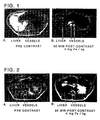
- Pre and 35 minutes post contrast T 1 -weighted MR images were recorded at 1.5T, Turbo-FLASH, TR/TE/TI/flip 15 ms/4.1 ms/846 ms/25° and appear as Figures 1A and 1B hereto.
- the liver appears bright in the left of the pre-contrast image. In the post contrast image, the liver parenchyma is dark while the blood vessels running through it appear very bright.
- a contrast agent composition produced according to Example 4 was administered intravenously into the pig at a dosage of 5 mg Fe/kg bodyweight.
- Pre and 20 minutes post contrast T 2 -weighted MR images were recorded at 1.5T fast spin echo, TR/TE 1800 ms/100 ms and appear as Figures 2A and 2B hereto.
- Pre contrast the liver parenchyma appears lighter than the blood vessels; post contrast both liver parenchyma and blood vessels appear dark.
- Comparison between Figures 1B and 2B allows differentiation between blood vessels in the liver and other non-parenchymal items.
- MPP coated particles produced according to Example 2 are combined with particles produced according to Example 1 in a weight ratio (of FeOx) of 5:1, diluted to an iron concentration of 10 mg FeOx/mL with 5% dextrose solution and sterile filtered.
- Example 1 In place of the particles of Example 1 one may use conventional MSM or commercially available magnetic particles such as those available from Guerbet SA under the trade name Endorem.
Landscapes
- Health & Medical Sciences (AREA)
- Chemical & Material Sciences (AREA)
- Nanotechnology (AREA)
- Engineering & Computer Science (AREA)
- Life Sciences & Earth Sciences (AREA)
- Nuclear Medicine, Radiotherapy & Molecular Imaging (AREA)
- Public Health (AREA)
- Epidemiology (AREA)
- Animal Behavior & Ethology (AREA)
- General Health & Medical Sciences (AREA)
- Veterinary Medicine (AREA)
- Molecular Biology (AREA)
- Radiology & Medical Imaging (AREA)
- Immunology (AREA)
- Medicines Containing Antibodies Or Antigens For Use As Internal Diagnostic Agents (AREA)
- Magnetic Resonance Imaging Apparatus (AREA)
- Medicines Containing Plant Substances (AREA)
- Medicines Containing Material From Animals Or Micro-Organisms (AREA)
Claims (23)
- Verfahren der kontrastverstärkten MR-Angiographie des menschlichen oder nichtmenschlichen tierischen Körpers, dessen Blutgefäßsystem vorausgehend mit einer kontrastwirksamen Menge einer Kontrastmittelzusammensetzung, welche magnetische Teilchen mit einem r2/r1-Verhältnis von nicht mehr als 5, gemessen bei 0,5 T und 37°C, umfaßt, verabreicht worden ist, wobei das Verfahren die Erzeugung eines T1-gewichteten Magnetresonanzbildes mindestens eines Organs des retikuloendothelialen Systems zu einer Zeit umfaßt, wenn genügend der magnetischen Teilchen in dem Blutgefäßsystem verbleiben, um eine positive Kontrastverstärkung hiervon in einem T1-gewichteten Bild vorzusehen, während genügend magnetische Teilchen in dem Organ aufgenommen worden sind. um eine negative Konstrastverstärkung hiervon in dem T1-gewichteten Bild vorzusehen.
- Verfahren nach Anspruch 1, wobei das Organ die Leber ist.
- Verfahren nach Anspruch 1, wobei das Organ ein Lymphknoten ist.
- Verfahren nach mindestens einem der vorangehenden Ansprüche, wobei ebenso ein T2-gewichtetes Bild mindestens des Organs erzeugt wird.
- Verfahren nach mindestens einem der vorangehenden Ansprüche, wobei die Zusammensetzung eine erste Vielzahl magnetischer Teilchen mit einem r2/r1-Verhältnis von nicht mehr als 5 und eine zweite Vielzahl magnetischer Teilchen mit einer kürzeren Bluthalbwertszeit als derjenigen der ersten Vielzahl umfaßt, wobei vorzugsweise die zweite Vielzahl magnetischer Teilchen ein höheres r2/r1-Verhältnis als das der ersten Vielzahl aufweist.
- Verfahren nach Anspruch 5, wobei die erste Vielzahl magnetischer Teilchen Teilchen umfaßt, die mit einem Opsonierungsinhibitor oder einem die Blutlebenszelt verlängernden Material versehen sind, vorzugsweise einem die Blutlebenszeit verlängernden Polyoxyalkylenmaterial.
- Verfahren nach Anspruch 5, wobei die erste Vielzahl magnetischer Teilchen eine Bluthalbwertszeit von mindestens 30 Minuten aufweist.
- Verfahren nach mindestens einem der vorangehenden Ansprüche, wobei die Zusammensetzung magnetische Teilchen mit einem r2/r1-Verhältnis von nicht mehr als 5 umfaßt, welche mit einem Opsonierungsinhibitor oder einem die Blutlebenszeit verlängernden Material versehen sind, vorzugsweise einem die Blutlebenszeit verlängernden Polyoxyalkylenmaterial.
- Verfahren nach mindestens einem der vorangehenden Ansprüche, wobei eine kontrastwirksame Menge einer ersten, magnetische Teilchen umfassenden Zusammensetzung dem Körper parenteral verabreicht worden ist, vor der Verabreichung in das Blutgefäßsystem des Körpers der kontrastwirksamen Menge der Zusammensetzung, welche magnetische Teilchen mit einem r2/r1-Verhältnis von nicht mehr als 5 (zweite Zusammensetzung) umfaßt, wobei vorzugsweise die erste Zusammensetzung in das Blutgefäßsystem des Körpers verabreicht worden ist.
- Verfahren nach Anspruch 9, wobei die erste Zusammensetzung in das Lymphknotensystem des Körpers verabreicht worden ist, und sowie das T1-gewichtete Bild erzeugt wird, ein Bild mindestens eines Teils des Systems erzeugt wird.
- Verfahren nach mindestens einem der vorangehenden Ansprüche, wobei die magnetischen Teilchen superparamagnetische Eigenschaften zeigen und vorzugsweise zusammengesetzte Teilchen sind, umfassend magnetische Kristalle und ein biotolerierbares Polymer, wobei das biotolerierbare Polymer vorzugsweise ein Oligo- oder Polysaccharid ist, weiter vorzugsweise eine oxidativ gespaltene Stärke.
- Verfahren nach Anspruch 11, wobei die zusammengesetzten Teilchen weiterhin einen Opsonierungsinhibitor oder ein die Blutlebenszeit verlängerndes Material umfassen, vorzugsweise ein die Blutlebenszeit verlängerndes Polyalkylenoxidmaterial.
- Verfahren nach mindestens einem der vorangehenden Ansprüche, wobei das T1-gewichtete Bild während einer Zeit von 1 Minute bis 24 Stunden, vorzugsweise bis zu 50 Minuten, nach Verabreichung der magnetischen Teilchen mit einem r2/r1-Verhältnis von nicht mehr als 5 erzeugt wird.
- Verfahren nach mindestens einem der vorangehenden Ansprüche, wobei das T1-gewichtete Bild zu einer Zeit erzeugt wird, wenn die positive Kontrastverstärkung einer Bildintensitätszunahme von mindestens 80%, vorzugsweise mindestens 100%, weiter vorzugsweise mindestens 150%, entspricht, und die negative Kontrastverstärkung einer Bildintensitätsabnahme von mindestens 20%, vorzugsweise mindestens 30%, entspricht.
- Verfahren nach mindestens einem der vorangehenden Ansprüche, wobei das r2/r1-Verhältnis der magnetischen Teilchen mit einem r2/r1-Verhältnis von nicht mehr als 5 nicht mehr als 3, vorzugsweise nicht mehr als 2,5 beträgt.
- Verfahren nach mindestens einem der vorangehenden Ansprüche, wobei das Gewichtsverhältnis von anorganischem magnetischem Material in der ersten Vielzahl zu demjenigen in der zweiten Vielzahl im Bereich von 10:1 bis 1:1 liegt.
- Verfahren nach mindestens einem der vorangehenden Ansprüche, wobei das Gewichtsverhältnis von anorganischem magnetischem Material in der kontrastwirksamen Menge der zweiten Zusammensetzung zu demjenigen in der kontrastwirksamen Menge der ersten Zusammensetzung im Bereich von 10:1 bis 1:1 liegt.
- Diagnostische Kontrastmittelzusammensetzung, umfassend magnetische Teilchen und einen physiologisch tolerierbaren Träger oder Exzipienten, dadurch gekennzeichnet, dass die Zusammensetzung eine erste Vielzahl magnetischer Teilchen mit einem r2/r1-Verhältnis von nicht mehr als 5, gemessen bei 0,5 T und 37°C, und einer Bluthalbwertszeit von bis zu 24 Stunden, und eine zweite Vielzahl magnetischer Teilchen mit einer geringeren Bluthalbwertszeit, vorzugsweise mindestens 50% geringeren, als derjenigen der ersten Vielzahl enthält.
- Zusammensetzung nach Anspruch 18, wobei die erste Vielzahl magnetischer Teilchen Teilchen umfaßt, welche mit einem Opsonierungsinhibitor oder einem die Blutlebenszeit verlängernden Material versehen sind.
- Zusammensetzung nach Anspruch 18, wobei die erste Vielzahl magnetischer Teilchen zusammengesetzte Teilchen umfaßt, die magnetische Kristalle, ein biotolerierbares Polymer und einen Opsonierungsinhibitor oder ein die Blutlebenszeit verlängerndes Material umfassen.
- Zusammensetzung nach Anspruch 20, wobei das biotolerierbare Polymer eine oxidativ gespaltene Stärke ist und das die Blutlebenszeit verlängernde Material ein Polyalkylenoxidmaterial ist.
- Zusammensetzung nach mindestens einem der Ansprüche 18 bis 21, wobei die magnetischen Teilchen eine mittlere Gesamtteilchengröße unterhalb 100 nm besitzen.
- Verwendung einer Zusammensetzung gemäß mindestens einem der Ansprüche 18 bis 22 bei der Herstellung eines Magnetresonanzbild-Kontrastmediums zur Verwendung bei einem Verfahren der MR-Angiographie gemäß mindestens einem der Ansprüche 1 bis 17.
Applications Claiming Priority (3)
| Application Number | Priority Date | Filing Date | Title |
|---|---|---|---|
| US08/625,223 US5855868A (en) | 1996-04-01 | 1996-04-01 | Method of T1 -weighted resonance imaging of RES organs |
| US625223 | 1996-04-01 | ||
| PCT/GB1997/000886 WO1997036617A2 (en) | 1996-04-01 | 1997-03-27 | Method of t1-weighted magnetic resonance imaging of res organs |
Publications (2)
| Publication Number | Publication Date |
|---|---|
| EP0896546A2 EP0896546A2 (de) | 1999-02-17 |
| EP0896546B1 true EP0896546B1 (de) | 2002-11-20 |
Family
ID=24505090
Family Applications (1)
| Application Number | Title | Priority Date | Filing Date |
|---|---|---|---|
| EP97914482A Expired - Lifetime EP0896546B1 (de) | 1996-04-01 | 1997-03-27 | T1-zeitgewichteten kernspintomogram der res-orgäne |
Country Status (14)
| Country | Link |
|---|---|
| US (1) | US5855868A (de) |
| EP (1) | EP0896546B1 (de) |
| JP (1) | JP4361970B2 (de) |
| KR (1) | KR20000005194A (de) |
| CN (1) | CN1219135A (de) |
| AT (1) | ATE228017T1 (de) |
| AU (1) | AU704926B2 (de) |
| CA (1) | CA2250818A1 (de) |
| DE (1) | DE69717269T2 (de) |
| EA (1) | EA000796B1 (de) |
| ES (1) | ES2188924T3 (de) |
| IL (1) | IL126412A0 (de) |
| NO (1) | NO984555D0 (de) |
| WO (1) | WO1997036617A2 (de) |
Cited By (1)
| Publication number | Priority date | Publication date | Assignee | Title |
|---|---|---|---|---|
| DE102004022061A1 (de) * | 2004-05-05 | 2005-12-08 | Siemens Ag | Verfahren zur verbesserten interventionallen Bildgebung in der Magnet-Resonanz-Tomographie |
Families Citing this family (10)
| Publication number | Priority date | Publication date | Assignee | Title |
|---|---|---|---|---|
| PT81498B (pt) * | 1984-11-23 | 1987-12-30 | Schering Ag | Processo para a preparacao de composicoes para diagnostico contendo particulas magneticas |
| DE59706019D1 (de) * | 1996-08-05 | 2002-02-21 | Schering Ag | Vorrichtung und verfahren zur abtrennung magnetischer materialien aus pharmazeutischen zubereitungen, deren ausgangs- oder zwischenprodukten sowie mit hilfe dieser vorrichtung hergestellte mittel |
| US7082326B2 (en) * | 2000-03-31 | 2006-07-25 | Amersham Health As | Method of magnetic resonance imaging |
| WO2001074245A1 (en) * | 2000-03-31 | 2001-10-11 | Amersham Health As | Method of magnetic resonance imaging |
| KR20020075513A (ko) * | 2001-03-24 | 2002-10-05 | 조용덕 | 전기분해법을 이용한 폐수의 처리방법 |
| WO2007035871A1 (en) * | 2005-09-21 | 2007-03-29 | Massachusetts Institute Of Technology | Systems and methods for tuning properties of nanoparticles |
| KR20090038337A (ko) * | 2007-10-15 | 2009-04-20 | 재단법인서울대학교산학협력재단 | 무기계 나노입자를 수계 매질에 분산시키는 생체적합성분산 안정화제 |
| WO2016004061A1 (en) * | 2014-06-30 | 2016-01-07 | University Of Washington | Mri signal supression agents, compositions, and methods |
| US10466326B2 (en) * | 2015-05-15 | 2019-11-05 | Stc. Unm | Quantitative [Fe]-MRI (femri) of anti-PSMA-conjugated SPIONs based on PSMA expression levels |
| JP2022548716A (ja) * | 2019-09-18 | 2022-11-21 | バイエル、アクチエンゲゼルシャフト | 肝臓のmri画像の生成 |
Family Cites Families (21)
| Publication number | Priority date | Publication date | Assignee | Title |
|---|---|---|---|---|
| US3935187A (en) * | 1973-10-19 | 1976-01-27 | Standard Brands Incorporated | Process for depolymerizing amylaceous polymers |
| WO1983001738A1 (en) * | 1981-11-12 | 1983-05-26 | SCHRÖDER, Ulf | Intravascularly administrable, magnetically responsive nanosphere or manoparticle, a process for the production thereof, and the use thereof |
| US4452773A (en) * | 1982-04-05 | 1984-06-05 | Canadian Patents And Development Limited | Magnetic iron-dextran microspheres |
| GB8408127D0 (en) * | 1984-03-29 | 1984-05-10 | Nyegaard & Co As | Contrast agents |
| US4767611A (en) * | 1984-07-03 | 1988-08-30 | Gordon Robert T | Method for affecting intracellular and extracellular electric and magnetic dipoles |
| US4849210A (en) * | 1985-05-08 | 1989-07-18 | Molecular Biosystems, Inc. | Magnetic resonance imaging of liver and spleen with superparamagnetic contrast agents |
| GB8601100D0 (en) * | 1986-01-17 | 1986-02-19 | Cosmas Damian Ltd | Drug delivery system |
| US4827945A (en) * | 1986-07-03 | 1989-05-09 | Advanced Magnetics, Incorporated | Biologically degradable superparamagnetic materials for use in clinical applications |
| US5314679A (en) * | 1986-07-03 | 1994-05-24 | Advanced Magnetics Inc. | Vascular magnetic resonance imaging agent comprising nanoparticles |
| US5069216A (en) * | 1986-07-03 | 1991-12-03 | Advanced Magnetics Inc. | Silanized biodegradable super paramagnetic metal oxides as contrast agents for imaging the gastrointestinal tract |
| US4925678A (en) * | 1987-04-01 | 1990-05-15 | Ranney David F | Endothelial envelopment drug carriers |
| DE3709851A1 (de) * | 1987-03-24 | 1988-10-06 | Silica Gel Gmbh Adsorptions Te | Nmr-diagnostische fluessigkeitszusammensetzungen |
| US5358702A (en) * | 1990-04-10 | 1994-10-25 | Unger Evan C | Methoxylated gel particle contrast media for improved diagnostic imaging |
| WO1992012735A1 (fr) * | 1991-01-19 | 1992-08-06 | Meito Sangyo Kabushiki Kaisha | Composition contenant des particules ultra fines d'oxyde de metal magnetique |
| US5225282A (en) * | 1991-12-13 | 1993-07-06 | Molecular Bioquest, Inc. | Biodegradable magnetic microcluster comprising non-magnetic metal or metal oxide particles coated with a functionalized polymer |
| DE69207589T2 (de) * | 1992-06-01 | 1996-05-23 | Basf Ag | Anwendung von Dispersionen von magneto-ionischen Partikeln in MRI-Kontrast-Mitteln |
| US5349957A (en) * | 1992-12-02 | 1994-09-27 | Sterling Winthrop Inc. | Preparation and magnetic properties of very small magnetite-dextran particles |
| DE59403734D1 (de) * | 1993-03-17 | 1997-09-18 | Silica Gel Gmbh | Superparamagnetische teilchen, verfahren zu ihrer herstellung und verwendung derselben |
| DE4428851C2 (de) * | 1994-08-04 | 2000-05-04 | Diagnostikforschung Inst | Eisen enthaltende Nanopartikel, ihre Herstellung und Anwendung in der Diagnostik und Therapie |
| AU687093B2 (en) * | 1994-09-27 | 1998-02-19 | Nycomed Imaging As | Contrast agent |
| HUP9901192A3 (en) * | 1996-01-10 | 2000-02-28 | Amersham Health As | Contrast media |
-
1996
- 1996-04-01 US US08/625,223 patent/US5855868A/en not_active Expired - Lifetime
-
1997
- 1997-03-27 WO PCT/GB1997/000886 patent/WO1997036617A2/en not_active Ceased
- 1997-03-27 AT AT97914482T patent/ATE228017T1/de not_active IP Right Cessation
- 1997-03-27 EP EP97914482A patent/EP0896546B1/de not_active Expired - Lifetime
- 1997-03-27 CA CA002250818A patent/CA2250818A1/en not_active Abandoned
- 1997-03-27 KR KR1019980707869A patent/KR20000005194A/ko not_active Withdrawn
- 1997-03-27 DE DE69717269T patent/DE69717269T2/de not_active Expired - Lifetime
- 1997-03-27 CN CN97194760A patent/CN1219135A/zh active Pending
- 1997-03-27 IL IL12641297A patent/IL126412A0/xx unknown
- 1997-03-27 JP JP53503697A patent/JP4361970B2/ja not_active Expired - Lifetime
- 1997-03-27 ES ES97914482T patent/ES2188924T3/es not_active Expired - Lifetime
- 1997-03-27 EA EA199800881A patent/EA000796B1/ru not_active IP Right Cessation
- 1997-03-27 AU AU21721/97A patent/AU704926B2/en not_active Ceased
-
1998
- 1998-09-29 NO NO984555A patent/NO984555D0/no not_active Application Discontinuation
Cited By (1)
| Publication number | Priority date | Publication date | Assignee | Title |
|---|---|---|---|---|
| DE102004022061A1 (de) * | 2004-05-05 | 2005-12-08 | Siemens Ag | Verfahren zur verbesserten interventionallen Bildgebung in der Magnet-Resonanz-Tomographie |
Also Published As
| Publication number | Publication date |
|---|---|
| AU2172197A (en) | 1997-10-22 |
| EP0896546A2 (de) | 1999-02-17 |
| EA000796B1 (ru) | 2000-04-24 |
| US5855868A (en) | 1999-01-05 |
| KR20000005194A (ko) | 2000-01-25 |
| JP4361970B2 (ja) | 2009-11-11 |
| NO984555L (no) | 1998-09-29 |
| DE69717269T2 (de) | 2003-08-28 |
| WO1997036617A2 (en) | 1997-10-09 |
| NO984555D0 (no) | 1998-09-29 |
| AU704926B2 (en) | 1999-05-06 |
| DE69717269D1 (de) | 2003-01-02 |
| ATE228017T1 (de) | 2002-12-15 |
| CN1219135A (zh) | 1999-06-09 |
| IL126412A0 (en) | 1999-05-09 |
| ES2188924T3 (es) | 2003-07-01 |
| WO1997036617A3 (en) | 1998-02-26 |
| CA2250818A1 (en) | 1997-10-09 |
| EA199800881A1 (ru) | 1999-04-29 |
| JP2000507567A (ja) | 2000-06-20 |
Similar Documents
| Publication | Publication Date | Title |
|---|---|---|
| EP0783325B1 (de) | Kontrastmittel | |
| Wang et al. | Superparamagnetic iron oxide contrast agents: physicochemical characteristics and applications in MR imaging | |
| Ferrucci et al. | Iron oxide-enhanced MR imaging of the liver and spleen: review of the first 5 years. | |
| KR100217208B1 (ko) | 진단 작용제 | |
| Jia et al. | Active-target T1-weighted MR imaging of tiny hepatic tumor via RGD modified ultra-small Fe3O4 nanoprobes | |
| JPH05506227A (ja) | 磁気共鳴画像用造影剤としてのポリマー | |
| EP0896546B1 (de) | T1-zeitgewichteten kernspintomogram der res-orgäne | |
| JP4965020B2 (ja) | 組織の潅流の造影剤増強磁気共鳴撮像法 | |
| Engelbrecht et al. | Contrast‐enhanced 3D‐TOF MRA of peripheral vessels: intravascular versus extracellular MR contrast media | |
| EP1126785B1 (de) | Manganzusammensetzungen und verfahren für mri | |
| US20020151787A1 (en) | Method of tumor imaging | |
| US20090317327A1 (en) | Aqueous Dispersion of Superparamagnetic Single-Domain Particles, Production and Use Thereof in Diagnosis and Therapy | |
| WO2012108856A1 (en) | Nanoparticulate manganese mri contrast agents | |
| US20240366804A1 (en) | Biocompatible imaging particles, their synthesis and use in imaging techniques | |
| US7082326B2 (en) | Method of magnetic resonance imaging | |
| Desser et al. | Interstitial MR and CT lymphography with Gd-DTPA-co-α, ω-diaminoPEG (1450) and Gd-DTPA-co-1, 6-diaminohexane polymers: Preliminary experience | |
| Woodward et al. | Magnetic Contrast Imaging: Magnetic nanoparticles as probes in living systems | |
| EP1267716B1 (de) | Verfahren zur magnetresonanzbildgebung | |
| Laurent et al. | Contrast agents for MRI: recent advances | |
| GB2311138A (en) | ESR-enhanced MRI using magnetic particles as contrast agents | |
| WO2001074245A1 (en) | Method of magnetic resonance imaging | |
| Reimer | Encyclopedia of Diagnostic Imaging Springer-Verlag Berlin Heidelberg New York 2008 | |
| Vogl | Contrast Media |
Legal Events
| Date | Code | Title | Description |
|---|---|---|---|
| PUAI | Public reference made under article 153(3) epc to a published international application that has entered the european phase |
Free format text: ORIGINAL CODE: 0009012 |
|
| 17P | Request for examination filed |
Effective date: 19981028 |
|
| AK | Designated contracting states |
Kind code of ref document: A2 Designated state(s): AT BE CH DE DK ES FI FR GB GR IE IT LI LU MC NL PT SE |
|
| AX | Request for extension of the european patent |
Free format text: AL PAYMENT 981028;LT PAYMENT 981028;LV PAYMENT 981028;RO PAYMENT 981028;SI PAYMENT 981028 |
|
| 17Q | First examination report despatched |
Effective date: 20010302 |
|
| GRAG | Despatch of communication of intention to grant |
Free format text: ORIGINAL CODE: EPIDOS AGRA |
|
| RAP1 | Party data changed (applicant data changed or rights of an application transferred) |
Owner name: AMERSHAM HEALTH AS |
|
| GRAG | Despatch of communication of intention to grant |
Free format text: ORIGINAL CODE: EPIDOS AGRA |
|
| GRAG | Despatch of communication of intention to grant |
Free format text: ORIGINAL CODE: EPIDOS AGRA |
|
| GRAH | Despatch of communication of intention to grant a patent |
Free format text: ORIGINAL CODE: EPIDOS IGRA |
|
| GRAH | Despatch of communication of intention to grant a patent |
Free format text: ORIGINAL CODE: EPIDOS IGRA |
|
| GRAA | (expected) grant |
Free format text: ORIGINAL CODE: 0009210 |
|
| AK | Designated contracting states |
Kind code of ref document: B1 Designated state(s): AT BE CH DE DK ES FI FR GB GR IE IT LI LU MC NL PT SE |
|
| AX | Request for extension of the european patent |
Free format text: AL PAYMENT 19981028;LT PAYMENT 19981028;LV PAYMENT 19981028;RO PAYMENT 19981028;SI PAYMENT 19981028 |
|
| PG25 | Lapsed in a contracting state [announced via postgrant information from national office to epo] |
Ref country code: NL Free format text: LAPSE BECAUSE OF FAILURE TO SUBMIT A TRANSLATION OF THE DESCRIPTION OR TO PAY THE FEE WITHIN THE PRESCRIBED TIME-LIMIT Effective date: 20021120 Ref country code: LI Free format text: LAPSE BECAUSE OF FAILURE TO SUBMIT A TRANSLATION OF THE DESCRIPTION OR TO PAY THE FEE WITHIN THE PRESCRIBED TIME-LIMIT Effective date: 20021120 Ref country code: GR Free format text: LAPSE BECAUSE OF FAILURE TO SUBMIT A TRANSLATION OF THE DESCRIPTION OR TO PAY THE FEE WITHIN THE PRESCRIBED TIME-LIMIT Effective date: 20021120 Ref country code: FI Free format text: LAPSE BECAUSE OF FAILURE TO SUBMIT A TRANSLATION OF THE DESCRIPTION OR TO PAY THE FEE WITHIN THE PRESCRIBED TIME-LIMIT Effective date: 20021120 Ref country code: CH Free format text: LAPSE BECAUSE OF FAILURE TO SUBMIT A TRANSLATION OF THE DESCRIPTION OR TO PAY THE FEE WITHIN THE PRESCRIBED TIME-LIMIT Effective date: 20021120 Ref country code: BE Free format text: LAPSE BECAUSE OF FAILURE TO SUBMIT A TRANSLATION OF THE DESCRIPTION OR TO PAY THE FEE WITHIN THE PRESCRIBED TIME-LIMIT Effective date: 20021120 Ref country code: AT Free format text: LAPSE BECAUSE OF FAILURE TO SUBMIT A TRANSLATION OF THE DESCRIPTION OR TO PAY THE FEE WITHIN THE PRESCRIBED TIME-LIMIT Effective date: 20021120 |
|
| REF | Corresponds to: |
Ref document number: 228017 Country of ref document: AT Date of ref document: 20021215 Kind code of ref document: T |
|
| REG | Reference to a national code |
Ref country code: GB Ref legal event code: FG4D |
|
| REG | Reference to a national code |
Ref country code: CH Ref legal event code: EP |
|
| REG | Reference to a national code |
Ref country code: IE Ref legal event code: FG4D |
|
| REF | Corresponds to: |
Ref document number: 69717269 Country of ref document: DE Date of ref document: 20030102 |
|
| PG25 | Lapsed in a contracting state [announced via postgrant information from national office to epo] |
Ref country code: SE Free format text: LAPSE BECAUSE OF FAILURE TO SUBMIT A TRANSLATION OF THE DESCRIPTION OR TO PAY THE FEE WITHIN THE PRESCRIBED TIME-LIMIT Effective date: 20030220 Ref country code: PT Free format text: LAPSE BECAUSE OF FAILURE TO SUBMIT A TRANSLATION OF THE DESCRIPTION OR TO PAY THE FEE WITHIN THE PRESCRIBED TIME-LIMIT Effective date: 20030220 Ref country code: DK Free format text: LAPSE BECAUSE OF FAILURE TO SUBMIT A TRANSLATION OF THE DESCRIPTION OR TO PAY THE FEE WITHIN THE PRESCRIBED TIME-LIMIT Effective date: 20030220 |
|
| PG25 | Lapsed in a contracting state [announced via postgrant information from national office to epo] |
Ref country code: LU Free format text: LAPSE BECAUSE OF NON-PAYMENT OF DUE FEES Effective date: 20030327 Ref country code: IE Free format text: LAPSE BECAUSE OF NON-PAYMENT OF DUE FEES Effective date: 20030327 |
|
| PG25 | Lapsed in a contracting state [announced via postgrant information from national office to epo] |
Ref country code: MC Free format text: LAPSE BECAUSE OF NON-PAYMENT OF DUE FEES Effective date: 20030331 |
|
| LTIE | Lt: invalidation of european patent or patent extension |
Effective date: 20021120 |
|
| NLV1 | Nl: lapsed or annulled due to failure to fulfill the requirements of art. 29p and 29m of the patents act | ||
| REG | Reference to a national code |
Ref country code: CH Ref legal event code: PL |
|
| ET | Fr: translation filed | ||
| REG | Reference to a national code |
Ref country code: ES Ref legal event code: FG2A Ref document number: 2188924 Country of ref document: ES Kind code of ref document: T3 |
|
| PLBE | No opposition filed within time limit |
Free format text: ORIGINAL CODE: 0009261 |
|
| STAA | Information on the status of an ep patent application or granted ep patent |
Free format text: STATUS: NO OPPOSITION FILED WITHIN TIME LIMIT |
|
| 26N | No opposition filed |
Effective date: 20030821 |
|
| REG | Reference to a national code |
Ref country code: IE Ref legal event code: MM4A |
|
| REG | Reference to a national code |
Ref country code: HK Ref legal event code: WD Ref document number: 1016900 Country of ref document: HK |
|
| REG | Reference to a national code |
Ref country code: ES Ref legal event code: PC2A |
|
| REG | Reference to a national code |
Ref country code: FR Ref legal event code: CD |
|
| REG | Reference to a national code |
Ref country code: DE Ref legal event code: R082 Ref document number: 69717269 Country of ref document: DE Representative=s name: J D REYNOLDS & CO., GB |
|
| REG | Reference to a national code |
Ref country code: FR Ref legal event code: PLFP Year of fee payment: 20 |
|
| PGFP | Annual fee paid to national office [announced via postgrant information from national office to epo] |
Ref country code: ES Payment date: 20160328 Year of fee payment: 20 |
|
| PGFP | Annual fee paid to national office [announced via postgrant information from national office to epo] |
Ref country code: GB Payment date: 20160329 Year of fee payment: 20 Ref country code: FR Payment date: 20160328 Year of fee payment: 20 |
|
| PGFP | Annual fee paid to national office [announced via postgrant information from national office to epo] |
Ref country code: DE Payment date: 20160331 Year of fee payment: 20 |
|
| PGFP | Annual fee paid to national office [announced via postgrant information from national office to epo] |
Ref country code: IT Payment date: 20160323 Year of fee payment: 20 |
|
| REG | Reference to a national code |
Ref country code: DE Ref legal event code: R071 Ref document number: 69717269 Country of ref document: DE |
|
| REG | Reference to a national code |
Ref country code: GB Ref legal event code: PE20 Expiry date: 20170326 |
|
| PG25 | Lapsed in a contracting state [announced via postgrant information from national office to epo] |
Ref country code: GB Free format text: LAPSE BECAUSE OF EXPIRATION OF PROTECTION Effective date: 20170326 |
|
| REG | Reference to a national code |
Ref country code: ES Ref legal event code: FD2A Effective date: 20170704 |
|
| PG25 | Lapsed in a contracting state [announced via postgrant information from national office to epo] |
Ref country code: ES Free format text: LAPSE BECAUSE OF EXPIRATION OF PROTECTION Effective date: 20170328 |