EP0902088A2 - Petide mit antiproliferativen Eigenschaften - Google Patents
Petide mit antiproliferativen Eigenschaften Download PDFInfo
- Publication number
- EP0902088A2 EP0902088A2 EP98118811A EP98118811A EP0902088A2 EP 0902088 A2 EP0902088 A2 EP 0902088A2 EP 98118811 A EP98118811 A EP 98118811A EP 98118811 A EP98118811 A EP 98118811A EP 0902088 A2 EP0902088 A2 EP 0902088A2
- Authority
- EP
- European Patent Office
- Prior art keywords
- peptide
- nucleic acid
- growth factor
- sequence
- amino acid
- Prior art date
- Legal status (The legal status is an assumption and is not a legal conclusion. Google has not performed a legal analysis and makes no representation as to the accuracy of the status listed.)
- Withdrawn
Links
- 108090000765 processed proteins & peptides Proteins 0.000 title claims abstract description 107
- 102000004196 processed proteins & peptides Human genes 0.000 title claims abstract description 52
- 230000001028 anti-proliverative effect Effects 0.000 title claims abstract description 8
- 239000003102 growth factor Substances 0.000 claims abstract description 21
- 150000007523 nucleic acids Chemical class 0.000 claims abstract description 21
- 102000039446 nucleic acids Human genes 0.000 claims abstract description 19
- 108020004707 nucleic acids Proteins 0.000 claims abstract description 19
- 108010009202 Growth Factor Receptors Proteins 0.000 claims abstract description 16
- 102000009465 Growth Factor Receptors Human genes 0.000 claims abstract description 15
- 206010028980 Neoplasm Diseases 0.000 claims abstract description 15
- 108010040002 Tumor Suppressor Proteins Proteins 0.000 claims abstract description 14
- 102000001742 Tumor Suppressor Proteins Human genes 0.000 claims abstract description 14
- 201000011510 cancer Diseases 0.000 claims abstract description 12
- 239000000225 tumor suppressor protein Substances 0.000 claims abstract description 12
- 239000008194 pharmaceutical composition Substances 0.000 claims abstract description 11
- 108091093037 Peptide nucleic acid Proteins 0.000 claims abstract description 8
- 201000010099 disease Diseases 0.000 claims abstract description 8
- 208000037265 diseases, disorders, signs and symptoms Diseases 0.000 claims abstract description 8
- 238000003745 diagnosis Methods 0.000 claims abstract description 6
- 238000002560 therapeutic procedure Methods 0.000 claims abstract description 6
- 201000001320 Atherosclerosis Diseases 0.000 claims abstract description 4
- 150000001413 amino acids Chemical class 0.000 claims description 16
- NOESYZHRGYRDHS-UHFFFAOYSA-N insulin Chemical compound N1C(=O)C(NC(=O)C(CCC(N)=O)NC(=O)C(CCC(O)=O)NC(=O)C(C(C)C)NC(=O)C(NC(=O)CN)C(C)CC)CSSCC(C(NC(CO)C(=O)NC(CC(C)C)C(=O)NC(CC=2C=CC(O)=CC=2)C(=O)NC(CCC(N)=O)C(=O)NC(CC(C)C)C(=O)NC(CCC(O)=O)C(=O)NC(CC(N)=O)C(=O)NC(CC=2C=CC(O)=CC=2)C(=O)NC(CSSCC(NC(=O)C(C(C)C)NC(=O)C(CC(C)C)NC(=O)C(CC=2C=CC(O)=CC=2)NC(=O)C(CC(C)C)NC(=O)C(C)NC(=O)C(CCC(O)=O)NC(=O)C(C(C)C)NC(=O)C(CC(C)C)NC(=O)C(CC=2NC=NC=2)NC(=O)C(CO)NC(=O)CNC2=O)C(=O)NCC(=O)NC(CCC(O)=O)C(=O)NC(CCCNC(N)=N)C(=O)NCC(=O)NC(CC=3C=CC=CC=3)C(=O)NC(CC=3C=CC=CC=3)C(=O)NC(CC=3C=CC(O)=CC=3)C(=O)NC(C(C)O)C(=O)N3C(CCC3)C(=O)NC(CCCCN)C(=O)NC(C)C(O)=O)C(=O)NC(CC(N)=O)C(O)=O)=O)NC(=O)C(C(C)CC)NC(=O)C(CO)NC(=O)C(C(C)O)NC(=O)C1CSSCC2NC(=O)C(CC(C)C)NC(=O)C(NC(=O)C(CCC(N)=O)NC(=O)C(CC(N)=O)NC(=O)C(NC(=O)C(N)CC=1C=CC=CC=1)C(C)C)CC1=CN=CN1 NOESYZHRGYRDHS-UHFFFAOYSA-N 0.000 claims description 16
- 238000000034 method Methods 0.000 claims description 12
- 108090000623 proteins and genes Proteins 0.000 claims description 12
- 108010039918 Polylysine Proteins 0.000 claims description 10
- 229920000656 polylysine Polymers 0.000 claims description 10
- 102000004169 proteins and genes Human genes 0.000 claims description 10
- 239000002299 complementary DNA Substances 0.000 claims description 9
- 102000004877 Insulin Human genes 0.000 claims description 8
- 230000000295 complement effect Effects 0.000 claims description 8
- 229940125396 insulin Drugs 0.000 claims description 8
- 108090001061 Insulin Proteins 0.000 claims description 7
- 201000000582 Retinoblastoma Diseases 0.000 claims description 6
- 239000003814 drug Substances 0.000 claims description 5
- 230000002068 genetic effect Effects 0.000 claims description 5
- 108050002653 Retinoblastoma protein Proteins 0.000 claims description 4
- 230000030648 nucleus localization Effects 0.000 claims description 4
- 108020003175 receptors Proteins 0.000 claims description 4
- 102000005962 receptors Human genes 0.000 claims description 4
- 108020004635 Complementary DNA Proteins 0.000 claims description 3
- 150000008574 D-amino acids Chemical class 0.000 claims description 3
- 238000010804 cDNA synthesis Methods 0.000 claims description 3
- 239000003795 chemical substances by application Substances 0.000 claims description 3
- 239000012634 fragment Substances 0.000 claims description 3
- 230000008569 process Effects 0.000 claims description 3
- 239000003826 tablet Substances 0.000 claims description 3
- 102000035195 Peptidases Human genes 0.000 claims description 2
- 108091005804 Peptidases Proteins 0.000 claims description 2
- 239000004365 Protease Substances 0.000 claims description 2
- 239000002156 adsorbate Substances 0.000 claims description 2
- 239000000443 aerosol Substances 0.000 claims description 2
- -1 agglomerates Substances 0.000 claims description 2
- 239000002775 capsule Substances 0.000 claims description 2
- 238000010367 cloning Methods 0.000 claims description 2
- 230000009918 complex formation Effects 0.000 claims description 2
- 239000006185 dispersion Substances 0.000 claims description 2
- 229940079593 drug Drugs 0.000 claims description 2
- 239000000839 emulsion Substances 0.000 claims description 2
- 239000006260 foam Substances 0.000 claims description 2
- 239000008187 granular material Substances 0.000 claims description 2
- 239000011325 microbead Substances 0.000 claims description 2
- 239000002674 ointment Substances 0.000 claims description 2
- 239000006187 pill Substances 0.000 claims description 2
- 239000000843 powder Substances 0.000 claims description 2
- 239000000243 solution Substances 0.000 claims description 2
- 125000003275 alpha amino acid group Chemical group 0.000 claims 7
- 238000004519 manufacturing process Methods 0.000 claims 4
- 230000035755 proliferation Effects 0.000 claims 3
- 230000002209 hydrophobic effect Effects 0.000 claims 2
- 239000002773 nucleotide Substances 0.000 claims 2
- 125000003729 nucleotide group Chemical group 0.000 claims 2
- 108010070716 Intercellular Signaling Peptides and Proteins Proteins 0.000 claims 1
- 102000005755 Intercellular Signaling Peptides and Proteins Human genes 0.000 claims 1
- 230000004663 cell proliferation Effects 0.000 claims 1
- BHONFOAYRQZPKZ-LCLOTLQISA-N chembl269478 Chemical compound C([C@H](NC(=O)[C@H](CC=1C2=CC=CC=C2NC=1)NC(=O)[C@H]([C@@H](C)CC)NC(=O)[C@H](CCCCN)NC(=O)[C@@H](NC(=O)[C@H](CCC(N)=O)NC(=O)[C@@H](N)CCCNC(N)=N)[C@@H](C)CC)C(=O)N[C@@H](CCC(N)=O)C(=O)N[C@@H](CC(N)=O)C(=O)N[C@@H](CCCNC(N)=N)C(=O)N[C@@H](CCCNC(N)=N)C(=O)N[C@@H](CCSC)C(=O)N[C@@H](CCCCN)C(=O)N[C@@H](CC=1C2=CC=CC=C2NC=1)C(=O)N[C@@H](CCCCN)C(=O)N[C@@H](CCCCN)C(O)=O)C1=CC=CC=C1 BHONFOAYRQZPKZ-LCLOTLQISA-N 0.000 claims 1
- 239000008298 dragée Substances 0.000 claims 1
- 238000000605 extraction Methods 0.000 claims 1
- 235000010603 pastilles Nutrition 0.000 claims 1
- 239000007787 solid Substances 0.000 claims 1
- 108091032973 (ribonucleotides)n+m Proteins 0.000 abstract description 20
- 108020004414 DNA Proteins 0.000 abstract description 20
- 102000040650 (ribonucleotides)n+m Human genes 0.000 abstract description 9
- 230000003463 hyperproliferative effect Effects 0.000 abstract description 3
- 210000004027 cell Anatomy 0.000 description 76
- 108090000723 Insulin-Like Growth Factor I Proteins 0.000 description 18
- 102000004218 Insulin-Like Growth Factor I Human genes 0.000 description 18
- 230000018199 S phase Effects 0.000 description 13
- 230000010190 G1 phase Effects 0.000 description 12
- 101800004193 Peptide P3 Proteins 0.000 description 12
- 230000001640 apoptogenic effect Effects 0.000 description 12
- 230000000694 effects Effects 0.000 description 11
- 125000003178 carboxy group Chemical group [H]OC(*)=O 0.000 description 10
- 230000007423 decrease Effects 0.000 description 9
- 108010043655 penetratin Proteins 0.000 description 8
- 102000009024 Epidermal Growth Factor Human genes 0.000 description 7
- 230000010337 G2 phase Effects 0.000 description 7
- 101710098940 Pro-epidermal growth factor Proteins 0.000 description 7
- 230000022131 cell cycle Effects 0.000 description 7
- MCYTYTUNNNZWOK-LCLOTLQISA-N penetratin Chemical compound C([C@H](NC(=O)[C@H](CC=1C2=CC=CC=C2NC=1)NC(=O)[C@H]([C@@H](C)CC)NC(=O)[C@H](CCCCN)NC(=O)[C@@H](NC(=O)[C@H](CCC(N)=O)NC(=O)[C@@H](N)CCCNC(N)=N)[C@@H](C)CC)C(=O)N[C@@H](CCC(N)=O)C(=O)N[C@@H](CC(N)=O)C(=O)N[C@@H](CCCNC(N)=N)C(=O)N[C@@H](CCCNC(N)=N)C(=O)N[C@@H](CCSC)C(=O)N[C@@H](CCCCN)C(=O)N[C@@H](CC=1C2=CC=CC=C2NC=1)C(=O)N[C@@H](CCCCN)C(=O)N[C@@H](CCCCN)C(N)=O)C1=CC=CC=C1 MCYTYTUNNNZWOK-LCLOTLQISA-N 0.000 description 7
- 230000032823 cell division Effects 0.000 description 6
- 231100000433 cytotoxic Toxicity 0.000 description 6
- 230000001472 cytotoxic effect Effects 0.000 description 6
- 239000002243 precursor Substances 0.000 description 6
- 230000027311 M phase Effects 0.000 description 5
- 206010006187 Breast cancer Diseases 0.000 description 4
- 208000026310 Breast neoplasm Diseases 0.000 description 4
- 230000035519 G0 Phase Effects 0.000 description 4
- 239000006146 Roswell Park Memorial Institute medium Substances 0.000 description 4
- 238000004891 communication Methods 0.000 description 4
- 238000011160 research Methods 0.000 description 4
- 239000006144 Dulbecco’s modified Eagle's medium Substances 0.000 description 3
- 102100022828 Retinoblastoma-like protein 2 Human genes 0.000 description 3
- 108050002651 Retinoblastoma-like protein 2 Proteins 0.000 description 3
- 102100033254 Tumor suppressor ARF Human genes 0.000 description 3
- 239000006143 cell culture medium Substances 0.000 description 3
- 210000003855 cell nucleus Anatomy 0.000 description 3
- 208000032839 leukemia Diseases 0.000 description 3
- 230000001360 synchronised effect Effects 0.000 description 3
- 101000599951 Homo sapiens Insulin-like growth factor I Proteins 0.000 description 2
- 102000048143 Insulin-Like Growth Factor II Human genes 0.000 description 2
- 108090001117 Insulin-Like Growth Factor II Proteins 0.000 description 2
- 102100037852 Insulin-like growth factor I Human genes 0.000 description 2
- 108091028043 Nucleic acid sequence Proteins 0.000 description 2
- 102000044209 Tumor Suppressor Genes Human genes 0.000 description 2
- 108700025716 Tumor Suppressor Genes Proteins 0.000 description 2
- 230000006369 cell cycle progression Effects 0.000 description 2
- 230000010261 cell growth Effects 0.000 description 2
- 230000002950 deficient Effects 0.000 description 2
- 238000011161 development Methods 0.000 description 2
- 230000004927 fusion Effects 0.000 description 2
- 201000008968 osteosarcoma Diseases 0.000 description 2
- 239000000126 substance Substances 0.000 description 2
- 238000012360 testing method Methods 0.000 description 2
- 238000011282 treatment Methods 0.000 description 2
- VOXZDWNPVJITMN-ZBRFXRBCSA-N 17β-estradiol Chemical compound OC1=CC=C2[C@H]3CC[C@](C)([C@H](CC4)O)[C@@H]4[C@@H]3CCC2=C1 VOXZDWNPVJITMN-ZBRFXRBCSA-N 0.000 description 1
- 108091092742 A-DNA Proteins 0.000 description 1
- 102100034540 Adenomatous polyposis coli protein Human genes 0.000 description 1
- 102100022987 Angiogenin Human genes 0.000 description 1
- 108700031308 Antennapedia Homeodomain Proteins 0.000 description 1
- 108700020463 BRCA1 Proteins 0.000 description 1
- 102000036365 BRCA1 Human genes 0.000 description 1
- 101150072950 BRCA1 gene Proteins 0.000 description 1
- 108700020462 BRCA2 Proteins 0.000 description 1
- 102000052609 BRCA2 Human genes 0.000 description 1
- 206010005949 Bone cancer Diseases 0.000 description 1
- 208000018084 Bone neoplasm Diseases 0.000 description 1
- 108091003079 Bovine Serum Albumin Proteins 0.000 description 1
- 101150008921 Brca2 gene Proteins 0.000 description 1
- 102100024209 CD177 antigen Human genes 0.000 description 1
- 108010009392 Cyclin-Dependent Kinase Inhibitor p16 Proteins 0.000 description 1
- 102100027834 DNA repair protein XRCC1 Human genes 0.000 description 1
- 102400001368 Epidermal growth factor Human genes 0.000 description 1
- 101800003838 Epidermal growth factor Proteins 0.000 description 1
- 101150021185 FGF gene Proteins 0.000 description 1
- 230000004668 G2/M phase Effects 0.000 description 1
- 229920002971 Heparan sulfate Polymers 0.000 description 1
- 241000282412 Homo Species 0.000 description 1
- 101001076292 Homo sapiens Insulin-like growth factor II Proteins 0.000 description 1
- 101000851176 Homo sapiens Pro-epidermal growth factor Proteins 0.000 description 1
- 101000695187 Homo sapiens Protein patched homolog 1 Proteins 0.000 description 1
- 108010001127 Insulin Receptor Proteins 0.000 description 1
- 102100025947 Insulin-like growth factor II Human genes 0.000 description 1
- 150000008575 L-amino acids Chemical class 0.000 description 1
- 101710128836 Large T antigen Proteins 0.000 description 1
- 102100030550 Menin Human genes 0.000 description 1
- 108010025020 Nerve Growth Factor Proteins 0.000 description 1
- 102000007999 Nuclear Proteins Human genes 0.000 description 1
- 108010089610 Nuclear Proteins Proteins 0.000 description 1
- 102100028680 Protein patched homolog 1 Human genes 0.000 description 1
- GLNADSQYFUSGOU-GPTZEZBUSA-J Trypan blue Chemical compound [Na+].[Na+].[Na+].[Na+].C1=C(S([O-])(=O)=O)C=C2C=C(S([O-])(=O)=O)C(/N=N/C3=CC=C(C=C3C)C=3C=C(C(=CC=3)\N=N\C=3C(=CC4=CC(=CC(N)=C4C=3O)S([O-])(=O)=O)S([O-])(=O)=O)C)=C(O)C2=C1N GLNADSQYFUSGOU-GPTZEZBUSA-J 0.000 description 1
- 102000015098 Tumor Suppressor Protein p53 Human genes 0.000 description 1
- 108010078814 Tumor Suppressor Protein p53 Proteins 0.000 description 1
- 239000000654 additive Substances 0.000 description 1
- 238000004458 analytical method Methods 0.000 description 1
- 108010072788 angiogenin Proteins 0.000 description 1
- 230000000118 anti-neoplastic effect Effects 0.000 description 1
- 238000013459 approach Methods 0.000 description 1
- 238000003556 assay Methods 0.000 description 1
- 230000015572 biosynthetic process Effects 0.000 description 1
- 208000023913 breast extraskeletal osteosarcoma Diseases 0.000 description 1
- 201000002858 breast osteosarcoma Diseases 0.000 description 1
- 230000005907 cancer growth Effects 0.000 description 1
- 239000012830 cancer therapeutic Substances 0.000 description 1
- 238000004113 cell culture Methods 0.000 description 1
- 238000002512 chemotherapy Methods 0.000 description 1
- 238000004587 chromatography analysis Methods 0.000 description 1
- 238000010276 construction Methods 0.000 description 1
- 239000000824 cytostatic agent Substances 0.000 description 1
- 230000001085 cytostatic effect Effects 0.000 description 1
- 230000007547 defect Effects 0.000 description 1
- 230000001419 dependent effect Effects 0.000 description 1
- 230000004069 differentiation Effects 0.000 description 1
- 239000002552 dosage form Substances 0.000 description 1
- 229940116977 epidermal growth factor Drugs 0.000 description 1
- 229960005309 estradiol Drugs 0.000 description 1
- 229930182833 estradiol Natural products 0.000 description 1
- 238000002474 experimental method Methods 0.000 description 1
- 239000012894 fetal calf serum Substances 0.000 description 1
- 238000011049 filling Methods 0.000 description 1
- 238000001943 fluorescence-activated cell sorting Methods 0.000 description 1
- 238000001415 gene therapy Methods 0.000 description 1
- 230000012010 growth Effects 0.000 description 1
- 201000005787 hematologic cancer Diseases 0.000 description 1
- 208000024200 hematopoietic and lymphoid system neoplasm Diseases 0.000 description 1
- 235000003642 hunger Nutrition 0.000 description 1
- 238000001727 in vivo Methods 0.000 description 1
- 238000011534 incubation Methods 0.000 description 1
- 230000003993 interaction Effects 0.000 description 1
- 238000001361 intraarterial administration Methods 0.000 description 1
- 238000007912 intraperitoneal administration Methods 0.000 description 1
- 238000001990 intravenous administration Methods 0.000 description 1
- 239000007937 lozenge Substances 0.000 description 1
- 239000011159 matrix material Substances 0.000 description 1
- 238000002844 melting Methods 0.000 description 1
- 230000008018 melting Effects 0.000 description 1
- 239000012528 membrane Substances 0.000 description 1
- 210000005087 mononuclear cell Anatomy 0.000 description 1
- 230000004660 morphological change Effects 0.000 description 1
- GVUGOAYIVIDWIO-UFWWTJHBSA-N nepidermin Chemical compound C([C@@H](C(=O)N[C@@H]([C@@H](C)CC)C(=O)NCC(=O)N[C@@H](CCC(O)=O)C(=O)N[C@@H](CCCNC(N)=N)C(=O)N[C@@H](CS)C(=O)N[C@@H](CCC(N)=O)C(=O)N[C@@H](CC=1C=CC(O)=CC=1)C(=O)N[C@@H](CCCNC(N)=N)C(=O)N[C@@H](CC(O)=O)C(=O)N[C@@H](CC(C)C)C(=O)N[C@@H](CCCCN)C(=O)N[C@@H](CC=1C2=CC=CC=C2NC=1)C(=O)N[C@@H](CC=1C2=CC=CC=C2NC=1)C(=O)N[C@@H](CCC(O)=O)C(=O)N[C@@H](CC(C)C)C(=O)N[C@@H](CCCNC(N)=N)C(O)=O)NC(=O)CNC(=O)[C@@H](NC(=O)[C@@H](NC(=O)[C@H](CS)NC(=O)[C@H](CC(N)=O)NC(=O)[C@H](CS)NC(=O)[C@H](C)NC(=O)[C@H](CC=1C=CC(O)=CC=1)NC(=O)[C@H](CCCCN)NC(=O)[C@H](CC(O)=O)NC(=O)[C@H](CC(C)C)NC(=O)[C@H](C)NC(=O)[C@H](CCC(O)=O)NC(=O)[C@@H](NC(=O)[C@H](CC=1C=CC(O)=CC=1)NC(=O)[C@H](CCSC)NC(=O)[C@H](CS)NC(=O)[C@@H](NC(=O)CNC(=O)[C@H](CC(O)=O)NC(=O)[C@H](CC=1NC=NC=1)NC(=O)[C@H](CC(C)C)NC(=O)[C@H](CS)NC(=O)[C@H](CC=1C=CC(O)=CC=1)NC(=O)CNC(=O)[C@H](CC(O)=O)NC(=O)[C@H](CC=1NC=NC=1)NC(=O)[C@H](CO)NC(=O)[C@H](CC(C)C)NC(=O)[C@H]1N(CCC1)C(=O)[C@H](CS)NC(=O)[C@H](CCC(O)=O)NC(=O)[C@H](CO)NC(=O)[C@H](CC(O)=O)NC(=O)[C@H](CO)NC(=O)[C@@H](N)CC(N)=O)C(C)C)[C@@H](C)CC)C(C)C)C(C)C)C1=CC=C(O)C=C1 GVUGOAYIVIDWIO-UFWWTJHBSA-N 0.000 description 1
- 210000004940 nucleus Anatomy 0.000 description 1
- 238000011275 oncology therapy Methods 0.000 description 1
- 108091008819 oncoproteins Proteins 0.000 description 1
- 102000027450 oncoproteins Human genes 0.000 description 1
- 210000000056 organ Anatomy 0.000 description 1
- 210000005259 peripheral blood Anatomy 0.000 description 1
- 239000011886 peripheral blood Substances 0.000 description 1
- 210000003819 peripheral blood mononuclear cell Anatomy 0.000 description 1
- 239000012071 phase Substances 0.000 description 1
- 229920001184 polypeptide Polymers 0.000 description 1
- 238000002360 preparation method Methods 0.000 description 1
- 230000002250 progressing effect Effects 0.000 description 1
- 125000006239 protecting group Chemical group 0.000 description 1
- 230000017854 proteolysis Effects 0.000 description 1
- 230000006337 proteolytic cleavage Effects 0.000 description 1
- 238000000746 purification Methods 0.000 description 1
- 230000005855 radiation Effects 0.000 description 1
- 238000001959 radiotherapy Methods 0.000 description 1
- 238000010532 solid phase synthesis reaction Methods 0.000 description 1
- 238000010561 standard procedure Methods 0.000 description 1
- 230000004936 stimulating effect Effects 0.000 description 1
- 230000000638 stimulation Effects 0.000 description 1
- 238000001356 surgical procedure Methods 0.000 description 1
- 230000004083 survival effect Effects 0.000 description 1
- 238000003786 synthesis reaction Methods 0.000 description 1
- 230000009885 systemic effect Effects 0.000 description 1
- 230000001225 therapeutic effect Effects 0.000 description 1
- 230000036962 time dependent Effects 0.000 description 1
- 230000005945 translocation Effects 0.000 description 1
- 238000011269 treatment regimen Methods 0.000 description 1
- 210000004881 tumor cell Anatomy 0.000 description 1
- VBEQCZHXXJYVRD-GACYYNSASA-N uroanthelone Chemical compound C([C@@H](C(=O)N[C@H](C(=O)N[C@@H](CS)C(=O)N[C@@H](CC(N)=O)C(=O)N[C@@H](CS)C(=O)N[C@H](C(=O)N[C@@H]([C@@H](C)CC)C(=O)NCC(=O)N[C@@H](CC=1C=CC(O)=CC=1)C(=O)N[C@@H](CO)C(=O)NCC(=O)N[C@@H](CC(O)=O)C(=O)N[C@@H](CCCNC(N)=N)C(=O)N[C@@H](CS)C(=O)N[C@@H](CCC(N)=O)C(=O)N[C@@H]([C@@H](C)O)C(=O)N[C@@H](CCCNC(N)=N)C(=O)N[C@@H](CC(O)=O)C(=O)N[C@@H](CC(C)C)C(=O)N[C@@H](CCCNC(N)=N)C(=O)N[C@@H](CC=1C2=CC=CC=C2NC=1)C(=O)N[C@@H](CC=1C2=CC=CC=C2NC=1)C(=O)N[C@@H](CCC(O)=O)C(=O)N[C@@H](CC(C)C)C(=O)N[C@@H](CCCNC(N)=N)C(O)=O)C(C)C)[C@@H](C)O)NC(=O)[C@H](CO)NC(=O)[C@H](CC(O)=O)NC(=O)[C@H](CC(C)C)NC(=O)[C@H](CO)NC(=O)[C@H](CCC(O)=O)NC(=O)[C@@H](NC(=O)[C@H](CC=1NC=NC=1)NC(=O)[C@H](CCSC)NC(=O)[C@H](CS)NC(=O)[C@@H](NC(=O)CNC(=O)CNC(=O)[C@H](CC(N)=O)NC(=O)[C@H](CC(C)C)NC(=O)[C@H](CS)NC(=O)[C@H](CC=1C=CC(O)=CC=1)NC(=O)CNC(=O)[C@H](CC(O)=O)NC(=O)[C@H](CC=1C=CC(O)=CC=1)NC(=O)[C@H](CO)NC(=O)[C@H](CO)NC(=O)[C@H]1N(CCC1)C(=O)[C@H](CS)NC(=O)CNC(=O)[C@H]1N(CCC1)C(=O)[C@H](CC=1C=CC(O)=CC=1)NC(=O)[C@H](CO)NC(=O)[C@@H](N)CC(N)=O)C(C)C)[C@@H](C)CC)C1=CC=C(O)C=C1 VBEQCZHXXJYVRD-GACYYNSASA-N 0.000 description 1
- 239000013598 vector Substances 0.000 description 1
Images


Classifications
-
- C—CHEMISTRY; METALLURGY
- C07—ORGANIC CHEMISTRY
- C07K—PEPTIDES
- C07K14/00—Peptides having more than 20 amino acids; Gastrins; Somatostatins; Melanotropins; Derivatives thereof
- C07K14/435—Peptides having more than 20 amino acids; Gastrins; Somatostatins; Melanotropins; Derivatives thereof from animals; from humans
- C07K14/46—Peptides having more than 20 amino acids; Gastrins; Somatostatins; Melanotropins; Derivatives thereof from animals; from humans from vertebrates
- C07K14/47—Peptides having more than 20 amino acids; Gastrins; Somatostatins; Melanotropins; Derivatives thereof from animals; from humans from vertebrates from mammals
- C07K14/4701—Peptides having more than 20 amino acids; Gastrins; Somatostatins; Melanotropins; Derivatives thereof from animals; from humans from vertebrates from mammals not used
- C07K14/4702—Regulators; Modulating activity
-
- C—CHEMISTRY; METALLURGY
- C07—ORGANIC CHEMISTRY
- C07K—PEPTIDES
- C07K14/00—Peptides having more than 20 amino acids; Gastrins; Somatostatins; Melanotropins; Derivatives thereof
- C07K14/001—Peptides having more than 20 amino acids; Gastrins; Somatostatins; Melanotropins; Derivatives thereof by chemical synthesis
-
- C—CHEMISTRY; METALLURGY
- C07—ORGANIC CHEMISTRY
- C07K—PEPTIDES
- C07K14/00—Peptides having more than 20 amino acids; Gastrins; Somatostatins; Melanotropins; Derivatives thereof
- C07K14/435—Peptides having more than 20 amino acids; Gastrins; Somatostatins; Melanotropins; Derivatives thereof from animals; from humans
- C07K14/46—Peptides having more than 20 amino acids; Gastrins; Somatostatins; Melanotropins; Derivatives thereof from animals; from humans from vertebrates
- C07K14/47—Peptides having more than 20 amino acids; Gastrins; Somatostatins; Melanotropins; Derivatives thereof from animals; from humans from vertebrates from mammals
- C07K14/4701—Peptides having more than 20 amino acids; Gastrins; Somatostatins; Melanotropins; Derivatives thereof from animals; from humans from vertebrates from mammals not used
- C07K14/4702—Regulators; Modulating activity
- C07K14/4703—Inhibitors; Suppressors
-
- A—HUMAN NECESSITIES
- A61—MEDICAL OR VETERINARY SCIENCE; HYGIENE
- A61K—PREPARATIONS FOR MEDICAL, DENTAL OR TOILETRY PURPOSES
- A61K38/00—Medicinal preparations containing peptides
Definitions
- the present invention relates to peptides with antiproliferative Properties that encode them Nucleic acid (DNAs / RNAs), to these nucleic acids structure-homologous peptide nucleic acids and the use the peptides, the nucleic acids, the peptide nucleic acids and / or their pharmaceutical composition in biotechnology, molecular biology, bioinformatics and the diagnosis and therapy of hyperproliferative diseases, especially of Cancer and atherosclerosis.
- the object of the present invention is therefore in providing a way that the growth of many different types of cancer efficiently can be braked and there is a regression the tumors are coming.
- a peptide according to claim 1 according to a nucleic acid (DNA / RNA) Claim 9, by a peptide nucleic acid according to claim 12, through a pharmaceutical composition according to claim 13 and by the use of the peptides according to claim 15, the use of DNA / RNA according to claim 17, the use of the peptide nucleic acid according to claim 18 and the pharmaceutical composition according to claim 21.
- Advantageous refinements result from the subclaims.
- the inventor has found a promising Approach in the treatment of cancer is to use specific peptides that are cancer-causing Substances, especially oncogenic proteins To block.
- the peptides according to the invention are characterized by the fact that they have a short sequence length have and thus from the point of view of synthesis seen are economical. They can also be efficient penetrate intracellularly so that the inside localized the cell, the cancer process immediately neutralize stimulating target proteins.
- the peptides according to the invention are therefore synthetic fusion polypeptides from the Building blocks (A) and (B), these building blocks in combination from AB or BA.
- the first Building block (A) contains an effective section of a Tumor suppressor protein - as for the first time by the inventor found out - or homologous to it hydropathically is.
- the second part (B) has a sequence which stabilizes the first part (A).
- Part (A) of the invention is also distinguished Peptide characterized in that it is in the sense of complementary peptide theory (J.E. Blalock, Trends in Biotechnology, Vol. 8, pp. 140-144, June 1990) and im Meaning of the three-dimensional configuration into one Fragment of a growth factor or growth factor receptor is hydropathically complementary.
- growth factors are: insulin, IGF1, IGF2, EGF, FGF, angiogenin, NGF and PDGF.
- Part (B) of the fusion peptide ensures that part (A) is stabilized, in particular that there is no proteolytic degradation in the cell. This can be done on the one hand by the fact that the peptides according to the invention quickly get into the cell nucleus and are less susceptible to proteases there (R. Fahraeus et al., Current Biology 1996, Vol. 6, No. 1, pp 84-91) or by branched parts (B), for example polylysine branches or core or D-amino acids. Part (B) is preferably a polylysine core (R. Radulescu et al., Biochemical and Biophysical Research Communications, 1995, Vol. 206, pp. 97-102 and G.
- Fassina EPO 0 481 930 A2
- a nuclear localization sequence NLS
- SV40-NLS or penetratin a nuclear localization sequence
- RNP A1 NLS RNP A1 NLS
- General information for the construction of peptides, which should preferably get into the cell nucleus, can be found in Sheldon et al., Proc. Natl., Acad. Sci. USA, Vol. 92, pp. 2056-2060 (1995); Dingwall et al., Trends in Biochemical Sciences 16, pp. 478-481 (1991) MS Moore, Current Biology, Vol. 6, No. 2, pp. 137-140 (1996); DA Jans, FASEB Journal, 1994, Vol.
- Part (A) preferably has the following sequence or is hydropathically homologous in at least two positions: NH 2 -LFYKKV-COOH
- This peptide is a combination of the sequence (P1) and the polylysine core [-GGG] 4 - [KRG] 2 -KG. NH 2 - [LFYKKV-GGG] 4 - [K] 2 -KG-COOH
- This peptide is a combination of the sequence P1 and the polylysine core [-GGG] 4 - [K] 2 -KG.
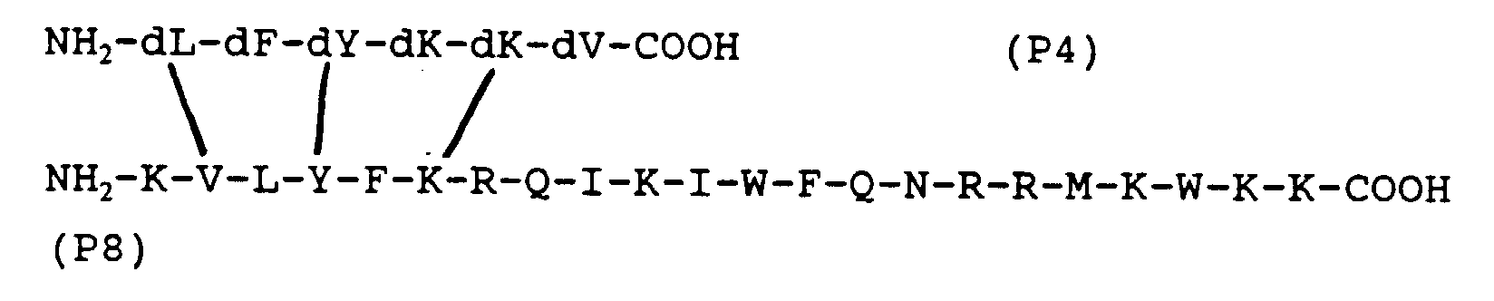
- This peptide is a combination of the sequence (P4) and the polylysine core [-GGG] 4 - [1K-dR-G] 2 -1K-G. NH 2 - [dL-dF-dY-dK-dK-dV-CGG] 4 -lK] 2 -lK-G-COOH
- This peptide is a combination of the sequence (P4) and the polylysine core [-GGG] 4 - [1K] 2 -1K-G. NH2-LFYKKVPKKKRKV-COOH
- This peptide (P5) is composed of the sequence (P1) and the nuclear localization sequence of the Large T antigen of the SV40 virus. NH 2 -LFYKKVRQIKIWFQNRRMKWK-K-COOH
- This peptide (P6) is a combination of the sequence (P1) and penetratin, a 16 amino acid sequence within the Antennapedia homeodomain, which is used for membrane translocation and thus also for nuclear localization (see above).
- This peptide (P7s) is a combination of the sequence dK-dV-dL-dY-dF-dK, which is hydropathically homologous to P1 and the polylysine core [-GGG] 4 - [lK-dR-G] 2 -lK -G. NH 2 - [dK-dV-dL-dY-dF-dK-GGG] 4 - [lK] 2 -lK-G-COOH
- This peptide (P7) is a combination of the sequence dK-dV-dL-dY-dF-dK, which is hydropathically homologous to P1 and the polylysine core [-GGG] 4 - [lK] 2 -lK-G.
- This peptide (P8) is a combination of the sequence dK-dV-dL-dY-dF-dK, the hydropathic homolog is to P1 and from penetratin.
- the peptides according to the invention have L and / or D amino acids.
- all-L forms, all-D forms, the retro-inverso isomers and the corresponding permutations of the L and D forms of each individual amino acid should be possible.
- all permutations of oxidized and reduced form of each individual amino acid and of free and protective group (s) bearing amino acid should be possible.
- the optimum active concentrations of the peptides according to the invention are in the range from 10 -4 M to 10 -5 M, however, other optimal active concentration ranges are also possible according to the application area. It has proven advantageous for branched peptides to be in the all-D form. Linear peptides containing an NLS most likely go into the nucleus; branched peptides probably go into the cell nucleus, but can also have an effect outside of it.
- the peptides according to the invention can be called "SCAPs" designate, i.e. "Synthetic cofactor-linked anioncogenic peptides ".
- the peptides according to the invention are produced preferably synthetically according to the usual solid phase method (see G.A. Grant, "Synthetic Peptides", W.H. Freeman and Company, New York, 1992). The subsequent one Purification of the peptides according to the invention was carried out and checked as already described (R. Radulescu et al., Biochemical and Biophysical Research Communications, 1995, vol. 206, pp. 97-102). The latter bibliography also provides an example how to use the peptides of the invention for biotechnological Pure growth factors, e.g. of insulin, and growth factor receptors can.
- the invention further relates to a DNA / RNA coding for the proteins according to the invention, the DNA / RNA sequence derived from the genetic code can.
- DNA / RNA preferably codes for the above-mentioned amino acid sequences (P1) and (P4) of part (A) of a peptide according to the invention:
- DNA / RNA preferably codes for the above-mentioned peptide (P5) according to the invention:
- DNA / RNA preferably codes for the above-mentioned peptide (P6) according to the invention:
- DNAs / RNAs can also be built into corresponding vectors are used for the purpose of gene therapy Cancer. An overview of the methodology can be found at R.C. Mulligan, Science 1993, vol. 260, pp. 926-932.
- DNAs / RNA sequences given above also close thus under stringent conditions, such as the Those skilled in the art are familiar with hybridizing DNAs / RNAs on. In particular DNAs / RNAs that are below at about 20 ° C the melting point of the DNA / RNA with those given above Hybridize DNAs / RNAs.
- the above close specified DNA / RNA sequences a DNAs / RNAs that with the above-mentioned DNA / RNA sequences DNAs / RNAs are related to the degenerate genetic code.
- Another application of the invention results for bioinformatics and molecular biology.
- the cDNA of the relevant growth factor or its receptor could be retrieved from the NCBI database and in the sense the complementary peptide strategy into a cDNA rewrite complementary DNA and then into a peptide translate using the DNA Strider software.
- For the resulting complementary peptides could to find homologous proteins / peptides (the ones you are looking for Tumor suppressors) using the BLAST algorithm in the OWL database navigator.
- you could too proceed from a tumor suppressor cDNA goes out and analogous to the above procedure for growth factors or their receptors as interaction partners tracing makes.
- the result of this search was that several nuclear proteins were homologous to the EGF precursor, including that structurally and functionally related to RB1 p130 protein, more precisely the p130 amino acids 290-313. More precisely: the EGF precursor amino acids 209-213 (REGSN) and p130 amino acids 305-309 (IGTLS) are hydropathically complementary to each other and thus potential binding sites in the possible Complex formation between the EGF precursor and p130 if these regions are antiparallel to each other orders.
- the amino acid sequence IGTLS or hydropathic homologous sequences as part of it (A) an antiproliferative peptide in the sense of serve present invention (s. Claim 1 and 2).
- the invention Peptides, individually or in combination of several, usually with auxiliary, filling and / or Additives together in a pharmaceutical composition used.
- a pharmaceutical composition used as dosage forms the pharmaceutical composition come into question: ointments, solutions, dispersions, Emulsions, aerosols, foams, particulate Agents (e.g. granules, agglomerates, powder, microbeads, Adsorbate), pills, lozenges, tablets, coated tablets or capsules.
- the peptides according to the invention can also in combination with other cytostatics or used in combination with radiation methods become.
- the administration is preferably local, intracutaneous or transcutaneous; for systemic application preferably intravenous, intraarterial, oral, rectal; for use in cavities, preferably intrathecally, intraperitoneal or intracavitary.
- the peptides, DNAs / RNAs according to the invention and pharmaceutical Compositions are suitable as cancer therapeutics and are according to the invention in this way used, with a pronounced cytotoxic Effect shows.
- Preferred efficacy is against breast cancer, Osteosarcoma and leukemia cells. Because of its conception the peptide according to the invention has a general effect against all tumor cells that have a defective retinoblastoma or protein.
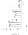
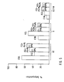
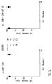
- FIG. 1 shows that the peptide P3 [10 -5 M] according to the invention reduces the G1 phase and increases the S phase in the G0 / G1 phase of synchronized MCF-7 cells under serum-free conditions.
- the morphology of the cells treated with P3 corresponds to that of apoptotic cells.
- Fig. 2 shows that peptide P3 according to the invention [10-5 M] to an increase in the G1 and G2 / M phases as well as a decrease in the S-phase of MCF-7 cells stimulated by insulin-like growth factor 1, IGF-1 for short, were stimulated in an optimal concentration of [10 -8 M].
- P3 thus blocks the effect of IGF-1 on MCF-7 cells, more precisely: the progression of the cell cycle caused by IGF-1 and thus cell division is slowed down by P3.
- the morphology of the cells treated with P3 corresponds to that of apoptotic cells.
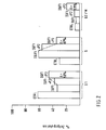
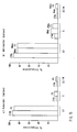
- Fig. 3 shows that each of the peptides P4, P5 and P6 according to the invention - each in a concentration of 10 -5 M - decreases the G1 phase and the S phase in the G0 / G1 phase of synchronized MCF-7 cells serum-free conditions increases.
- the morphology, in particular of the cells treated with P6, corresponds to that of apoptotic cells.
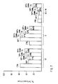
- Fig. 4 shows that the peptide of the invention P6 [10-5 M] leads to an increase in the G1 phase and a decrease in the S-phase of MCF-7 cells by IGF-1 in an optimal concentration of [10 -8 M] were stimulated. P6 thus blocks the effect of IGF-1 on MCF-7 cells, more precisely: the progression of the cell cycle caused by IGF-1 and thus cell division is slowed down by P6.
- the morphology of the cells treated with P6 corresponds to that of apoptotic cells.
- FIG. 5 shows that the combination of the peptides P3 [10 -5 M] and P5 [10 -5 M] and the combination of the peptides P3 [10 -5 M] and P6 [10 -5 M] according to the invention each increase an increase in the G1 and G2 / M phases and a decrease in the S phase of MCF-7 cells stimulated by 10% fetal calf serum (short: FCS).
- FCS 10% fetal calf serum
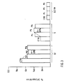
- Fig. 6 shows that the peptide P3 according to the invention [10-5 M] leads to an increase in the G1 and G2 phases as well as a decrease in the S-phase of MCF-7 cells by estradiol, short E2, in a optimal concentration of [10 -9 M] or by epidermal growth factor, in short: EGF, in an optimal concentration of [10 -8 ].
- the morphology of the cells treated with P3 corresponds to that of apoptotic cells.
- FIG. 7 shows that the peptide P3 according to the invention blocks the progression of the cell cycle caused by IGF-1 [10 -8 M] in a concentration-dependent manner.
- the morphology of the cells treated with P3 [5 x 10 -6 M] and in particular with P3 [10 -5 M] corresponds to that of the apoptotic cells.
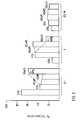
- FIG. 8 shows that the peptide P3 [10 -5 M] according to the invention reduces the G1 phase and increases the S and G2 phases in the G0 / G1 phase of synchronized SAOS-2 cells under serum-free conditions.
- the morphology of the cells treated with P3 corresponds to that of apoptotic cells.
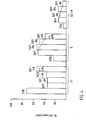
- Fig. Shows that the peptide of the invention P3 [10-5 M] 9 leads to an increase in the G1 phase and a decrease in the S phase in SAOS-2 cells, which were stimulated by 10% FS.
- the morphology of the cells treated with P3 corresponds to that of apoptotic cells.
- FIG. 10 shows that the peptide P3 [10 -5 M] according to the invention reduces the G1 phase and increases the S phase of asynchronous MCF-7 cells which have been incubated in DMEM cell culture medium with 10% FCS.
- the morphology of the cells treated with P3 corresponds to that of apoptotic cells.
- FIG. 11 shows that the peptide P8 [10 -5 M] according to the invention leads to an increase in the G1 and G2 / M phases and a decrease in the S phase of MCF-7 cells, which is caused by IGF-1 in one optimal concentration of [10 - 8 M] were stimulated.
- P8 thus blocks the effect of IGF-1 on MCF-7 cells, more precisely: the progression of the cell cycle caused by IGF-1 and thus cell division is slowed down by P8.
- the morphology of the cells treated with P8 corresponds to that of apoptotic cells.
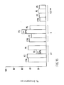
- FIG. 12 shows that the peptide P7s [10 -5 M] according to the invention leads to an increase in the G1 and G2 / M phases and a decrease in the S phase of MCF-7 cells, which is caused by IGF-1 in an optimal concentration of [10 -8 M] were stimulated.
- P7s thus blocks the effect of IGF-1 on MCF-7 cells, more precisely: the progression of the cell cycle caused by IGF-1 and thus cell division is slowed down by P7s.
- the morphology of the cells treated with P7s corresponds to that of apoptotic cells.
- FIG. 13 shows that the cytotoxic effect of the peptide P3 [10 -5 M] according to the invention on asynchronous K562 cells which were incubated in RPMI cell culture medium with 10% FCS is time-dependent. The results were plotted as living cells treated with P3 as a percentage of the living cells not treated with P3. The number of living cells was determined by counting at least 200 cells using the trypan blue method at each time.
- FIG., 14 shows that the peptide P3 [10 -5] of the present invention to cytotoxic asynchronous CCRF-CEM sensitive - and CCRF-CEM / ACT 400 resistant cells acts, which were incubated in RPMI cell culture medium containing 10% FCS.
- FIG. 15 shows that the peptide P3 [10 -5 ] according to the invention has no effect on normal human mononuclear cells of the peripheral blood, regardless of whether these are in a dormant (a) or activated (b) state. Accordingly, the cells treated with P3 also show no morphological changes. The cells were incubated with P3 for a period of 24 hours.
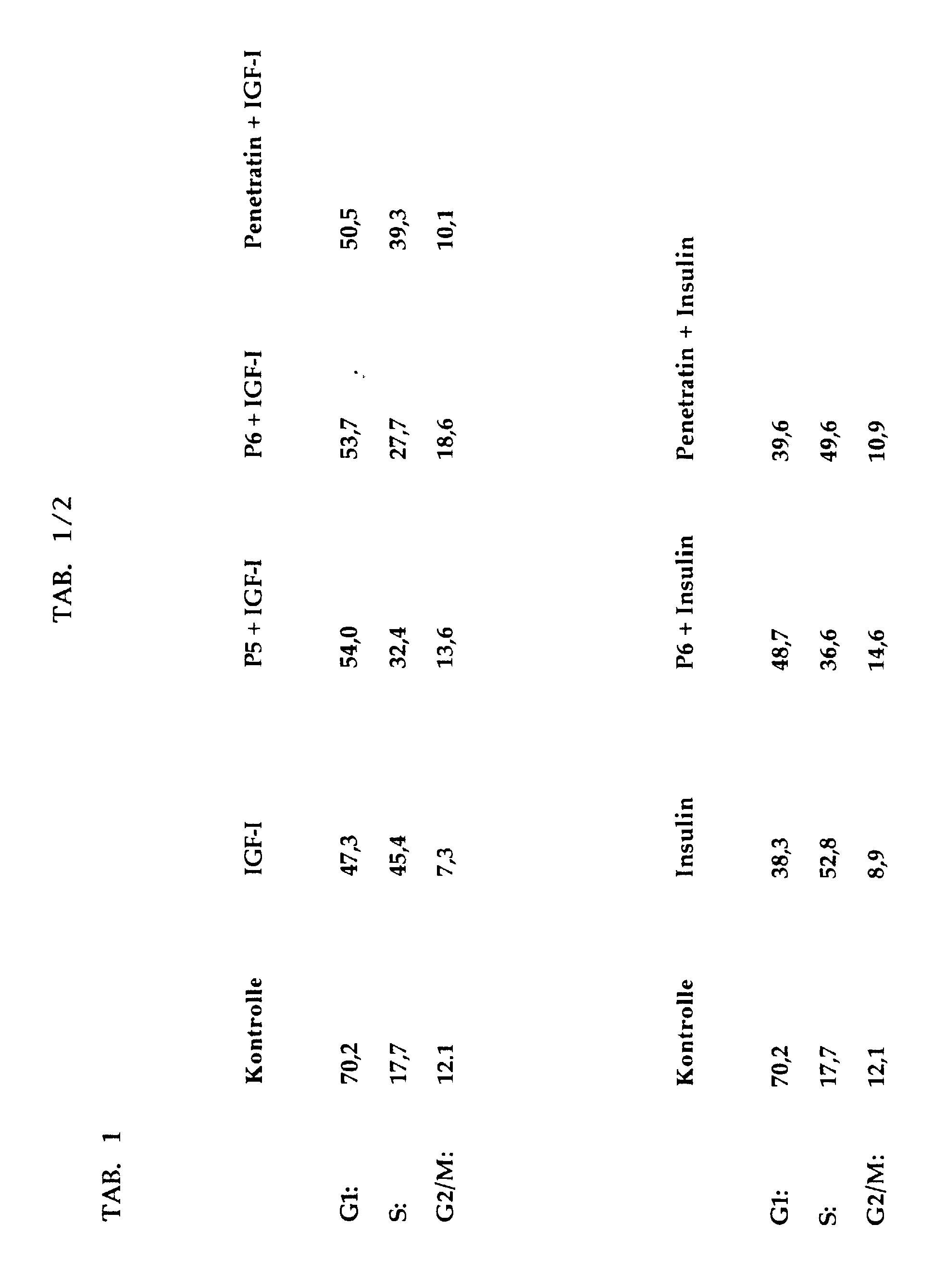
- Tab. 1 shows that the peptides P5 and P6 according to the invention, each in a concentration of 10 -5 M, reduce the S phase of MCF-7 cells by IGF-1 [10 -8 M] and by insulin [10 -6 M] were stimulated.
- the peptide penetratin [10 -5 M] had no effect in this assay as expected.
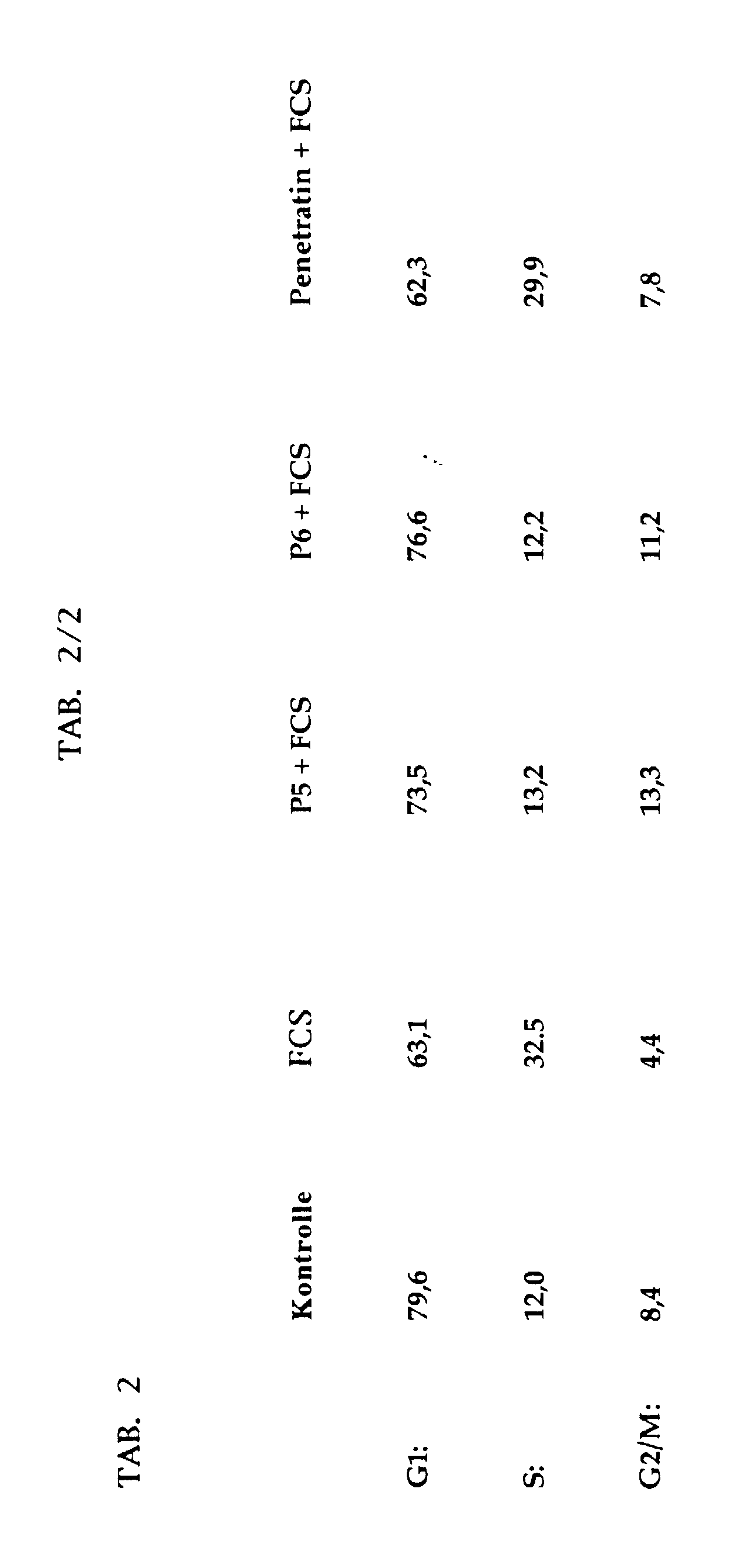
- Tab. 2 shows that the peptides P5 and P6 according to the invention, each in a concentration of 5 ⁇ 10 -5 M, lead to an increase in the G1 phase and a decrease in the S phase of SAOS-2 cells, which are caused by 10 % FCS were stimulated.
- the data shown here for IGF-1 [10 -8 M] also apply to insulin [10 -6 M].
- the one test arrangement contains tests with the breast cancer cell line MCF-7 containing human intact retinoblastoma protein and the osteosarcoma cell line SAOS-2 containing human defective retinoblastoma protein.
- the control group (G0 / G1-synchronized cells) is fixed at time 0 after the three-day hunger phase, the remaining cells (+/- peptide) are fixed after 24 hours.
- the cells are then analyzed in the FACS for cell cycle analysis. Analogous methods can be found, for example, in R. Fahraeus et al., Current Biology, 1996, Vol. 6, No. 1, pp 84-91 and L. Zhu et al., Genes & Development, 1993, Vol. 7, pp. 1111-1125.
- the second set of experiments concerns the leukemia cell lines K562 and CCRF-CEM and CCRF-CEM / ACT400.
- As Readout applies to the number of trypan stains (L.D. Attardi et al., The EMBO Journal, 1996, Vol. 15, No. 14, pp. 3693-3701 and M.K. Shipowners & H.C. Isom, Cell Growth & Differentiation, 1996, Vol. 7, pp. 449-460) after 48 hours of incubation dead cells / 200 counted cells. Each the more dead cells, the more cytotoxic the peptide and so all the more effective.
Landscapes
- Chemical & Material Sciences (AREA)
- Health & Medical Sciences (AREA)
- Organic Chemistry (AREA)
- Life Sciences & Earth Sciences (AREA)
- General Health & Medical Sciences (AREA)
- Proteomics, Peptides & Aminoacids (AREA)
- Biochemistry (AREA)
- Biophysics (AREA)
- Gastroenterology & Hepatology (AREA)
- Genetics & Genomics (AREA)
- Medicinal Chemistry (AREA)
- Molecular Biology (AREA)
- Zoology (AREA)
- Toxicology (AREA)
- Chemical Kinetics & Catalysis (AREA)
- General Chemical & Material Sciences (AREA)
- Medicines That Contain Protein Lipid Enzymes And Other Medicines (AREA)
- Peptides Or Proteins (AREA)
- Pharmaceuticals Containing Other Organic And Inorganic Compounds (AREA)
Abstract
Description


- Fig. 1 bis Fig. 12:
- Auftrag der % Zellpopulationen in der G1-, S- und G2/M-Phase von MCF-7- oder SAOS-2-Zellen und in der An- oder Abwesenheit verschiedener erfindungsgemäßer Peptide
- Fig. 13:
- Zytotoxischer Effekt des P3-Peptids auf K562-Zellen
- Fig. 14:
- Zytotoxischer Effekt des P3-Peptids auf CCRF-CEM und CCRF-CEM/ACT 400 - Zellen
- Fig. 15:
- Effekt des P3-Peptids auf normale periphere mononukleäre Blutzellen
- Tab 1:
- Bremsen der Zellzyklusprogression von MCF-7-Zellen durch P5 und P6, nicht aber durch Penetratin
- Tab 2:
- Bremsen der Zellzyklusprogression von SAOS-2-Zellen durch P5 und P6, nicht aber durch Penetratin
Claims (27)
- Antiproliferatives Peptid AB oder BA, bestehend aus:einem antiproliferativen Teil (A), umfassend ein Fragment eines Tumorsuppressorproteins oder ein dazu hydropathisch homologes Peptidsegment, das an ein Wachstumsfaktor- oder Wachstumsfaktorrezeptor-S bindet,einem Teil (B), der als Kofaktor den Teil (A) gegen Proteasen stabilisiert.
- Peptid nach Anspruch 1,
wobei der antiproliferative Teil (A) zu einem Wachstumsfaktor- oder einem Wachstumsfaktorrezeptor-Segment hydropathisch komplementär ist. - Peptid nach Anspruch 1 oder 2,
wobei der antiproliferative Teil (A) ein von dem Retinoblastomprotein (RB1) abgeleitetes Tumorsuppressorprotein-Fragment ist. - Peptid nach einem der Ansprüche 1 bis 3,
wobei der Teil (A) die Aminosäuresequenz FYKK oder eine in mindestens zwei Positionen dazu hydropathisch homologe Aminosäuresequenz umfaßt. - Peptid nach Anspruch 4,
wobei der Teil (A) die Aminosäuresequenz LFYKKV oder eine in mindestens zwei Positionen dazu hydropathisch homologe Aminosäuresequenz umfaßt. - Peptid nach einem der vorhergehenden Ansprüche,
wobei der Teil (B) ausgewählt ist unter einem verzweigten Peptid, einem Polylysin-Core, D-Aminosäuren und einer nukleären Lokalisationssequenz (NLS). - Peptid nach Anspruch 6,
wobei der Teil (B) ein Polylysin-Core ist, der ausgewählt ist untera) [GGG]4[KRG]2KGb) [GGG]4[K]2KGc) [GGG]4[IKdRG]2lKG. - Peptid nach Anspruch 6.
wobei der Teil (B) eine NLS ist, die ausgewählt ist untera) PKKKRKVb) RQIKIWFQNRRMKWKKc) einer bipartiten NLSd) der RNP A1 NLS. - Peptid nach einem der Ansprüche 1 bis 8, wobei das Wachstumsfaktor-Segment oder das Wachstumsfaktorrezeptor-Segment eine Aminosäuresequenz mit dem folgenden hydropathischen Profil umfaßt:
hydrophobe Aminosäure - X - hydrophobe Aminosäure - X - hydrophile Aminosäure, worin X eine beliebige Aminosäure darstellt. - Peptid nach Anspruch 9,
wobei das Wachstumsfaktor-Segment oder das Wachstumsfaktorrezeptor-Segment die Aminosäuresequenz LXCXE oder FVCGD umfaßt,
wobei X eine beliebige Aminosäure darstellt. - Peptid nach Anspruch 10,
wobei die Aminosäuresequenz LXCXE von Insulin abgeleitet ist. - Peptid nach einem der Ansprüche 1 bis 11,
wobei das Peptid ausgewählt ist untera) [LFYKKVGGG]4[KRG]2KGb) [dLdFdYdKdKdVGGG]4[IKdRG]21KGc) [dLdFdYdKdKdVGGG]4[lK]21KGd) LFYKKVPKKKRKVe) (all-D) LFYKKVPKKKRKVf) LFYKKVRQIKIWFQNRRMKWKKg) (all-D) LFYKKVRQIKIWFQNRRMKWKKh) [dKdVdLdYdFdKGGG]4[IKdRG]2lKGi) [dKdVdLdYdFdKGGG]4[1K]2lKGj) KVLYFKRQIKIWFQNRRMKWKKk) (all-D) KVLYFKRQIKIWFQNRRMKWKK. - Nukleinsäure kodierend für ein Peptid nach einem der vorhergehenden Ansprüche, entsprechend dem genetischen Code.
- Nukleinsäure nach Anspruch 13,
wobei die Nukleinsäure die folgende Sequenz umfaßt:a)oder eine für das gleiche Peptid kodierende Nukleinsäure, b) in der ein oder mehrere Nukleotide ersetzt sind, oderc) die mit der Nukleinsäure (D0) hybridisiert, oderd) die mit der Nukleinsäure (D0) über den degenerierten genetischen Code verwandt ist.
 - Nukleinsäure nach Anspruch 13,
wobei die Nukleinsäure die folgende Sequenz umfaßt:a)oder eine für das gleiche Peptid kodierende Nukleinsäure, in der b) ein oder mehrere Nukleotide ersetzt sind, oderc) die mit der Nukleinsäure (D1) hybridisiert, oderd) die mit der Nukleinsäure (D1) über den degenerierten genetischen Code verwandt ist.
 - Peptidnukleinsäure, deren Struktur homolog ist zu der einer Nukleinsäure nach einem der Ansprüche 13 bis 15.
- Pharmazeutische Zusammensetzung, welche ein oder mehrere Peptide nach einem der Ansprüche 1 bis 12 enthält.
- Pharmazeutische Zusammensetzung nach Anspruch 17 in Form von Salben, Lösungen, Dispersionen, Emulsionen, Aerosolen, Schäumen, teilchenförmigen Mitteln, Pillen, Pastillen, Tabletten, Dragees oder Kapseln.
- Pharmazeutische Zusammensetzung nach Anspruch 17,
bei der die teilchenförmigen Mittel ausgewählt werden unter Granulaten, Agglomeraten, Puder, Mikroperlen und Adsorbaten. - Verwendung eines Peptids nach einem der Ansprüche 1 bis 12 zur Herstellung eines Arzneimittels zur Diagnose und/oder Therapie von Erkrankungen, die mit einer gesteigerten Proliferation bzw. Hyperproliferation von Zellen einhergehen.
- Verwendung einer Nukleinsäure nach einem der Ansprüche 13 bis 15 zur Herstellung eines Arzneimittels zur Diagnose und/oder Therapie von Erkrankungen, die mit einer gesteigerten Proliferation bzw. Hyperproliferation von Zellen einhergehen.
- Verwendung einer Peptidnukleinsäure nach Anspruch 16 zur Herstellung eines Arzneimittels zur Diagnose und/oder Therapie von Erkrankungen, die mit einer gesteigerten Proliferation bzw. Hyperproliferation von Zellen einhergehen.
- Verwendung einer pharmazeutischen Zusammensetzung nach einem der Ansprüche 17 bis 19 zur Herstellung eines Arzneimittels zur Diagnose und/oder Therapie von Erkrankungen, die mit einer gesteigerten Proliferation bzw. Hyperproliferation von Zellen einhergehen.
- Verwendung nach einem der Ansprüche 20 bis 23, wobei die Erkrankung ein benigner Tumor, ein maligner Tumor oder Atherosklerose ist.
- Verfahren zur Reingewinnung von Wachstumsfaktoren und/oder Wachstumsfaktor-rezeptoren, wobei ein Peptid nach einem der Ansprüche 1 bis 12 an einen festen Träger gekoppelt wird, mit der Maßgabe, daß das Peptid [LFYKKVGGG]4[K]2KG ausgeschlossen ist.
- Verfahren zur Identifikation oder Klonierung von Wachstumsfaktoren, Wachstumsfaktorrezeptoren und/oder Tumorsuppressorproteinen, wobei die Sequenz eines Peptids nach einem der Ansprüche 1 bis 12, die Sequenz einer Nukleinsäure nach einem der Ansprüche 13 bis 15 und/oder die Sequenz einer Peptidnukleinsäure nach Anspruch 16 verwendet wird/werden.
- Verfahren zur Vorhersage von Komplexbildungen zwischen Wachstumsfaktoren/ Wachstumsfaktorrezeptoren und Tumorsuppressorproteinen, wobei die cDNA, die für einen Wachstumsfaktor, einen Wachstumsfaktorrezeptor oder ein Tumorsuppressorprotein kodiert, in eine komplementäre DNA-Sequenz übersetzt wird, die in eine Peptidsequenz übersetzt wird, welche dazu verwendet wird, mit dieser Sequenz identische oder homologe Segmente von Proteinen oder Peptiden in Datenbanken zu finden.
Applications Claiming Priority (5)
| Application Number | Priority Date | Filing Date | Title |
|---|---|---|---|
| DE19611939 | 1996-03-26 | ||
| DE19611939 | 1996-03-26 | ||
| DE19653445 | 1996-12-20 | ||
| DE19653445A DE19653445C1 (de) | 1996-03-26 | 1996-12-20 | Peptide mit antineoplastischen Eigenschaften |
| EP97918058A EP0892811B1 (de) | 1996-03-26 | 1997-03-26 | Peptide mit antiproliferativen eigenschaften |
Related Parent Applications (1)
| Application Number | Title | Priority Date | Filing Date |
|---|---|---|---|
| EP97918058A Division EP0892811B1 (de) | 1996-03-26 | 1997-03-26 | Peptide mit antiproliferativen eigenschaften |
Publications (2)
| Publication Number | Publication Date |
|---|---|
| EP0902088A2 true EP0902088A2 (de) | 1999-03-17 |
| EP0902088A3 EP0902088A3 (de) | 1999-09-15 |
Family
ID=7789481
Family Applications (1)
| Application Number | Title | Priority Date | Filing Date |
|---|---|---|---|
| EP98118811A Withdrawn EP0902088A3 (de) | 1996-03-26 | 1997-03-26 | Petide mit antiproliferativen Eigenschaften |
Country Status (3)
| Country | Link |
|---|---|
| EP (1) | EP0902088A3 (de) |
| DE (2) | DE19653445C1 (de) |
| EA (1) | EA199800862A1 (de) |
Family Cites Families (9)
| Publication number | Priority date | Publication date | Assignee | Title |
|---|---|---|---|---|
| WO1989006703A1 (en) * | 1988-01-21 | 1989-07-27 | Massachusetts Eye And Ear Infirmary | Diagnosis of retinoblastoma |
| ATE154609T1 (de) * | 1990-10-15 | 1997-07-15 | Tecnogen Scpa | Nicht-lineare peptide, die hydropatisch komplementär zu bekannten aminosäuresequenzen sind, verfahren zur herstellung und verwendung davon |
| US5539082A (en) * | 1993-04-26 | 1996-07-23 | Nielsen; Peter E. | Peptide nucleic acids |
| DE4237129A1 (de) * | 1992-11-03 | 1994-05-05 | Radulescu Razvan Tudor Dr Med | Anwendung der Komplementärpeptid-Theorie in der Onkologie/Kartiologie: 1. VPH = NH¶2¶-len alphaN-Me-Phe Tyr Lys alphaN-Me-Lys Val D-Tyr-COOH 2. Kalziumantagonist auf der Basis eines Komplementärpeptides zur Calmodulinbindungsstelle 3.hSNI¶B¶¶1¶¶6¶¶-¶¶2¶¶1¶[3'->5'] = NH¶2¶-Len Pro Thr His Asp Ala-COOH |
| US5625031A (en) * | 1994-02-08 | 1997-04-29 | Bristol-Myers Squibb Company | Peptide inhibitors of the p33cdk2 and p34cdc2 cell cycle regulatory kinases and human papillomavirus E7 oncoprotein |
| EP0685493A1 (de) * | 1994-06-01 | 1995-12-06 | CANJI, Inc. | Tumorsuppressor-Fusionsproteine |
| WO1995034647A1 (en) * | 1994-06-13 | 1995-12-21 | Vanderbilt University | Compositions for and methods of enhancing delivery of nucleic acids to cells |
| DE4432942A1 (de) * | 1994-09-15 | 1996-03-21 | Radulescu Razvan T Dr Med M D | Synthetic Inducible Biological Response Amplifiers (SIBRAs) und ihre potentiellen Anwendungen in der Medizin |
| DE19526174C2 (de) * | 1995-07-18 | 1998-02-26 | Friedhelm Prof Dr Herrmann | Mittel gegen Tumorwachstum |
-
1996
- 1996-12-20 DE DE19653445A patent/DE19653445C1/de not_active Expired - Fee Related
-
1997
- 1997-03-26 EP EP98118811A patent/EP0902088A3/de not_active Withdrawn
- 1997-03-26 DE DE59709014T patent/DE59709014D1/de not_active Expired - Lifetime
- 1997-03-26 EA EA199800862A patent/EA199800862A1/ru unknown
Also Published As
| Publication number | Publication date |
|---|---|
| EP0902088A3 (de) | 1999-09-15 |
| DE59709014D1 (de) | 2003-01-30 |
| DE19653445C1 (de) | 1997-12-11 |
| EA199800862A1 (ru) | 1999-02-25 |
Similar Documents
| Publication | Publication Date | Title |
|---|---|---|
| EP0892811B1 (de) | Peptide mit antiproliferativen eigenschaften | |
| DE69122956T2 (de) | Verfahren und zusammensetzungen zur hemmung der angiogenese | |
| DE69333670T2 (de) | Vom mage-3-gen abgeleitetes und von hla-a1 präsentiertes, isoliertes nonapeptid und dessen anwendungen | |
| DE69433518T4 (de) | Nukleinsäure, die für einen tumor abstossungsantigenvorläufer kodiert | |
| DE69334186T2 (de) | Vom MAGE-3-Gen en abgeleitetes und von HLA-A1 präsentiertes, isoliertes Nonapeptid und dessen Anwendungen | |
| DE69030956T2 (de) | Inhibierung des Transformierenden Wachstumsfaktors zur Verhinderung der Anhäufung Extrazellulärer Matrix | |
| DE60129202T2 (de) | Caspasen-aktivatoren | |
| DE19757250A1 (de) | Insulin-like growth factor binding protein und seine Verwendung | |
| DE69325383T2 (de) | Testreihenanalyse-Verfahren mit transkriptionsfaktor DP-1 | |
| EP0875567A2 (de) | Myc-bindende Zinkfinger-Proteine, ihre Herstellung und ihre Verwendung | |
| EP1805215A2 (de) | Chemisch modifizierte iapp-peptidanaloga | |
| EP0736095B1 (de) | Humanes zirkulierendes cytokin cc-1 | |
| EP0870024A2 (de) | Nukleinsäuresequenzen von genen der high mobility group proteine sowie verwendungen derselben | |
| DE69607034T2 (de) | Pcna bindende substanz | |
| DE69310145T2 (de) | Polypeptide mit Fibronectin-Bindungstellungen als Modulator der Matrixe-Zusammenstellung | |
| EP1056467A2 (de) | Verfahren zur behandlung von erkrankungen oder störungen des innenohrs | |
| DE60030587T2 (de) | Mit bh4 fusionierte polypeptide | |
| WO2001034640A2 (de) | Humanes zirkulierendes virus inhibierendes peptid (virip) und seine verwendung | |
| EP0902088A2 (de) | Petide mit antiproliferativen Eigenschaften | |
| DE60117788T2 (de) | Menschliche wingless-ähnliche gene | |
| EP1220921B1 (de) | Nukleinsäuresequenzen von hyperplasien und tumoren der schilddrüse | |
| DE69707001T2 (de) | Ein zellfreies system zum start der dna replikation | |
| DE69832447T2 (de) | Gen kodierend für Afadin-1 | |
| EP0820511A1 (de) | Sonde zur früherkennung von epithelialen dysplasien des mehrschichtigen plattenepithels sowie zur tumordiagnostik und tumortherapie | |
| DE69826255T2 (de) | uPAR-IMITIERENDE PEPTIDE |
Legal Events
| Date | Code | Title | Description |
|---|---|---|---|
| PUAI | Public reference made under article 153(3) epc to a published international application that has entered the european phase |
Free format text: ORIGINAL CODE: 0009012 |
|
| AC | Divisional application: reference to earlier application |
Ref document number: 892811 Country of ref document: EP |
|
| AK | Designated contracting states |
Kind code of ref document: A2 Designated state(s): AT BE CH DE DK ES FI FR GB GR IE IT LI LU MC NL PT SE |
|
| RAX | Requested extension states of the european patent have changed |
Free format text: LT PAYMENT 990125;LV PAYMENT 990125;RO PAYMENT 990125;SI PAYMENT 990125 |
|
| PUAL | Search report despatched |
Free format text: ORIGINAL CODE: 0009013 |
|
| AK | Designated contracting states |
Kind code of ref document: A3 Designated state(s): AT BE CH DE DK ES FI FR GB GR IE IT LI LU MC NL PT SE |
|
| AX | Request for extension of the european patent |
Free format text: LT PAYMENT 19990125;LV PAYMENT 19990125;RO PAYMENT 19990125;SI PAYMENT 19990125 |
|
| RIC1 | Information provided on ipc code assigned before grant |
Free format text: 6C 07K 14/47 A, 6C 12N 15/62 B, 6C 12N 15/12 B, 6C 07K 14/475 B, 6C 07K 14/71 B, 6C 12Q 1/68 B, 6A 61K 38/18 B, 6A 61K 31/70 B, 6G 01N 33/68 B |
|
| 17P | Request for examination filed |
Effective date: 19991022 |
|
| 17Q | First examination report despatched |
Effective date: 20030206 |
|
| STAA | Information on the status of an ep patent application or granted ep patent |
Free format text: STATUS: THE APPLICATION IS DEEMED TO BE WITHDRAWN |
|
| 18D | Application deemed to be withdrawn |
Effective date: 20030607 |