EP0919190A2 - Instrument de biopsie avec chambre de réception de tissu continue - Google Patents
Instrument de biopsie avec chambre de réception de tissu continue Download PDFInfo
- Publication number
- EP0919190A2 EP0919190A2 EP98309545A EP98309545A EP0919190A2 EP 0919190 A2 EP0919190 A2 EP 0919190A2 EP 98309545 A EP98309545 A EP 98309545A EP 98309545 A EP98309545 A EP 98309545A EP 0919190 A2 EP0919190 A2 EP 0919190A2
- Authority
- EP
- European Patent Office
- Prior art keywords
- tissue
- elongated tube
- cutter
- distal end
- receiving chamber
- Prior art date
- Legal status (The legal status is an assumption and is not a legal conclusion. Google has not performed a legal analysis and makes no representation as to the accuracy of the status listed.)
- Withdrawn
Links
- 238000001574 biopsy Methods 0.000 title claims abstract description 52
- 230000000149 penetrating effect Effects 0.000 claims abstract description 29
- 230000033001 locomotion Effects 0.000 claims abstract description 6
- 238000003780 insertion Methods 0.000 claims description 9
- 230000037431 insertion Effects 0.000 claims description 9
- 238000004891 communication Methods 0.000 claims description 2
- 208000014674 injury Diseases 0.000 abstract description 6
- 230000008733 trauma Effects 0.000 abstract description 6
- 210000001519 tissue Anatomy 0.000 description 136
- 238000000034 method Methods 0.000 description 14
- 210000000481 breast Anatomy 0.000 description 12
- 230000003902 lesion Effects 0.000 description 11
- 238000007387 excisional biopsy Methods 0.000 description 6
- 206010006187 Breast cancer Diseases 0.000 description 5
- 208000026310 Breast neoplasm Diseases 0.000 description 5
- 238000013459 approach Methods 0.000 description 5
- 238000001514 detection method Methods 0.000 description 4
- 238000005516 engineering process Methods 0.000 description 4
- 238000003384 imaging method Methods 0.000 description 4
- 206010061619 Deformity Diseases 0.000 description 3
- 238000005070 sampling Methods 0.000 description 3
- 206010028980 Neoplasm Diseases 0.000 description 2
- 201000011510 cancer Diseases 0.000 description 2
- 231100000504 carcinogenesis Toxicity 0.000 description 2
- 230000034994 death Effects 0.000 description 2
- 231100000517 death Toxicity 0.000 description 2
- 238000003745 diagnosis Methods 0.000 description 2
- 201000010099 disease Diseases 0.000 description 2
- 208000037265 diseases, disorders, signs and symptoms Diseases 0.000 description 2
- 230000036210 malignancy Effects 0.000 description 2
- 230000037390 scarring Effects 0.000 description 2
- 238000002604 ultrasonography Methods 0.000 description 2
- 201000009030 Carcinoma Diseases 0.000 description 1
- 230000015572 biosynthetic process Effects 0.000 description 1
- 230000000740 bleeding effect Effects 0.000 description 1
- 238000007796 conventional method Methods 0.000 description 1
- 230000003247 decreasing effect Effects 0.000 description 1
- 230000001419 dependent effect Effects 0.000 description 1
- 238000011161 development Methods 0.000 description 1
- 238000002224 dissection Methods 0.000 description 1
- 230000000694 effects Effects 0.000 description 1
- 238000002695 general anesthesia Methods 0.000 description 1
- 238000002690 local anesthesia Methods 0.000 description 1
- 238000002324 minimally invasive surgery Methods 0.000 description 1
- 238000010899 nucleation Methods 0.000 description 1
- 231100000241 scar Toxicity 0.000 description 1
- 238000012216 screening Methods 0.000 description 1
- 238000010008 shearing Methods 0.000 description 1
- 210000004003 subcutaneous fat Anatomy 0.000 description 1
- 230000004083 survival effect Effects 0.000 description 1
Images


Classifications
-
- A—HUMAN NECESSITIES
- A61—MEDICAL OR VETERINARY SCIENCE; HYGIENE
- A61B—DIAGNOSIS; SURGERY; IDENTIFICATION
- A61B10/00—Instruments for taking body samples for diagnostic purposes; Other methods or instruments for diagnosis, e.g. for vaccination diagnosis, sex determination or ovulation-period determination; Throat striking implements
- A61B10/02—Instruments for taking cell samples or for biopsy
- A61B10/0233—Pointed or sharp biopsy instruments
- A61B10/0266—Pointed or sharp biopsy instruments means for severing sample
Definitions
- This invention relates to an instrument for excising a targeted tissue mass. Specifically, it relates to an instrument for excising an appropriately sized tissue mass from a targeted lesion which is to be biopsied for determination of malignancy, particularly for breast cancer.
- Biopsy requests stem from a screening process generally performed via a physical examination to detect a palpable lesion or a mammogram for the detection of a nonpalpable lesion. Five out of six biopsies performed return benign indications.
- the goal of the biopsy is the discovery and accurate diagnosis of benignity or malignancy of suspect tissue. Survival rates are significantly greater with early detection. However, as detection is attempted in earlier stages of carcinoma development, accurate sampling of the lesion becomes more difficult due to small lesion size. Ideally, a sufficiently large sample size is taken to accurately diagnosis disease state with minimal trauma (physical deformity or scarring) to the patient.
- the current standard for care is a significantly invasive procedure known as "Open Excisional Biopsy”.
- an interventional radiologist Prior to an open excisional biopsy for a nonpalpable mass, an interventional radiologist will initially take mammograms to place a guidewire which directs the surgeon to the area of excision. Mammograms are then taken to assure that the guidewire is accurately placed in or near the desired biopsy site.
- the guidewire has a barbed end in order maintain its position at the desired site and is fed through an elongated needle.
- the general surgeon applies local or, more commonly, general anesthesia, during an open excisional biopsy. Dissection is made through the skin and subcutaneous fat and the flap is undercut and retracted. Incision length, during an open excisional biopsy generally runs between 2-5 inches and is dependent on the location of the lesion, breast characteristics, and surgeon technique. The breast parenchyma is then dissected along the guidewire until the site of the specimen is exposed. The specimen, generally spherical in shape centered about the barbed end of the guidewire, and roughly 1 inch in diameter, is manually excised.
- New technologies are being developed to assist the early detection of breast cancer.
- the technologies are primarily focused on imaging of nonpalpable tissue and accessing the tissue with minimally invasive techniques for biopsy. The cost and trauma associated with open excisional biopsy is minimized with these technologies.
- the two primary imaging technologies for assisting minimally invasive breast biopsy of a nonpalpable mass are ultrasound and stereotactic X-rays.
- Ultrasound accurately guides a hand-held core biopsy needle (CBN) to the biopsy site.
- CBN core biopsy needle
- Stereotactic imaging is performed with the patient lying prone on a stereotactic table and X-rays are taken at 15 degrees off-axis on each side of the breast.
- the X-rays are fed into a digital signal imaging system to create a 3-D reference; the location for biopsy is triangulated and coordinates are fed to the stereotactic table.
- the actual specimen removal in the minimally invasive approach is performed in any one of the following ways.
- TruCut® Needle Method utilizes a small diameter needle which is inserted to a desired depth, actuated, and removed.
- the product for practicing this method has two co-axial tubular members.
- the outer member is retracted exposing a lateral opening in the inner member.
- the elastic property of the tissue causes tissue to enter the hollow core of the inner member.
- the outer member is then advanced forward, shearing the tissue sample which has been trapped in the inner member.
- Inherent disadvantages to this technique are the need for multiple insertions with questionable accuracy and extremely limited tissue sample sizes.
- the TruCut® needle and its method of use are described in U.S. Patent 4,958,625.
- the SSCS method attempts to maximize accuracy through the increase in sample size while maintaining controlled guidance.
- a cylindrical cutting tube is advanced through an opening in the skin. As it moves forward, it creates a continuous cylindrical core sample until it reaches the depth desired by the surgeon. Upon completion of the linear motion, the distal end of the sample is transected, and the device is withdrawn with the sample within the cutting element.
- the critical disadvantage to this approach is the trauma and disfigurement associated with excision of the healthy tissue superior to the site of questionable tissue.
- PCB Percutaneous Core Biopsy
- U.S. Patent 5,526,822 A recent minimally invasive approach for excising a portion of a targeted lesion is described in U.S. Patent 5,526,822.
- the biopsy apparatus described in this patent has an outer biopsy cannula with a penetrating member for positioning adjacent the targeted lesion.
- the biopsy cannula has a lateral tissue receiving port, and a vacuum is drawn through the port for pulling tissue into the port.
- An inner cutting cannula moves forwardly to excise the tissue received in the port. With the aid of a vacuum drawn on the inner cutting cannula, the inner cutting cannula retains the excised tissue as the inner cannula is retracted for tissue removal, and the outer biopsy cannula remains stationary.
- the outer biopsy cannula can be rotated relative to a fixed housing, and multiple samples can therefore be taken circumferentially without the need for multiple insertions and withdrawals of the biopsy cannula.
- the biopsy apparatus and its method of use as described in the '822 patent are exemplified in the Mammotome® Breast Biopsy System.
- Another device for minimally invasive biopsy is disclosed in Japanese Kokoku Patent No. Sho 57 [1982]-15892.
- This device has a tubular outer needle with an annular beveled cutting edge at its distal end, an inner needle with a conical distal tip and a small diameter portion occupying a length proximal to the conical tip, and a means for adjustably fixing the relative axial position between the outer and inner needle.
- the conical tip protrudes from the distal end of the outer needle to create a gap between the outer diameter of the conical tip and the inner diameter of the outer needle cutting edge. Tissue is cut by the outer needle cutting edge and passes through this gap into the small diameter portion of the inner needle.
- the inner needle is released and withdrawn from the outer needle.
- the withdrawal force is intended to sever the tissue sample collected continuously about the small diameter portion of the inner needle from the surrounding tissue.
- this device is intended to collect a sample continuously about an inner tubular needle, it relies on tissue passively entering the small gap between the conical tip of the inner needle and the outer needle cutting edge. The effect of the gap is that tissue samples cannot be completely severed from surrounding tissue without relying on tearing.
- a biopsy instrument which can be inserted minimally invasively to the site of the targeted lesion and remove a sufficiently large tissue sample is needed.
- an instrument is needed which will facilitate the capture and cutting of the tissue sample once received within the instrument.
- an instrument which can severe the collected sample without unnecessarily and undesirably tearing the tissue as it is being severed.
- the invention is a surgical instrument for excising a targeted tissue mass to be biopsied from adjacent bodily tissue of a surgical patient.
- the instrument comprises an elongated tube, a penetrating member, first and second tissue stops and a tissue cutter.
- the elongated tube of the surgical instrument has proximal and distal ends.
- the proximal end of the elongated tube is positioned externally of the surgical patient and the distal end of the elongated tube is positioned adjacent the targeted tissue mass.
- the penetrating member is fixed to the distal end of the elongated tube.
- the penetrating member has a penetrating tip at a distal end of the penetrating member to facilitate the insertion of the distal end of the elongated tube adjacent the targeted tissue mass.
- a first tissue stop at a proximal end of the penetrating member extends radially outwardly from the elongated tube.
- a second tissue stop extends radially outwardly from the elongated tube.
- the second tissue stop faces the first tissue stop and is disposed proximally of the first tissue stop.
- the first and second tissue stops define a tissue receiving chamber therebetween continuously about a 360° arc at the distal end of the elongated tube for receiving at least a portion of the targeted tissue mass within the tissue receiving chamber.
- the tissue cutter is mounted concentrically relative to the elongated tube.
- the cutter has a distal cutting edge surface for cutting tissue.
- the cutter is movable relative to the penetrating member from a first non-cutting position where the cutting edge surface is spaced from the first tissue stop to a second tissue-cut position where the cutting edge surface is adjacent the first tissue stop.
- the penetrating member When the penetrating member is positioned adjacent the targeted tissue mass to be excised for biopsy and the cutter is in the first non-cutting position, the targeted tissue mass or at least the portion thereof is received continuously about the distal end of the elongated tube within the tissue receiving chamber.
- the cutter When the cutter is subsequently moved from the first non-cutting position to the second tissue-cut position, the cutter encloses the tissue receiving chamber and the cutting edge surface of the cutter has excised the tissue received in the tissue receiving chamber from the adjacent bodily tissue.
- the surgical instrument of this invention does not rely on the passive capture of tissue into the tissue receiving chamber, but rather relies on the movement of the tissue cutter relative to the elongated tube from its first to second positions to actively sever the captured tissue. In this way, tearing of the tissue, and the undesirable consequences such tearing may cause, can be avoided or minimized.
- the surgical instrument further comprises a vacuum source in communication with the tissue receiving chamber for pulling and holding the targeted tissue mass or the portion thereof into the tissue receiving chamber and against the distal end of the elongated tube.
- a vacuum source in communication with the tissue receiving chamber for pulling and holding the targeted tissue mass or the portion thereof into the tissue receiving chamber and against the distal end of the elongated tube.
- the instrument of this invention can be used whenever it is necessary or desirable to excise a targeted lesion from an adjacent tissue mass for the purpose of obtaining a biopsy of the excised tissue.
- this instrument is adapted for procuring a biopsy sample in a minimally invasive manner under local anesthesia.
- tissue is captured without tearing, therefore creating less trauma at the biopsy site during sampling, which in turn minimizes bleeding and also the formation of dense scar tissue that could be misinterpreted as a lesion.
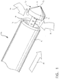
- FIG. 1 A perspective view of the preferred surgical instrument, specifically the tissue biopsy device 10 which is advantageously used with an expandable sleeve 11 to readily provide access to the targeted tissue mass, is shown in Figure 1.
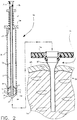
- Figure 2 provides additional details in a sectional view of the tissue biopsy device and the expandable sleeve, as well as depicting the interrelationship between the device and the sleeve.
- the biopsy device 10 has an elongated tube 12 with proximal and distal ends which extend from the proximal and distal ends of the device. At the distal end of the tube, there are a plurality of vacuum orifices 13 to pull a vacuum against the distal end of the tube. At the proximal end of the tube, there is a tube base 14 and a vacuum adapter 15 which is connected to a vacuum source for drawing the vacuum through the vacuum orifices and through the longitudinal channel 16 of the elongated tube (the vacuum source is not shown in Figures 1 or 2).
- the penetrating member has a sharp penetrating tip 18, and the penetrating tip has a conical configuration
- the penetrating member has a circular base 19 at its proximal end. The base is spaced from the apex 19 of the penetrating tip.
- the underside of the base of the penetrating member acts as a first tissue stop 20.
- the first tissue stop extends outwardly in the radial direction from the elongated tube.
- a second tissue stop 21 extends from the elongated tube and faces the first tissue stop (see Figure 2). Similarly to the first tissue stop, the second tissue stop extends outwardly in the radial direction from the elongated tube, and is located proximally of the first tissue stop.
- the annular tissue cutter 22 of the biopsy device is mounted concentrically, about the elongated tube. It is movable longitudinally relative to the penetrating member affixed to the tube from a first non-cutting position to a second tissue-cut position.
- the tissue cutter is shown in its first non-cutting position. In this position, the tissue cutter is located adjacent the base of the penetrating member, and the gap between the first and second tissue stops is enclosed.
- the first and second tissue stops define a tissue receiving chamber 40 between the stops.
- This receiving chamber can receive tissue continuously about a 360° arc at the distal end of the tube where the vacuum orifices are placed. A greater amount of tissue can be loaded into the receiving chamber than what is capable if a conventional lateral tissue receiving port were used. Further, the outward radial dimension provided by the first and second tissue stops also increases the depth of tissue which can be loaded into the chamber.
- the annular cutter 22 of the device has a cutter base 40 at a proximal end and a cutter stop 24 located adjacent to the cutter base. Interposed between the tube base and the cutter base is a cutter spring 25. The spring biases the annular cutter in the distal direction. Consequently, the spring biases the cutter toward its first non-cutting position.
- the sleeve 11 which is used to receive the tissue biopsy device for providing the access portal to the target tissue mass is expandable.
- the sleeve is sized to receive the biopsy device and has a lengthwise dimension sufficient to extend to a position where the distal end of the sleeve is adjacent the targeted tissue mass.
- At a proximal end of the sleeve there is a sleeve handle 26 for gripping and manipulating the sleeve.
- the sleeve When a breast biopsy is desired, the sleeve is inserted through the outer skin layer 29 of the breast and into the percutaneous breast tissue 30. When the sleeve is fully inserted, the sleeve handle 26 is located exteriorly of the outer skin layer 29 of the breast.
- the sleeve is expandable from a first original diameter to a greater, second expanded diameter to accomodate the diameter of the annular cutter of the biopsy device.
- the sleeve is originally inserted into the breast at its first original diameter, and then it is expanded to its second diameter when the biopsy device is inserted through the sleeve.
- the expanded second diameter is substantially the same as the diameter of the annular cutter of the device.
- the tissue biopsy device 10 is inserted in the direction depicted by directional arrow A1 to take advantage of the conical tip configuration of the penetrating member to facilitate ease of insertion.
- tissue can be pulled into the tissue receiving chamber in the direction depicted by the directional arrows A2 as shown.
- the tissue biopsy device can then be inserted through the sleeve, using the sleeve as an access portal to provide ease of insertion of the tissue biopsy device to a location where the tissue receiving chamber can be placed within the targeted tissue mass.
- the sleeve expands from its first original diameter to its second expanded diameter which corresponds to the diameter of the annular tissue cutter. In so doing, the tissue is dialated from the first original diameter to the second expanded diameter, causing less trauma to the patient than if an incision the size of the second expanded diameter were made.
- the biopsy device is fully inserted, and the penetrating tip of the penetrating member affixed to the elongated tube extends from the distal end of the expanded sleeve. Additionally, when fully inserted, the proximal end of the tube of the biopsy device is positioned externally of the tissue, and the tube distal end is positioned percutaneously adjacent the targeted tissue mass.
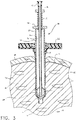
- the annular cutter 22 is retracted from its first non-cutting position to its second tissue-cut position.
- a distal cutting edge surface 31 at the distal end of the annular cutter is exposed. This surface is located adjacent the first tissue stop when the cutter is retracted to its second tissue-cut position.
- the cutter is retracted by exerting an upper proximal force against the spring 25 (using the cutter base 40 as a grip to apply the force) to compress the spring.
- the cutter is then held in its second position.
- the elongated tube 12 of the tissue biopsy device, and the expandable sleeve 11 remain stationary relative to the movement of the annular tissue cutter 22.
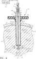
- the vacuum adapter 15 is connected to a vacuum source 32 (see Figure 6), and vacuum is applied.
- a vacuum source 32 see Figure 6
- vacuum is applied, at least a portion of the targeted tissue mass is pulled into the tissue receiving chamber and against the distal end of the elongated tube as illustrated by the directional arrows depicted in Figure 6.
- the vacuum is drawn through the vacuum orifices and into the longitudinal channel 16 of the elongated tube 12.
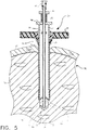
- the distal cutting edge surface 31 of the annular cutter excises the tissue loaded into the chamber as the cutter slides longitudinally from its second tissue-cut position to its first non-cutting position.
- the desired portion of the targeted tissue mass is fully enclosed within the continuous tissue receiving chamber of the device as depicted in Figure 7. From this point, the device can be removed from the sleeve, and the sample of tissue in the chamber can be extracted from the chamber using conventional techniques.
Landscapes
- Health & Medical Sciences (AREA)
- Life Sciences & Earth Sciences (AREA)
- Medical Informatics (AREA)
- Engineering & Computer Science (AREA)
- Biomedical Technology (AREA)
- Heart & Thoracic Surgery (AREA)
- Pathology (AREA)
- Molecular Biology (AREA)
- Surgery (AREA)
- Animal Behavior & Ethology (AREA)
- General Health & Medical Sciences (AREA)
- Public Health (AREA)
- Veterinary Medicine (AREA)
- Surgical Instruments (AREA)
- Sampling And Sample Adjustment (AREA)
Applications Claiming Priority (2)
| Application Number | Priority Date | Filing Date | Title |
|---|---|---|---|
| US97670597A | 1997-11-24 | 1997-11-24 | |
| US976705 | 2001-10-12 |
Publications (2)
| Publication Number | Publication Date |
|---|---|
| EP0919190A2 true EP0919190A2 (fr) | 1999-06-02 |
| EP0919190A3 EP0919190A3 (fr) | 2000-11-02 |
Family
ID=25524376
Family Applications (1)
| Application Number | Title | Priority Date | Filing Date |
|---|---|---|---|
| EP98309545A Withdrawn EP0919190A3 (fr) | 1997-11-24 | 1998-11-23 | Instrument de biopsie avec chambre de réception de tissu continue |
Country Status (5)
| Country | Link |
|---|---|
| EP (1) | EP0919190A3 (fr) |
| JP (1) | JPH11235345A (fr) |
| CN (1) | CN1203810C (fr) |
| AU (1) | AU9326498A (fr) |
| CA (1) | CA2254303A1 (fr) |
Cited By (9)
| Publication number | Priority date | Publication date | Assignee | Title |
|---|---|---|---|---|
| US6712773B1 (en) | 2000-09-11 | 2004-03-30 | Tyco Healthcare Group Lp | Biopsy system |
| US6860860B2 (en) | 2000-11-27 | 2005-03-01 | Tyco Healthcare Group, Lp | Tissue sampling and removal apparatus and method |
| US7189206B2 (en) | 2003-02-24 | 2007-03-13 | Senorx, Inc. | Biopsy device with inner cutter |
| US9157637B2 (en) | 2010-08-27 | 2015-10-13 | Alstom Technology Ltd. | Burner arrangement with deflection elements for deflecting cooling air flow |
| WO2017066771A1 (fr) * | 2015-10-15 | 2017-04-20 | Indiana University Research And Technology Corporation | Système et procédé de biopsie par rasage quantifiable |
| US9750487B2 (en) | 2005-05-23 | 2017-09-05 | Senorx, Inc. | Tissue cutting member for a biopsy device |
| US10105125B2 (en) | 2004-12-16 | 2018-10-23 | Senorx, Inc. | Biopsy device with aperture orientation and improved tip |
| CN112089454A (zh) * | 2020-09-28 | 2020-12-18 | 孙宝柱 | 一种微创外科切除组织取出装置 |
| US11589849B2 (en) | 2003-02-24 | 2023-02-28 | Senorx, Inc. | Biopsy device with selectable tissue receiving aperature orientation and site illumination |
Families Citing this family (7)
| Publication number | Priority date | Publication date | Assignee | Title |
|---|---|---|---|---|
| US6497706B1 (en) | 1998-03-03 | 2002-12-24 | Senorx, Inc. | Biopsy device and method of use |
| US9408592B2 (en) | 2003-12-23 | 2016-08-09 | Senorx, Inc. | Biopsy device with aperture orientation and improved tip |
| KR101649713B1 (ko) * | 2014-04-10 | 2016-08-19 | 주식회사 메디칼파크 | 생체 검사 장치 및 시스템 |
| CN109938779A (zh) * | 2019-03-20 | 2019-06-28 | 南阳市中心医院 | 一种肿瘤内科用穿刺取样器 |
| EP3818945A1 (fr) * | 2019-11-08 | 2021-05-12 | Saga Surgical Ab | Système d'aiguille d'injection et d'aspiration |
| CN111297443B (zh) * | 2020-03-24 | 2025-03-07 | 深圳核心医疗科技股份有限公司 | 手术开孔装置 |
| CN116509465B (zh) * | 2023-06-26 | 2023-09-01 | 四川大学华西第二医院 | 一种带冲洗装置的盆腔内囊肿活检穿刺器 |
Citations (8)
| Publication number | Priority date | Publication date | Assignee | Title |
|---|---|---|---|---|
| US3789852A (en) | 1972-06-12 | 1974-02-05 | S Kim | Expandable trochar, especially for medical purposes |
| JPS5715892A (en) | 1980-07-01 | 1982-01-27 | Mitsubishi Heavy Ind Ltd | Treatment of high concentration waste liquid |
| US4716901A (en) | 1984-09-27 | 1988-01-05 | Pratt Burnerd International Limited | Surgical appliance for forming an opening through the skin |
| US4899729A (en) | 1985-05-30 | 1990-02-13 | Gill Steven S | Expansible cannula |
| US4958625A (en) | 1989-07-18 | 1990-09-25 | Boston Scientific Corporation | Biopsy needle instrument |
| US5183464A (en) | 1991-05-17 | 1993-02-02 | Interventional Thermodynamics, Inc. | Radially expandable dilator |
| US5320611A (en) | 1993-02-04 | 1994-06-14 | Peter M. Bonutti | Expandable cannula having longitudinal wire and method of use |
| US5526822A (en) | 1994-03-24 | 1996-06-18 | Biopsys Medical, Inc. | Method and apparatus for automated biopsy and collection of soft tissue |
Family Cites Families (2)
| Publication number | Priority date | Publication date | Assignee | Title |
|---|---|---|---|---|
| US4926858A (en) * | 1984-05-30 | 1990-05-22 | Devices For Vascular Intervention, Inc. | Atherectomy device for severe occlusions |
| BE1009902A3 (nl) * | 1995-12-28 | 1997-10-07 | Janssens Jacques Phillibert | Inrichting voor het nemen van een weefselstaal. |
-
1998
- 1998-11-19 AU AU93264/98A patent/AU9326498A/en not_active Abandoned
- 1998-11-20 JP JP10346683A patent/JPH11235345A/ja active Pending
- 1998-11-23 EP EP98309545A patent/EP0919190A3/fr not_active Withdrawn
- 1998-11-23 CA CA 2254303 patent/CA2254303A1/fr not_active Abandoned
- 1998-11-24 CN CN 98122635 patent/CN1203810C/zh not_active Expired - Fee Related
Patent Citations (8)
| Publication number | Priority date | Publication date | Assignee | Title |
|---|---|---|---|---|
| US3789852A (en) | 1972-06-12 | 1974-02-05 | S Kim | Expandable trochar, especially for medical purposes |
| JPS5715892A (en) | 1980-07-01 | 1982-01-27 | Mitsubishi Heavy Ind Ltd | Treatment of high concentration waste liquid |
| US4716901A (en) | 1984-09-27 | 1988-01-05 | Pratt Burnerd International Limited | Surgical appliance for forming an opening through the skin |
| US4899729A (en) | 1985-05-30 | 1990-02-13 | Gill Steven S | Expansible cannula |
| US4958625A (en) | 1989-07-18 | 1990-09-25 | Boston Scientific Corporation | Biopsy needle instrument |
| US5183464A (en) | 1991-05-17 | 1993-02-02 | Interventional Thermodynamics, Inc. | Radially expandable dilator |
| US5320611A (en) | 1993-02-04 | 1994-06-14 | Peter M. Bonutti | Expandable cannula having longitudinal wire and method of use |
| US5526822A (en) | 1994-03-24 | 1996-06-18 | Biopsys Medical, Inc. | Method and apparatus for automated biopsy and collection of soft tissue |
Cited By (22)
| Publication number | Priority date | Publication date | Assignee | Title |
|---|---|---|---|---|
| US8128577B2 (en) | 2000-09-11 | 2012-03-06 | Tyco Healthcare Group Lp | Biopsy system |
| US6712773B1 (en) | 2000-09-11 | 2004-03-30 | Tyco Healthcare Group Lp | Biopsy system |
| US7189207B2 (en) | 2000-09-11 | 2007-03-13 | Tyco Healthcare Group Lp | Biopsy system having a single use loading unit operable with a trocar driver, a knife driver and firing module |
| US6860860B2 (en) | 2000-11-27 | 2005-03-01 | Tyco Healthcare Group, Lp | Tissue sampling and removal apparatus and method |
| US7513877B2 (en) | 2000-11-27 | 2009-04-07 | Tyco Healthcare Group Lp | Tissue sampling and removal apparatus and method |
| US10335127B2 (en) | 2003-02-24 | 2019-07-02 | Senorx, Inc. | Biopsy device with selectable tissue receiving aperature orientation and site illumination |
| US10231715B2 (en) | 2003-02-24 | 2019-03-19 | Senorx, Inc. | Biopsy device with inner cutting member |
| US8460204B2 (en) | 2003-02-24 | 2013-06-11 | Senorx, Inc. | Biopsy device with inner cutting member |
| US9044215B2 (en) | 2003-02-24 | 2015-06-02 | Senorx, Inc. | Biopsy device with selectable tissue receiving aperature orientation and site illumination |
| US11589849B2 (en) | 2003-02-24 | 2023-02-28 | Senorx, Inc. | Biopsy device with selectable tissue receiving aperature orientation and site illumination |
| US11534147B2 (en) | 2003-02-24 | 2022-12-27 | Senorx, Inc. | Biopsy device with a removable sample recieving cartridge |
| US7819819B2 (en) | 2003-02-24 | 2010-10-26 | Senorx, Inc. | Biopsy device with inner cutting member |
| US7189206B2 (en) | 2003-02-24 | 2007-03-13 | Senorx, Inc. | Biopsy device with inner cutter |
| US10172595B2 (en) | 2003-02-24 | 2019-01-08 | Senorx, Inc. | Biopsy device with selectable tissue receiving aperture orientation and site illumination |
| US11246574B2 (en) | 2004-12-16 | 2022-02-15 | Senorx, Inc. | Biopsy device with aperture orientation and improved tip |
| US10105125B2 (en) | 2004-12-16 | 2018-10-23 | Senorx, Inc. | Biopsy device with aperture orientation and improved tip |
| US10478161B2 (en) | 2005-05-23 | 2019-11-19 | Senorx, Inc. | Tissue cutting member for a biopsy device |
| US9750487B2 (en) | 2005-05-23 | 2017-09-05 | Senorx, Inc. | Tissue cutting member for a biopsy device |
| US11426149B2 (en) | 2005-05-23 | 2022-08-30 | SenoRx., Inc. | Tissue cutting member for a biopsy device |
| US9157637B2 (en) | 2010-08-27 | 2015-10-13 | Alstom Technology Ltd. | Burner arrangement with deflection elements for deflecting cooling air flow |
| WO2017066771A1 (fr) * | 2015-10-15 | 2017-04-20 | Indiana University Research And Technology Corporation | Système et procédé de biopsie par rasage quantifiable |
| CN112089454A (zh) * | 2020-09-28 | 2020-12-18 | 孙宝柱 | 一种微创外科切除组织取出装置 |
Also Published As
| Publication number | Publication date |
|---|---|
| CA2254303A1 (fr) | 1999-05-24 |
| EP0919190A3 (fr) | 2000-11-02 |
| CN1203810C (zh) | 2005-06-01 |
| AU9326498A (en) | 1999-06-10 |
| JPH11235345A (ja) | 1999-08-31 |
| CN1221602A (zh) | 1999-07-07 |
Similar Documents
| Publication | Publication Date | Title |
|---|---|---|
| US6083237A (en) | Biopsy instrument with tissue penetrating spiral | |
| US6488636B2 (en) | Biopsy apparatus | |
| EP0919191A2 (fr) | Ensemble d'instrument de biopsie avec manchon extensible | |
| US5823970A (en) | Biopsy needle set | |
| US6280398B1 (en) | Methods and devices for collection of soft tissue | |
| EP0829232B1 (fr) | Dispositif de biopsie invasible minimal | |
| US6019733A (en) | Biopsy apparatus and method | |
| US5810806A (en) | Methods and devices for collection of soft tissue | |
| US5449001A (en) | Biopsy needle | |
| US7510535B2 (en) | Biopsy method | |
| US5913857A (en) | Methods and devices for collection of soft tissue | |
| US20050113716A1 (en) | Biopsy device having endoscope | |
| AU9326498A (en) | Biopsy instrument with continuous tissue receiving chamber | |
| US6981949B2 (en) | Perimeter cut biopsy probe | |
| WO2002062226A1 (fr) | Appareil et methode de biopsie | |
| EP0919192A2 (fr) | Instrument de biopsie avec pointe pour dilatation de tissue | |
| WO2002062231A2 (fr) | Appareil et procede de biopsie | |
| WO2002062229A2 (fr) | Methode et appareil de biopsie | |
| WO2002062230A1 (fr) | Instrument et methode de biopsie |
Legal Events
| Date | Code | Title | Description |
|---|---|---|---|
| PUAI | Public reference made under article 153(3) epc to a published international application that has entered the european phase |
Free format text: ORIGINAL CODE: 0009012 |
|
| AK | Designated contracting states |
Kind code of ref document: A2 Designated state(s): AT BE CH CY DE DK ES FI FR GB GR IE IT LI LU MC NL PT SE |
|
| AX | Request for extension of the european patent |
Free format text: AL;LT;LV;MK;RO;SI |
|
| PUAL | Search report despatched |
Free format text: ORIGINAL CODE: 0009013 |
|
| AK | Designated contracting states |
Kind code of ref document: A3 Designated state(s): AT BE CH CY DE DK ES FI FR GB GR IE IT LI LU MC NL PT SE |
|
| AX | Request for extension of the european patent |
Free format text: AL;LT;LV;MK;RO;SI |
|
| AKX | Designation fees paid | ||
| REG | Reference to a national code |
Ref country code: DE Ref legal event code: 8566 |
|
| STAA | Information on the status of an ep patent application or granted ep patent |
Free format text: STATUS: THE APPLICATION IS DEEMED TO BE WITHDRAWN |
|
| 18D | Application deemed to be withdrawn |
Effective date: 20010503 |