EP0920831A1 - Appareil et procédé d'imagerie par autofluorescence intégrée, destinés à des tissus malades - Google Patents
Appareil et procédé d'imagerie par autofluorescence intégrée, destinés à des tissus malades Download PDFInfo
- Publication number
- EP0920831A1 EP0920831A1 EP99103430A EP99103430A EP0920831A1 EP 0920831 A1 EP0920831 A1 EP 0920831A1 EP 99103430 A EP99103430 A EP 99103430A EP 99103430 A EP99103430 A EP 99103430A EP 0920831 A1 EP0920831 A1 EP 0920831A1
- Authority
- EP
- European Patent Office
- Prior art keywords
- light
- image
- tissue
- autofluorescence
- excitation light
- Prior art date
- Legal status (The legal status is an assumption and is not a legal conclusion. Google has not performed a legal analysis and makes no representation as to the accuracy of the status listed.)
- Granted
Links
- 238000003384 imaging method Methods 0.000 title claims abstract description 18
- 238000000034 method Methods 0.000 title abstract description 12
- 230000005284 excitation Effects 0.000 claims abstract description 71
- 239000000523 sample Substances 0.000 claims abstract description 35
- 238000005286 illumination Methods 0.000 claims abstract description 23
- 230000001965 increasing effect Effects 0.000 claims description 11
- 230000003595 spectral effect Effects 0.000 claims description 10
- 206010034960 Photophobia Diseases 0.000 claims description 6
- 238000001914 filtration Methods 0.000 claims description 6
- 208000013469 light sensitivity Diseases 0.000 claims description 6
- 238000012545 processing Methods 0.000 claims description 6
- 230000035945 sensitivity Effects 0.000 claims description 5
- 230000003247 decreasing effect Effects 0.000 claims description 4
- 239000011248 coating agent Substances 0.000 claims description 2
- 238000000576 coating method Methods 0.000 claims description 2
- 238000002073 fluorescence micrograph Methods 0.000 abstract description 21
- 201000010099 disease Diseases 0.000 abstract description 14
- 208000037265 diseases, disorders, signs and symptoms Diseases 0.000 abstract description 14
- 230000003287 optical effect Effects 0.000 abstract description 7
- 238000001444 catalytic combustion detection Methods 0.000 description 35
- 238000001228 spectrum Methods 0.000 description 15
- 206010028980 Neoplasm Diseases 0.000 description 7
- 230000002159 abnormal effect Effects 0.000 description 7
- 230000008859 change Effects 0.000 description 4
- 230000001747 exhibiting effect Effects 0.000 description 4
- 238000001727 in vivo Methods 0.000 description 4
- 238000010521 absorption reaction Methods 0.000 description 3
- 239000008280 blood Substances 0.000 description 3
- 210000004369 blood Anatomy 0.000 description 3
- 201000011510 cancer Diseases 0.000 description 3
- 230000003292 diminished effect Effects 0.000 description 3
- 230000004044 response Effects 0.000 description 3
- 230000001427 coherent effect Effects 0.000 description 2
- 239000002131 composite material Substances 0.000 description 2
- 230000007423 decrease Effects 0.000 description 2
- 238000001514 detection method Methods 0.000 description 2
- 239000003814 drug Substances 0.000 description 2
- 229940079593 drug Drugs 0.000 description 2
- 238000001839 endoscopy Methods 0.000 description 2
- 238000012986 modification Methods 0.000 description 2
- 230000004048 modification Effects 0.000 description 2
- 238000010606 normalization Methods 0.000 description 2
- 230000008569 process Effects 0.000 description 2
- 206010058314 Dysplasia Diseases 0.000 description 1
- 238000013459 approach Methods 0.000 description 1
- 230000008901 benefit Effects 0.000 description 1
- 230000015556 catabolic process Effects 0.000 description 1
- 210000001072 colon Anatomy 0.000 description 1
- 238000006731 degradation reaction Methods 0.000 description 1
- 230000001419 dependent effect Effects 0.000 description 1
- 238000011161 development Methods 0.000 description 1
- 238000005516 engineering process Methods 0.000 description 1
- 230000002708 enhancing effect Effects 0.000 description 1
- 210000003238 esophagus Anatomy 0.000 description 1
- 239000000835 fiber Substances 0.000 description 1
- 238000002189 fluorescence spectrum Methods 0.000 description 1
- 239000003365 glass fiber Substances 0.000 description 1
- 238000003780 insertion Methods 0.000 description 1
- 230000037431 insertion Effects 0.000 description 1
- 230000010354 integration Effects 0.000 description 1
- 210000000867 larynx Anatomy 0.000 description 1
- 210000004072 lung Anatomy 0.000 description 1
- 238000005259 measurement Methods 0.000 description 1
- 239000000203 mixture Substances 0.000 description 1
- 210000001989 nasopharynx Anatomy 0.000 description 1
- 239000013307 optical fiber Substances 0.000 description 1
- 230000003685 thermal hair damage Effects 0.000 description 1
- 238000012546 transfer Methods 0.000 description 1
Images


Classifications
-
- A—HUMAN NECESSITIES
- A61—MEDICAL OR VETERINARY SCIENCE; HYGIENE
- A61B—DIAGNOSIS; SURGERY; IDENTIFICATION
- A61B1/00—Instruments for performing medical examinations of the interior of cavities or tubes of the body by visual or photographical inspection, e.g. endoscopes; Illuminating arrangements therefor
- A61B1/04—Instruments for performing medical examinations of the interior of cavities or tubes of the body by visual or photographical inspection, e.g. endoscopes; Illuminating arrangements therefor combined with photographic or television appliances
- A61B1/043—Instruments for performing medical examinations of the interior of cavities or tubes of the body by visual or photographical inspection, e.g. endoscopes; Illuminating arrangements therefor combined with photographic or television appliances for fluorescence imaging
-
- A—HUMAN NECESSITIES
- A61—MEDICAL OR VETERINARY SCIENCE; HYGIENE
- A61B—DIAGNOSIS; SURGERY; IDENTIFICATION
- A61B1/00—Instruments for performing medical examinations of the interior of cavities or tubes of the body by visual or photographical inspection, e.g. endoscopes; Illuminating arrangements therefor
- A61B1/04—Instruments for performing medical examinations of the interior of cavities or tubes of the body by visual or photographical inspection, e.g. endoscopes; Illuminating arrangements therefor combined with photographic or television appliances
- A61B1/05—Instruments for performing medical examinations of the interior of cavities or tubes of the body by visual or photographical inspection, e.g. endoscopes; Illuminating arrangements therefor combined with photographic or television appliances characterised by the image sensor, e.g. camera, being in the distal end portion
-
- A—HUMAN NECESSITIES
- A61—MEDICAL OR VETERINARY SCIENCE; HYGIENE
- A61B—DIAGNOSIS; SURGERY; IDENTIFICATION
- A61B5/00—Measuring for diagnostic purposes; Identification of persons
- A61B5/0059—Measuring for diagnostic purposes; Identification of persons using light, e.g. diagnosis by transillumination, diascopy, fluorescence
- A61B5/0071—Measuring for diagnostic purposes; Identification of persons using light, e.g. diagnosis by transillumination, diascopy, fluorescence by measuring fluorescence emission
-
- A—HUMAN NECESSITIES
- A61—MEDICAL OR VETERINARY SCIENCE; HYGIENE
- A61B—DIAGNOSIS; SURGERY; IDENTIFICATION
- A61B5/00—Measuring for diagnostic purposes; Identification of persons
- A61B5/0059—Measuring for diagnostic purposes; Identification of persons using light, e.g. diagnosis by transillumination, diascopy, fluorescence
- A61B5/0082—Measuring for diagnostic purposes; Identification of persons using light, e.g. diagnosis by transillumination, diascopy, fluorescence adapted for particular medical purposes
- A61B5/0084—Measuring for diagnostic purposes; Identification of persons using light, e.g. diagnosis by transillumination, diascopy, fluorescence adapted for particular medical purposes for introduction into the body, e.g. by catheters
- A61B5/0086—Measuring for diagnostic purposes; Identification of persons using light, e.g. diagnosis by transillumination, diascopy, fluorescence adapted for particular medical purposes for introduction into the body, e.g. by catheters using infrared radiation
-
- H—ELECTRICITY
- H04—ELECTRIC COMMUNICATION TECHNIQUE
- H04N—PICTORIAL COMMUNICATION, e.g. TELEVISION
- H04N23/00—Cameras or camera modules comprising electronic image sensors; Control thereof
- H04N23/50—Constructional details
- H04N23/555—Constructional details for picking-up images in sites, inaccessible due to their dimensions or hazardous conditions, e.g. endoscopes or borescopes
Definitions
- This invention relates to an endoscope apparatus and method for viewing tissue within the body that uses autofluorescence of the tissue to detect and delineate diseased tissue.
- Fluorescence endoscopy involves introducing excitation light into the body and collecting the emitted fluorescence light by means of a probe that is inserted into the body to the location of interest.
- the probe comprises a lens fitted onto a coherent bundle of glass fibres which brings the fluorescent image out of the body cavity.
- the probe can be a rigid endoscope without fibre optics.
- the image brought out of the body is captured by very sensitive photodetectors and further processed in an attempt to delineate diseased tissue on the basis that diseased tissue has a different fluorescence intensity than normal tissue.
- United States Patents 5,131,398 to Alfano, United States Patent 4,930,516 to Alfano, and United States Patent 4,786,813 to Svanberg et al. disclose various equipment and methods that acquire and process fluorescence images in an attempt to detect and delineate diseased tissue.
- image intensified cameras are very large, they employ high voltage circuitry and they cannot be made to fit the end of the endoscope.
- the fluorescence images must therefore be brought out of tissue cavities through the coherent optical fibers of the endoscope before processing of the images and/or displaying them on a video monitor.
- a first spectral bad comprises light whereby the intensity of the autofluorescence light for healthy tissue is substantially different than the intensity of light produced for diseased tissue.
- a second band includes light whereby the autofluorescence intensity for diseased and healthy tissue are substantially the same. Light in each spectral band is imaged and applied to a color monitor that produces a pseudo-color image of the tissue under examination.
- the acquired fluorescence image of endoscope systems would be of better quality if it could be collected by a sensor at the end of the endoscope probe inserted into the body.
- the outer diameter of a endoscope probe must be small to allow insertion into various body cavities thereby limiting the size of the sensor that can be mounted at the distal end of the apparatus.
- fluorescence images are generally extremely faint and it is not possible for these small image sensors to capture the fluorescence images. Theoretically, increasing the excitation irradiance would increase the fluorescence intensity, however, this may also result in unwanted thermal damage or photobleaching of the tissue under examination.
- the present invention seeks to provide an apparatus and method that addresses the problems of the prior art by providing a new system for acquiring fluorescence images that uses integration of autofluorescence intensity over a broad band of wavelengths to acquire a well-defined autofluorescence image that is combined with a remittance light image to create a merged image in which diseased and normal tissue are readily distinguishable.
- the remittance light image is used to normalize the autofluorescence image to account for image non-uniformity due to changes in distance, angle and illumination intensity.
- the present invention exploits applicant's discovery that in vivo tissue autofluorescence of diseased tissue is greatly reduced through a significant range of the visible light spectrum (from approximately 500nm to approximately 700nm).
- the present novel system was designed and developed to use the integrated fluorescence over this large spectral range (500 nm - 700 nm) and display the fluorescence image along with a remittance light image such that, to the observer, normal tissue appears in a different colour from diseased tissue.
- remittance light refers to reflected and back scattered light. Fluorescence signal intensity is dependent on such factors as the excitation light intensity, the proximity of the excitation light source to the tissue, and the angle of incidence of the excitation light to the tissue.
- two wavelengths of light for imaging a tissue site can be used.
- blue light is used as excitation light to induce tissue autofluorescence and red/near-infrared light (approx. 700 nm) is used to produce a remittance light image.
- red/near-infrared light approximately 700 nm
- a single wavelength of light can be used as excitation light and a remittance light image can be collected from the remitted excitation light.
- the integrated autofluorescence image and the remittance light image are combined and displayed in a form to allow the observer to intuitively adjust for the excitation light intensity variations.
- the present invention provides apparatus for imaging diseases in tissue comprising:
- the present invention provides, a method for imaging diseases in tissue comprising:
- the present invention provides a fluorescence endoscope apparatus having an image sensing device that is positioned at the end of a probe comprised of light guides.
- the probe is introduced into the body thereby eliminating the problem of fluorescence image degradation due to light loss while travelling through the collecting light guides.
- the image sensing device can be positioned adjacent to the tissue surface being examined so that there is little loss of light.
- the apparatus of the present invention employs an image sensing device such as a CCD (or equivalent such as CID) array having a sensitivity that can be varied.
- a preferred way to achieve increased sensitivity of the CCD array is to combine individual pixel sensing elements to create larger to very large sensing units. Individual sensing elements can be combined together into 2x2, 4x4, 8x8, 16x16 or even larger groups creating new larger sensing units, particularly if CCDs are used with 100% fill factor and high quantum efficiency.
- very sensitive detectors can be made with very low noise, i.e. very high signal-to-noise ratio (SNR), well within parameters to detect the low fluorescence emitted by tissues.
- SNR signal-to-noise ratio
- the present invention provides an endoscope apparatus for imaging a disease site within a body comprising:
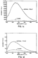
- Figure 1a shows a typical graph of the autofluorescence response spectrum of tissue in vivo when exposed to blue excitation light (400nm - 450nm). Applicant has discovered that autofluorescence of diseased tissue in vivo is greatly reduced as compared to normal tissue through the entire range of visible light (from approximately 500nm to approximately 700nm).
- Figures 1b-1d are examples of actual autofluorescence curves for tissue illuminated by blue excitation light.
- the autofluorescence spectrum for bronchial tissue is shown. Note that the autofluorescence intensity for normal tissue and tumorous tissue both peak in the green range of the spectrum (approximately 530nm), however, the peak for tumorous tissue is much lower than the peak for normal tissue.
- Figure 1c shows the autofluorescence spectrum for larynx tissue that is induced to fluoresce using blue laser light. Again, there is a pronounced peak in the green region of the spectrum for normal tissue and a greatly reduced spectrum for tumor tissue having a low peak in the green region. In the region of the spectrum greater than 700 nm, the spectrum is substantially the same for both tissues.
- Figure 1d the autofluorescence spectrum for nasopharynx tissue is shown having a green region peak for normal tissue and a reduced overall spectrum for diseased tissue. While there are some differences in the autofluorescence spectrum for specific types of tissue, in general, the diseased tissue autofluorescence is significantly reduced in comparison to the normal tissue autofluorescence over a broad spectral band.
- Figure 2 is a schematic view of apparatus according to a first embodiment that has been developed to exploit the autofluorescence response of the tissue to differentiate between diseased and normal tissue.
- the apparatus of Figure 2 employs simultaneous illumination of the tissue with both excitation and illumination light.
- the apparatus includes a light source 30 for producing excitation light of a first pre-selected wavelength, preferably blue light (400-450nm), to excite the tissue to autofluoresce.
- the excited tissue produces a predominantly green autofluorescence light as indicated in Figures 1a-1d.
- Light source 30 also produces non-excitation light of a different wavelength to illuminate the tissue.
- the non-excitation light is preferably far red to infrared light ( ⁇ R ) including wavelengths greater than 700nm and is used to produced a remittance light image of the tissue.
- This non-excitation light is preferred as it falls outside the relatively broad spectral range of generally green emitted autofluorescence light ( ⁇ f ) in which autofluorescence intensity for normal tissue is substantially greater than for diseased tissue so as not to interfere with detection of the relatively faint autofluorescence image.
- ⁇ f generally green emitted autofluorescence light
- far red to infrared light has a low absorption coefficient in most tissue and it has a large scattering coefficient.
- red and infrared light can be used to generate an image to correct for non-uniformity in the autofluorescence image that might arise due to changes in the proximity of the excitation light to the tissue, the angle of incidence of the excitation light to the tissue and the intensity of the excitation light.
- intensity of remittance light ( ⁇ R ) is much greater than the intensity of the autofluorescence light and a vertical break in the range over which remittance light is collected is provided in Figure 1a to account for this intensity difference.
- the light is delivered to the tissue under examination by a conventional endoscope probe 32 comprising a bundle of optical fibres.
- Probe 32 also delivers autofluorescence light and remittance light from the tissue to optical means in the form of CCD cameras 34 and 36.
- CCD camera 36 has associated means for integrating the intensity of the autofluorescence light over a range of wavelengths in which the autofluorescence intensity for normal tissue is substantially different from the autofluorescence intensity for diseased tissue to establish an integrated autofluorescence intensity image of the tissue.
- the means for integrating the intensity of autofluorescence light preferably comprises a broad band filter 49 to admit autofluorescence light over a broad range of wavelengths.
- the resulting autofluorescence tends to be mainly green light and the intensity is integrated over the range of wavelengths from 500-650nm designated ( ⁇ f ) in Figure 1.
- the need for an image intensifier is obviated as the integrated autofluorescence signal is nearly an order of magnitude stronger compared to the natural autofluorescence intensity in a narrow wave band.
- the detected change for the apparatus and method of the present invention is generally 2 to 5 times greater than the detected change of the prior art.
- the images captured by CCD cameras 34 and 36 are processed and displayed on display means in the form of a monitor 40.
- the images are preferably processed in an imaging board in a computer.
- the integrated autofluorescence image captured by camera 36 and the remittance light image captured by camera 34 are combined to produce a normalized image in which diseased tissue is distinguishable from normal tissue.
- Camera 36 provides a substantially green integrated autofluorescence image that is fed to the green channel of the display and camera 34 provides a substantially red remittance light image to normalize the autofluorescence image that is fed to the red channel of the display to produce a normalized pseudo-colour image.
- the intensity of the green autofluorescence image will be reduced over the diseased area while the remittance light image will be substantially unaffected. If tissue is normal, the intensity of the green autofluorescence image will be increased relative to the diseased tissue and the remittance light image will continue to be substantially unaffected. Combining and display of the two images on monitor 40 is arranged such that the relatively constant red remittance light image is used to establish a "underlying" image that accurately displays contours in the tissue and image variations that arise due to the proximity and intensity of the light source to the tissue. The mix of the images is such that normal tissue will appear green and diseased tissue will appear red due to the reduced levels of green autofluorescence in the vicinity of the diseased tissue allowing the red remittance light to predominate.
- the apparatus of Figure 2 is set up such that light source 30 illuminates tissue 42 with the excitation light and illumination light simultaneously and separate CCD cameras 34 and 36 are provided to receive the autofluorescence image and the remittance light image.
- light splitting means in the form of dichroic mirrors 44 and 46 are provided to direct the remittance light and the autofluorescence light to the appropriate CCD camera. While traditional 50%, 50% or X%, 100-X% beam splitters may be used, dichroics increase the fluorescence image intensity available to be detected.
- a first dichroic mirror 44 passes the red remittance light (R) to CCD camera 34 and diverts the green autofluorescence light (G) and reflected blue excitation light (B) to second dichroic mirror 46.
- Second dichroic mirror 46 diverts the green autofluorescence light (G) to CCD camera 36 and passes the blue excitation light (B).
- Filter 49 in the path of the autofluorescence light is preferably adjustable to vary the range of wavelengths of autofluorescence light that are received by CCD camera 36.
- the "window" over which autofluorescence is integrated can be varied to fine tune the apparatus to detect different disease conditions in different tissues that tend to exhibit characteristic autofluorescence spectra as illustrated in Figures 1b to 1d.
- Filter 49 can be varied by inserting an appropriate filter into place for the particular tissue and the type of tumour under examination.
- an optional filter means 50 can be included in the light path to prevent blue excitation light from being received by the CCD cameras.
- FIG. 3 schematically illustrates imaging apparatus according to a second embodiment of the present invention that illuminates the tissue sequentially over a timing cycle.
- Light source 30 illuminates tissue 42 through endoscope 32 with excitation light for a first portion of the timing cycle.
- Autofluorescence light is delivered via endoscope 32 and filter 60 to a single CCD camera 62 in order to capture an integrated autofluorescence image.
- Filter 60 is positionable before camera 62 to filter out the excitation light and pass broadband autofluorescence light.
- the captured autofluorescence image is transferred to image processing means and then to image storage means located in a computer 63.
- Light source 30 then illuminates the tissue with illumination light to generate a remittance light image. If necessary, filter 65 is moved into position to filter the remittance light. Filters 60 and 65 are mounted on a turntable arrangement 67 that is controlled by computer 63. The remittance light image is transferred to the image processing and storage means. The two images collected in the timing cycle are then combined and displayed on monitor 40 in the same manner as previously described using colour to delineate normal and diseased tissue. The cycle is then repeated under the control of computer 63.
- the foregoing embodiments of the present invention employ blue excitation light and red illumination light to produce an image that distinguishes normal and diseased tissue.
- the foregoing systems may cause false positive readings.
- a third embodiment of the present invention has been developed that employs blue light as both the excitation light and the illumination light.
- Figure 3 is a schematic drawing of the apparatus of the third embodiment that is designed to reduce the rate of false positive readings without sacrificing true diseased tissue (true positive) detection rates.
- light source 30 provides blue light for both excitation and illumination light in a manner identical to the embodiment of Figure 2.
- Green autofluorescence light (G) and blue remittance light (B) are collected from the tissue site. The combined light is split at dichroic mirror 44.
- a blue remittance light image is captured by CCD camera 34 and a green autofluorescence image is reflected through integrating filter 49 for capture by CCD camera 49.
- the intensity of the detected blue remittance light image tends to drop off almost as much as the green integrated fluorescence image, consequently, the displayed image of inflamed tissue does not differ significantly from normal tissue.
- An additional modification of the apparatus of the third embodiment can be used to make the presence of increased blood at a tissue site appear as a recognizable colour on the displayed image that would not be confused with normal or diseased tissue.
- blue excitation light and red illumination light are used and a blue remittance light image, a red remittance light image and a green integrated autofluorescence image are collected.
- the red and blue remittance images are captured by camera 34.
- Camera 34 sends the red remittance image via its red channel and the blue remittance image via its blue channel to display 40.
- the green integrated autofluorescence image is captured by camera 36 and sent to the green channel of the display.
- normal tissue appears white, inflamed tissue has a reddish hue and abnormal tissue has a purplish hue.
- the technique of the present invention also allows development of an imaging system that is sufficiently small and compact to be positioned at the end of an endoscope probe.
- an endoscope system 2 for imaging a disease site within a body according to a fourth embodiment of the present invention.
- the apparatus includes a probe 4 having an inner end 6 that is insertable into a body to be located near the disease site.
- the outer end of probe 4 remains outside the body for connection to a light source 8 for generating light and display means in the form of a colour monitor 10 for displaying the image collected by probe 4.
- Light source 8 generates light at the outer end of the probe that includes excitation light to induce tissue fluorescence.
- the probe is comprised of a bundle of optical fibres 10 that convey the eight of source 8 from the outer end to inner end 6 to illuminate the disease site.
- image sensing means preferably in the form of CCD sensor 12 to detect and collect an image of the diseased site using remittance light and emitted fluorescent light.
- CCD array 12 communicates with the outer end of probe 4 via electrical connection 14 that extends through probe 4.
- inner end 6 is provided with a lens 15 to focus light on CCD sensor 12.
- Filtering means in the form of a filter module 16 is positioned before CCD sensor 12.
- Filter module 16 can be fixed in place or removable remotely.
- the filtering means can be a coating applied directly to surface of CCD sensor 12.
- filter module 16 can be associated with lens 15. The filter module 16 acts to exclude excitation light and pass light of other wavelengths to the image sensing means, particularly emitted fluorescent light.
- the image collected by CCD sensor 12 is transmitted via line 14 to the outer end of probe 4 for viewing on display means in the form of a colour monitor 18.
- the image displayed on monitor 18 delineates diseased and normal tissue for viewing by the endoscope operator.
- the endoscope apparatus of the fourth embodiment is able to collect the faint fluorescence images that provide information necessary for delineating diseased an normal tissue by virtue of the fact that CCD sensor 12 is provided with a light sensitivity that can be increased to acquire low resolution images at low fluorescent light intensities and decreased to acquire high resolution images at other light intensities.
- the light sensitivity of CCD sensor 12 is adjusted by binning the pixels of the sensor array. Binning the pixels decreases spatial resolution. For example, a CCD sensor comprised of 250x250, (i.e. 62,500 active pixels) if binned to form 8x8 pixels per each new sensor unit would yield an effective CCD of approximately 30x30 pixels (each pixel being 64 times larger than the original pixels) providing a very low resolution image.
- dynamic binning can solve the problem of the low spatial resolution as will be described below.
- light source 8 can provide a broad spectral band of excitation light ranging in wavelength between 400-480nm (broad band blue light). Such an excitation light tends to generate a strong fluorescence response in most tissues such as the lungs, the esophagus, the colon, the bladder and the skin. The peak fluorescence is in the green region at approximately 510nm and it has been shown that the fluorescence signal is greatly diminished or virtually disappears in abnormal tissue due to early cancer or dysplasia.
- the excitation light can be in a narrow spectral band of blue or violet light.
- the blue excitation light of light source 8 is conveyed to inner probe end 6 to illuminate an internal disease site to induce maximum tissue fluorescence particularly in the green and longer wavelengths. Scattered/reflected excitation light and fluorescence light are focused by lens 15 on CCD sensor through filter module 16. Filter module 16 is selected to filter out the blue band excitation light such that only the fluorescence green and longer wavelengths (e.g. greater than 480nm) are collected by the CCD sensor. Since abnormal tissue has greatly diminished (or a lack of) fluorescence particulary in the green region, the abnormal tissue will be detected as a darker area on the image captured by the CCD sensor. The darker abnormal regions of tissue will be readily apparent on display monitor 18. The light sensitivity of the CCD sensor can be increased by binning to acquire low resolution images at low fluorescent light intensities. While the images are of reduced resolution, they are generally sufficiently detailed to accurately determine the extent and location of the diseased tissue.
- the arrangement of the fourth embodiment of the present invention reduces the complexity of the apparatus as there is generally no longer the need for an image intensifier to enhance the image collected by the CCD sensor since the sensor is located immediately adjacent the image site and the binning of the CCD sensor permits acquisition of very faint fluorescence images albeit at reduced resolution.
- high resolution non-fluorescence images and low resolution fluorescence images of the disease site can be acquired sequentially and combined for display on monitor 18.
- the endoscope apparatus is identical to the fourth embodiment except light source 8 is modified to sequentially illuminate the disease site with two or more wavelengths of light over a timing cycle.
- One wavelength is a fluorescence excitation light that is on for the majority of the timing cycle and the other wavelength is a non-excitation light that is on for a short period of the timing cycle.
- This arrangement requires means to increase the image sensing means sensitivity to acquire a low resolution fluorescence image during illumination by the fluorescence excitation light and decrease the image sensing means sensitivity to acquire a high resolution image during illumination by the non-excitation light.
- This arrangement also requires image processing means for storing sequential images captured by the image sensing means and combining the images for display as a pseudo-colour image on the display means.
- the endoscope apparatus of the present embodiment is operated in the following manner:
- Light source 8 sequentially illuminates the disease site briefly through probe 6 with red non-excitation light and blue fluorescence excitation light over a single cycle of 30msec which is consistent with video display rates.
- the scattered and/or reflected red light is passed through filter module 16 in front of the CCD sensor 12.
- the red light is sufficiently intense that CCD sensor 12 does not have to be binned and an image of the site at high resolution is captured by the sensor.
- the illumination with red light is of short duration (e.g. 5msec pulse) and the image is stored in a buffer of the image processing means comprising an imaging board 20.
- the blue light is of much longer duration (e.g.25 msec) to collect sufficient fluorescence light generated by the blue light.
- the CCD is now binned if necessary to detect and capture the fluorescence image of the site at reduced resolution.
- Filter module 16 excludes the blue excitation light and permits passage of green and longer wavelengths.
- the captured fluorescence image is fed into another buffer of the imaging board.
- a pseudo image is then formed by the imaging board for display on RGB monitor 18.
- the pseudo image is a composite image that includes a very high resolution colour image (generated using the red light) superimposed on a green to dark green lower resolution image (generated using the blue light).
- normal tissue will be green and diseased or abnormal tissue will show up as red or pink areas.
- This combination of fluorescence and reflectance imaging has the additional advantage of compensating for changes in fluorescence intensity due to variation in the distance and angle between the probe and the surface of the body cavity.
Landscapes
- Health & Medical Sciences (AREA)
- Life Sciences & Earth Sciences (AREA)
- Surgery (AREA)
- Engineering & Computer Science (AREA)
- Medical Informatics (AREA)
- Animal Behavior & Ethology (AREA)
- Biophysics (AREA)
- Pathology (AREA)
- Biomedical Technology (AREA)
- Heart & Thoracic Surgery (AREA)
- Veterinary Medicine (AREA)
- Molecular Biology (AREA)
- Public Health (AREA)
- Physics & Mathematics (AREA)
- General Health & Medical Sciences (AREA)
- Nuclear Medicine, Radiotherapy & Molecular Imaging (AREA)
- Optics & Photonics (AREA)
- Radiology & Medical Imaging (AREA)
- Multimedia (AREA)
- Signal Processing (AREA)
- Endoscopes (AREA)
- Investigating, Analyzing Materials By Fluorescence Or Luminescence (AREA)
Priority Applications (1)
| Application Number | Priority Date | Filing Date | Title |
|---|---|---|---|
| EP04018813A EP1472972B1 (fr) | 1994-03-28 | 1995-03-24 | Appareil et procédé d'imagerie par autofluorescence intégrée, destinés à des tissus malades |
Applications Claiming Priority (3)
| Application Number | Priority Date | Filing Date | Title |
|---|---|---|---|
| US218662 | 1994-03-28 | ||
| US08/218,662 US5590660A (en) | 1994-03-28 | 1994-03-28 | Apparatus and method for imaging diseased tissue using integrated autofluorescence |
| EP95912989A EP0752825B1 (fr) | 1994-03-28 | 1995-03-24 | Appareil et procede d'imagerie par autofluorescence integree, destines a des tissus malades |
Related Parent Applications (1)
| Application Number | Title | Priority Date | Filing Date |
|---|---|---|---|
| EP95912989A Division EP0752825B1 (fr) | 1994-03-28 | 1995-03-24 | Appareil et procede d'imagerie par autofluorescence integree, destines a des tissus malades |
Related Child Applications (1)
| Application Number | Title | Priority Date | Filing Date |
|---|---|---|---|
| EP04018813A Division EP1472972B1 (fr) | 1994-03-28 | 1995-03-24 | Appareil et procédé d'imagerie par autofluorescence intégrée, destinés à des tissus malades |
Publications (2)
| Publication Number | Publication Date |
|---|---|
| EP0920831A1 true EP0920831A1 (fr) | 1999-06-09 |
| EP0920831B1 EP0920831B1 (fr) | 2005-08-17 |
Family
ID=22815983
Family Applications (3)
| Application Number | Title | Priority Date | Filing Date |
|---|---|---|---|
| EP99103430A Expired - Lifetime EP0920831B1 (fr) | 1994-03-28 | 1995-03-24 | Appareil d'imagerie par autofluorescence intégrée, destinés à des tissus malades |
| EP95912989A Expired - Lifetime EP0752825B1 (fr) | 1994-03-28 | 1995-03-24 | Appareil et procede d'imagerie par autofluorescence integree, destines a des tissus malades |
| EP04018813A Expired - Lifetime EP1472972B1 (fr) | 1994-03-28 | 1995-03-24 | Appareil et procédé d'imagerie par autofluorescence intégrée, destinés à des tissus malades |
Family Applications After (2)
| Application Number | Title | Priority Date | Filing Date |
|---|---|---|---|
| EP95912989A Expired - Lifetime EP0752825B1 (fr) | 1994-03-28 | 1995-03-24 | Appareil et procede d'imagerie par autofluorescence integree, destines a des tissus malades |
| EP04018813A Expired - Lifetime EP1472972B1 (fr) | 1994-03-28 | 1995-03-24 | Appareil et procédé d'imagerie par autofluorescence intégrée, destinés à des tissus malades |
Country Status (5)
| Country | Link |
|---|---|
| US (2) | US5590660A (fr) |
| EP (3) | EP0920831B1 (fr) |
| JP (3) | JP3683271B2 (fr) |
| DE (3) | DE69518915T2 (fr) |
| WO (1) | WO1995026673A2 (fr) |
Cited By (15)
| Publication number | Priority date | Publication date | Assignee | Title |
|---|---|---|---|---|
| US6185443B1 (en) | 1997-09-29 | 2001-02-06 | Boston Scientific Corporation | Visible display for an interventional device |
| US6289229B1 (en) | 1998-01-20 | 2001-09-11 | Scimed Life Systems, Inc. | Readable probe array for in vivo use |
| US6343227B1 (en) | 1996-11-21 | 2002-01-29 | Boston Scientific Corporation | Miniature spectrometer |
| US6444970B1 (en) | 1998-06-26 | 2002-09-03 | Scimed Life Systems, Inc. | Miniature low-noise photodiode system |
| US6580941B2 (en) | 2000-02-08 | 2003-06-17 | Cornell Research Foundation, Inc. | Use of multiphoton excitation through optical fibers for fluorescence spectroscopy in conjunction with optical biopsy needles and endoscopes |
| EP1161919A3 (fr) * | 2000-06-06 | 2003-12-10 | Fuji Photo Film Co., Ltd. | Méthode d'affichage d'image de sujets éclairés par de la lumière fluorescente et appareil correspondant |
| EP1467197A1 (fr) * | 2000-06-26 | 2004-10-13 | Fuji Photo Film Co., Ltd. | Dispositif pour obtenir un image fluorescent |
| WO2004093673A1 (fr) * | 2003-04-10 | 2004-11-04 | Stookey George K | Detection optique de caries dentaires |
| US7270543B2 (en) | 2004-06-29 | 2007-09-18 | Therametric Technologies, Inc. | Handpiece for caries detection |
| US7668586B2 (en) | 2000-11-02 | 2010-02-23 | Cornell Research Foundation, Inc. | In vivo multiphoton diagnostic detection and imaging of a neurodegenerative disease |
| US7702381B2 (en) | 2003-08-19 | 2010-04-20 | Cornell Research Foundation, Inc. | Optical fiber delivery and collection method for biological applications such as multiphoton microscopy, spectroscopy, and endoscopy |
| US8360771B2 (en) | 2006-12-28 | 2013-01-29 | Therametric Technologies, Inc. | Handpiece for detection of dental demineralization |
| CN103327880A (zh) * | 2011-01-31 | 2013-09-25 | 奥林巴斯株式会社 | 荧光观察装置 |
| EP2679137A4 (fr) * | 2011-06-07 | 2014-10-22 | Olympus Medical Systems Corp | Appareil de type endoscope et procédé de réglage de la quantité de lumière pour l'observation de la lumière fluorescente |
| US9588046B2 (en) | 2011-09-07 | 2017-03-07 | Olympus Corporation | Fluorescence observation apparatus |
Families Citing this family (511)
| Publication number | Priority date | Publication date | Assignee | Title |
|---|---|---|---|---|
| US5845639A (en) * | 1990-08-10 | 1998-12-08 | Board Of Regents Of The University Of Washington | Optical imaging methods |
| US6196226B1 (en) | 1990-08-10 | 2001-03-06 | University Of Washington | Methods and apparatus for optically imaging neuronal tissue and activity |
| US6198532B1 (en) | 1991-02-22 | 2001-03-06 | Applied Spectral Imaging Ltd. | Spectral bio-imaging of the eye |
| US6304771B1 (en) * | 1993-10-29 | 2001-10-16 | The Trustees Of The University Of Pennsylvania | Systems and methods for imaging fluorophores |
| US5590660A (en) * | 1994-03-28 | 1997-01-07 | Xillix Technologies Corp. | Apparatus and method for imaging diseased tissue using integrated autofluorescence |
| US5991653A (en) * | 1995-03-14 | 1999-11-23 | Board Of Regents, The University Of Texas System | Near-infrared raman spectroscopy for in vitro and in vivo detection of cervical precancers |
| US6258576B1 (en) | 1996-06-19 | 2001-07-10 | Board Of Regents, The University Of Texas System | Diagnostic method and apparatus for cervical squamous intraepithelial lesions in vitro and in vivo using fluorescence spectroscopy |
| US5697373A (en) * | 1995-03-14 | 1997-12-16 | Board Of Regents, The University Of Texas System | Optical method and apparatus for the diagnosis of cervical precancers using raman and fluorescence spectroscopies |
| US5840017A (en) * | 1995-08-03 | 1998-11-24 | Asahi Kogaku Kogyo Kabushiki Kaisha | Endoscope system |
| US7328059B2 (en) | 1996-08-23 | 2008-02-05 | The Texas A & M University System | Imaging of light scattering tissues with fluorescent contrast agents |
| WO1998034533A1 (fr) * | 1996-08-23 | 1998-08-13 | Purdue Research Foundation | Imagerie de tissus photodiffusants a l'aide d'agents de contraste fluorescents |
| JP3819032B2 (ja) * | 1995-08-24 | 2006-09-06 | ザ・テキサス・エイ・アンド・エム・ユニバーシティ・システム | 組織およびその他のランダム媒体における蛍光寿命に基づく撮像および分光分析 |
| WO1997011636A2 (fr) * | 1995-09-26 | 1997-04-03 | Karl Storz Gmbh & Co. | Dispositif de diagnostic photodynamique |
| US5953477A (en) * | 1995-11-20 | 1999-09-14 | Visionex, Inc. | Method and apparatus for improved fiber optic light management |
| US6174424B1 (en) | 1995-11-20 | 2001-01-16 | Cirrex Corp. | Couplers for optical fibers |
| US5804389A (en) * | 1995-12-29 | 1998-09-08 | Phanos Technologies, Inc. | Method for detecting abnormal epithelial cell shedding |
| US5647368A (en) * | 1996-02-28 | 1997-07-15 | Xillix Technologies Corp. | Imaging system for detecting diseased tissue using native fluorsecence in the gastrointestinal and respiratory tract |
| US6571119B2 (en) | 1996-03-06 | 2003-05-27 | Fuji Photo Film Co., Ltd. | Fluorescence detecting apparatus |
| JP3796635B2 (ja) * | 1996-03-06 | 2006-07-12 | 富士写真フイルム株式会社 | 蛍光検出装置 |
| CA2251924C (fr) * | 1996-04-19 | 2006-05-30 | The Dow Chemical Company | Chelates fluorescents utilises en tant qu'agents visuels destines a l'imagerie et specifiques de certains tissus |
| US5842995A (en) * | 1996-06-28 | 1998-12-01 | Board Of Regents, The Univerisity Of Texas System | Spectroscopic probe for in vivo measurement of raman signals |
| JP3713347B2 (ja) * | 1996-11-25 | 2005-11-09 | オリンパス株式会社 | 蛍光内視鏡装置 |
| US6293911B1 (en) * | 1996-11-20 | 2001-09-25 | Olympus Optical Co., Ltd. | Fluorescent endoscope system enabling simultaneous normal light observation and fluorescence observation in infrared spectrum |
| US7179222B2 (en) * | 1996-11-20 | 2007-02-20 | Olympus Corporation | Fluorescent endoscope system enabling simultaneous achievement of normal light observation based on reflected light and fluorescence observation based on light with wavelengths in infrared spectrum |
| CA2192036A1 (fr) * | 1996-12-04 | 1998-06-04 | Harvey Lui | Systeme de visualisation par fluorescence pour diagnostic dermatologique |
| US7865230B1 (en) | 1997-02-07 | 2011-01-04 | Texas A&M University System | Method and system for detecting sentinel lymph nodes |
| JP3654325B2 (ja) * | 1997-02-13 | 2005-06-02 | 富士写真フイルム株式会社 | 蛍光検出装置 |
| EP1011446A1 (fr) | 1997-02-28 | 2000-06-28 | Electro-Optical Sciences, Inc. | Systemes et procedes d'imagerie multispectrale et de caracterisation d'un tissu cutane |
| US6081612A (en) * | 1997-02-28 | 2000-06-27 | Electro Optical Sciences Inc. | Systems and methods for the multispectral imaging and characterization of skin tissue |
| US6208783B1 (en) | 1997-03-13 | 2001-03-27 | Cirrex Corp. | Optical filtering device |
| CA2283949A1 (fr) | 1997-03-13 | 1998-09-17 | Haishan Zeng | Procede et dispositif de detection du rejet d'un tissu greffe |
| US6008889A (en) * | 1997-04-16 | 1999-12-28 | Zeng; Haishan | Spectrometer system for diagnosis of skin disease |
| US6317624B1 (en) | 1997-05-05 | 2001-11-13 | The General Hospital Corporation | Apparatus and method for demarcating tumors |
| US6280386B1 (en) * | 1997-06-16 | 2001-08-28 | The Research Foundation Of The City University Of New York | Apparatus for enhancing the visibility of a luminous object inside tissue and methods for same |
| US6422994B1 (en) * | 1997-09-24 | 2002-07-23 | Olympus Optical Co., Ltd. | Fluorescent diagnostic system and method providing color discrimination enhancement |
| US6289236B1 (en) * | 1997-10-10 | 2001-09-11 | The General Hospital Corporation | Methods and apparatus for distinguishing inflamed and tumorous bladder tissue |
| US6937885B1 (en) | 1997-10-30 | 2005-08-30 | Hypermed, Inc. | Multispectral/hyperspectral medical instrument |
| DE19854833B4 (de) | 1997-11-27 | 2009-06-04 | Hoya Corp. | Fluoreszenzdiagnoseeinrichtung |
| US8974363B2 (en) * | 1997-12-11 | 2015-03-10 | Provectus Pharmatech, Inc. | Topical medicaments and methods for photodynamic treatment of disease |
| US6055451A (en) | 1997-12-12 | 2000-04-25 | Spectrx, Inc. | Apparatus and method for determining tissue characteristics |
| US20030135122A1 (en) * | 1997-12-12 | 2003-07-17 | Spectrx, Inc. | Multi-modal optical tissue diagnostic system |
| DE19800312A1 (de) * | 1998-01-07 | 1999-07-08 | Wolf Gmbh Richard | Diagnosegerät zur bildgebenden Aufnahme fluoreszierender biologischer Gewebebereiche |
| DE69938493T2 (de) * | 1998-01-26 | 2009-05-20 | Massachusetts Institute Of Technology, Cambridge | Endoskop zur erfassung von fluoreszenzbilder |
| US6364829B1 (en) * | 1999-01-26 | 2002-04-02 | Newton Laboratories, Inc. | Autofluorescence imaging system for endoscopy |
| US6129667A (en) * | 1998-02-02 | 2000-10-10 | General Electric Company | Luminal diagnostics employing spectral analysis |
| US6174291B1 (en) | 1998-03-09 | 2001-01-16 | Spectrascience, Inc. | Optical biopsy system and methods for tissue diagnosis |
| US6462770B1 (en) * | 1998-04-20 | 2002-10-08 | Xillix Technologies Corp. | Imaging system with automatic gain control for reflectance and fluorescence endoscopy |
| US6447527B1 (en) * | 1998-04-23 | 2002-09-10 | Ronald J. Thompson | Apparatus and methods for the penetration of tissue |
| US6371908B1 (en) * | 1998-05-01 | 2002-04-16 | Asahi Kogaku Kogyo Kabushiki Kaisha | Video endoscopic apparatus for fluorescent diagnosis |
| US6592847B1 (en) * | 1998-05-14 | 2003-07-15 | The General Hospital Corporation | Intramolecularly-quenched near infrared flourescent probes |
| GB9810471D0 (en) * | 1998-05-16 | 1998-07-15 | Helmet Hund Gmbh | Toothbrush |
| US6110106A (en) * | 1998-06-24 | 2000-08-29 | Biomax Technologies, Inc. | Endoscopes and methods relating to direct viewing of a target tissue |
| US8557298B2 (en) * | 1998-08-06 | 2013-10-15 | Provectus Pharmatech, Inc. | Medicaments for chemotherapeutic treatment of disease |
| US20090117199A1 (en) * | 1998-08-06 | 2009-05-07 | Scott Timothy C | Method of treatment of cancer |
| US8024027B2 (en) | 1998-09-03 | 2011-09-20 | Hyperspectral Imaging, Inc. | Infrared endoscopic balloon probes |
| AU5908699A (en) | 1998-09-03 | 2000-03-27 | Hypermed Imaging, Inc. | Infrared endoscopic balloon probes |
| AU6139199A (en) * | 1998-09-11 | 2000-04-03 | Spectrx, Inc. | Multi-modal optical tissue diagnostic system |
| US6256530B1 (en) | 1998-09-15 | 2001-07-03 | Denvu, L.L.C. | Optical instrument and technique for cancer diagnosis using in-vivo fluorescence emission of test tissue |
| US6377842B1 (en) | 1998-09-22 | 2002-04-23 | Aurora Optics, Inc. | Method for quantitative measurement of fluorescent and phosphorescent drugs within tissue utilizing a fiber optic probe |
| US6544370B1 (en) | 1998-10-06 | 2003-04-08 | Impulse Wear, Inc. | Puff heat transfer |
| US6178346B1 (en) * | 1998-10-23 | 2001-01-23 | David C. Amundson | Infrared endoscopic imaging in a liquid with suspended particles: method and apparatus |
| CA2359637A1 (fr) * | 1999-01-26 | 2000-07-27 | Stephen F. Fulghum, Jr. | Systeme d'imagerie a autofluorescence pour endoscopie |
| US7914442B1 (en) | 1999-03-01 | 2011-03-29 | Gazdzinski Robert F | Endoscopic smart probe and method |
| US8636648B2 (en) | 1999-03-01 | 2014-01-28 | West View Research, Llc | Endoscopic smart probe |
| US8068897B1 (en) | 1999-03-01 | 2011-11-29 | Gazdzinski Robert F | Endoscopic smart probe and method |
| US10973397B2 (en) | 1999-03-01 | 2021-04-13 | West View Research, Llc | Computerized information collection and processing apparatus |
| US6580935B1 (en) | 1999-03-12 | 2003-06-17 | Cirrex Corp. | Method and system for stabilizing reflected light |
| JP3309276B2 (ja) * | 1999-03-17 | 2002-07-29 | エーカポット・パンナチェート | 蛍光電子内視鏡システム |
| US20040147843A1 (en) * | 1999-11-05 | 2004-07-29 | Shabbir Bambot | System and method for determining tissue characteristics |
| US6902527B1 (en) * | 1999-05-18 | 2005-06-07 | Olympus Corporation | Endoscope system with charge multiplying imaging device and automatic gain control |
| US6205354B1 (en) | 1999-06-18 | 2001-03-20 | University Of Utah | Method and apparatus for noninvasive measurement of carotenoids and related chemical substances in biological tissue |
| US6697666B1 (en) | 1999-06-22 | 2004-02-24 | Board Of Regents, The University Of Texas System | Apparatus for the characterization of tissue of epithelial lined viscus |
| WO2001017272A1 (fr) * | 1999-09-01 | 2001-03-08 | Hamamatsu Photonics K.K. | Dispositif d'imagerie en couleur a faible lumiere |
| US7054002B1 (en) | 1999-10-08 | 2006-05-30 | The Texas A&M University System | Characterization of luminescence in a scattering medium |
| US6652452B1 (en) | 1999-10-25 | 2003-11-25 | Advanced Medical Electronics Corporation | Infrared endoscope with sensor array at the distal tip |
| EP1097670B1 (fr) * | 1999-11-02 | 2010-10-27 | FUJIFILM Corporation | Appareil de visualisation de fluorescence |
| JP3983947B2 (ja) | 1999-11-16 | 2007-09-26 | 富士フイルム株式会社 | 蛍光画像表示方法および装置 |
| EP1101438B1 (fr) * | 1999-11-18 | 2006-10-18 | Fuji Photo Film Co., Ltd. | Procédé et dispositif d'acquisition d'images fluorescentes |
| US6603552B1 (en) * | 1999-12-22 | 2003-08-05 | Xillix Technologies Corp. | Portable system for detecting skin abnormalities based on characteristic autofluorescence |
| DE60014702T2 (de) * | 1999-12-22 | 2006-02-02 | Xillix Technologies Corp., Richmond | Tragbares system zur ermittlung von hautanomalien |
| US20020138008A1 (en) * | 2000-01-13 | 2002-09-26 | Kazuhiro Tsujita | Method and apparatus for displaying fluorescence images and method and apparatus for acquiring endoscope images |
| JP4433347B2 (ja) * | 2000-01-17 | 2010-03-17 | 富士フイルム株式会社 | 蛍光撮像装置 |
| KR100789305B1 (ko) * | 2000-01-29 | 2007-12-28 | 폴 이. 톰슨 | 관절과 조직의 염증 검사 및 양측정 |
| JP4265851B2 (ja) * | 2000-02-07 | 2009-05-20 | 富士フイルム株式会社 | 蛍光撮像装置 |
| US7039453B2 (en) * | 2000-02-08 | 2006-05-02 | Tarun Mullick | Miniature ingestible capsule |
| AU2001251164B2 (en) | 2000-03-28 | 2006-11-02 | Board Of Regents, The University Of Texas System | Methods and apparatus for diagnostic multispectral digital imaging |
| US20040044287A1 (en) * | 2000-03-31 | 2004-03-04 | Wei-Chiang Lin | Identification of human tissue using optical spectroscopy |
| US6377841B1 (en) | 2000-03-31 | 2002-04-23 | Vanderbilt University | Tumor demarcation using optical spectroscopy |
| US6858005B2 (en) * | 2000-04-03 | 2005-02-22 | Neo Guide Systems, Inc. | Tendon-driven endoscope and methods of insertion |
| US8888688B2 (en) | 2000-04-03 | 2014-11-18 | Intuitive Surgical Operations, Inc. | Connector device for a controllable instrument |
| US8517923B2 (en) * | 2000-04-03 | 2013-08-27 | Intuitive Surgical Operations, Inc. | Apparatus and methods for facilitating treatment of tissue via improved delivery of energy based and non-energy based modalities |
| EP1662972A4 (fr) * | 2000-04-03 | 2010-08-25 | Intuitive Surgical Inc | Instruments articules a polymere active, et methodes d'introduction |
| US6610007B2 (en) * | 2000-04-03 | 2003-08-26 | Neoguide Systems, Inc. | Steerable segmented endoscope and method of insertion |
| US6984203B2 (en) * | 2000-04-03 | 2006-01-10 | Neoguide Systems, Inc. | Endoscope with adjacently positioned guiding apparatus |
| US6468203B2 (en) * | 2000-04-03 | 2002-10-22 | Neoguide Systems, Inc. | Steerable endoscope and improved method of insertion |
| EP1301118B1 (fr) * | 2000-07-14 | 2006-09-06 | Xillix Technologies Corp. | Systeme video compact d'endoscopie en fluorescence |
| US7068316B1 (en) * | 2000-09-29 | 2006-06-27 | Ess Technology, Inc. | Selectable resolution image capture system |
| AU2001293592A1 (en) * | 2000-10-06 | 2002-04-15 | Peter R. Herman | Multi-spectral fluorescence imaging and spectroscopy device |
| US20020107448A1 (en) * | 2000-10-06 | 2002-08-08 | Gandjbakhche Amir H. | Probe using diffuse-reflectance spectroscopy |
| ATE454845T1 (de) | 2000-10-30 | 2010-01-15 | Gen Hospital Corp | Optische systeme zur gewebeanalyse |
| JP4520014B2 (ja) * | 2000-11-10 | 2010-08-04 | Hoya株式会社 | 電子内視鏡装置 |
| US9295391B1 (en) | 2000-11-10 | 2016-03-29 | The General Hospital Corporation | Spectrally encoded miniature endoscopic imaging probe |
| US7383076B2 (en) * | 2000-11-27 | 2008-06-03 | The General Hospital Corporation | Fluorescence-mediated molecular tomography |
| US6615063B1 (en) * | 2000-11-27 | 2003-09-02 | The General Hospital Corporation | Fluorescence-mediated molecular tomography |
| EP1210907A1 (fr) * | 2000-12-04 | 2002-06-05 | Fuji Photo Film Co., Ltd. | Dispositif d'imagerie de fluorescence |
| US6826424B1 (en) * | 2000-12-19 | 2004-11-30 | Haishan Zeng | Methods and apparatus for fluorescence and reflectance imaging and spectroscopy and for contemporaneous measurements of electromagnetic radiation with multiple measuring devices |
| US20030044353A1 (en) * | 2001-01-05 | 2003-03-06 | Ralph Weissleder | Activatable imaging probes |
| US6529769B2 (en) | 2001-03-08 | 2003-03-04 | Apti, Inc. | Apparatus for performing hyperspectral endoscopy |
| ATE509328T1 (de) * | 2001-03-14 | 2011-05-15 | Given Imaging Ltd | Verfahren und system zum erkennen colorimetrischer anomalien |
| US6600947B2 (en) | 2001-03-16 | 2003-07-29 | Nymox Corporation | Method of detecting amyloid-containing lesions by autofluorescence |
| DE10116859C2 (de) * | 2001-04-04 | 2003-10-09 | Wolf Gmbh Richard | Vorrichtung zur bildgebenden Diagnose von Gewebe |
| US7123756B2 (en) * | 2001-04-27 | 2006-10-17 | Fuji Photo Film Co., Ltd. | Method and apparatus for standardized fluorescence image generation |
| WO2002088684A1 (fr) * | 2001-04-30 | 2002-11-07 | The General Hospital Corporation | Procede et appareil permettant d'ameliorer la clarte et la sensibilite de l'image en tomographie a coherence optique au moyen d'une interaction permettant de controler les proprietes focales et la synchronisation de coherence |
| WO2002088705A2 (fr) * | 2001-05-01 | 2002-11-07 | The General Hospital Corporation | Procede et appareil pour determiner un type de plaque d'atherosclerose par mesure de proprietes optiques de tissu |
| US7172553B2 (en) * | 2001-05-16 | 2007-02-06 | Olympus Corporation | Endoscope system using normal light and fluorescence |
| DE60228165D1 (de) * | 2001-05-16 | 2008-09-25 | Olympus Corp | Endoskop mit Bildverarbeitungseinrichtung |
| JP2002345733A (ja) * | 2001-05-29 | 2002-12-03 | Fuji Photo Film Co Ltd | 撮像装置 |
| JP3862582B2 (ja) * | 2001-06-29 | 2006-12-27 | 富士フイルムホールディングス株式会社 | 蛍光画像取得方法および装置並びにプログラム |
| US9113846B2 (en) | 2001-07-26 | 2015-08-25 | Given Imaging Ltd. | In-vivo imaging device providing data compression |
| JP4316170B2 (ja) * | 2001-09-05 | 2009-08-19 | 富士フイルム株式会社 | 画像データ作成方法および装置 |
| JP4772235B2 (ja) * | 2001-09-13 | 2011-09-14 | オリンパス株式会社 | 内視鏡装置 |
| US6756586B2 (en) * | 2001-10-15 | 2004-06-29 | Vanderbilt University | Methods and apparatus for analyzing biological samples by mass spectrometry |
| US6980299B1 (en) | 2001-10-16 | 2005-12-27 | General Hospital Corporation | Systems and methods for imaging a sample |
| KR100411631B1 (ko) * | 2001-10-18 | 2003-12-18 | 주식회사 메디미르 | 형광 내시경 장치 및 그 장치를 이용한 진단부위 조상 방법 |
| DE10153900B4 (de) | 2001-11-02 | 2006-04-27 | Richard Wolf Gmbh | Vorrichtung zur bildgebenden Diagnose von Gewebe |
| US20040146290A1 (en) * | 2001-11-08 | 2004-07-29 | Nikiforos Kollias | Method of taking images of the skin using blue light and the use thereof |
| US6922523B2 (en) * | 2001-11-08 | 2005-07-26 | Johnson & Johnson Consumer Companies, Inc. | Method of promoting skin care products |
| US7738032B2 (en) * | 2001-11-08 | 2010-06-15 | Johnson & Johnson Consumer Companies, Inc. | Apparatus for and method of taking and viewing images of the skin |
| US20030109787A1 (en) * | 2001-12-12 | 2003-06-12 | Michael Black | Multiple laser diagnostics |
| US7039452B2 (en) | 2002-12-19 | 2006-05-02 | The University Of Utah Research Foundation | Method and apparatus for Raman imaging of macular pigments |
| AU2002360767A1 (en) * | 2002-01-09 | 2003-07-30 | Neoguide Systems, Inc | Apparatus and method for spectroscopic examination of the colon |
| JP2005514145A (ja) | 2002-01-09 | 2005-05-19 | ネオガイド システムズ, インコーポレイテッド | 内視鏡結腸切除のための装置および方法 |
| AU2003207507A1 (en) * | 2002-01-11 | 2003-07-30 | Gen Hospital Corp | Apparatus for oct imaging with axial line focus for improved resolution and depth of field |
| US6899675B2 (en) * | 2002-01-15 | 2005-05-31 | Xillix Technologies Corp. | Fluorescence endoscopy video systems with no moving parts in the camera |
| US20060241496A1 (en) * | 2002-01-15 | 2006-10-26 | Xillix Technologies Corp. | Filter for use with imaging endoscopes |
| US7355716B2 (en) | 2002-01-24 | 2008-04-08 | The General Hospital Corporation | Apparatus and method for ranging and noise reduction of low coherence interferometry LCI and optical coherence tomography OCT signals by parallel detection of spectral bands |
| US8229548B2 (en) * | 2002-03-12 | 2012-07-24 | Beth Israel Deaconess Medical Center | Medical imaging systems |
| US8620410B2 (en) * | 2002-03-12 | 2013-12-31 | Beth Israel Deaconess Medical Center | Multi-channel medical imaging system |
| US9155544B2 (en) * | 2002-03-20 | 2015-10-13 | P Tech, Llc | Robotic systems and methods |
| US8131332B2 (en) * | 2002-04-04 | 2012-03-06 | Veralight, Inc. | Determination of a measure of a glycation end-product or disease state using tissue fluorescence of various sites |
| CN1474175B (zh) * | 2002-04-15 | 2010-05-26 | 黄鹰 | 超精细光谱成像仪或系统 |
| US20110201924A1 (en) * | 2002-04-30 | 2011-08-18 | The General Hospital Corporation | Method and Apparatus for Improving Image Clarity and Sensitivity in Optical Tomography Using Dynamic Feedback to Control Focal Properties and Coherence Gating |
| EP1514093B1 (fr) | 2002-06-04 | 2021-04-28 | Visen Medical, Inc. | Tomographie sans contact : volumes d'imagerie avec geometries arbitraires |
| DE10362402B3 (de) | 2002-08-28 | 2022-03-03 | Carl Zeiss Meditec Ag | Mikroskopiesystem und Mikroskopieverfahren |
| EP1538967A1 (fr) | 2002-09-16 | 2005-06-15 | Joule Microsystems Canada Inc. | Systeme optique et son utilisation pour detecter des modeles dans un tissu biologique |
| DE10252313B9 (de) * | 2002-11-11 | 2006-10-19 | Carl Zeiss | Untersuchungssystem zur gleichzeitigen direkten Sichtbarmachung einer Fluoreszenzmarkierung und eines die Fluoreszenzmarkierung umgebenden Gewebebereichs und Untersuchungsverfahren dafür |
| US7643153B2 (en) * | 2003-01-24 | 2010-01-05 | The General Hospital Corporation | Apparatus and method for ranging and noise reduction of low coherence interferometry LCI and optical coherence tomography OCT signals by parallel detection of spectral bands |
| US7761139B2 (en) * | 2003-01-24 | 2010-07-20 | The General Hospital Corporation | System and method for identifying tissue using low-coherence interferometry |
| US7647091B2 (en) * | 2003-02-05 | 2010-01-12 | The General Hospital Corporation | Method and system for free space optical tomography of diffuse media |
| US7613335B2 (en) * | 2003-02-12 | 2009-11-03 | The University Of Iowa Research Foundation | Methods and devices useful for analyzing color medical images |
| US20040254479A1 (en) * | 2003-02-20 | 2004-12-16 | John Fralick | Bio-photonic feedback control software and database |
| US20040186813A1 (en) * | 2003-02-26 | 2004-09-23 | Tedesco Daniel E. | Image analysis method and apparatus in a network that is structured with multiple layers and differentially weighted neurons |
| US8882657B2 (en) | 2003-03-07 | 2014-11-11 | Intuitive Surgical Operations, Inc. | Instrument having radio frequency identification systems and methods for use |
| WO2004082474A1 (fr) * | 2003-03-18 | 2004-09-30 | Koninklijke Philips Electronics N.V. | Analyse et surveillance d'une composition |
| JP3898661B2 (ja) * | 2003-03-18 | 2007-03-28 | 株式会社ミワテック | 蛍光標識用センサー装置。 |
| US20040210107A1 (en) * | 2003-03-31 | 2004-10-21 | Pentax Corporation | Endoscope system for fluorescent observation |
| CA2519937C (fr) | 2003-03-31 | 2012-11-20 | Guillermo J. Tearney | Reduction de granularite dans la tomographie par coherence optique au moyen d'une composition angulaire par codage de longueur de trajet |
| US20050245789A1 (en) | 2003-04-01 | 2005-11-03 | Boston Scientific Scimed, Inc. | Fluid manifold for endoscope system |
| US7591783B2 (en) | 2003-04-01 | 2009-09-22 | Boston Scientific Scimed, Inc. | Articulation joint for video endoscope |
| US20040199052A1 (en) | 2003-04-01 | 2004-10-07 | Scimed Life Systems, Inc. | Endoscopic imaging system |
| US7578786B2 (en) | 2003-04-01 | 2009-08-25 | Boston Scientific Scimed, Inc. | Video endoscope |
| US8118732B2 (en) | 2003-04-01 | 2012-02-21 | Boston Scientific Scimed, Inc. | Force feedback control system for video endoscope |
| US20040225222A1 (en) * | 2003-05-08 | 2004-11-11 | Haishan Zeng | Real-time contemporaneous multimodal imaging and spectroscopy uses thereof |
| US20040254478A1 (en) * | 2003-05-22 | 2004-12-16 | De Josselin De Jong Elbert | Fluorescence filter for tissue examination and imaging |
| US7697975B2 (en) * | 2003-06-03 | 2010-04-13 | British Colombia Cancer Agency | Methods and apparatus for fluorescence imaging using multiple excitation-emission pairs and simultaneous multi-channel image detection |
| ES2310744T3 (es) * | 2003-06-06 | 2009-01-16 | The General Hospital Corporation | Fuente de luz sintonizable en longitudes de onda. |
| EP1654531A1 (fr) * | 2003-06-20 | 2006-05-10 | The Texas A & M University System | Procede et systeme d'imagerie amelioree par contraste a fluorescence infrarouge proche avec zone d'exposition et zone de detection |
| FR2859279B1 (fr) * | 2003-09-03 | 2005-11-25 | Jobin Yvon Sas | Dispositif et procede de mesure spectroscopique avec un dispositif d'imagerie comprenant une matrice de photodetecteurs |
| AU2003272531A1 (en) * | 2003-09-15 | 2005-04-27 | Beth Israel Deaconess Medical Center | Medical imaging systems |
| US8634607B2 (en) | 2003-09-23 | 2014-01-21 | Cambridge Research & Instrumentation, Inc. | Spectral imaging of biological samples |
| US7321791B2 (en) * | 2003-09-23 | 2008-01-22 | Cambridge Research And Instrumentation, Inc. | Spectral imaging of deep tissue |
| EP2278287B1 (fr) | 2003-10-27 | 2016-09-07 | The General Hospital Corporation | Procédé et appareil de l'imagerie optique avec de l'interférométrie dans le domaine de fréquence |
| ATE445356T1 (de) * | 2003-11-19 | 2009-10-15 | Wolf Gmbh Richard | Vorrichtung zur bildgebenden diagnose von gewebe |
| EP1687587B1 (fr) * | 2003-11-28 | 2020-01-08 | The General Hospital Corporation | Procede et appareil d'imagerie codee de maniere spectrale tridimensionnelle |
| DE10357184A1 (de) * | 2003-12-08 | 2005-07-07 | Siemens Ag | Verfahren zur fusionierten Bilddarstellung |
| US20050278184A1 (en) * | 2004-06-10 | 2005-12-15 | John Fralick | Bio-photonic feedback control software and database |
| US8092549B2 (en) | 2004-09-24 | 2012-01-10 | The Invention Science Fund I, Llc | Ciliated stent-like-system |
| US8337482B2 (en) | 2004-04-19 | 2012-12-25 | The Invention Science Fund I, Llc | System for perfusion management |
| US7857767B2 (en) * | 2004-04-19 | 2010-12-28 | Invention Science Fund I, Llc | Lumen-traveling device |
| US8353896B2 (en) | 2004-04-19 | 2013-01-15 | The Invention Science Fund I, Llc | Controllable release nasal system |
| US8019413B2 (en) | 2007-03-19 | 2011-09-13 | The Invention Science Fund I, Llc | Lumen-traveling biological interface device and method of use |
| US9801527B2 (en) | 2004-04-19 | 2017-10-31 | Gearbox, Llc | Lumen-traveling biological interface device |
| US9011329B2 (en) | 2004-04-19 | 2015-04-21 | Searete Llc | Lumenally-active device |
| US8361013B2 (en) | 2004-04-19 | 2013-01-29 | The Invention Science Fund I, Llc | Telescoping perfusion management system |
| US7998060B2 (en) | 2004-04-19 | 2011-08-16 | The Invention Science Fund I, Llc | Lumen-traveling delivery device |
| US8018598B2 (en) | 2004-05-29 | 2011-09-13 | The General Hospital Corporation | Process, system and software arrangement for a chromatic dispersion compensation using reflective layers in optical coherence tomography (OCT) imaging |
| US9968290B2 (en) * | 2004-06-30 | 2018-05-15 | Given Imaging Ltd. | Apparatus and methods for capsule endoscopy of the esophagus |
| AU2005270037B2 (en) | 2004-07-02 | 2012-02-09 | The General Hospital Corporation | Endoscopic imaging probe comprising dual clad fibre |
| JP5053845B2 (ja) | 2004-08-06 | 2012-10-24 | ザ ジェネラル ホスピタル コーポレイション | 光学コヒーレンス断層撮影法を使用して試料中の少なくとも1つの位置を決定するための方法、システムおよびソフトウェア装置 |
| JP5324095B2 (ja) | 2004-08-24 | 2013-10-23 | ザ ジェネラル ホスピタル コーポレイション | 血管セグメントを画像化する方法および装置 |
| EP1989997A1 (fr) * | 2004-08-24 | 2008-11-12 | The General Hospital Corporation | Procèdé, Système et logiciel pour la mesure de la contrainte mécanique et des propriétés élastiques d'un echantillon |
| KR100961591B1 (ko) | 2004-08-30 | 2010-06-04 | 올림푸스 가부시키가이샤 | 내시경 장치 |
| JP4384626B2 (ja) * | 2004-09-02 | 2009-12-16 | オリンパス株式会社 | 内視鏡装置 |
| JP4009626B2 (ja) * | 2004-08-30 | 2007-11-21 | オリンパス株式会社 | 内視鏡用映像信号処理装置 |
| US7365859B2 (en) * | 2004-09-10 | 2008-04-29 | The General Hospital Corporation | System and method for optical coherence imaging |
| EP1804638B1 (fr) * | 2004-09-29 | 2012-12-19 | The General Hospital Corporation | Systeme et procede d'imagerie a coherence optique |
| CA2581079A1 (fr) | 2004-09-30 | 2006-04-13 | Boston Scientific Scimed, Inc. | Systeme endoscopique multifonctionnel pour une utilisation dans des applications electrochirurgicales |
| WO2006039511A2 (fr) | 2004-09-30 | 2006-04-13 | Boston Scientific Scimed, Inc. | Systeme et procede pour retirer une obstruction |
| US7241263B2 (en) | 2004-09-30 | 2007-07-10 | Scimed Life Systems, Inc. | Selectively rotatable shaft coupler |
| US7597662B2 (en) | 2004-09-30 | 2009-10-06 | Boston Scientific Scimed, Inc. | Multi-fluid delivery system |
| US8083671B2 (en) | 2004-09-30 | 2011-12-27 | Boston Scientific Scimed, Inc. | Fluid delivery system for use with an endoscope |
| US8199187B2 (en) | 2004-09-30 | 2012-06-12 | Boston Scientific Scimed, Inc. | Adapter for use with digital imaging medical device |
| US7479106B2 (en) | 2004-09-30 | 2009-01-20 | Boston Scientific Scimed, Inc. | Automated control of irrigation and aspiration in a single-use endoscope |
| US8026942B2 (en) | 2004-10-29 | 2011-09-27 | Johnson & Johnson Consumer Companies, Inc. | Skin imaging system with probe |
| EP1819270B1 (fr) * | 2004-10-29 | 2012-12-19 | The General Hospital Corporation | Systeme et procede d'analyse a base de matrice de jones pour determiner des parametres de polarisation/non polarisation en utilisant la tco sensible a la polarisation |
| EP1807722B1 (fr) * | 2004-11-02 | 2022-08-10 | The General Hospital Corporation | Dispositif rotationnel a fibres optiques, systeme optique pour imager un echantillon |
| US7365839B2 (en) * | 2004-11-03 | 2008-04-29 | Nu Skin International, Inc. | Process and compositions for synthetic calibration of bio-photonic scanners |
| US7763058B2 (en) * | 2004-11-20 | 2010-07-27 | Dick Sterenborg | Device and method for photodynamic therapy of the nasopharyngeal cavity |
| EP2278266A3 (fr) * | 2004-11-24 | 2011-06-29 | The General Hospital Corporation | Interféromètre à chemin commun pour OCT endoscopique |
| WO2006058346A1 (fr) * | 2004-11-29 | 2006-06-01 | The General Hospital Corporation | Ensembles, dispositifs, endoscopes, catheters et methodes d'imagerie optique permettant d'eclairer et de detecter simultanement plusieurs points sur un echantillon |
| WO2006063246A1 (fr) * | 2004-12-08 | 2006-06-15 | The General Hospital Corporation | Systeme et procede d'imagerie par fluorescence ou bioluminescence normalisee |
| WO2006081547A1 (fr) | 2005-01-27 | 2006-08-03 | Cambridge Research And Instrumentation, Inc. | Classification de caracteristiques d'images |
| DE102005013043A1 (de) * | 2005-03-18 | 2006-09-28 | Siemens Ag | Mobiler Fluoreszenz-Scanner für molekulare Signaturen |
| US20060217594A1 (en) * | 2005-03-24 | 2006-09-28 | Ferguson Gary W | Endoscopy device with removable tip |
| IL174531A0 (en) * | 2005-04-06 | 2006-08-20 | Given Imaging Ltd | System and method for performing capsule endoscopy diagnosis in remote sites |
| US20060235457A1 (en) * | 2005-04-15 | 2006-10-19 | Amir Belson | Instruments having a rigidizable external working channel |
| KR20080013919A (ko) * | 2005-04-22 | 2008-02-13 | 더 제너럴 하스피탈 코포레이션 | 스펙트럼 도메인 편광 민감형 광간섭 단층촬영을 제공할 수있는 장치, 시스템 및 방법 |
| EP2325803A1 (fr) * | 2005-04-28 | 2011-05-25 | The General Hospital Corporation | Evaluation des informations de tomographie par cohérence optique pour une structure anatomique |
| US8097003B2 (en) | 2005-05-13 | 2012-01-17 | Boston Scientific Scimed, Inc. | Endoscopic apparatus with integrated variceal ligation device |
| US7846107B2 (en) | 2005-05-13 | 2010-12-07 | Boston Scientific Scimed, Inc. | Endoscopic apparatus with integrated multiple biopsy device |
| WO2006124860A1 (fr) * | 2005-05-13 | 2006-11-23 | The General Hospital Corporation | Arrangements, systemes et procedes permettant d'obtenir une reflectrometrie de coherence optique du domaine spectral pour une detection sensible d'un echantillon chimique et biologique |
| US20060293556A1 (en) * | 2005-05-16 | 2006-12-28 | Garner David M | Endoscope with remote control module or camera |
| EP1887926B1 (fr) * | 2005-05-31 | 2014-07-30 | The General Hospital Corporation | Systeme et procede qui utilisent des techniques d'interferometrie d'heterodyne a codage spectral pour l'imagerie |
| EP1889037A2 (fr) * | 2005-06-01 | 2008-02-20 | The General Hospital Corporation | Appareil, methode et systeme pour effectuer une imagerie de domaine de frequence optique a resolution de phase |
| JP5191090B2 (ja) * | 2005-07-15 | 2013-04-24 | オリンパスメディカルシステムズ株式会社 | 内視鏡装置 |
| DE602006017558D1 (de) * | 2005-08-09 | 2010-11-25 | Gen Hospital Corp | Gerät und verfahren zur durchführung von polarisationsbasierter quadraturdemodulation bei optischer kohärenztomographie |
| WO2007022220A2 (fr) * | 2005-08-16 | 2007-02-22 | The General Hospital Corporation | Ensembles et procedes pour la mise en image dans des vaisseaux sanguins |
| US8451327B2 (en) * | 2005-08-18 | 2013-05-28 | Hoya Corporation | Electronic endoscope, endoscope light unit, endoscope processor, and electronic endoscope system |
| JP4681981B2 (ja) * | 2005-08-18 | 2011-05-11 | Hoya株式会社 | 電子内視鏡装置 |
| US8052597B2 (en) | 2005-08-30 | 2011-11-08 | Boston Scientific Scimed, Inc. | Method for forming an endoscope articulation joint |
| JP5114024B2 (ja) | 2005-08-31 | 2013-01-09 | オリンパス株式会社 | 光イメージング装置 |
| US20070122344A1 (en) | 2005-09-02 | 2007-05-31 | University Of Rochester Medical Center Office Of Technology Transfer | Intraoperative determination of nerve location |
| WO2007033379A2 (fr) * | 2005-09-14 | 2007-03-22 | Neoguide Systems, Inc. | Procédés et appareil pour effectuer des procédures transluminales et autres |
| EP1940286A1 (fr) * | 2005-09-29 | 2008-07-09 | General Hospital Corporation | Procede et appareil destines a un procede pour visualiser et analyser un ou plusieurs echantillons biologiques avec des resolutions augmentant progressivement |
| JP5203951B2 (ja) * | 2005-10-14 | 2013-06-05 | ザ ジェネラル ホスピタル コーポレイション | スペクトル及び周波数符号化蛍光画像形成 |
| KR20080064155A (ko) | 2005-10-14 | 2008-07-08 | 어플라이드 리써치 어쏘시에이츠 뉴질랜드 리미티드 | 표면 특징을 모니터링하는 방법 및 장치 |
| JP2007111357A (ja) * | 2005-10-21 | 2007-05-10 | Olympus Medical Systems Corp | 生体撮像装置及び生体観測システム |
| US7596253B2 (en) | 2005-10-31 | 2009-09-29 | Carestream Health, Inc. | Method and apparatus for detection of caries |
| US20100020164A1 (en) * | 2005-11-04 | 2010-01-28 | Ronald Perrault | Surface Analysis Method and System |
| WO2007059139A2 (fr) * | 2005-11-11 | 2007-05-24 | Barbour Randall L | Imagerie fonctionnelle d'autoregulation |
| EP1956962B1 (fr) | 2005-11-22 | 2020-09-16 | Intuitive Surgical Operations, Inc. | SYSTèME DE DéTERMINATION DE LA FORME D'UN INSTRUMENT PLIABLE |
| EP1958294A4 (fr) * | 2005-11-23 | 2011-09-21 | Neoguide Systems Inc | Cable de commande multibrin, non metallique, pour instruments orientables |
| US7796270B2 (en) | 2006-01-10 | 2010-09-14 | The General Hospital Corporation | Systems and methods for generating data based on one or more spectrally-encoded endoscopy techniques |
| US7967759B2 (en) | 2006-01-19 | 2011-06-28 | Boston Scientific Scimed, Inc. | Endoscopic system with integrated patient respiratory status indicator |
| WO2007084945A1 (fr) * | 2006-01-19 | 2007-07-26 | The General Hospital Corporation | Systemes et procedes pour realiser des mesures rapides de durees de vies de la fluorescence, de l'excitation et de l'emission spectrale |
| CN101384212A (zh) | 2006-01-19 | 2009-03-11 | 通用医疗公司 | 通过上皮内腔器官束扫描对上皮内腔器官进行光学成像的方法和系统 |
| WO2007084903A2 (fr) | 2006-01-19 | 2007-07-26 | The General Hospital Corporation | Dispositif de collecte d'information pour une structure utilisant des techniques d'endoscopie à codage spectral, et procédé d'élaboration correspondant |
| US20070171430A1 (en) * | 2006-01-20 | 2007-07-26 | The General Hospital Corporation | Systems and methods for providing mirror tunnel micropscopy |
| US20080002211A1 (en) * | 2006-01-20 | 2008-01-03 | The General Hospital Corporation | System, arrangement and process for providing speckle reductions using a wave front modulation for optical coherence tomography |
| US20070171433A1 (en) * | 2006-01-20 | 2007-07-26 | The General Hospital Corporation | Systems and processes for providing endogenous molecular imaging with mid-infrared light |
| WO2007149601A2 (fr) * | 2006-02-01 | 2007-12-27 | The General Hospital Corporation | Appareil destiné à commander au moins l'une d'au moins deux sections d'au moins une fibre |
| US10426548B2 (en) * | 2006-02-01 | 2019-10-01 | The General Hosppital Corporation | Methods and systems for providing electromagnetic radiation to at least one portion of a sample using conformal laser therapy procedures |
| US9186066B2 (en) * | 2006-02-01 | 2015-11-17 | The General Hospital Corporation | Apparatus for applying a plurality of electro-magnetic radiations to a sample |
| US20090303317A1 (en) | 2006-02-07 | 2009-12-10 | Novadaq Technologies Inc. | Near infrared imaging |
| EP3143926B1 (fr) | 2006-02-08 | 2020-07-01 | The General Hospital Corporation | Procédés, agencements et systèmes pour obtenir des informations associées à un prélèvement anatomique utilisant la microscopie optique |
| US20070224694A1 (en) * | 2006-02-10 | 2007-09-27 | Puchalski Daniel M | Method and system for hyperspectral detection of animal diseases |
| JP2009527770A (ja) | 2006-02-24 | 2009-07-30 | ザ ジェネラル ホスピタル コーポレイション | 角度分解型のフーリエドメイン光干渉断層撮影法を遂行する方法及びシステム |
| JP4818753B2 (ja) * | 2006-02-28 | 2011-11-16 | オリンパス株式会社 | 内視鏡システム |
| US20070208400A1 (en) * | 2006-03-01 | 2007-09-06 | The General Hospital Corporation | System and method for providing cell specific laser therapy of atherosclerotic plaques by targeting light absorbers in macrophages |
| WO2007109540A2 (fr) * | 2006-03-17 | 2007-09-27 | The General Hospital Corporation | Appareil, procédé et support accessible par ordinateur pour l'identification de caractéristiques d'au moins une partie d'un vaisseau sanguin compris à l'intérieur d'un tissu au moyen d'une interférométrie faible cohérence à domaine spectral |
| US8888684B2 (en) | 2006-03-27 | 2014-11-18 | Boston Scientific Scimed, Inc. | Medical devices with local drug delivery capabilities |
| US7742173B2 (en) * | 2006-04-05 | 2010-06-22 | The General Hospital Corporation | Methods, arrangements and systems for polarization-sensitive optical frequency domain imaging of a sample |
| US9408530B2 (en) | 2006-04-12 | 2016-08-09 | Gearbox, Llc | Parameter-based navigation by a lumen traveling device |
| US20080058786A1 (en) * | 2006-04-12 | 2008-03-06 | Searete Llc, A Limited Liability Corporation Of The State Of Delaware | Autofluorescent imaging and target ablation |
| US20080058788A1 (en) * | 2006-04-12 | 2008-03-06 | Searete Llc., A Limited Liability Corporation Of The State Of Delaware | Autofluorescent imaging and target ablation |
| US8202265B2 (en) | 2006-04-20 | 2012-06-19 | Boston Scientific Scimed, Inc. | Multiple lumen assembly for use in endoscopes or other medical devices |
| US7955255B2 (en) | 2006-04-20 | 2011-06-07 | Boston Scientific Scimed, Inc. | Imaging assembly with transparent distal cap |
| EP3150110B1 (fr) | 2006-05-10 | 2020-09-02 | The General Hospital Corporation | Processus, agencements et systèmes pour fournir une imagerie de domaine de fréquence d'un échantillon |
| US7782464B2 (en) * | 2006-05-12 | 2010-08-24 | The General Hospital Corporation | Processes, arrangements and systems for providing a fiber layer thickness map based on optical coherence tomography images |
| US8140141B2 (en) * | 2006-05-17 | 2012-03-20 | University Of Utah Research Foundation | Devices and methods for fluorescent inspection and/or removal of material in a sample |
| WO2007137208A2 (fr) | 2006-05-19 | 2007-11-29 | Neoguide Systems, Inc. | Procédés et appareil pour afficher l'orientation tridimensionnelle d'une extrémité distale orientable d'un endoscope |
| JP2008005984A (ja) * | 2006-06-28 | 2008-01-17 | Univ Of Tokushima | 青紫色レーザー光が誘導の自家蛍光を指標に用いる粘膜炎の定量的評価方法とその装置 |
| US8078265B2 (en) | 2006-07-11 | 2011-12-13 | The General Hospital Corporation | Systems and methods for generating fluorescent light images |
| US8408269B2 (en) | 2006-07-28 | 2013-04-02 | Novadaq Technologies, Inc. | System and method for deposition and removal of an optical element on an endoscope objective |
| WO2008016927A2 (fr) * | 2006-08-01 | 2008-02-07 | The General Hospital Corporation | Systèmes et procédés pour recevoir et/ou analyser des informations associées à un rayonnement électromagnétique |
| FR2904927B1 (fr) * | 2006-08-17 | 2018-05-18 | Mauna Kea Technologies | Utilisation d'un systeme d'imagerie par fluorescence confocale fibre in vivo in situ, systeme et procede d'imagerie par fluorescence confocale fibres in vivo in situ |
| EP2054712B1 (fr) * | 2006-08-25 | 2015-10-07 | The General Hospital Corporation | Appareil et procédés permettant d'améliorer l'imagerie de tomographie par cohérence optique mettant en oeuvre des techniques de filtrage volumétrique |
| WO2008028298A1 (fr) * | 2006-09-06 | 2008-03-13 | University Health Network | Quantification de fluorescence et acquisition d'image dans un support extrêmement trouble |
| US20080161744A1 (en) | 2006-09-07 | 2008-07-03 | University Of Rochester Medical Center | Pre-And Intra-Operative Localization of Penile Sentinel Nodes |
| US20080287808A1 (en) * | 2006-09-12 | 2008-11-20 | The General Hospital Corporation | Apparatus, probe and method for providing depth assessment in an anatomical structure |
| US9079762B2 (en) | 2006-09-22 | 2015-07-14 | Ethicon Endo-Surgery, Inc. | Micro-electromechanical device |
| US20080081990A1 (en) * | 2006-09-28 | 2008-04-03 | The Research Foundation Of State University Of New York | Apparatus, system, kit and method for heart mapping |
| WO2008049118A2 (fr) * | 2006-10-19 | 2008-04-24 | The General Hospital Corporation | Dispositif et procédé d'obtention et de fourniture d'informations d'image associées à au moins une portion d' échantillon et permettant de réaliser une telle portion |
| US7561317B2 (en) * | 2006-11-03 | 2009-07-14 | Ethicon Endo-Surgery, Inc. | Resonant Fourier scanning |
| US20080146898A1 (en) * | 2006-12-19 | 2008-06-19 | Ethicon Endo-Surgery, Inc. | Spectral windows for surgical treatment through intervening fluids |
| US8498695B2 (en) | 2006-12-22 | 2013-07-30 | Novadaq Technologies Inc. | Imaging system with a single color image sensor for simultaneous fluorescence and color video endoscopy |
| US20080151343A1 (en) * | 2006-12-22 | 2008-06-26 | Ethicon Endo-Surgery, Inc. | Apparatus including a scanned beam imager having an optical dome |
| US7713265B2 (en) * | 2006-12-22 | 2010-05-11 | Ethicon Endo-Surgery, Inc. | Apparatus and method for medically treating a tattoo |
| US8273015B2 (en) * | 2007-01-09 | 2012-09-25 | Ethicon Endo-Surgery, Inc. | Methods for imaging the anatomy with an anatomically secured scanner assembly |
| US8801606B2 (en) | 2007-01-09 | 2014-08-12 | Ethicon Endo-Surgery, Inc. | Method of in vivo monitoring using an imaging system including scanned beam imaging unit |
| US7589316B2 (en) * | 2007-01-18 | 2009-09-15 | Ethicon Endo-Surgery, Inc. | Scanning beam imaging with adjustable detector sensitivity or gain |
| JP5074044B2 (ja) * | 2007-01-18 | 2012-11-14 | オリンパス株式会社 | 蛍光観察装置および蛍光観察装置の作動方法 |
| WO2008089342A1 (fr) * | 2007-01-19 | 2008-07-24 | The General Hospital Corporation | Réflexion de disque rotatif pour balayage de longueurs d'onde rapide de lumière à large bande dispersée |
| WO2008089406A2 (fr) | 2007-01-19 | 2008-07-24 | The General Hospital Corporation | Appareil et procédé pour contrôler la profondeur de télémétrie dans une imagerie de domaine de fréquence optique |
| US20080206804A1 (en) * | 2007-01-19 | 2008-08-28 | The General Hospital Corporation | Arrangements and methods for multidimensional multiplexed luminescence imaging and diagnosis |
| US20080177140A1 (en) | 2007-01-23 | 2008-07-24 | Xillix Technologies Corp. | Cameras for fluorescence and reflectance imaging |
| US7655004B2 (en) | 2007-02-15 | 2010-02-02 | Ethicon Endo-Surgery, Inc. | Electroporation ablation apparatus, system, and method |
| US8216214B2 (en) | 2007-03-12 | 2012-07-10 | Ethicon Endo-Surgery, Inc. | Power modulation of a scanning beam for imaging, therapy, and/or diagnosis |
| US20080226029A1 (en) * | 2007-03-12 | 2008-09-18 | Weir Michael P | Medical device including scanned beam unit for imaging and therapy |
| WO2008116010A1 (fr) * | 2007-03-19 | 2008-09-25 | The General Hospital Corporation | Système et procédé permettant un diagnostic non invasif du syndrome de compartiment, exemples de procédure d'imagerie de points par laser |
| WO2008115965A1 (fr) * | 2007-03-19 | 2008-09-25 | The General Hospital Corporation | Appareil et methode de diagnostic non invasif d'une hemorragie interne |
| JP5436757B2 (ja) * | 2007-03-20 | 2014-03-05 | オリンパス株式会社 | 蛍光観察装置 |
| JP2008229024A (ja) * | 2007-03-20 | 2008-10-02 | Olympus Corp | 蛍光観察装置 |
| EP2602651A3 (fr) * | 2007-03-23 | 2014-08-27 | The General Hospital Corporation | Procédés, agencements et appareil pour utiliser un laser à balayage de longueur d'ondes utilisant un balayage angulaire et des procédures de dispersion |
| US10534129B2 (en) * | 2007-03-30 | 2020-01-14 | The General Hospital Corporation | System and method providing intracoronary laser speckle imaging for the detection of vulnerable plaque |
| JP5388371B2 (ja) * | 2007-04-02 | 2014-01-15 | クック メディカル テクノロジーズ エルエルシー | 外部レールを有する内視鏡装置 |
| US7995045B2 (en) | 2007-04-13 | 2011-08-09 | Ethicon Endo-Surgery, Inc. | Combined SBI and conventional image processor |
| US8626271B2 (en) | 2007-04-13 | 2014-01-07 | Ethicon Endo-Surgery, Inc. | System and method using fluorescence to examine within a patient's anatomy |
| US8045177B2 (en) * | 2007-04-17 | 2011-10-25 | The General Hospital Corporation | Apparatus and methods for measuring vibrations using spectrally-encoded endoscopy |
| US8115919B2 (en) * | 2007-05-04 | 2012-02-14 | The General Hospital Corporation | Methods, arrangements and systems for obtaining information associated with a sample using optical microscopy |
| US8160678B2 (en) | 2007-06-18 | 2012-04-17 | Ethicon Endo-Surgery, Inc. | Methods and devices for repairing damaged or diseased tissue using a scanning beam assembly |
| US7558455B2 (en) * | 2007-06-29 | 2009-07-07 | Ethicon Endo-Surgery, Inc | Receiver aperture broadening for scanned beam imaging |
| US7982776B2 (en) * | 2007-07-13 | 2011-07-19 | Ethicon Endo-Surgery, Inc. | SBI motion artifact removal apparatus and method |
| US20090021818A1 (en) * | 2007-07-20 | 2009-01-22 | Ethicon Endo-Surgery, Inc. | Medical scanning assembly with variable image capture and display |
| US9125552B2 (en) * | 2007-07-31 | 2015-09-08 | Ethicon Endo-Surgery, Inc. | Optical scanning module and means for attaching the module to medical instruments for introducing the module into the anatomy |
| WO2009018456A2 (fr) * | 2007-07-31 | 2009-02-05 | The General Hospital Corporation | Systèmes et procédés pour fournir des motifs de balayage de faisceau pour une imagerie dans le domaine de la fréquence optique doppler de vitesse élevée |
| US7983739B2 (en) | 2007-08-27 | 2011-07-19 | Ethicon Endo-Surgery, Inc. | Position tracking and control for a scanning assembly |
| US7925333B2 (en) | 2007-08-28 | 2011-04-12 | Ethicon Endo-Surgery, Inc. | Medical device including scanned beam unit with operational control features |
| EP2191254B1 (fr) * | 2007-08-31 | 2017-07-19 | The General Hospital Corporation | Systeme et procede pour une microscopie par fluorescence a auto-interference, et support lisible par ordinateur associe à ceux-ci |
| WO2009036453A1 (fr) * | 2007-09-15 | 2009-03-19 | The General Hospital Corporation | Appareil, support accessible par ordinateur et procédé de mesure de compositions chimiques et/ou moléculaires de plaques athéroscléreuses coronariennes dans des structures anatomiques |
| US9220398B2 (en) | 2007-10-11 | 2015-12-29 | Intuitive Surgical Operations, Inc. | System for managing Bowden cables in articulating instruments |
| JP2011500173A (ja) * | 2007-10-12 | 2011-01-06 | ザ ジェネラル ホスピタル コーポレイション | 管腔解剖構造の光学イメージングのためのシステムおよびプロセス |
| WO2009059034A1 (fr) * | 2007-10-30 | 2009-05-07 | The General Hospital Corporation | Système et procédé permettant une détection de mode de gaine |
| US20100268025A1 (en) * | 2007-11-09 | 2010-10-21 | Amir Belson | Apparatus and methods for capsule endoscopy of the esophagus |
| US20090225324A1 (en) * | 2008-01-17 | 2009-09-10 | The General Hospital Corporation | Apparatus for providing endoscopic high-speed optical coherence tomography |
| US8815214B2 (en) * | 2008-01-18 | 2014-08-26 | Visen Medical, Inc. | Fluorescent imaging agents |
| US20090192390A1 (en) * | 2008-01-24 | 2009-07-30 | Lifeguard Surgical Systems | Common bile duct surgical imaging system |
| US9072445B2 (en) * | 2008-01-24 | 2015-07-07 | Lifeguard Surgical Systems Inc. | Common bile duct surgical imaging system |
| US8406860B2 (en) | 2008-01-25 | 2013-03-26 | Novadaq Technologies Inc. | Method for evaluating blush in myocardial tissue |
| US9332942B2 (en) * | 2008-01-28 | 2016-05-10 | The General Hospital Corporation | Systems, processes and computer-accessible medium for providing hybrid flourescence and optical coherence tomography imaging |
| US11123047B2 (en) | 2008-01-28 | 2021-09-21 | The General Hospital Corporation | Hybrid systems and methods for multi-modal acquisition of intravascular imaging data and counteracting the effects of signal absorption in blood |
| KR101707924B1 (ko) | 2008-02-06 | 2017-02-17 | 인튜어티브 서지컬 오퍼레이션즈 인코포레이티드 | 제동 능력을 가지고 있는 체절식 기구 |
| DE102008011013B4 (de) | 2008-02-25 | 2014-11-13 | Mevitec Gmbh | Verfahren und Einrichtung zur komplexen Stoffwechselanalyse |
| US8182418B2 (en) * | 2008-02-25 | 2012-05-22 | Intuitive Surgical Operations, Inc. | Systems and methods for articulating an elongate body |
| MX2010010292A (es) | 2008-03-18 | 2011-01-25 | Novadaq Technologies Inc | Sistema de formacion de imagenes para la reflectancia combinada de color completo y formacion de imagenes cercanas al infrarrojo. |
| US8050520B2 (en) * | 2008-03-27 | 2011-11-01 | Ethicon Endo-Surgery, Inc. | Method for creating a pixel image from sampled data of a scanned beam imager |
| US10219742B2 (en) | 2008-04-14 | 2019-03-05 | Novadaq Technologies ULC | Locating and analyzing perforator flaps for plastic and reconstructive surgery |
| JP4987790B2 (ja) * | 2008-04-15 | 2012-07-25 | オリンパスメディカルシステムズ株式会社 | 撮像装置 |
| US8332014B2 (en) * | 2008-04-25 | 2012-12-11 | Ethicon Endo-Surgery, Inc. | Scanned beam device and method using same which measures the reflectance of patient tissue |
| US8228368B2 (en) * | 2008-04-26 | 2012-07-24 | Intuitive Surgical Operations, Inc. | Augmented stereoscopic visualization for a surgical robot using a captured fluorescence image and captured stereoscopic visible images |
| EP3372250B1 (fr) | 2008-05-02 | 2019-12-25 | Novadaq Technologies ULC | Procédés de production et d'utilisation d'érythrocytes chargés de substance pour l'observation et le traitement de l'hémodynamique vasculaire |
| US8593619B2 (en) | 2008-05-07 | 2013-11-26 | The General Hospital Corporation | System, method and computer-accessible medium for tracking vessel motion during three-dimensional coronary artery microscopy |
| US8956591B2 (en) * | 2008-05-15 | 2015-02-17 | Osaka Prefectural Hospital Organization | Method for detecting cancer using ICG fluorescence method |
| CA3162577C (fr) | 2008-05-20 | 2023-09-26 | University Health Network | Dispositif et procede pour imagerie et surveillance par fluorescence |
| JP5110702B2 (ja) * | 2008-05-22 | 2012-12-26 | 富士フイルム株式会社 | 蛍光画像取得装置 |
| JP5795531B2 (ja) * | 2008-06-20 | 2015-10-14 | ザ ジェネラル ホスピタル コーポレイション | フューズドファイバオプティックカプラ構造、及びその使用方法 |
| US20100010294A1 (en) * | 2008-07-10 | 2010-01-14 | Ethicon Endo-Surgery, Inc. | Temporarily positionable medical devices |
| US8888792B2 (en) | 2008-07-14 | 2014-11-18 | Ethicon Endo-Surgery, Inc. | Tissue apposition clip application devices and methods |
| WO2010009136A2 (fr) * | 2008-07-14 | 2010-01-21 | The General Hospital Corporation | Appareil et procédés d'endoscopie couleur |
| WO2010022330A2 (fr) * | 2008-08-21 | 2010-02-25 | University Of Florida Research Foundation, Inc. | Perturbation différentielle induite par laser (dlip) pour bioimagerie et détection chimique |
| JP5264382B2 (ja) * | 2008-09-17 | 2013-08-14 | 富士フイルム株式会社 | 画像取得装置 |
| US8157834B2 (en) | 2008-11-25 | 2012-04-17 | Ethicon Endo-Surgery, Inc. | Rotational coupling device for surgical instrument with flexible actuators |
| EP2359121A4 (fr) | 2008-12-10 | 2013-08-14 | Gen Hospital Corp | Systèmes, appareil et procédés d'extension de la plage de profondeur d'imagerie de tomographie par cohérence optique, par le biais du sous-échantillonnage optique |
| DE102008062650B9 (de) * | 2008-12-17 | 2021-10-28 | Carl Zeiss Meditec Ag | Operationsmikroskop zur Beobachtung einer Infrarot-Fluoreszenz und Verfahren hierzu |
| CN106137138A (zh) * | 2009-01-07 | 2016-11-23 | 基文影像公司 | 用于检测体内病变的装置和方法 |
| US8361066B2 (en) | 2009-01-12 | 2013-01-29 | Ethicon Endo-Surgery, Inc. | Electrical ablation devices |
| JP5315561B2 (ja) * | 2009-01-14 | 2013-10-16 | 富士フイルム株式会社 | 撮像装置、撮像方法、及びプログラム |
| JP2012515930A (ja) * | 2009-01-26 | 2012-07-12 | ザ ジェネラル ホスピタル コーポレーション | 広視野の超解像顕微鏡を提供するためのシステム、方法及びコンピューターがアクセス可能な媒体 |
| US9178330B2 (en) * | 2009-02-04 | 2015-11-03 | The General Hospital Corporation | Apparatus and method for utilization of a high-speed optical wavelength tuning source |
| US9351642B2 (en) | 2009-03-12 | 2016-05-31 | The General Hospital Corporation | Non-contact optical system, computer-accessible medium and method for measurement at least one mechanical property of tissue using coherent speckle technique(s) |
| US7896498B2 (en) * | 2009-03-30 | 2011-03-01 | Ottawa Hospital Research Institute | Apparatus and method for optical measurements |
| DE102009018141A1 (de) * | 2009-04-08 | 2010-10-21 | Karl Storz Gmbh & Co. Kg | Vorrichtung zur Fluoreszenzdiagnose |
| US10492671B2 (en) | 2009-05-08 | 2019-12-03 | Novadaq Technologies ULC | Near infra red fluorescence imaging for visualization of blood vessels during endoscopic harvest |
| US11864734B2 (en) | 2009-06-18 | 2024-01-09 | Endochoice, Inc. | Multi-camera endoscope |
| US8926502B2 (en) | 2011-03-07 | 2015-01-06 | Endochoice, Inc. | Multi camera endoscope having a side service channel |
| US10165929B2 (en) | 2009-06-18 | 2019-01-01 | Endochoice, Inc. | Compact multi-viewing element endoscope system |
| US9101268B2 (en) | 2009-06-18 | 2015-08-11 | Endochoice Innovation Center Ltd. | Multi-camera endoscope |
| US11547275B2 (en) | 2009-06-18 | 2023-01-10 | Endochoice, Inc. | Compact multi-viewing element endoscope system |
| US9706903B2 (en) | 2009-06-18 | 2017-07-18 | Endochoice, Inc. | Multiple viewing elements endoscope system with modular imaging units |
| US9402533B2 (en) | 2011-03-07 | 2016-08-02 | Endochoice Innovation Center Ltd. | Endoscope circuit board assembly |
| US9872609B2 (en) | 2009-06-18 | 2018-01-23 | Endochoice Innovation Center Ltd. | Multi-camera endoscope |
| US9901244B2 (en) | 2009-06-18 | 2018-02-27 | Endochoice, Inc. | Circuit board assembly of a multiple viewing elements endoscope |
| US9642513B2 (en) | 2009-06-18 | 2017-05-09 | Endochoice Inc. | Compact multi-viewing element endoscope system |
| EP2442706B1 (fr) | 2009-06-18 | 2014-11-12 | EndoChoice Innovation Center Ltd. | Endoscope à caméras multiples |
| US9492063B2 (en) | 2009-06-18 | 2016-11-15 | Endochoice Innovation Center Ltd. | Multi-viewing element endoscope |
| US9713417B2 (en) | 2009-06-18 | 2017-07-25 | Endochoice, Inc. | Image capture assembly for use in a multi-viewing elements endoscope |
| US12137873B2 (en) | 2009-06-18 | 2024-11-12 | Endochoice, Inc. | Compact multi-viewing element endoscope system |
| US9101287B2 (en) | 2011-03-07 | 2015-08-11 | Endochoice Innovation Center Ltd. | Multi camera endoscope assembly having multiple working channels |
| US11278190B2 (en) | 2009-06-18 | 2022-03-22 | Endochoice, Inc. | Multi-viewing element endoscope |
| WO2012077116A1 (fr) | 2010-12-09 | 2012-06-14 | Peermedical Ltd. | Carte de circuit électronique flexible pour endoscope à caméras multiples |
| BR112012001042A2 (pt) | 2009-07-14 | 2016-11-22 | Gen Hospital Corp | equipamento e método de medição do fluxo de fluído dentro de estrutura anatômica. |
| US8553975B2 (en) | 2009-09-21 | 2013-10-08 | Boston Scientific Scimed, Inc. | Method and apparatus for wide-band imaging based on narrow-band image data |
| JP5856061B2 (ja) * | 2009-10-06 | 2016-02-09 | ザ ジェネラル ホスピタル コーポレイション | スペクトル符号化共焦点顕微鏡法を用いた特定の細胞を撮像するための装置及び方法 |
| WO2011044248A2 (fr) * | 2009-10-06 | 2011-04-14 | Cardiofocus, Inc. | Système et procédé d'analyse d'images d'ablation cardiaque |
| US20110098704A1 (en) | 2009-10-28 | 2011-04-28 | Ethicon Endo-Surgery, Inc. | Electrical ablation devices |
| US20110224541A1 (en) * | 2009-12-08 | 2011-09-15 | The General Hospital Corporation | Methods and arrangements for analysis, diagnosis, and treatment monitoring of vocal folds by optical coherence tomography |
| US9028483B2 (en) | 2009-12-18 | 2015-05-12 | Ethicon Endo-Surgery, Inc. | Surgical instrument comprising an electrode |
| EP2542145B1 (fr) | 2010-03-05 | 2020-08-12 | The General Hospital Corporation | Systèmes qui procurent des images microscopiques d'au moins une structure anatomique à une résolution particulière |
| JP5558178B2 (ja) * | 2010-04-07 | 2014-07-23 | オリンパス株式会社 | 蛍光観察装置 |
| US9069130B2 (en) | 2010-05-03 | 2015-06-30 | The General Hospital Corporation | Apparatus, method and system for generating optical radiation from biological gain media |
| EP2575597B1 (fr) | 2010-05-25 | 2022-05-04 | The General Hospital Corporation | Appareil pour fournir une imagerie optique de structures et de compositions |
| US9795301B2 (en) | 2010-05-25 | 2017-10-24 | The General Hospital Corporation | Apparatus, systems, methods and computer-accessible medium for spectral analysis of optical coherence tomography images |
| WO2011153434A2 (fr) | 2010-06-03 | 2011-12-08 | The General Hospital Corporation | Appareil et procédé pour dispositifs de structures d'imagerie, dans ou sur un ou plusieurs organes luminaux |
| US12220105B2 (en) | 2010-06-16 | 2025-02-11 | Endochoice, Inc. | Circuit board assembly of a multiple viewing elements endoscope |
| US9211058B2 (en) * | 2010-07-02 | 2015-12-15 | Intuitive Surgical Operations, Inc. | Method and system for fluorescent imaging with background surgical image composed of selective illumination spectra |
| JP5592715B2 (ja) | 2010-07-02 | 2014-09-17 | オリンパス株式会社 | 画像処理装置および画像処理方法 |
| US10682198B2 (en) | 2010-07-02 | 2020-06-16 | Intuitive Surgical Operations, Inc. | Method and system for fluorescent imaging with background surgical image composed of selective illumination spectra |
| EP4233680B1 (fr) | 2010-09-20 | 2025-06-18 | EndoChoice, Inc. | Section distal d'endoscope comprenant un composant unique de canalisation de fluides |
| US9560953B2 (en) | 2010-09-20 | 2017-02-07 | Endochoice, Inc. | Operational interface in a multi-viewing element endoscope |
| JP5604248B2 (ja) * | 2010-09-28 | 2014-10-08 | 富士フイルム株式会社 | 内視鏡画像表示装置 |
| JP5496852B2 (ja) * | 2010-10-26 | 2014-05-21 | 富士フイルム株式会社 | 電子内視鏡システム、電子内視鏡システムのプロセッサ装置、及び電子内視鏡システムの作動方法 |
| JP5883018B2 (ja) | 2010-10-27 | 2016-03-09 | ザ ジェネラル ホスピタル コーポレイション | 少なくとも1つの血管内部の血圧を測定するための装置、システム、および方法 |
| US12204087B2 (en) | 2010-10-28 | 2025-01-21 | Endochoice, Inc. | Optical systems for multi-sensor endoscopes |
| EP2635932B1 (fr) | 2010-10-28 | 2019-06-05 | EndoChoice Innovation Center Ltd. | Systèmes optiques pour endoscopes à capteurs multiples |
| US11889986B2 (en) | 2010-12-09 | 2024-02-06 | Endochoice, Inc. | Flexible electronic circuit board for a multi-camera endoscope |
| EP3420886B8 (fr) | 2010-12-09 | 2020-07-15 | EndoChoice, Inc. | Endoscope multicaméra à carte de circuit électronique souple |
| JP5570963B2 (ja) | 2010-12-17 | 2014-08-13 | 株式会社ミツトヨ | 光学式測定装置 |
| US9522396B2 (en) | 2010-12-29 | 2016-12-20 | S.D. Sight Diagnostics Ltd. | Apparatus and method for automatic detection of pathogens |
| CN103491854B (zh) | 2011-02-07 | 2016-08-24 | 恩多卓斯创新中心有限公司 | 用于多摄影机内窥镜的多元件罩 |
| US9254169B2 (en) | 2011-02-28 | 2016-02-09 | Ethicon Endo-Surgery, Inc. | Electrical ablation devices and methods |
| US9233241B2 (en) | 2011-02-28 | 2016-01-12 | Ethicon Endo-Surgery, Inc. | Electrical ablation devices and methods |
| KR101723762B1 (ko) | 2011-03-08 | 2017-04-06 | 노바다크 테크놀러지즈 인코포레이티드 | 풀 스펙트럼 led 조명기 |
| US9049987B2 (en) | 2011-03-17 | 2015-06-09 | Ethicon Endo-Surgery, Inc. | Hand held surgical device for manipulating an internal magnet assembly within a patient |
| US8873816B1 (en) | 2011-04-06 | 2014-10-28 | Given Imaging Ltd. | Method and system for identification of red colored pathologies in vivo |
| US9134241B2 (en) * | 2011-04-11 | 2015-09-15 | Li-Cor, Inc. | Differential scan imaging systems and methods |
| US8721077B2 (en) | 2011-04-29 | 2014-05-13 | The General Hospital Corporation | Systems, methods and computer-readable medium for determining depth-resolved physical and/or optical properties of scattering media by analyzing measured data over a range of depths |
| JP5296930B2 (ja) * | 2011-06-21 | 2013-09-25 | オリンパスメディカルシステムズ株式会社 | 医療機器 |
| WO2013013049A1 (fr) | 2011-07-19 | 2013-01-24 | The General Hospital Corporation | Systèmes, procédés, appareils et supports accessibles par ordinateur permettant de produire une compensation de dispersion en mode polarisation dans la tomographie à cohérence optique |
| EP2748587B1 (fr) | 2011-08-25 | 2021-01-13 | The General Hospital Corporation | Procédés et arrangements permettant de mettre en oeuvre des procédures de tomographie par cohérence micro-optique |
| CN104066368B (zh) | 2011-09-22 | 2017-02-22 | 乔治华盛顿大学 | 用于使经消融组织可视化的系统和方法 |
| ES2727868T3 (es) | 2011-09-22 | 2019-10-21 | Univ George Washington | Sistemas para visualizar el tejido ablacionado |
| US9341783B2 (en) | 2011-10-18 | 2016-05-17 | The General Hospital Corporation | Apparatus and methods for producing and/or providing recirculating optical delay(s) |
| US9179844B2 (en) | 2011-11-28 | 2015-11-10 | Aranz Healthcare Limited | Handheld skin measuring or monitoring device |
| CA2798729A1 (fr) | 2011-12-13 | 2013-06-13 | Peermedical Ltd. | Connecteur rotatif pour un endoscope |
| CA2798716A1 (fr) | 2011-12-13 | 2013-06-13 | Endochoice Innovation Center Ltd. | Endoscope a pointe amovible |
| CN104169719B (zh) | 2011-12-29 | 2017-03-08 | 思迪赛特诊断有限公司 | 用于检测生物样品中病原体的方法和系统 |
| DE102012002086A1 (de) | 2012-02-06 | 2013-08-08 | Carl Zeiss Meditec Ag | Verfahren zum Untersuchen von biologischem Gewebe und Vorrichtungen zum Untersuchen und Behandeln des Gewebes |
| WO2013148360A1 (fr) * | 2012-03-30 | 2013-10-03 | Trustees Of Boston University | Microscopie à contraste de phase à rétroéclairage oblique |
| US9629528B2 (en) | 2012-03-30 | 2017-04-25 | The General Hospital Corporation | Imaging system, method and distal attachment for multidirectional field of view endoscopy |
| US10835127B2 (en) * | 2012-04-13 | 2020-11-17 | Baker Idi Heart & Diabetes Institute Holdings Limited | Atherosclerotic plaque detection |
| US20150105619A1 (en) * | 2012-04-18 | 2015-04-16 | Oncofluor, Inc. | Light emitting diode endoscopic devices for visualization of diseased tissue in humans and animals |
| US9427255B2 (en) | 2012-05-14 | 2016-08-30 | Ethicon Endo-Surgery, Inc. | Apparatus for introducing a steerable camera assembly into a patient |
| EP2852315A4 (fr) | 2012-05-21 | 2016-06-08 | Gen Hospital Corp | Appareil, dispositif et procédé pour microscopie par capsule |
| JP6028096B2 (ja) | 2012-06-21 | 2016-11-16 | ノバダック テクノロジーズ インコーポレイテッド | 血管造影及びかん流の定量化並びに解析手法 |
| US9078662B2 (en) | 2012-07-03 | 2015-07-14 | Ethicon Endo-Surgery, Inc. | Endoscopic cap electrode and method for using the same |
| JP5623469B2 (ja) * | 2012-07-06 | 2014-11-12 | 富士フイルム株式会社 | 内視鏡システム、内視鏡システムのプロセッサ装置、及び内視鏡用制御プログラム |
| WO2014014956A1 (fr) | 2012-07-17 | 2014-01-23 | The Arizona Board Of Regents On Behalf Of The University Of Arizona | Imagerie classique pour le diagnostic de tissus |
| US9560954B2 (en) | 2012-07-24 | 2017-02-07 | Endochoice, Inc. | Connector for use with endoscope |
| US9545290B2 (en) | 2012-07-30 | 2017-01-17 | Ethicon Endo-Surgery, Inc. | Needle probe guide |
| US10314649B2 (en) | 2012-08-02 | 2019-06-11 | Ethicon Endo-Surgery, Inc. | Flexible expandable electrode and method of intraluminal delivery of pulsed power |
| US9572623B2 (en) | 2012-08-02 | 2017-02-21 | Ethicon Endo-Surgery, Inc. | Reusable electrode and disposable sheath |
| US9277957B2 (en) | 2012-08-15 | 2016-03-08 | Ethicon Endo-Surgery, Inc. | Electrosurgical devices and methods |
| JP6227652B2 (ja) | 2012-08-22 | 2017-11-08 | ザ ジェネラル ホスピタル コーポレイション | ソフトリソグラフィを用いてミニチュア内視鏡を製作するためのシステム、方法、およびコンピュータ・アクセス可能媒体 |
| CN103006168A (zh) * | 2012-12-29 | 2013-04-03 | 上海乾衡生物科技有限公司 | 集束光纤快速成像装置 |
| US10893806B2 (en) | 2013-01-29 | 2021-01-19 | The General Hospital Corporation | Apparatus, systems and methods for providing information regarding the aortic valve |
| WO2014121082A1 (fr) | 2013-02-01 | 2014-08-07 | The General Hospital Corporation | Agencement d'objectif pour endomicroscopie confocale |
| US10098527B2 (en) | 2013-02-27 | 2018-10-16 | Ethidcon Endo-Surgery, Inc. | System for performing a minimally invasive surgical procedure |
| WO2014144709A2 (fr) | 2013-03-15 | 2014-09-18 | The General Hospital Corporation | Procédés et systèmes de caractérisation d'objet |
| US9986899B2 (en) | 2013-03-28 | 2018-06-05 | Endochoice, Inc. | Manifold for a multiple viewing elements endoscope |
| US9993142B2 (en) | 2013-03-28 | 2018-06-12 | Endochoice, Inc. | Fluid distribution device for a multiple viewing elements endoscope |
| US10499794B2 (en) | 2013-05-09 | 2019-12-10 | Endochoice, Inc. | Operational interface in a multi-viewing element endoscope |
| EP2997354A4 (fr) | 2013-05-13 | 2017-01-18 | The General Hospital Corporation | Détection de la phase et de l'amplitude d'une fluorescence auto-interférente |
| EP2999988A4 (fr) | 2013-05-23 | 2017-01-11 | S.D. Sight Diagnostics Ltd. | Procédé et système d'imagerie de prélèvement cellulaire |
| IL227276A0 (en) | 2013-07-01 | 2014-03-06 | Parasight Ltd | A method and system for preparing a monolayer of cells, particularly suitable for diagnosis |
| EP3021735A4 (fr) | 2013-07-19 | 2017-04-19 | The General Hospital Corporation | Détermination de mouvement oculaire au moyen d'une imagerie de la rétine avec rétroaction d'un mouvement de l'oeil par imagerie de la rétine et fournir des informations en retour pour l'acquisition de signaux venant de la rétine |
| WO2015009932A1 (fr) | 2013-07-19 | 2015-01-22 | The General Hospital Corporation | Appareil d'imagerie et procédé utilisant une endoscopie à champ de vision multidirectionnel |
| WO2015013651A2 (fr) | 2013-07-26 | 2015-01-29 | The General Hospital Corporation | Système, appareil et procédé utilisant une dispersion optique pour réaliser une tomographie en cohérence optique dans le domaine de fourier |
| US9324145B1 (en) | 2013-08-08 | 2016-04-26 | Given Imaging Ltd. | System and method for detection of transitions in an image stream of the gastrointestinal tract |
| US10831013B2 (en) | 2013-08-26 | 2020-11-10 | S.D. Sight Diagnostics Ltd. | Digital microscopy systems, methods and computer program products |
| WO2015042529A2 (fr) | 2013-09-20 | 2015-03-26 | The Regents Of The University Of California | Procédés, systèmes et dispositifs permettant d'imager des tumeurs microscopiques |
| JP6737705B2 (ja) | 2013-11-14 | 2020-08-12 | ザ・ジョージ・ワシントン・ユニバーシティThe George Washingtonuniversity | 損傷部位の深さを決定するシステムの動作方法及び心臓組織の画像を生成するシステム |
| US20150141847A1 (en) | 2013-11-20 | 2015-05-21 | The George Washington University | Systems and methods for hyperspectral analysis of cardiac tissue |
| US10602917B2 (en) | 2013-12-31 | 2020-03-31 | Karl Storz Imaging, Inc. | Switching between white light imaging and excitation light imaging leaving last video frame displayed |
| US10602918B2 (en) | 2013-12-31 | 2020-03-31 | Karl Storz Imaging, Inc. | Switching between white light imaging and excitation light imaging leaving last video frame displayed |
| WO2015105870A1 (fr) | 2014-01-08 | 2015-07-16 | The General Hospital Corporation | Procédé et appareil pour imagerie microscopique |
| US10117563B2 (en) * | 2014-01-09 | 2018-11-06 | Gyrus Acmi, Inc. | Polyp detection from an image |
| US10736494B2 (en) | 2014-01-31 | 2020-08-11 | The General Hospital Corporation | System and method for facilitating manual and/or automatic volumetric imaging with real-time tension or force feedback using a tethered imaging device |
| US10228556B2 (en) | 2014-04-04 | 2019-03-12 | The General Hospital Corporation | Apparatus and method for controlling propagation and/or transmission of electromagnetic radiation in flexible waveguide(s) |
| WO2015156153A1 (fr) * | 2014-04-08 | 2015-10-15 | オリンパス株式会社 | Système d'endoscopie à fluorescence |
| WO2016011534A1 (fr) | 2014-07-24 | 2016-01-28 | University Health Network | Collecte et analyse de données à des fins de diagnostic |
| JP2017525435A (ja) | 2014-07-25 | 2017-09-07 | ザ ジェネラル ホスピタル コーポレイション | インビボ・イメージングおよび診断のための機器、デバイスならびに方法 |
| US10482595B2 (en) | 2014-08-27 | 2019-11-19 | S.D. Sight Diagnostics Ltd. | System and method for calculating focus variation for a digital microscope |
| CN107209118B (zh) | 2014-09-29 | 2021-05-28 | 史赛克欧洲运营有限公司 | 在自体荧光存在下生物材料中目标荧光团的成像 |
| EP3203902B1 (fr) | 2014-10-09 | 2021-07-07 | Novadaq Technologies ULC | Quantification de circulation sanguine absolue dans un tissu à l'aide de photopléthysmographie induite par fluorescence |
| EP3215002B1 (fr) | 2014-11-03 | 2024-03-20 | The George Washington University | Systèmes d'évaluation de lésion |
| JP6771731B2 (ja) | 2014-11-03 | 2020-10-21 | 460メディカル・インコーポレイテッド460Medical, Inc. | 接触性評価システム及び方法 |
| EP3232975B1 (fr) * | 2014-12-16 | 2020-07-15 | Intuitive Surgical Operations, Inc. | Détection d'uretère à l'aide d'une imagerie à sélection de gammes d'ondes |
| US10779904B2 (en) | 2015-07-19 | 2020-09-22 | 460Medical, Inc. | Systems and methods for lesion formation and assessment |
| DE102015011441A1 (de) * | 2015-09-01 | 2017-03-02 | Carl Zeiss Meditec Ag | Fluoreszenzlichtdetektionssystem und Mikroskopiesystem |
| JPWO2017047141A1 (ja) * | 2015-09-14 | 2017-09-14 | オリンパス株式会社 | 内視鏡装置及び内視鏡システム |
| AU2016322966B2 (en) | 2015-09-17 | 2021-10-14 | S.D. Sight Diagnostics Ltd | Methods and apparatus for detecting an entity in a bodily sample |
| EP4442222A3 (fr) | 2015-11-13 | 2024-12-18 | Stryker Corporation | Systèmes et procédés d'éclairage et d'imagerie d'une cible |
| WO2017127929A1 (fr) | 2016-01-26 | 2017-08-03 | Novadaq Technologies Inc. | Plateforme configurable |
| US10293122B2 (en) | 2016-03-17 | 2019-05-21 | Novadaq Technologies ULC | Endoluminal introducer with contamination avoidance |
| US11733150B2 (en) | 2016-03-30 | 2023-08-22 | S.D. Sight Diagnostics Ltd. | Distinguishing between blood sample components |
| MX2018012631A (es) | 2016-04-13 | 2019-07-08 | Inspektor Res Systems B V | Examinación dental de dos frecuencias. |
| USD916294S1 (en) | 2016-04-28 | 2021-04-13 | Stryker European Operations Limited | Illumination and imaging device |
| US10013527B2 (en) | 2016-05-02 | 2018-07-03 | Aranz Healthcare Limited | Automatically assessing an anatomical surface feature and securely managing information related to the same |
| US11307196B2 (en) | 2016-05-11 | 2022-04-19 | S.D. Sight Diagnostics Ltd. | Sample carrier for optical measurements |
| EP3455626B1 (fr) | 2016-05-11 | 2025-08-06 | S.D. Sight Diagnostics Ltd. | Exécution des mesures optiques sur un échantillon |
| US10869645B2 (en) | 2016-06-14 | 2020-12-22 | Stryker European Operations Limited | Methods and systems for adaptive imaging for low light signal enhancement in medical visualization |
| US11116407B2 (en) | 2016-11-17 | 2021-09-14 | Aranz Healthcare Limited | Anatomical surface assessment methods, devices and systems |
| DE102016125524A1 (de) | 2016-12-22 | 2018-06-28 | Arnold & Richter Cine Technik Gmbh & Co. Betriebs Kg | Elektronisches Mikroskop |
| CA3049922A1 (fr) | 2017-02-10 | 2018-08-16 | Novadaq Technologies ULC | Systemes et procedes d'imagerie a fluorescence portative a champ ouvert |
| US10395770B2 (en) * | 2017-02-16 | 2019-08-27 | General Electric Company | Systems and methods for monitoring a patient |
| DE102017203452B9 (de) | 2017-03-02 | 2021-12-09 | Carl Zeiss Meditec Ag | Fluoreszenzbeobachtungssystem |
| EP4183328A1 (fr) | 2017-04-04 | 2023-05-24 | Aranz Healthcare Limited | Procédés, dispositifs et systèmes d'évaluation de surface anatomique |
| AU2018369859B2 (en) | 2017-11-14 | 2024-01-25 | S.D. Sight Diagnostics Ltd | Sample carrier for optical measurements |
| CN108107032B (zh) * | 2018-01-29 | 2024-03-15 | 北京博晖创新光电技术股份有限公司 | 一种原子荧光光谱仪 |
| US11382487B2 (en) | 2018-04-03 | 2022-07-12 | Curadel, LLC | Micro CMOS scopes for medical imaging |
| US11625825B2 (en) | 2019-01-30 | 2023-04-11 | Covidien Lp | Method for displaying tumor location within endoscopic images |
| US12039726B2 (en) | 2019-05-20 | 2024-07-16 | Aranz Healthcare Limited | Automated or partially automated anatomical surface assessment methods, devices and systems |
| EP3998936B1 (fr) | 2019-07-17 | 2025-09-03 | Nxgenport Inc | Chambre à cathéter implantable à capacités de surveillance physiologique à distance |
| AU2020372024B2 (en) | 2019-10-22 | 2026-03-12 | S.D. Sight Diagnostics Ltd | Accounting for errors in optical measurements |
| AU2020400400B2 (en) | 2019-12-12 | 2026-03-19 | S.D. Sight Diagnostics Ltd | Detecting platelets in a blood sample |
| MX2022007127A (es) | 2019-12-12 | 2022-07-11 | S D Sight Diagnostics Ltd | Generacion artificial de una imagen de frotis sanguineo en color. |
| BR112022011312A2 (pt) | 2019-12-12 | 2022-08-23 | S D Sight Diagnostics Ltd | Análise de um analito disposto dentro de um meio |
| US12076081B2 (en) | 2020-01-08 | 2024-09-03 | 460Medical, Inc. | Systems and methods for optical interrogation of ablation lesions |
| WO2021243061A1 (fr) | 2020-05-27 | 2021-12-02 | The George Washington University | Visualisation de lésions par une approche à deux longueurs d'onde |
| AU2021321456B2 (en) | 2020-08-04 | 2022-06-30 | Baker Heart and Diabetes Institute | Guidewire sensing apparatus |
| JP7520362B2 (ja) * | 2020-11-24 | 2024-07-23 | 国立大学法人山形大学 | 判別装置、判別端末装置、判別装置の制御方法及び制御プログラム、並びに判別システム |
Citations (9)
| Publication number | Priority date | Publication date | Assignee | Title |
|---|---|---|---|---|
| US4631582A (en) * | 1984-08-31 | 1986-12-23 | Olympus Optical Co., Ltd. | Endoscope using solid state image pick-up device |
| US4768089A (en) * | 1986-09-25 | 1988-08-30 | Kabushiki Kaisha Toshiba | Endoscopic system |
| US4786813A (en) | 1984-10-22 | 1988-11-22 | Hightech Network Sci Ab | Fluorescence imaging system |
| US4878113A (en) * | 1987-08-11 | 1989-10-31 | Olympus Optical Co., Ltd. | Endoscope apparatus |
| US4928172A (en) * | 1988-01-07 | 1990-05-22 | Olympus Optical Co., Ltd. | Endoscope output signal control device and endoscope apparatus making use of the same |
| US4930516A (en) | 1985-11-13 | 1990-06-05 | Alfano Robert R | Method for detecting cancerous tissue using visible native luminescence |
| EP0487143A2 (fr) * | 1990-11-21 | 1992-05-27 | Philips Electronics Uk Limited | Spectromètre |
| US5131398A (en) | 1990-01-22 | 1992-07-21 | Mediscience Technology Corp. | Method and apparatus for distinguishing cancerous tissue from benign tumor tissue, benign tissue or normal tissue using native fluorescence |
| EP0512965A1 (fr) | 1991-05-08 | 1992-11-11 | Xillix Technologies Corporation | Système de visualisation endoscopique pour tissus malade |
Family Cites Families (49)
| Publication number | Priority date | Publication date | Assignee | Title |
|---|---|---|---|---|
| JPS58141135A (ja) * | 1981-10-20 | 1983-08-22 | 富士写真フイルム株式会社 | 固体イメ−ジセンサを用いた内視鏡の画像伝送方式 |
| JPS58222331A (ja) * | 1982-06-21 | 1983-12-24 | Sony Corp | 文字情報再生装置 |
| JPS5940830A (ja) * | 1982-08-31 | 1984-03-06 | 浜松ホトニクス株式会社 | レ−ザ光パルスを用いた癌の診断装置 |
| JPS59139237A (ja) * | 1983-01-31 | 1984-08-10 | 熊谷 博彰 | 螢光分光画像解析装置 |
| US4541438A (en) * | 1983-06-02 | 1985-09-17 | The Johns Hopkins University | Localization of cancerous tissue by monitoring infrared fluorescence emitted by intravenously injected porphyrin tumor-specific markers excited by long wavelength light |
| JPS60256443A (ja) * | 1984-05-31 | 1985-12-18 | オムロン株式会社 | 画像計測装置 |
| US5125404A (en) * | 1985-03-22 | 1992-06-30 | Massachusetts Institute Of Technology | Apparatus and method for obtaining spectrally resolved spatial images of tissue |
| US4718417A (en) * | 1985-03-22 | 1988-01-12 | Massachusetts Institute Of Technology | Visible fluorescence spectral diagnostic for laser angiosurgery |
| US5318024A (en) * | 1985-03-22 | 1994-06-07 | Massachusetts Institute Of Technology | Laser endoscope for spectroscopic imaging |
| US4913142A (en) * | 1985-03-22 | 1990-04-03 | Massachusetts Institute Of Technology | Catheter for laser angiosurgery |
| CN85100424B (zh) * | 1985-04-01 | 1986-10-29 | 上海医疗器械研究所 | 恶性肿瘤固有荧光诊断仪 |
| AT387860B (de) * | 1985-09-16 | 1989-03-28 | Optical Sensing Technology | Verfahren und vorrichtung zur tumordiagnose mittels sera |
| US4719508A (en) * | 1985-10-02 | 1988-01-12 | Olympus Optical Co., Ltd. | Endoscopic photographing apparatus |
| US5042494A (en) * | 1985-11-13 | 1991-08-27 | Alfano Robert R | Method and apparatus for detecting cancerous tissue using luminescence excitation spectra |
| US4774568A (en) * | 1986-01-27 | 1988-09-27 | Kabushiki Kaisha Toshiba | Endoscopic apparatus |
| JPS62247232A (ja) * | 1986-04-21 | 1987-10-28 | Agency Of Ind Science & Technol | 蛍光測定装置 |
| GB2203831B (en) * | 1986-07-07 | 1991-02-06 | Academy Of Applied Sciences | Apparatus and method for the diagnosis of malignant tumours |
| JPH0763443B2 (ja) * | 1986-09-30 | 1995-07-12 | 株式会社東芝 | 電子内視鏡 |
| JPS63122421A (ja) * | 1986-11-12 | 1988-05-26 | 株式会社東芝 | 内視鏡装置 |
| US5255087A (en) * | 1986-11-29 | 1993-10-19 | Olympus Optical Co., Ltd. | Imaging apparatus and endoscope apparatus using the same |
| JP2572394B2 (ja) * | 1987-03-19 | 1997-01-16 | オリンパス光学工業株式会社 | 電子内視鏡 |
| JPH0783B2 (ja) * | 1987-03-30 | 1995-01-11 | 株式会社東芝 | 電子内視鏡装置 |
| US4986262A (en) * | 1987-03-31 | 1991-01-22 | Kabushiki Kaisha Toshiba | Measuring endoscope |
| US4852579A (en) * | 1987-04-20 | 1989-08-01 | Karl Storz Endoscopy Gmbh And Company | Photocharacterization and treatment of normal abnormal and ectopic endometrium |
| US4900934A (en) * | 1987-07-15 | 1990-02-13 | University Of Utah | Apparatus for simultaneous visualization and measurement of fluorescence from fluorescent dye-treated cell preparations and solutions |
| JPH0824668B2 (ja) * | 1987-09-14 | 1996-03-13 | オリンパス光学工業株式会社 | 電子内視鏡装置 |
| US4858001A (en) * | 1987-10-08 | 1989-08-15 | High-Tech Medical Instrumentation, Inc. | Modular endoscopic apparatus with image rotation |
| JPH01160525A (ja) * | 1987-12-17 | 1989-06-23 | Olympus Optical Co Ltd | 内視鏡 |
| JPH06105190B2 (ja) * | 1988-03-31 | 1994-12-21 | 工業技術院長 | 信号解析装置 |
| US4998972A (en) * | 1988-04-28 | 1991-03-12 | Thomas J. Fogarty | Real time angioscopy imaging system |
| US5078150A (en) * | 1988-05-02 | 1992-01-07 | Olympus Optical Co., Ltd. | Spectral diagnosing apparatus with endoscope |
| US4938205A (en) * | 1988-05-27 | 1990-07-03 | The University Of Connecticut | Endoscope with traced raster and elemental photodetectors |
| US4981138A (en) * | 1988-06-30 | 1991-01-01 | Yale University | Endoscopic fiberoptic fluorescence spectrometer |
| US5442438A (en) * | 1988-12-22 | 1995-08-15 | Renishaw Plc | Spectroscopic apparatus and methods |
| SE8900612D0 (sv) * | 1989-02-22 | 1989-02-22 | Jonas Johansson | Vaevnadskarakterisering utnyttjande ett blodfritt fluorescenskriterium |
| US5421337A (en) * | 1989-04-14 | 1995-06-06 | Massachusetts Institute Of Technology | Spectral diagnosis of diseased tissue |
| WO1990012536A1 (fr) * | 1989-04-14 | 1990-11-01 | Massachusetts Institute Of Technology | Diagnostic spectral des tissus malades |
| US4993404A (en) * | 1989-06-26 | 1991-02-19 | Lane Timothy G | Fluoroscopy switching device |
| US5091652A (en) * | 1990-01-12 | 1992-02-25 | The Regents Of The University Of California | Laser excited confocal microscope fluorescence scanner and method |
| US5071417A (en) * | 1990-06-15 | 1991-12-10 | Rare Earth Medical Lasers, Inc. | Laser fusion of biological materials |
| JPH04307024A (ja) * | 1991-04-02 | 1992-10-29 | Olympus Optical Co Ltd | 電子内視鏡装置 |
| US5377676A (en) * | 1991-04-03 | 1995-01-03 | Cedars-Sinai Medical Center | Method for determining the biodistribution of substances using fluorescence spectroscopy |
| US5318023A (en) * | 1991-04-03 | 1994-06-07 | Cedars-Sinai Medical Center | Apparatus and method of use for a photosensitizer enhanced fluorescence based biopsy needle |
| US5117466A (en) * | 1991-04-30 | 1992-05-26 | The United States Of America As Represented By The United States Department Of Energy | Integrated fluorescence analysis system |
| US5408996A (en) * | 1993-03-25 | 1995-04-25 | Salb; Jesse | System and method for localization of malignant tissue |
| US5421339A (en) * | 1993-05-12 | 1995-06-06 | Board Of Regents, The University Of Texas System | Diagnosis of dysplasia using laser induced fluoroescence |
| US5408998A (en) * | 1994-03-10 | 1995-04-25 | Ethicon Endo-Surgery | Video based tissue oximetry |
| US5590660A (en) * | 1994-03-28 | 1997-01-07 | Xillix Technologies Corp. | Apparatus and method for imaging diseased tissue using integrated autofluorescence |
| US5697373A (en) * | 1995-03-14 | 1997-12-16 | Board Of Regents, The University Of Texas System | Optical method and apparatus for the diagnosis of cervical precancers using raman and fluorescence spectroscopies |
-
1994
- 1994-03-28 US US08/218,662 patent/US5590660A/en not_active Expired - Lifetime
-
1995
- 1995-03-24 EP EP99103430A patent/EP0920831B1/fr not_active Expired - Lifetime
- 1995-03-24 EP EP95912989A patent/EP0752825B1/fr not_active Expired - Lifetime
- 1995-03-24 WO PCT/CA1995/000171 patent/WO1995026673A2/fr not_active Ceased
- 1995-03-24 DE DE69518915T patent/DE69518915T2/de not_active Expired - Lifetime
- 1995-03-24 JP JP52531795A patent/JP3683271B2/ja not_active Expired - Lifetime
- 1995-03-24 EP EP04018813A patent/EP1472972B1/fr not_active Expired - Lifetime
- 1995-03-24 DE DE69535270T patent/DE69535270T2/de not_active Expired - Lifetime
- 1995-03-24 DE DE69534388T patent/DE69534388T2/de not_active Expired - Lifetime
-
1996
- 1996-08-19 US US08/699,607 patent/US5827190A/en not_active Expired - Lifetime
-
2001
- 2001-11-19 JP JP2001353886A patent/JP3694667B2/ja not_active Expired - Fee Related
-
2004
- 2004-01-27 JP JP2004019085A patent/JP2004154592A/ja active Pending
Patent Citations (10)
| Publication number | Priority date | Publication date | Assignee | Title |
|---|---|---|---|---|
| US4631582A (en) * | 1984-08-31 | 1986-12-23 | Olympus Optical Co., Ltd. | Endoscope using solid state image pick-up device |
| US4786813A (en) | 1984-10-22 | 1988-11-22 | Hightech Network Sci Ab | Fluorescence imaging system |
| US4930516A (en) | 1985-11-13 | 1990-06-05 | Alfano Robert R | Method for detecting cancerous tissue using visible native luminescence |
| US4930516B1 (en) | 1985-11-13 | 1998-08-04 | Laser Diagnostic Instr Inc | Method for detecting cancerous tissue using visible native luminescence |
| US4768089A (en) * | 1986-09-25 | 1988-08-30 | Kabushiki Kaisha Toshiba | Endoscopic system |
| US4878113A (en) * | 1987-08-11 | 1989-10-31 | Olympus Optical Co., Ltd. | Endoscope apparatus |
| US4928172A (en) * | 1988-01-07 | 1990-05-22 | Olympus Optical Co., Ltd. | Endoscope output signal control device and endoscope apparatus making use of the same |
| US5131398A (en) | 1990-01-22 | 1992-07-21 | Mediscience Technology Corp. | Method and apparatus for distinguishing cancerous tissue from benign tumor tissue, benign tissue or normal tissue using native fluorescence |
| EP0487143A2 (fr) * | 1990-11-21 | 1992-05-27 | Philips Electronics Uk Limited | Spectromètre |
| EP0512965A1 (fr) | 1991-05-08 | 1992-11-11 | Xillix Technologies Corporation | Système de visualisation endoscopique pour tissus malade |
Cited By (25)
| Publication number | Priority date | Publication date | Assignee | Title |
|---|---|---|---|---|
| US6343227B1 (en) | 1996-11-21 | 2002-01-29 | Boston Scientific Corporation | Miniature spectrometer |
| US8660637B2 (en) | 1996-11-21 | 2014-02-25 | Boston Scientific Scimed, Inc. | Miniature spectrometer |
| US6185443B1 (en) | 1997-09-29 | 2001-02-06 | Boston Scientific Corporation | Visible display for an interventional device |
| US6882875B1 (en) | 1997-09-29 | 2005-04-19 | Boston Scientific Corporation | Visible display for an interventional device |
| US6289229B1 (en) | 1998-01-20 | 2001-09-11 | Scimed Life Systems, Inc. | Readable probe array for in vivo use |
| US8140148B2 (en) | 1998-01-20 | 2012-03-20 | Boston Scientific Scimed Ltd. | Readable probe array for in vivo use |
| US6444970B1 (en) | 1998-06-26 | 2002-09-03 | Scimed Life Systems, Inc. | Miniature low-noise photodiode system |
| US6580941B2 (en) | 2000-02-08 | 2003-06-17 | Cornell Research Foundation, Inc. | Use of multiphoton excitation through optical fibers for fluorescence spectroscopy in conjunction with optical biopsy needles and endoscopes |
| US6839586B2 (en) | 2000-02-08 | 2005-01-04 | Cornell Research Foundation, Inc. | Use of multiphoton excitation through optical fibers for fluorescence spectroscopy in conjunction with optical biopsy needles and endoscopes |
| US7283858B2 (en) | 2000-06-06 | 2007-10-16 | Fujifilm Corporation | Fluorescent-light image display method and apparatus therefor |
| EP1161919A3 (fr) * | 2000-06-06 | 2003-12-10 | Fuji Photo Film Co., Ltd. | Méthode d'affichage d'image de sujets éclairés par de la lumière fluorescente et appareil correspondant |
| EP1759631A3 (fr) * | 2000-06-06 | 2007-05-09 | FUJIFILM Corporation | Méthode d'affichage d'image de sujets éclairés par de la lumière fluorescente et appareil correspondant |
| EP1467197A1 (fr) * | 2000-06-26 | 2004-10-13 | Fuji Photo Film Co., Ltd. | Dispositif pour obtenir un image fluorescent |
| US7349725B2 (en) | 2000-06-26 | 2008-03-25 | Fujifilm Corporation | Fluorescent image obtaining apparatus |
| US7668586B2 (en) | 2000-11-02 | 2010-02-23 | Cornell Research Foundation, Inc. | In vivo multiphoton diagnostic detection and imaging of a neurodegenerative disease |
| WO2004093673A1 (fr) * | 2003-04-10 | 2004-11-04 | Stookey George K | Detection optique de caries dentaires |
| US7702381B2 (en) | 2003-08-19 | 2010-04-20 | Cornell Research Foundation, Inc. | Optical fiber delivery and collection method for biological applications such as multiphoton microscopy, spectroscopy, and endoscopy |
| US7270543B2 (en) | 2004-06-29 | 2007-09-18 | Therametric Technologies, Inc. | Handpiece for caries detection |
| US8360771B2 (en) | 2006-12-28 | 2013-01-29 | Therametric Technologies, Inc. | Handpiece for detection of dental demineralization |
| CN103327880A (zh) * | 2011-01-31 | 2013-09-25 | 奥林巴斯株式会社 | 荧光观察装置 |
| EP2656774A4 (fr) * | 2011-01-31 | 2014-02-26 | Olympus Corp | Dispositif d'observation de fluorescence |
| CN103327880B (zh) * | 2011-01-31 | 2015-08-26 | 奥林巴斯株式会社 | 荧光观察装置 |
| US9513221B2 (en) | 2011-01-31 | 2016-12-06 | Olympus Corporation | Fluorescence observation apparatus |
| EP2679137A4 (fr) * | 2011-06-07 | 2014-10-22 | Olympus Medical Systems Corp | Appareil de type endoscope et procédé de réglage de la quantité de lumière pour l'observation de la lumière fluorescente |
| US9588046B2 (en) | 2011-09-07 | 2017-03-07 | Olympus Corporation | Fluorescence observation apparatus |
Also Published As
| Publication number | Publication date |
|---|---|
| JP3683271B2 (ja) | 2005-08-17 |
| WO1995026673A3 (fr) | 1995-11-23 |
| EP0752825B1 (fr) | 2000-09-20 |
| DE69534388D1 (de) | 2005-09-22 |
| WO1995026673A2 (fr) | 1995-10-12 |
| JP2002301009A (ja) | 2002-10-15 |
| EP0920831B1 (fr) | 2005-08-17 |
| DE69534388T2 (de) | 2006-05-24 |
| EP0752825A1 (fr) | 1997-01-15 |
| DE69535270D1 (de) | 2006-11-23 |
| US5590660A (en) | 1997-01-07 |
| JPH10500588A (ja) | 1998-01-20 |
| JP2004154592A (ja) | 2004-06-03 |
| EP1472972A1 (fr) | 2004-11-03 |
| JP3694667B2 (ja) | 2005-09-14 |
| DE69518915T2 (de) | 2001-04-19 |
| DE69518915D1 (de) | 2000-10-26 |
| DE69535270T2 (de) | 2007-04-19 |
| US5827190A (en) | 1998-10-27 |
| EP1472972B1 (fr) | 2006-10-11 |
Similar Documents
| Publication | Publication Date | Title |
|---|---|---|
| EP0920831B1 (fr) | Appareil d'imagerie par autofluorescence intégrée, destinés à des tissus malades | |
| US7697975B2 (en) | Methods and apparatus for fluorescence imaging using multiple excitation-emission pairs and simultaneous multi-channel image detection | |
| US6678398B2 (en) | Dual mode real-time screening and rapid full-area, selective-spectral, remote imaging and analysis device and process | |
| JP3317409B2 (ja) | 撮像装置 | |
| EP0792618B1 (fr) | Système d'imagerie pour détecter de tissus maligne par la fluorescence à l'état naturel dans les voies gastro-intestinales et respiratoires | |
| US5769792A (en) | Endoscopic imaging system for diseased tissue | |
| US5986271A (en) | Fluorescence imaging system | |
| US6465968B1 (en) | Method and apparatus for displaying fluorescence information | |
| US20040225222A1 (en) | Real-time contemporaneous multimodal imaging and spectroscopy uses thereof | |
| US20160262602A1 (en) | Laparoscopic Cholecystectomy With Fluorescence Cholangiography | |
| US7613505B2 (en) | Device for the detection and characterization of biological tissue | |
| JP2001128925A (ja) | 蛍光表示方法および装置 | |
| JP3810337B2 (ja) | 撮像装置 |
Legal Events
| Date | Code | Title | Description |
|---|---|---|---|
| PUAI | Public reference made under article 153(3) epc to a published international application that has entered the european phase |
Free format text: ORIGINAL CODE: 0009012 |
|
| 17P | Request for examination filed |
Effective date: 19990222 |
|
| AC | Divisional application: reference to earlier application |
Ref document number: 752825 Country of ref document: EP |
|
| AK | Designated contracting states |
Kind code of ref document: A1 Designated state(s): DE FR GB IT |
|
| RIN1 | Information on inventor provided before grant (corrected) |
Inventor name: JAGGI, BRUNO Inventor name: LAM, STEPHEN Inventor name: MACAULAY, CALUM Inventor name: PALCIC, BRANKO |
|
| 17Q | First examination report despatched |
Effective date: 20000225 |
|
| RIN1 | Information on inventor provided before grant (corrected) |
Inventor name: JAGGI, BRUNO Inventor name: LAM, STEPHEN Inventor name: MACAULAY, CALUM Inventor name: PALCIC, BRANKO |
|
| RTI1 | Title (correction) |
Free format text: APPARATUS FOR IMAGING DISEASED TISSUE USING INTEGRATED AUTOFLUORESCENCE |
|
| RTI1 | Title (correction) |
Free format text: APPARATUS FOR IMAGING DISEASED TISSUE USING INTEGRATED AUTOFLUORESCENCE |
|
| RTI1 | Title (correction) |
Free format text: APPARATUS FOR IMAGING DISEASED TISSUE USING INTEGRATED AUTOFLUORESCENCE |
|
| GRAP | Despatch of communication of intention to grant a patent |
Free format text: ORIGINAL CODE: EPIDOSNIGR1 |
|
| RTI1 | Title (correction) |
Free format text: APPARATUS FOR IMAGING DISEASED TISSUE USING INTEGRATED AUTOFLUORESCENCE |
|
| DAX | Request for extension of the european patent (deleted) | ||
| GRAS | Grant fee paid |
Free format text: ORIGINAL CODE: EPIDOSNIGR3 |
|
| GRAA | (expected) grant |
Free format text: ORIGINAL CODE: 0009210 |
|
| AC | Divisional application: reference to earlier application |
Ref document number: 0752825 Country of ref document: EP Kind code of ref document: P |
|
| AK | Designated contracting states |
Kind code of ref document: B1 Designated state(s): DE FR GB IT |
|
| REG | Reference to a national code |
Ref country code: GB Ref legal event code: FG4D |
|
| REF | Corresponds to: |
Ref document number: 69534388 Country of ref document: DE Date of ref document: 20050922 Kind code of ref document: P |
|
| ET | Fr: translation filed | ||
| PLBE | No opposition filed within time limit |
Free format text: ORIGINAL CODE: 0009261 |
|
| STAA | Information on the status of an ep patent application or granted ep patent |
Free format text: STATUS: NO OPPOSITION FILED WITHIN TIME LIMIT |
|
| 26N | No opposition filed |
Effective date: 20060518 |
|
| REG | Reference to a national code |
Ref country code: GB Ref legal event code: 732E |
|
| REG | Reference to a national code |
Ref country code: FR Ref legal event code: TP |
|
| PGFP | Annual fee paid to national office [announced via postgrant information from national office to epo] |
Ref country code: IT Payment date: 20070628 Year of fee payment: 13 |
|
| PG25 | Lapsed in a contracting state [announced via postgrant information from national office to epo] |
Ref country code: IT Free format text: LAPSE BECAUSE OF NON-PAYMENT OF DUE FEES Effective date: 20080324 |
|
| PGFP | Annual fee paid to national office [announced via postgrant information from national office to epo] |
Ref country code: FR Payment date: 20140311 Year of fee payment: 20 |
|
| PGFP | Annual fee paid to national office [announced via postgrant information from national office to epo] |
Ref country code: GB Payment date: 20140319 Year of fee payment: 20 |
|
| PGFP | Annual fee paid to national office [announced via postgrant information from national office to epo] |
Ref country code: DE Payment date: 20140417 Year of fee payment: 20 |
|
| REG | Reference to a national code |
Ref country code: DE Ref legal event code: R071 Ref document number: 69534388 Country of ref document: DE |
|
| REG | Reference to a national code |
Ref country code: GB Ref legal event code: PE20 Expiry date: 20150323 |
|
| PG25 | Lapsed in a contracting state [announced via postgrant information from national office to epo] |
Ref country code: GB Free format text: LAPSE BECAUSE OF EXPIRATION OF PROTECTION Effective date: 20150323 |